



Content Curator
Human Eye is one of the five sensory organs of the human body. It is the organ that provides us vision, allows us to perceive and differentiate colors, and acts as the biological clock of the human body.
Structure of the Human Eye is highly complex. It is made up of a number of nerves and muscles all of which have specific functions. The Human Eye is not perfectly spherical as is popularly believed. Rather, two separate segments are fused together to form a roughly spherical shape.
| Table of Content |
Key Terms: Diagram of the Eye, Human Eye, Human Eye Diagram, Real Human Eye, Parts of the Human Eye, Human Eye Anatomy, Vision and Human Eye
The Human Eye
The Human Eye is a complex sensory organ. Its function is to let humans see the world. Additionally, it helps with light, colour, and depth perception. It is part of our sensory nervous system. The human eye absorbs light from the surroundings, which is received by the rod and cone cells in the retina. These photoreceptive cells detect the visible light and convey information to the brain. The brain, in turn, processes the information and reacts accordingly.
The video below explains this:
Human Eye One Shot Detailed Video Explanation:
Structure of Human Eye
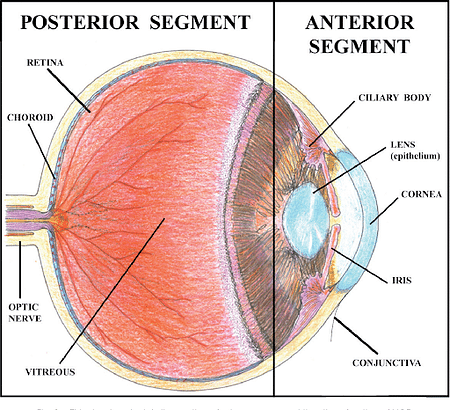
Every human being has two eyes. They sit in a bony cavity in the skull, called the orbit. The eye is composed of two segments: Anterior and Posterior. The detailed explanation of each component of the eye is as follows:
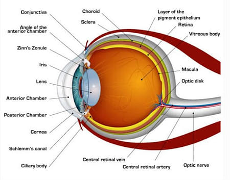
Sclera: The outer white part of the eye is called Sclera. It is the outermost protective layer of the eye.
Cornea: The front-facing transparent part of the eye is called Cornea. It is made up of six layers. The Cornea is thinner in the centre and its thickness increases as you move outwards. Due to the curvature of the cornea, it refracts the light up to around 45 dioptres. To protect the eye, the cornea has a surrounding of tear fluids that are produced in the tear glands.
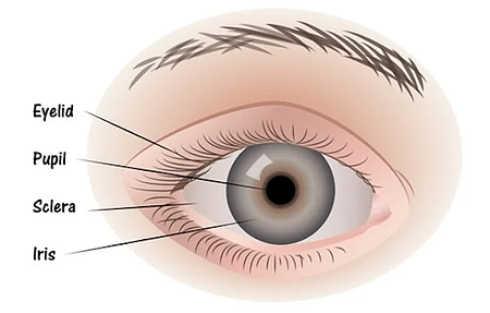
Iris: The iris is situated behind the cornea, in its centre. It has a ring-like structure. It imparts colour to the eye. It also controls the exposure of the eye to light by adjusting the size of the pupil.
Pupil: The iris has a small opening called the pupil. It determines the exposure of the eye to light.
Lens: Lens is a transparent structure that is situated behind the pupil. It has the ability to change its refractive power. This is achieved with the help of ciliary muscles. The ciliary muscles help in changing the shape of the lens to focus the light on the retina. It becomes thinner and thicker to focus on distant and nearby objects respectively.
Ciliary Muscles: Ciliary Muscles have a ring-shaped structure and are present behind the cornea. They are responsible for the curving of the eye lens. The diameter of the lens increases and decreases as the ciliary muscles contract and relax respectively.
Vitreous Chamber: Vitreous Chamber is filled with a gel-like clear liquid. It is responsible for the stability of the eyeball. The liquid also creates intraocular pressure which protects the eye from breakdown and peels off. Some diseases like glaucoma are caused by abnormal intraocular pressure.
Retina: Retina is responsible for the conversion of light into electrical impulses. It consists of numerous nerve cells of two types: Rods and Cones.
- Rods: Rods help in peripheral vision and they are sensitive to dim light.
- Cones: Cones help in central and colour vision and they are sensitive to bright light.
Different Muscles in the Human Eye
There are six muscles present in the eye which are responsible for different torques and tensions in the eye. These muscles are listed as follows:
- Medial Rectus
- Lateral Rectus
- Superior Rectus
- Inferior Rectus
- Superior Oblique
- Inferior Oblique

Functions of the Human Eye
The function of the eye is to let human beings see the world. It is achieved through the process of refraction.

The refractive index of air is 1 and the refractive index of the cornea is 1.376. Due to the difference in refractive index, the light rays bend and fall on the retina. The presence of rod and cone cells in the retina help in detecting the intensity and frequency of the light. The nerve cells convert the light into electrical impulses. These impulses are transferred to the brain through the optic nerve. The brain processes the impulses and reacts accordingly.
Things to Remember based on Human Eye
- Human Eye is the vision providing organ of the body. It allows us to see and perceive objects, distinguish color and acts as a biological clock.
- The structure of the human eye is roughly spherical. It is composed of two segments fused together.
- Some of the most important parts of the eye are: Sclera, Retina, Cornea, Iris, Pupil, Lens etc.
- There are six muscles present in the eye: Medial Rectus, Lateral Rectus, Superior Rectus, Inferior Rectus, Superior Oblique, Inferior Oblique.
- The refractive index of air is 1 while the refractive index of the cornea is 1.376. Due to the difference in refractive index, the light rays bend and fall on the retina. The presence of rod and cone cells in the retina help in detecting the intensity and frequency of the light.
Sample Questions based on Human Eye
Ques. How does the human eye work? (2 marks)
Ans- Light rays enter the eye through the cornea. With the help of ciliary muscles, the lens contracts and relaxes to focus the light ray on the retina. Millions of rod and cone cells present on the retina convert the light rays into electrical impulses. These electrical impulses are transmitted to the brain through the optic nerve.
Ques. What is the function of the Cornea? (3 marks)
Ans- The functions of the cornea are as follows:
- It shields the eye from germs and dust.
- It plays an important role in maintaining the amount of light entering the eye.
- The human eye absorbs light from the surroundings, which is received by the rod and cone cells in the retina. Photoreceptive cells detect visible light and send it to the brain. The brain then processes and responds to the information conveyed.
Ques. Define Blind Spot. (2 marks)
Ans- There are no sensory nerve cells present at the junction of the retina and optic nerve. Due to the absence of nerve cells, no vision is possible at this point. This point is known as the Blind Spot.
Ques. What is Stereoscopic Vision? (2 marks)
Ans- Stereoscopic Vision refers to the ability of our brain to determine the shape and form of an object through visual inputs. The information relayed to the brain by both the yes differs due to differences in the angle of incidence of the refracted light rays. Our brain takes advantage of this difference to determine the three-dimensional form of the object.
Ques. What are the different types of Cone Cells? (3 marks)
Ans- The different types of cone cells are:
- Pins for red vision (46% of all pins)
- Cones for green vision (46% of all cones)
- Cones for blue vision (8% of all cones)



Comments