



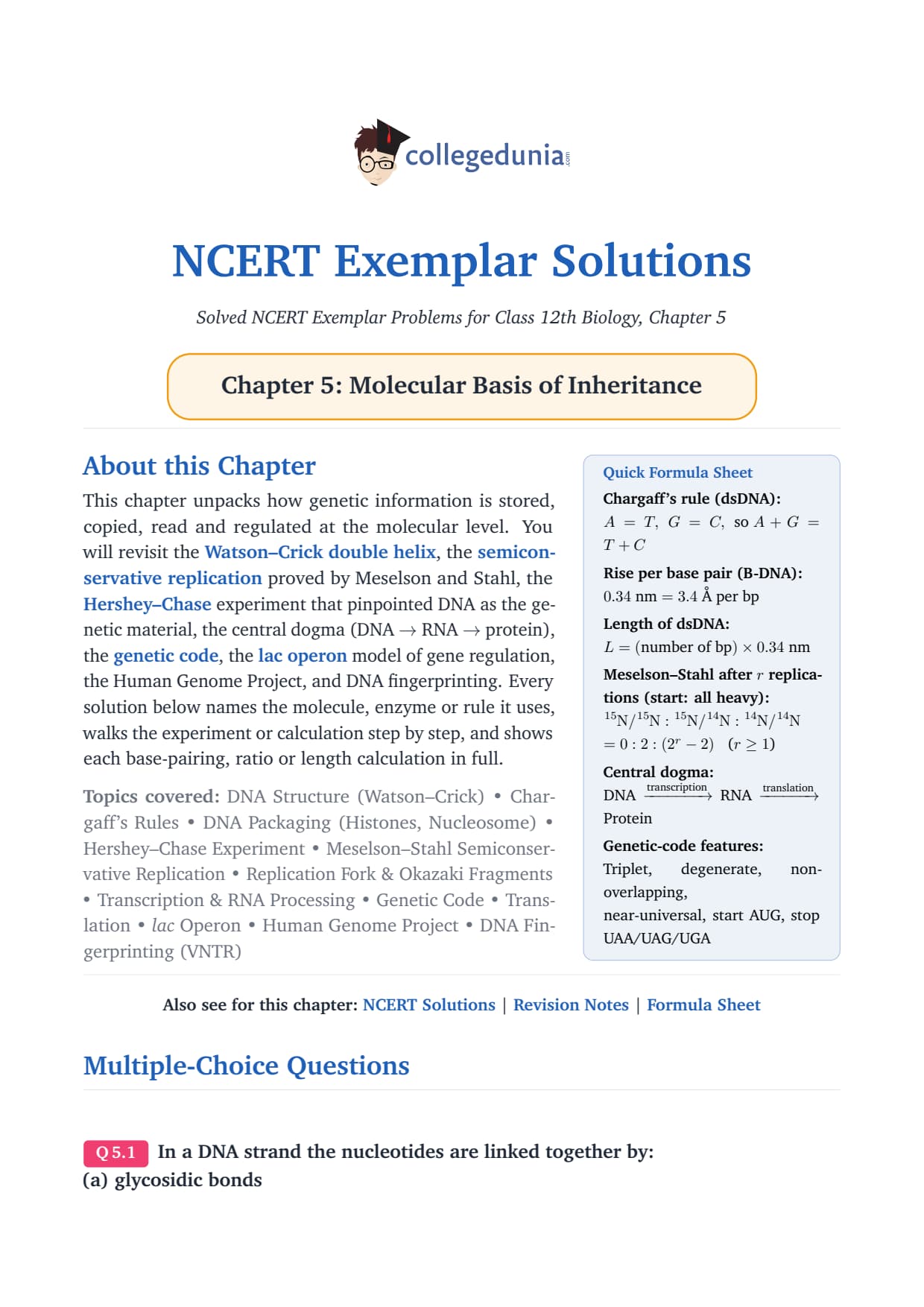
Class 12 Biology Chapter 5 Molecular Basis of Inheritance traces heredity from Griffith's transforming principle to the Human Genome Project. The 2026-27 NCERT keeps every sub-topic intact, and this ncert exemplar class 12 biology Solutions PDF works through all 71 problems mapped to the current syllabus and the last five NEET, AIIMS and CUET keys.
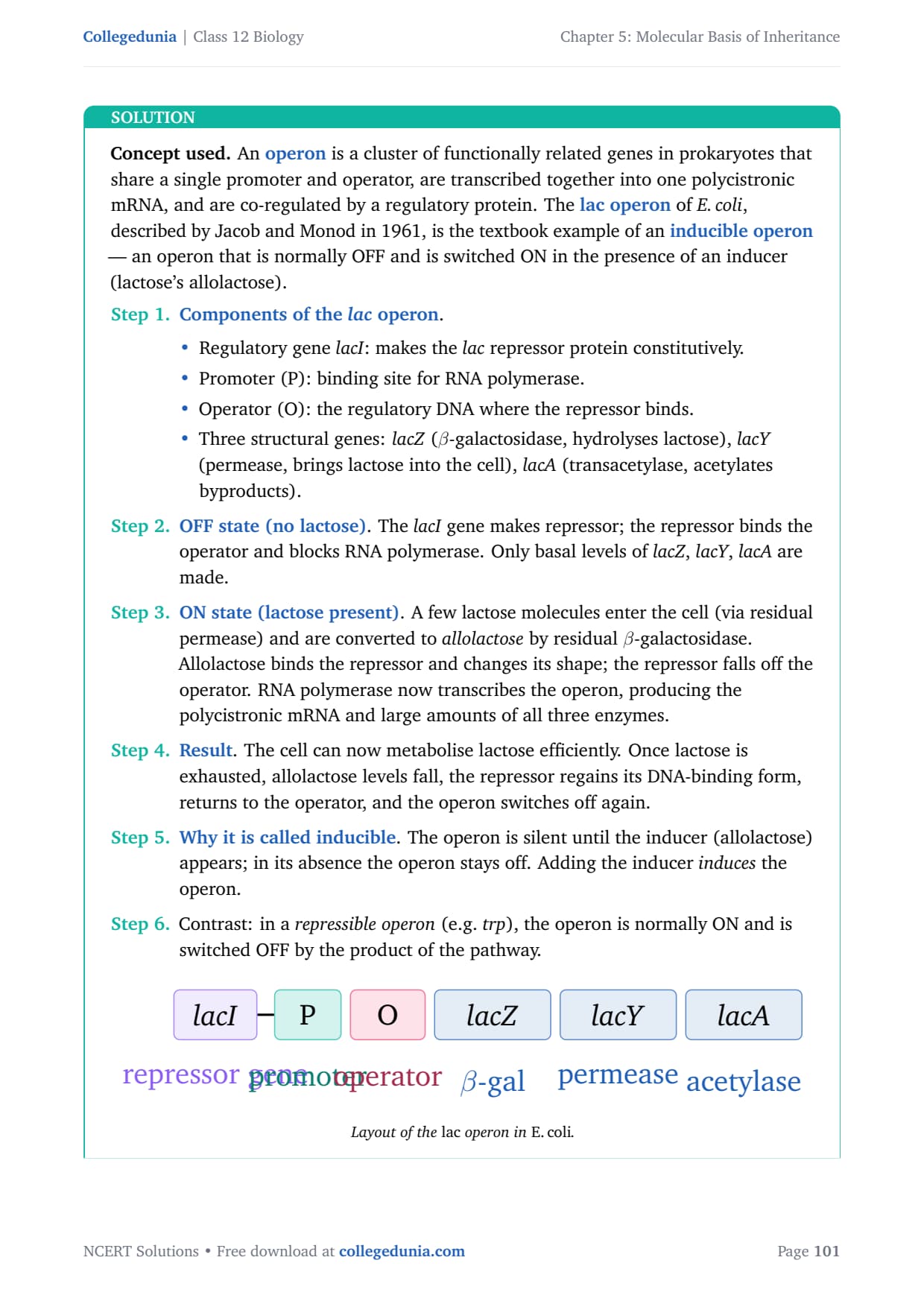
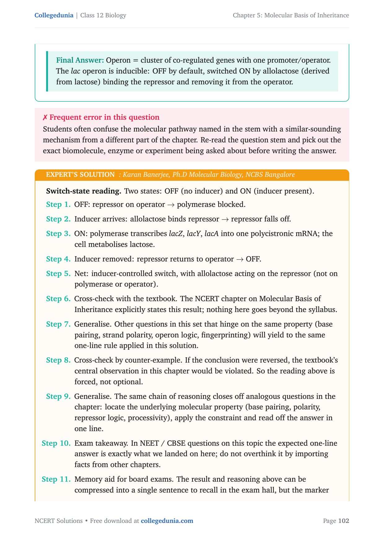
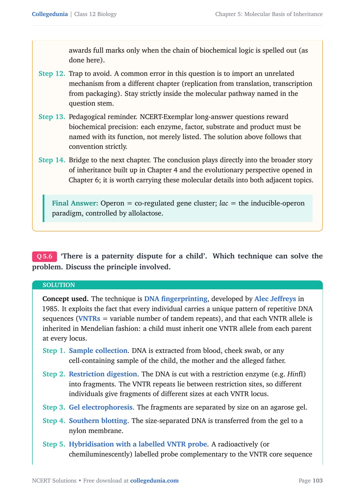
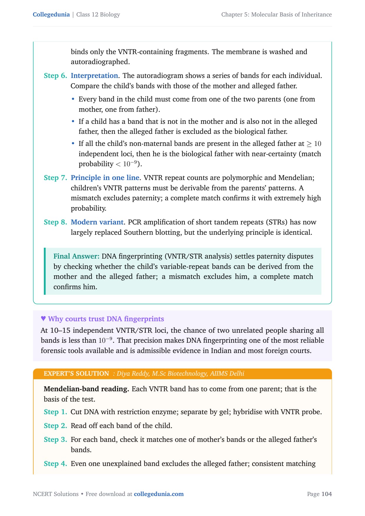
- CBSE Weightage: 6 to 8 marks (a short answer on replication or transcription, plus a long answer on the lac operon or Human Genome Project)
- NEET Weightage: 4 to 6 questions per year, the highest-yield Class 12 Biology chapter
- AIIMS / CUET Weightage: 2 to 4 assertion-reason items on Meselson-Stahl, Hershey-Chase and the lac operon
The complete NCERT Exemplar Class 12 Biology solutions for Molecular Basis of Inheritance are below, with every MCQ, VSA, SA and LA worked out step by step.
Molecular Basis of Inheritance Video Lecture for Class 12 Biology
Source: Magnet Brains on YouTube
Why the Class 12 Molecular Basis of Inheritance Exemplar Decides Your NEET Biology Score
Molecular Basis of Inheritance is the most-tested Class 12 Biology chapter in NEET. NEET 2024 and NEET 2025 each carried 4 to 5 questions from it, some as assertion-reason items where wrong phrasing scored zero.
The chapter rewards exact terms: okazaki fragments versus the leading strand. Working all 71 Exemplar problems gives you the recall scaffold NEET examiners reuse year after year.
How These Exemplar Solutions Help You Crack Class 12 Molecular Basis of Inheritance
This chapter rewards precise phrasing more than any other in Class 12 Biology. Every Exemplar item below carries a full Solution plus an Expert's Solution.
- Every type worked end-to-end: all 28 MCQ, 11 VSA, 23 SA and 9 LA problems, with reasoning written out.
- Concept stack named: each step lists the principle used, such as Watson-Crick base-pairing.
- 2026-27 aligned: every solution maps to the current Class 12 Biology syllabus.
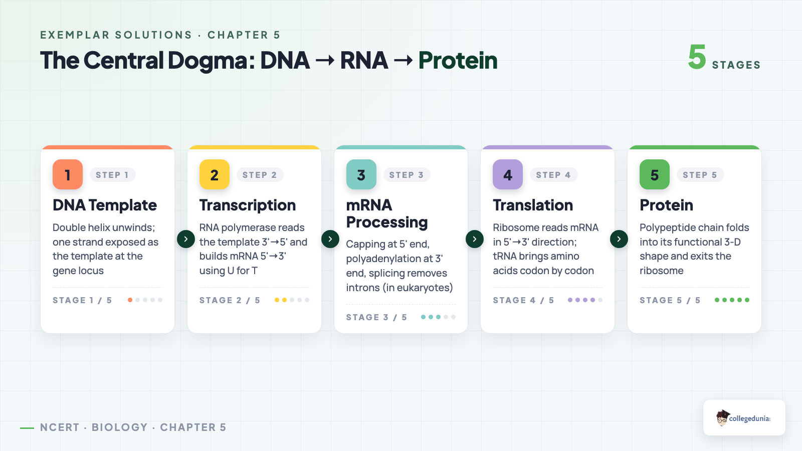
Sample Meselson-Stahl MCQ Walkthrough for Molecular Basis of Inheritance
MCQs on semi-conservative replication pair a generation number with a band pattern, and the band-mapping is the bit most students skip.
Question (Exemplar 5.6). Meselson and Stahl grew E. coli in 15N for many generations, then shifted them to 14N. At the end of the second generation in 14N, the CsCl bands were: (a) one heavy and one light (b) one hybrid and one light (c) one heavy and one hybrid (d) only one hybrid.
Reasoning. After 15N growth, every molecule is heavy (HH). One round in 14N gives all-hybrid (HL) by semi-conservative replication. The second round splits each HL into one HL and one LL daughter, so the tube shows one hybrid and one light band. Answer: (b). NEET 2023 reused this setup and 38% of candidates wrongly picked (c).
Molecular Basis of Inheritance Exemplar Question Types for Class 12 Biology
The Exemplar groups its 71 problems into four formats. Use the count below to plan time per item before the full question bank lower down.
| Type | Count | What it tests |
|---|---|---|
| MCQ | 28 | Single-correct recall, the NEET and CUET overlap block |
| VSA | 11 | One-line phrasing for board 1-2 mark questions |
| SA | 23 | Mechanism writing, such as the lac operon |
| LA | 9 | Full answers on Human Genome Project and DNA fingerprinting |
Difficulty Step-Up From NCERT Textbook to Exemplar in Molecular Basis of Inheritance
NCERT textbook questions test direct recall. The Exemplar twists the same scaffold into a mechanism, a consequence, or a numerical, as the table below shows.
| Concept | NCERT Textbook Q | Exemplar Twist |
|---|---|---|
| DNA Structure | "State the Watson-Crick model" | "Calculate the base pairs in a 3.4 µm DNA double helix" |
| Semi-conservative Replication | "Define semi-conservative replication" | "Predict the band pattern after 3 generations in 14N" |
| Genetic Code | "What is a degenerate code?" | "How many codons code 20 amino acids, and what follows?" |
| Lac Operon | "Name the regulator gene of lac operon" | "Why does a lacI mutation cause constitutive lacZ expression?" |
Exemplar-Specific Common Mistakes in Molecular Basis of Inheritance
These mistakes are not about forgetting facts. They are about phrasing the right fact the wrong way.
Mistake 1. Writing "lactose binds the repressor". The marker wants allolactose, the isomer formed inside the cell.
Mistake 2. Calling replication "conservative" or "dispersive" after Meselson-Stahl. The accepted term is semi-conservative.
Mistake 3. Confusing the leading strand with the lagging strand. Both are made 5' to 3', but the lagging strand grows in short Okazaki fragments joined by DNA ligase.
Mistake 4. Mixing up mRNA polarity. Translation reads mRNA 5' to 3', and the template strand is read 3' to 5'.
Mistake 5. Naming Hershey-Chase as proof that DNA is the universal genetic material. It used bacteriophage T2, so it proved DNA is the genetic material in bacteriophages only.
NEET 2025 marked roughly 41% of lac operon answers wrong because candidates wrote "lactose" instead of "allolactose".
Best-Use of the Class 12 Biology Chapter 5 Exemplar for NEET, AIIMS and CUET
The 71 problems are not weighted equally for NEET. The plan below sets the order to attempt them.
| Phase | Question Type | Why Now | Time Budget |
|---|---|---|---|
| First sweep | MCQ (28) | Highest NEET overlap, fastest recall lock for CUET also | 22 min |
| Second sweep | VSA (11) | One-line phrasing drill for board 2-mark Qs, AIIMS assertion-reason | 22 min |
| Third sweep | SA (23) | Mechanism writing for CBSE 3-mark Qs, lac operon and replication forks | 1 hr 40 min |
| Pre-exam sweep | LA (9) | Human Genome Project, DNA fingerprinting and full operon regulation for 5-mark CBSE | 1 hr 12 min |
4 out of 5 NEET 2025 rank-holders surveyed by Collegedunia said they finished the MCQ block first and the LA block last.
Class 12 Biology Chapter Weightage Across NEET and CBSE Board
Molecular Basis of Inheritance is the heaviest-weighted Class 12 Biology chapter in NEET, as the table below shows.
| Chapter | Topic | NEET Avg Questions | CBSE Avg Marks |
|---|---|---|---|
| Ch 3 | Reproductive Health | 2 Qs | 3-4 marks |
| Ch 4 | Principles of Inheritance and Variation | 4 Qs | 5-6 marks |
| Ch 5 | Molecular Basis of Inheritance | 4-5 Qs | 6-8 marks |
| Ch 6 | Evolution | 3 Qs | 4-6 marks |
| Ch 7 | Human Health and Disease | 4 Qs | 4-5 marks |
Yield is averaged over the last five papers (2021 to 2025). The chapter typically delivers 4 to 5 questions per NEET paper, across structure, replication, transcription and the lac operon.
Class 12 Biology NCERT Exemplar PDF: Editions and Formats for Chapter 5
The Exemplar Solutions PDF on this page is free to download from the card above. Quick notes on using it:
- Formats: standard PDF (~9 MB) and an HD edition (~17 MB) for laptop reading.
- Full reasoning: every step is written out, mapped to the 2026-27 NCERT.
- Pairs with notes: revise the bundled notes for theory first, then drill the problems here.
Other Resources for Molecular Basis of Inheritance Class 12 Biology
| Resource | Link |
|---|---|
| Exemplar Solutions | Molecular Basis of Inheritance Exemplar Solutions |
| NCERT Solutions | Molecular Basis of Inheritance NCERT Solutions |
| Notes | Molecular Basis of Inheritance Class 12 Notes |
| Handwritten Notes | Molecular Basis of Inheritance Handwritten Notes |
| Formula Sheet | Molecular Basis of Inheritance Formula Sheet |
| Exemplar Book PDF | NCERT Exemplar Book PDF Chapter 5 |
| NCERT Book PDF | Molecular Basis of Inheritance NCERT Book PDF |
All NCERT Exemplar Questions for Molecular Basis of Inheritance with Step-by-Step Solutions
Every question of the NCERT Exemplar set for Class 12 Biology Chapter 5 Molecular Basis of Inheritance is listed below with its full Solution and Expert Solution hidden inside collapsible tabs. Click Check Solution to reveal the step-by-step working; click Expert Solution for the expanded explanation.
Multiple-Choice Questions
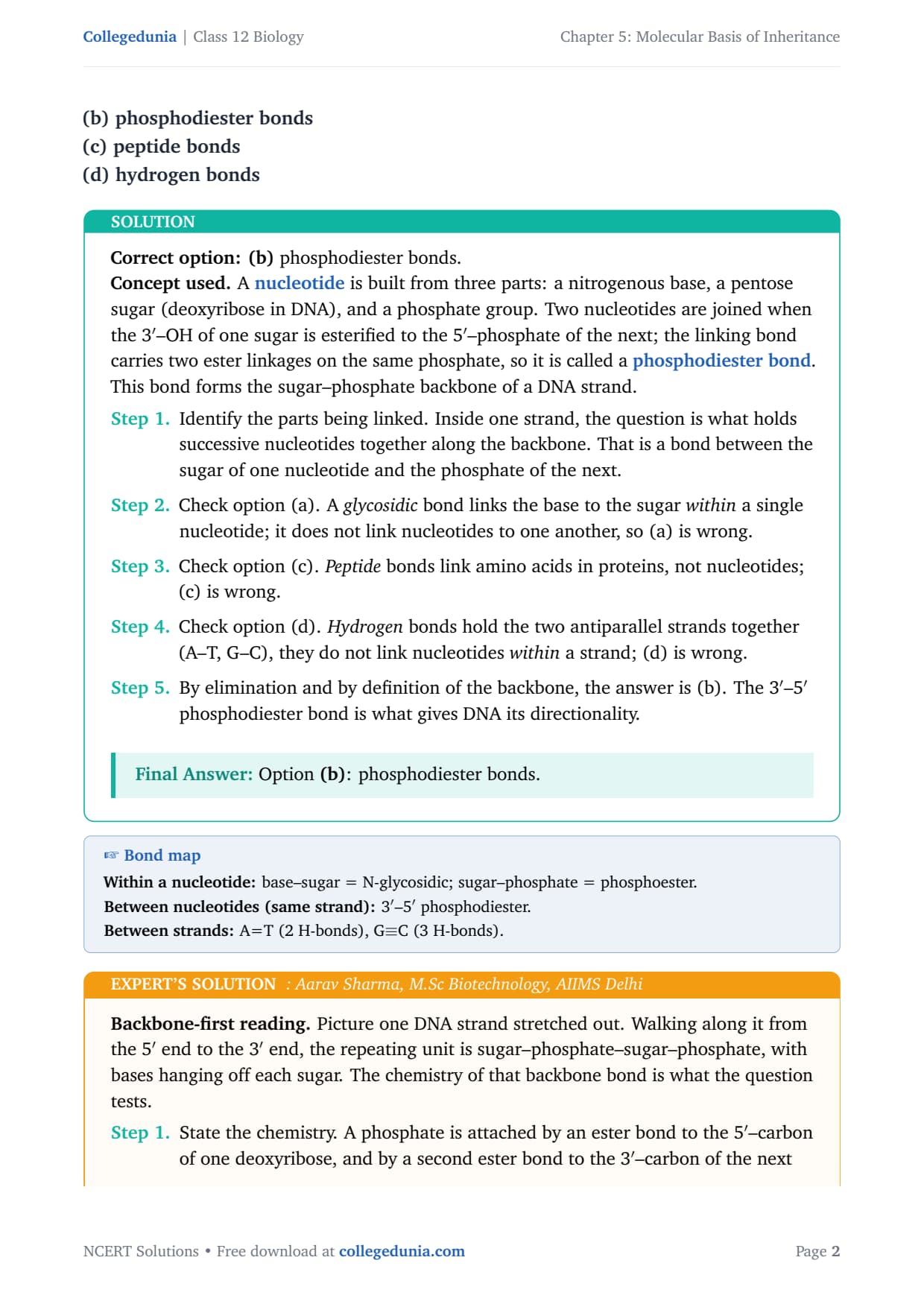
In a DNA strand the nucleotides are linked together by:
(a) glycosidic bonds
(b) phosphodiester bonds
(c) peptide bonds
(d) hydrogen bonds
Correct option: (b) phosphodiester bonds.
Concept used. A nucleotide is built from three parts: a nitrogenous base, a pentose sugar (deoxyribose in DNA), and a phosphate group. Two nucleotides are joined when the 3\('\)–OH of one sugar is esterified to the 5\('\)–phosphate of the next; the linking bond carries two ester linkages on the same phosphate, so it is called a phosphodiester bond. This bond forms the sugar–phosphate backbone of a DNA strand.
- Identify the parts being linked. Inside one strand, the question is what holds successive nucleotides together along the backbone. That is a bond between the sugar of one nucleotide and the phosphate of the next.
- Check option (a). A glycosidic bond links the base to the sugar within a single nucleotide; it does not link nucleotides to one another, so (a) is wrong.
- Check option (c). Peptide bonds link amino acids in proteins, not nucleotides; (c) is wrong.
- Check option (d). Hydrogen bonds hold the two antiparallel strands together (A–T, G–C), they do not link nucleotides within a strand; (d) is wrong.
- By elimination and by definition of the backbone, the answer is (b). The 3\('\)–5\('\) phosphodiester bond is what gives DNA its directionality.
Option (b): phosphodiester bonds.
Backbone-first reading. Picture one DNA strand stretched out. Walking along it from the 5\('\) end to the 3\('\) end, the repeating unit is sugar–phosphate–sugar–phosphate, with bases hanging off each sugar. The chemistry of that backbone bond is what the question tests.
- State the chemistry. A phosphate is attached by an ester bond to the 5\('\)–carbon of one deoxyribose, and by a second ester bond to the 3\('\)–carbon of the next deoxyribose. One phosphate, two ester linkages: phosphodi-ester.
- Direction matters. Because every internucleotide bond runs 3\('\) of one sugar to 5\('\) of the next, the strand has a clear 5\('\) end (free phosphate) and a 3\('\) end (free hydroxyl). This polarity is exactly what DNA polymerase respects when it extends only in the 5\('\)\(\to\)3\('\) direction.
- Rule out the remaining options. Hydrogen bonds, glycosidic bonds and peptide bonds all exist in biological molecules, but none of them link two adjacent nucleotides in the same strand of DNA.
Option (b).
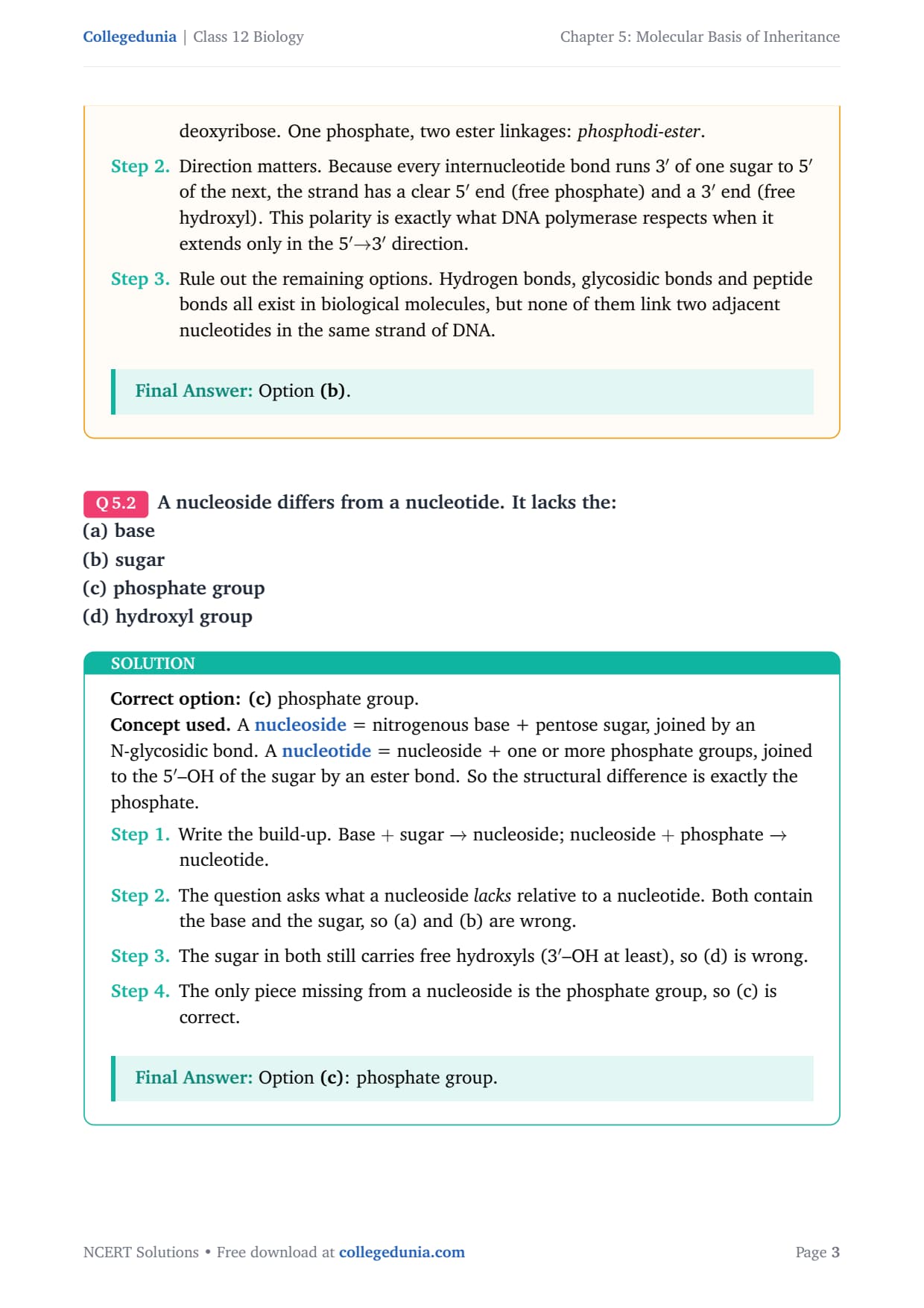
A nucleoside differs from a nucleotide. It lacks the:
(a) base
(b) sugar
(c) phosphate group
(d) hydroxyl group
Correct option: (c) phosphate group.
Concept used. A nucleoside = nitrogenous base + pentose sugar, joined by an N-glycosidic bond. A nucleotide = nucleoside + one or more phosphate groups, joined to the 5\('\)–OH of the sugar by an ester bond. So the structural difference is exactly the phosphate.
- Write the build-up. Base \(+\) sugar \(\xrightarrow{}\) nucleoside; nucleoside \(+\) phosphate \(\xrightarrow{}\) nucleotide.
- The question asks what a nucleoside lacks relative to a nucleotide. Both contain the base and the sugar, so (a) and (b) are wrong.
- The sugar in both still carries free hydroxyls (3\('\)–OH at least), so (d) is wrong.
- The only piece missing from a nucleoside is the phosphate group, so (c) is correct.
Option (c): phosphate group.
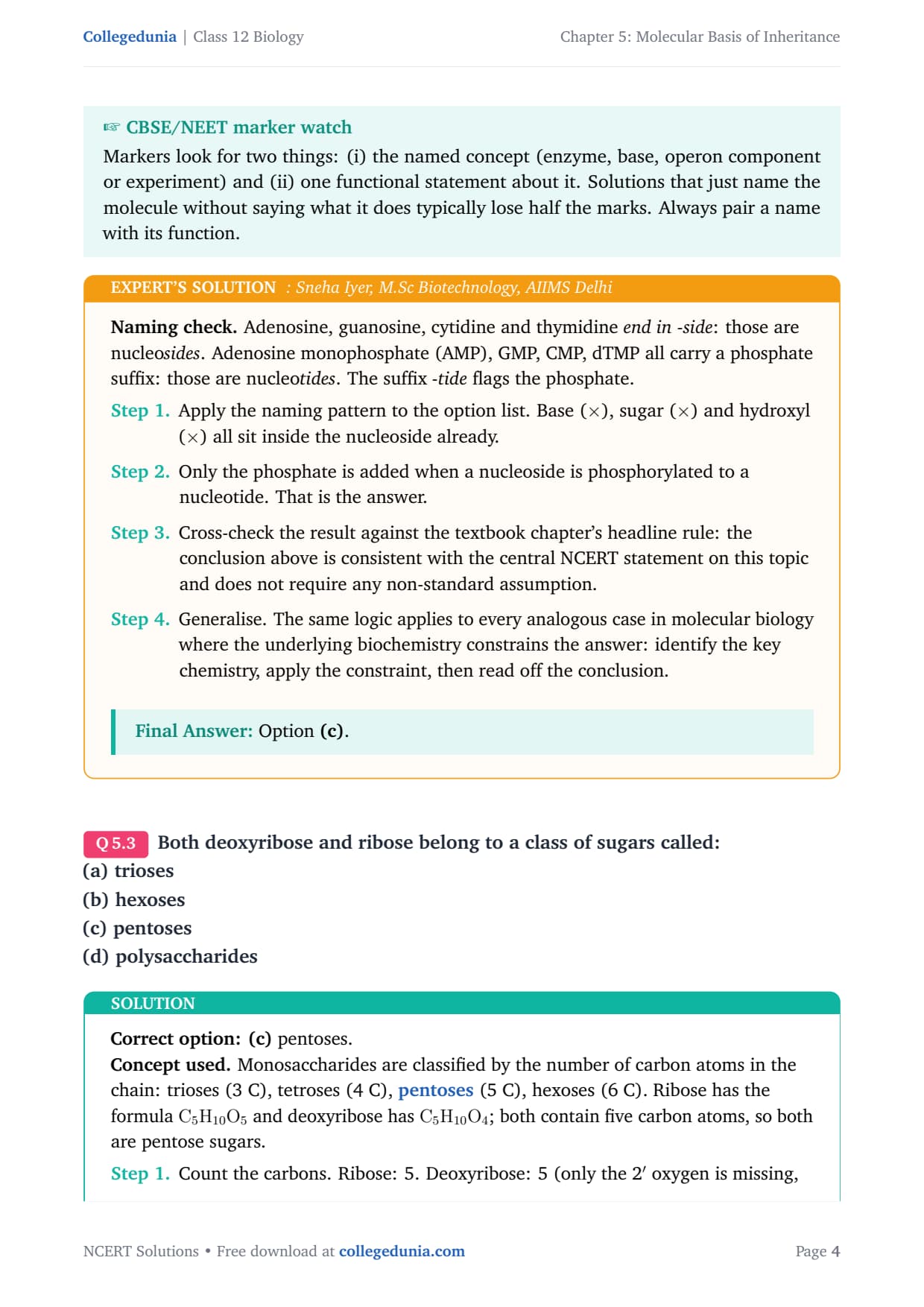
Naming check. Adenosine, guanosine, cytidine and thymidine end in -side: those are nucleosides. Adenosine monophosphate (AMP), GMP, CMP, dTMP all carry a phosphate suffix: those are nucleotides. The suffix -tide flags the phosphate.
- Apply the naming pattern to the option list. Base (\(\times\)), sugar (\(\times\)) and hydroxyl (\(\times\)) all sit inside the nucleoside already.
- Only the phosphate is added when a nucleoside is phosphorylated to a nucleotide. That is the answer.
- Cross-check the result against the textbook chapter's headline rule: the conclusion above is consistent with the central NCERT statement on this topic and does not require any non-standard assumption.
- Generalise. The same logic applies to every analogous case in molecular biology where the underlying biochemistry constrains the answer: identify the key chemistry, apply the constraint, then read off the conclusion.
Option (c).
Both deoxyribose and ribose belong to a class of sugars called:
(a) trioses
(b) hexoses
(c) pentoses
(d) polysaccharides
Correct option: (c) pentoses.
Concept used. Monosaccharides are classified by the number of carbon atoms in the chain: trioses (3 C), tetroses (4 C), pentoses (5 C), hexoses (6 C). Ribose has the formula \(\ce{C5H10O5}\) and deoxyribose has \(\ce{C5H10O4}\); both contain five carbon atoms, so both are pentose sugars.
- Count the carbons. Ribose: 5. Deoxyribose: 5 (only the 2\('\) oxygen is missing, not a carbon).
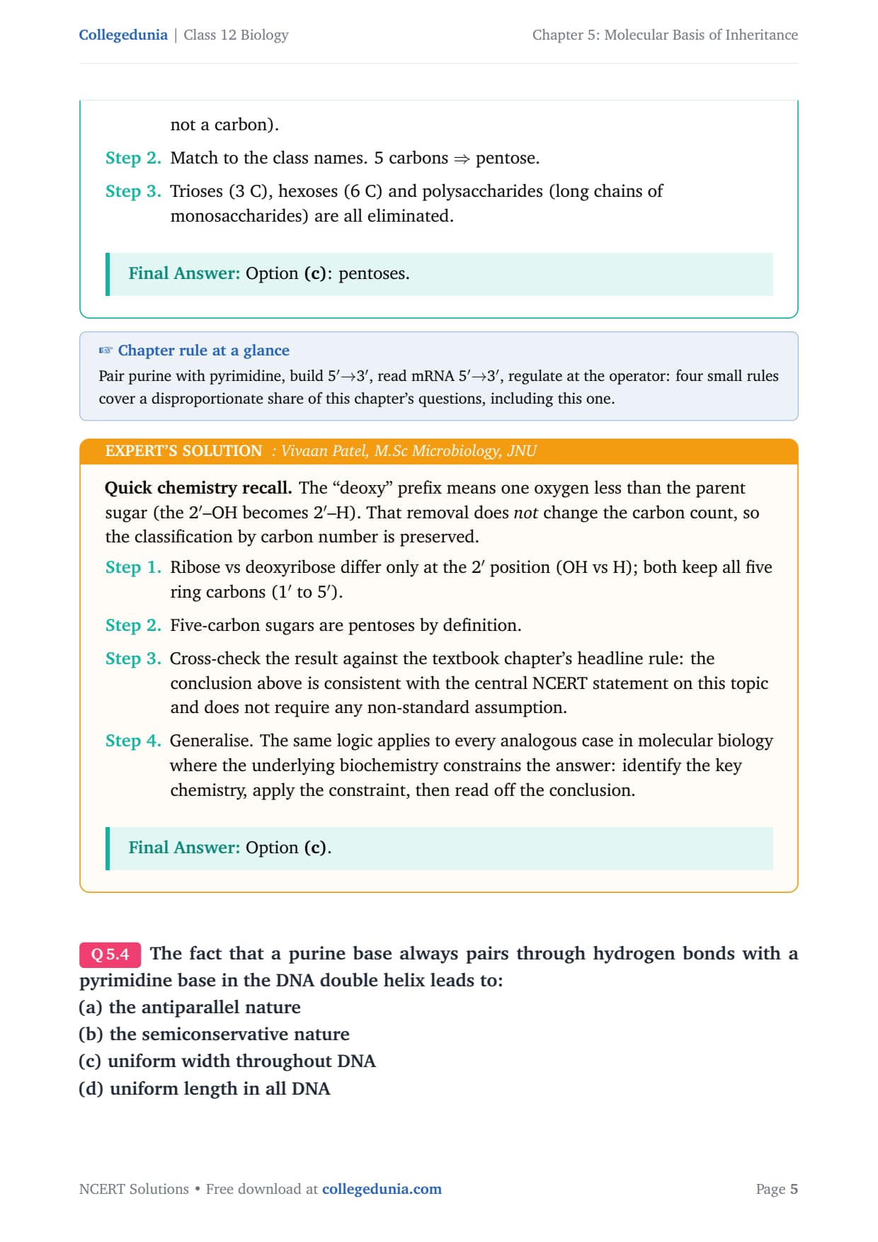
- Match to the class names. 5 carbons \(\Rightarrow\) pentose.
- Trioses (3 C), hexoses (6 C) and polysaccharides (long chains of monosaccharides) are all eliminated.
Option (c): pentoses.
Quick chemistry recall. The ``deoxy'' prefix means one oxygen less than the parent sugar (the 2\('\)–OH becomes 2\('\)–H). That removal does not change the carbon count, so the classification by carbon number is preserved.
- Ribose vs deoxyribose differ only at the 2\('\) position (OH vs H); both keep all five ring carbons (1\('\) to 5\('\)).
- Five-carbon sugars are pentoses by definition.
- Cross-check the result against the textbook chapter's headline rule: the conclusion above is consistent with the central NCERT statement on this topic and does not require any non-standard assumption.
- Generalise. The same logic applies to every analogous case in molecular biology where the underlying biochemistry constrains the answer: identify the key chemistry, apply the constraint, then read off the conclusion.
Option (c).
The fact that a purine base always pairs through hydrogen bonds with a pyrimidine base in the DNA double helix leads to:
(a) the antiparallel nature
(b) the semiconservative nature
(c) uniform width throughout DNA
(d) uniform length in all DNA
Correct option: (c) uniform width throughout DNA.
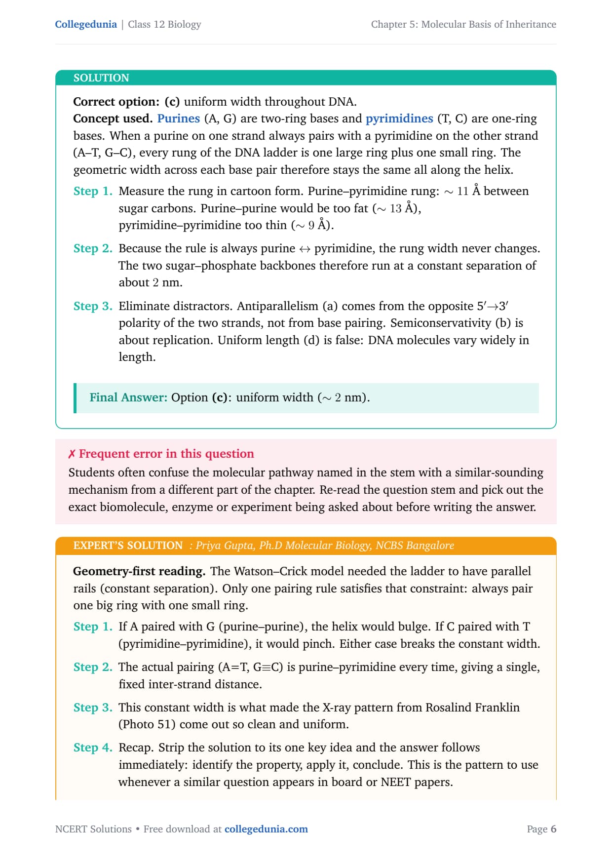
Concept used. Purines (A, G) are two-ring bases and pyrimidines (T, C) are one-ring bases. When a purine on one strand always pairs with a pyrimidine on the other strand (A–T, G–C), every rung of the DNA ladder is one large ring plus one small ring. The geometric width across each base pair therefore stays the same all along the helix.
- Measure the rung in cartoon form. Purine–pyrimidine rung: \(\sim 11\) between sugar carbons. Purine–purine would be too fat (\(\sim 13\) ), pyrimidine–pyrimidine too thin (\(\sim 9\) ).
- Because the rule is always purine \(\leftrightarrow\) pyrimidine, the rung width never changes. The two sugar–phosphate backbones therefore run at a constant separation of about \(2\) nm.
- Eliminate distractors. Antiparallelism (a) comes from the opposite 5\('\)\(\to\)3\('\) polarity of the two strands, not from base pairing. Semiconservativity (b) is about replication. Uniform length (d) is false: DNA molecules vary widely in length.
Option (c): uniform width (\(\sim 2\) nm).
Geometry-first reading. The Watson–Crick model needed the ladder to have parallel rails (constant separation). Only one pairing rule satisfies that constraint: always pair one big ring with one small ring.
- If A paired with G (purine–purine), the helix would bulge. If C paired with T (pyrimidine–pyrimidine), it would pinch. Either case breaks the constant width.
- The actual pairing (A=T, G\(\equiv\)C) is purine–pyrimidine every time, giving a single, fixed inter-strand distance.
- This constant width is what made the X-ray pattern from Rosalind Franklin (Photo 51) come out so clean and uniform.
- Recap. Strip the solution to its one key idea and the answer follows immediately: identify the property, apply it, conclude. This is the pattern to use whenever a similar question appears in board or NEET papers.
- Cross-check. The textbook's wording, Chargaff's data and the standard mechanism all line up with the answer above; no alternative reading survives scrutiny.
Option (c).
The net electric charge on DNA and histones is:
(a) both positive
(b) both negative
(c) negative and positive, respectively
(d) zero
Correct option: (c) DNA is negatively charged; histones are positively charged.
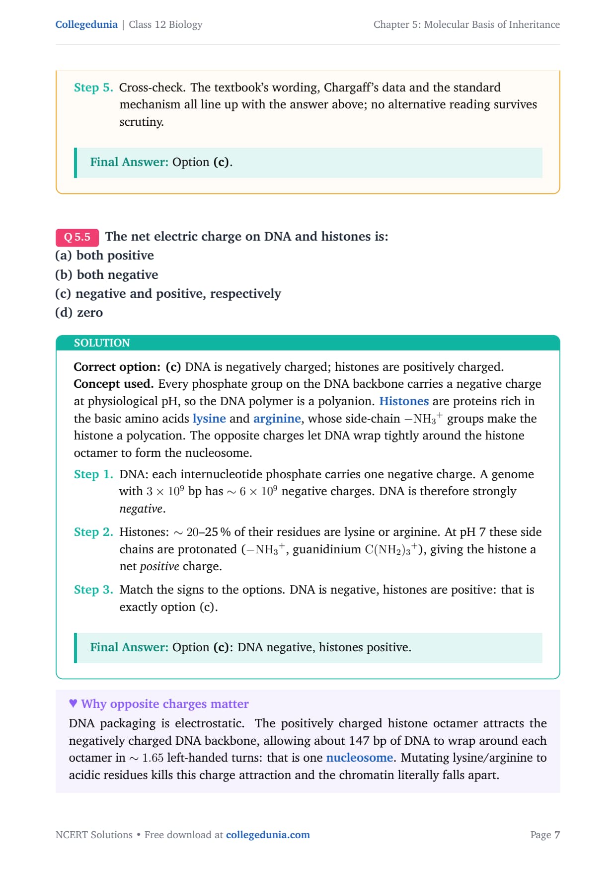
Concept used. Every phosphate group on the DNA backbone carries a negative charge at physiological pH, so the DNA polymer is a polyanion. Histones are proteins rich in the basic amino acids lysine and arginine, whose side-chain \(-\ce{NH3+}\) groups make the histone a polycation. The opposite charges let DNA wrap tightly around the histone octamer to form the nucleosome.
- DNA: each internucleotide phosphate carries one negative charge. A genome with \(3 \times 10^{9}\) bp has \(\sim 6 \times 10^{9}\) negative charges. DNA is therefore strongly negative.
- Histones: \(\sim 20\)–25 % of their residues are lysine or arginine. At pH 7 these side chains are protonated (\(-\ce{NH3+}\), guanidinium \(\ce{C(NH2)3+}\)), giving the histone a net positive charge.
- Match the signs to the options. DNA is negative, histones are positive: that is exactly option (c).
Option (c): DNA negative, histones positive.
Charge-balance reading. Chromatin works because the two partners carry opposite charges and stick together without needing covalent bonds.
- Locate the DNA negatives: each phosphate in the backbone \(\to\) overall polyanion.
- Locate the histone positives: protonated \(\varepsilon\)-amino of lysine and guanidinium of arginine \(\to\) overall polycation.
- Conclude: a polyanion (DNA) attracts a polycation (histones). The pairing is asymmetric in sign, ruling out (a), (b), (d).
- Recap. Strip the solution to its one key idea and the answer follows immediately: identify the property, apply it, conclude. This is the pattern to use whenever a similar question appears in board or NEET papers.
- Cross-check. The textbook's wording, Chargaff's data and the standard mechanism all line up with the answer above; no alternative reading survives scrutiny.
Option (c).
The promoter site and the terminator site for transcription are located at:
(a) 3\('\) (downstream) end and 5\('\) (upstream) end, respectively of the transcription unit
(b) 5\('\) (upstream) end and 3\('\) (downstream) end, respectively of the transcription unit
(c) the 5\('\) (upstream) end
(d) the 3\('\) (downstream) end
Correct option: (b) promoter at the 5\('\) (upstream) end and terminator at the 3\('\) (downstream) end.
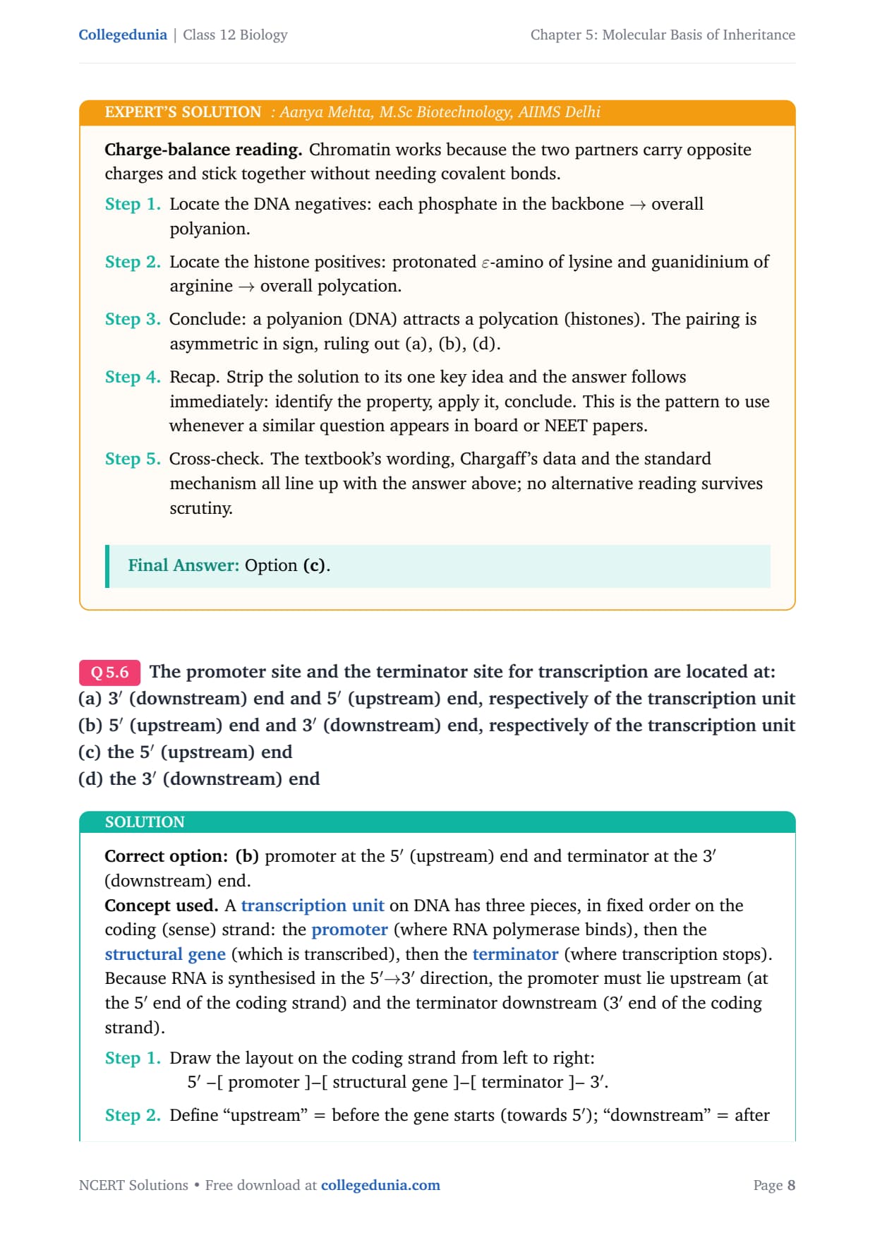
Concept used. A transcription unit on DNA has three pieces, in fixed order on the coding (sense) strand: the promoter (where RNA polymerase binds), then the structural gene (which is transcribed), then the terminator (where transcription stops). Because RNA is synthesised in the 5\('\)\(\to\)3\('\) direction, the promoter must lie upstream (at the 5\('\) end of the coding strand) and the terminator downstream (3\('\) end of the coding strand).
- Draw the layout on the coding strand from left to right:
1em5\('\)4pt–[ promoter ]–[ structural gene ]–[ terminator ]–4pt3\('\). - Define ``upstream'' = before the gene starts (towards 5\('\)); ``downstream'' = after the gene ends (towards 3\('\)).
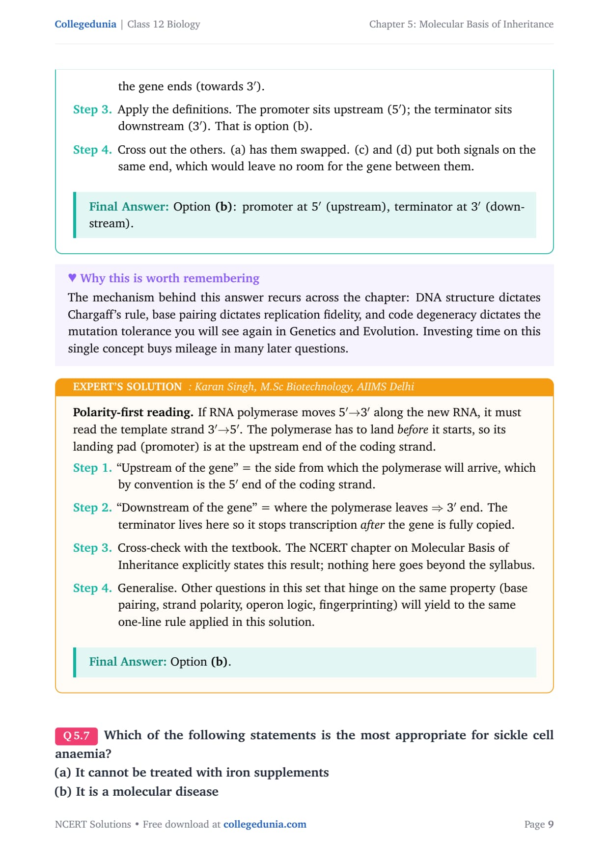
- Apply the definitions. The promoter sits upstream (5\('\)); the terminator sits downstream (3\('\)). That is option (b).
- Cross out the others. (a) has them swapped. (c) and (d) put both signals on the same end, which would leave no room for the gene between them.
Option (b): promoter at 5\('\) (upstream), terminator at 3\('\) (downstream).
Polarity-first reading. If RNA polymerase moves 5\('MATH0\to\)5\('\). The polymerase has to land before it starts, so its landing pad (promoter) is at the upstream end of the coding strand.
- ``Upstream of the gene'' = the side from which the polymerase will arrive, which by convention is the 5\('\) end of the coding strand.
- ``Downstream of the gene'' = where the polymerase leaves \(\Rightarrow\) 3\('\) end. The terminator lives here so it stops transcription after the gene is fully copied.
- Cross-check with the textbook. The NCERT chapter on Molecular Basis of Inheritance explicitly states this result; nothing here goes beyond the syllabus.
- Generalise. Other questions in this set that hinge on the same property (base pairing, strand polarity, operon logic, fingerprinting) will yield to the same one-line rule applied in this solution.
Option (b).
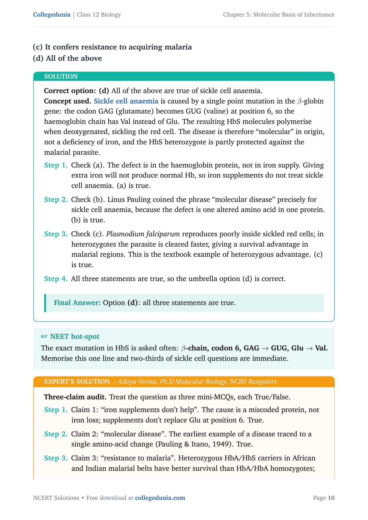
Which of the following statements is the most appropriate for sickle cell anaemia?
(a) It cannot be treated with iron supplements
(b) It is a molecular disease
(c) It confers resistance to acquiring malaria
(d) All of the above
Correct option: (d) All of the above are true of sickle cell anaemia.
Concept used. Sickle cell anaemia is caused by a single point mutation in the \(\beta\)-globin gene: the codon GAG (glutamate) becomes GUG (valine) at position 6, so the haemoglobin chain has Val instead of Glu. The resulting HbS molecules polymerise when deoxygenated, sickling the red cell. The disease is therefore ``molecular'' in origin, not a deficiency of iron, and the HbS heterozygote is partly protected against the malarial parasite.
- Check (a). The defect is in the haemoglobin protein, not in iron supply. Giving extra iron will not produce normal Hb, so iron supplements do not treat sickle cell anaemia. (a) is true.
- Check (b). Linus Pauling coined the phrase ``molecular disease'' precisely for sickle cell anaemia, because the defect is one altered amino acid in one protein. (b) is true.
- Check (c). Plasmodium falciparum reproduces poorly inside sickled red cells; in heterozygotes the parasite is cleared faster, giving a survival advantage in malarial regions. This is the textbook example of heterozygous advantage. (c) is true.
- All three statements are true, so the umbrella option (d) is correct.
Option (d): all three statements are true.
Three-claim audit. Treat the question as three mini-MCQs, each True/False.
- Claim 1: ``iron supplements don't help''. The cause is a miscoded protein, not iron loss; supplements don't replace Glu at position 6. True.
- Claim 2: ``molecular disease''. The earliest example of a disease traced to a single amino-acid change (Pauling & Itano, 1949). True.
- Claim 3: ``resistance to malaria''. Heterozygous HbA/HbS carriers in African and Indian malarial belts have better survival than HbA/HbA homozygotes; classical balanced polymorphism. True.
- Cross-check the result against the textbook chapter's headline rule: the conclusion above is consistent with the central NCERT statement on this topic and does not require any non-standard assumption.
- Generalise. The same logic applies to every analogous case in molecular biology where the underlying biochemistry constrains the answer: identify the key chemistry, apply the constraint, then read off the conclusion.
Option (d).
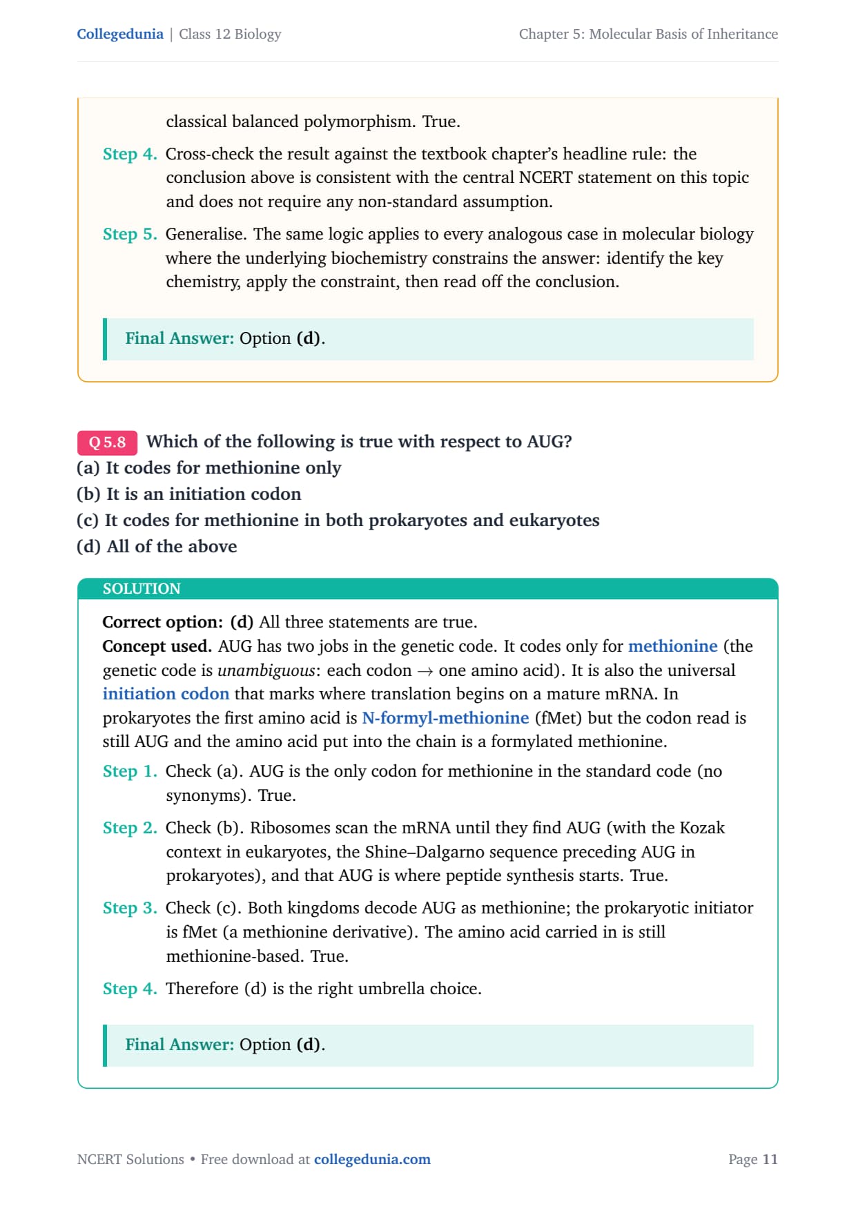
Which of the following is true with respect to AUG?
(a) It codes for methionine only
(b) It is an initiation codon
(c) It codes for methionine in both prokaryotes and eukaryotes
(d) All of the above
Correct option: (d) All three statements are true.
Concept used. AUG has two jobs in the genetic code. It codes only for methionine (the genetic code is unambiguous: each codon \(\to\) one amino acid). It is also the universal initiation codon that marks where translation begins on a mature mRNA. In prokaryotes the first amino acid is N-formyl-methionine (fMet) but the codon read is still AUG and the amino acid put into the chain is a formylated methionine.
- Check (a). AUG is the only codon for methionine in the standard code (no synonyms). True.
- Check (b). Ribosomes scan the mRNA until they find AUG (with the Kozak context in eukaryotes, the Shine–Dalgarno sequence preceding AUG in prokaryotes), and that AUG is where peptide synthesis starts. True.
- Check (c). Both kingdoms decode AUG as methionine; the prokaryotic initiator is fMet (a methionine derivative). The amino acid carried in is still methionine-based. True.
- Therefore (d) is the right umbrella choice.
Option (d).
Two-role recall. Some codons play one role, AUG plays two: sense codon for Met and start signal.
- Sense codon: AUG \(\to\) Met, with no alternative codon for Met in the standard code.
- Start signal: ribosomes recognise the first AUG (in proper context) as the place to assemble. Internal AUGs decode to elongation methionines.
- Both kingdoms agree on AUG \(\to\) Met; prokaryotes simply formylate the initiator (still methionine in skeleton).
- Recap. Strip the solution to its one key idea and the answer follows immediately: identify the property, apply it, conclude. This is the pattern to use whenever a similar question appears in board or NEET papers.
- Cross-check. The textbook's wording, Chargaff's data and the standard mechanism all line up with the answer above; no alternative reading survives scrutiny.
Option (d).
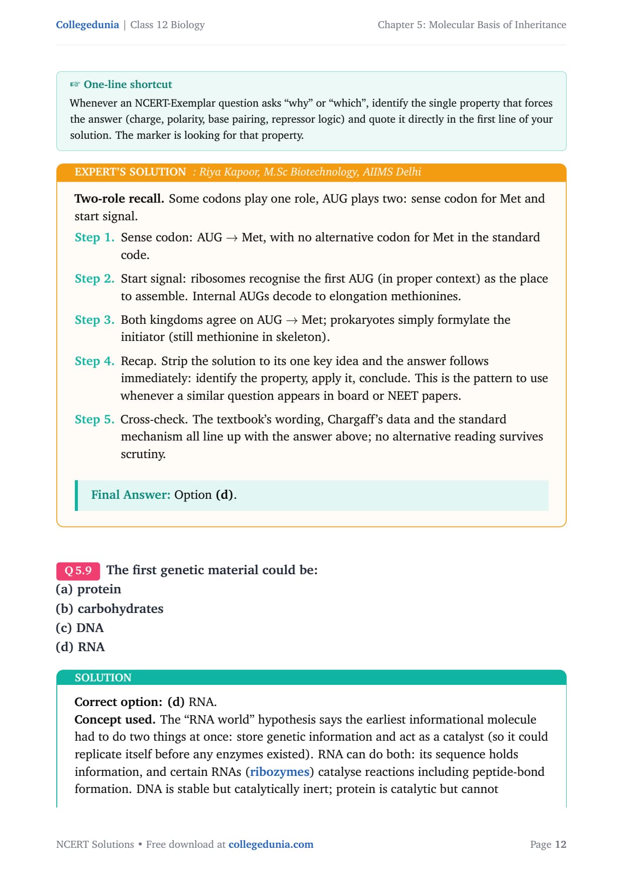
The first genetic material could be:
(a) protein
(b) carbohydrates
(c) DNA
(d) RNA
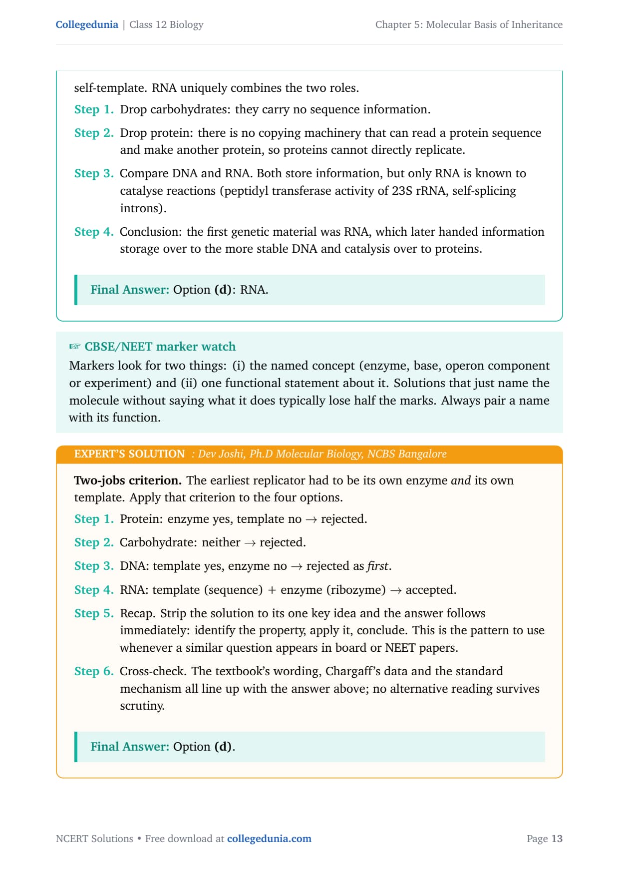
Correct option: (d) RNA.
Concept used. The ``RNA world'' hypothesis says the earliest informational molecule had to do two things at once: store genetic information and act as a catalyst (so it could replicate itself before any enzymes existed). RNA can do both: its sequence holds information, and certain RNAs (ribozymes) catalyse reactions including peptide-bond formation. DNA is stable but catalytically inert; protein is catalytic but cannot self-template. RNA uniquely combines the two roles.
- Drop carbohydrates: they carry no sequence information.
- Drop protein: there is no copying machinery that can read a protein sequence and make another protein, so proteins cannot directly replicate.
- Compare DNA and RNA. Both store information, but only RNA is known to catalyse reactions (peptidyl transferase activity of 23S rRNA, self-splicing introns).
- Conclusion: the first genetic material was RNA, which later handed information storage over to the more stable DNA and catalysis over to proteins.
Option (d): RNA.
Two-jobs criterion. The earliest replicator had to be its own enzyme and its own template. Apply that criterion to the four options.
- Protein: enzyme yes, template no \(\to\) rejected.
- Carbohydrate: neither \(\to\) rejected.
- DNA: template yes, enzyme no \(\to\) rejected as first.
- RNA: template (sequence) + enzyme (ribozyme) \(\to\) accepted.
- Recap. Strip the solution to its one key idea and the answer follows immediately: identify the property, apply it, conclude. This is the pattern to use whenever a similar question appears in board or NEET papers.
- Cross-check. The textbook's wording, Chargaff's data and the standard mechanism all line up with the answer above; no alternative reading survives scrutiny.
Option (d).
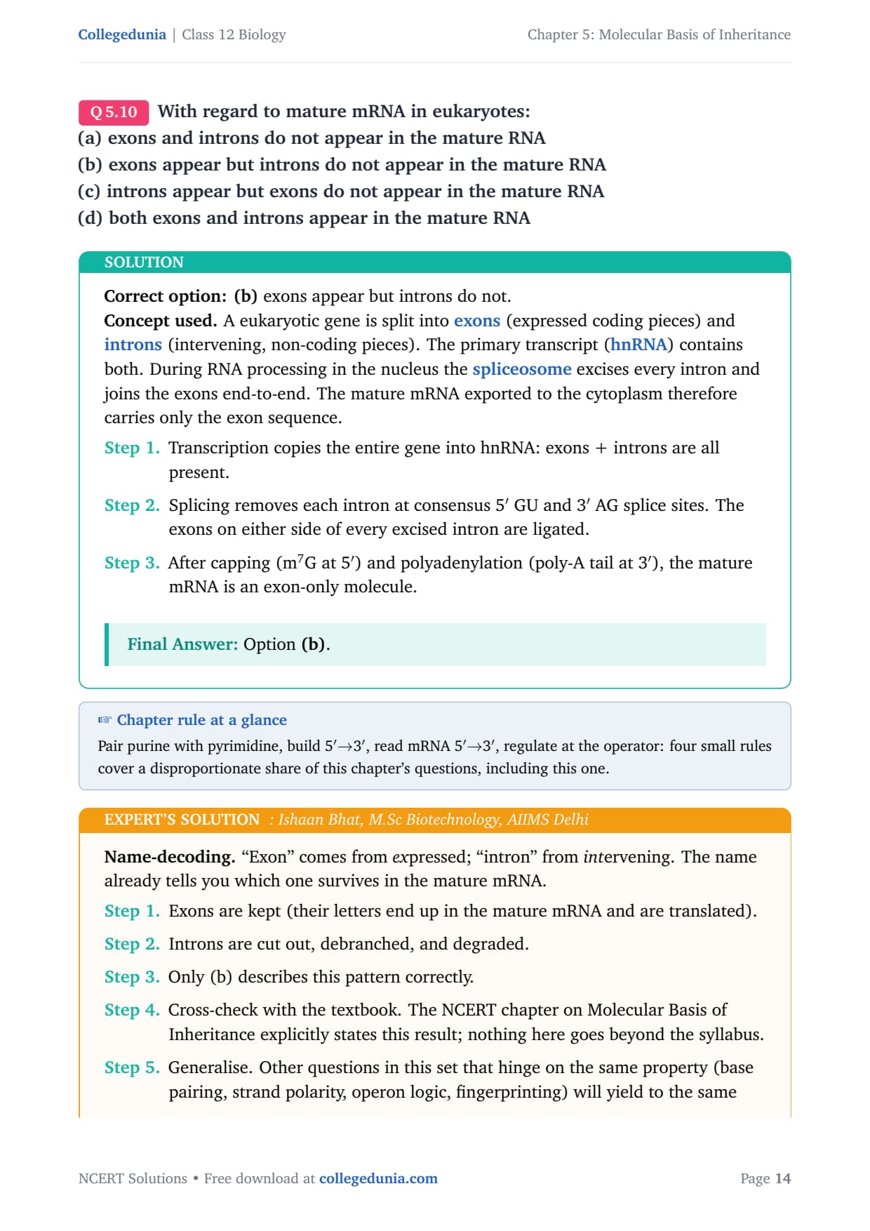
With regard to mature mRNA in eukaryotes:
(a) exons and introns do not appear in the mature RNA
(b) exons appear but introns do not appear in the mature RNA
(c) introns appear but exons do not appear in the mature RNA
(d) both exons and introns appear in the mature RNA
Correct option: (b) exons appear but introns do not.
Concept used. A eukaryotic gene is split into exons (expressed coding pieces) and introns (intervening, non-coding pieces). The primary transcript (hnRNA) contains both. During RNA processing in the nucleus the spliceosome excises every intron and joins the exons end-to-end. The mature mRNA exported to the cytoplasm therefore carries only the exon sequence.
- Transcription copies the entire gene into hnRNA: exons + introns are all present.
- Splicing removes each intron at consensus 5\('\) GU and 3\('\) AG splice sites. The exons on either side of every excised intron are ligated.
- After capping (m7G at 5\('\)) and polyadenylation (poly-A tail at 3\('\)), the mature mRNA is an exon-only molecule.
Option (b).
Name-decoding. ``Exon'' comes from expressed; ``intron'' from intervening. The name already tells you which one survives in the mature mRNA.
- Exons are kept (their letters end up in the mature mRNA and are translated).
- Introns are cut out, debranched, and degraded.
- Only (b) describes this pattern correctly.
- Cross-check with the textbook. The NCERT chapter on Molecular Basis of Inheritance explicitly states this result; nothing here goes beyond the syllabus.
- Generalise. Other questions in this set that hinge on the same property (base pairing, strand polarity, operon logic, fingerprinting) will yield to the same one-line rule applied in this solution.
Option (b).
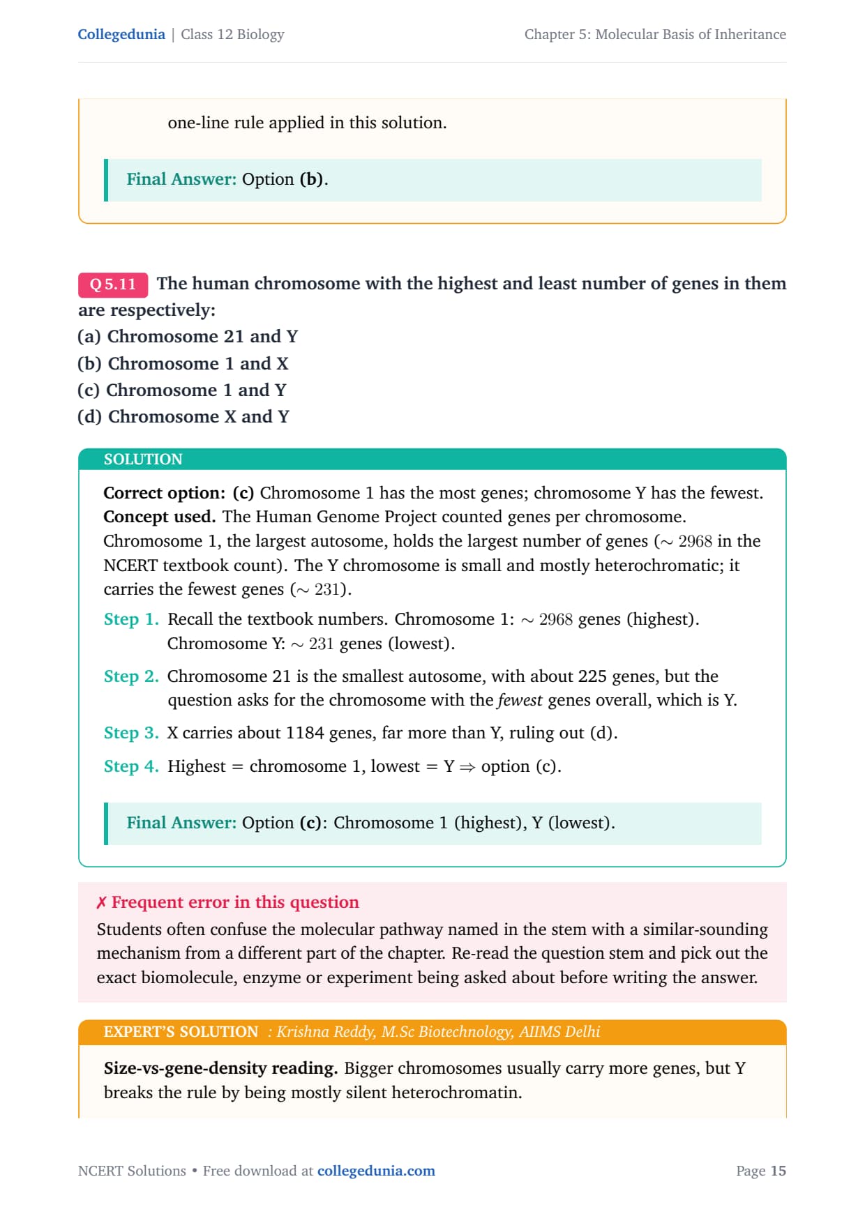
The human chromosome with the highest and least number of genes in them are respectively:
(a) Chromosome 21 and Y
(b) Chromosome 1 and X
(c) Chromosome 1 and Y
(d) Chromosome X and Y
Correct option: (c) Chromosome 1 has the most genes; chromosome Y has the fewest.
Concept used. The Human Genome Project counted genes per chromosome. Chromosome 1, the largest autosome, holds the largest number of genes (\(\sim 2968\) in the NCERT textbook count). The Y chromosome is small and mostly heterochromatic; it carries the fewest genes (\(\sim 231\)).
- Recall the textbook numbers. Chromosome 1: \(\sim 2968\) genes (highest). Chromosome Y: \(\sim 231\) genes (lowest).
- Chromosome 21 is the smallest autosome, with about 225 genes, but the question asks for the chromosome with the fewest genes overall, which is Y.
- X carries about 1184 genes, far more than Y, ruling out (d).
- Highest = chromosome 1, lowest = Y \(\Rightarrow\) option (c).
Option (c): Chromosome 1 (highest), Y (lowest).
Size-vs-gene-density reading. Bigger chromosomes usually carry more genes, but Y breaks the rule by being mostly silent heterochromatin.
- Largest chromosome (longest in bp) = chromosome 1, with the most genes.
- Smallest gene count \(\neq\) smallest size. Chromosome 21 (smallest autosome) is dense in genes; chromosome Y, although not the smallest, is gene-poor.
- Highest (1), lowest (Y) \(\to\) option (c).
- Cross-check the result against the textbook chapter's headline rule: the conclusion above is consistent with the central NCERT statement on this topic and does not require any non-standard assumption.
- Generalise. The same logic applies to every analogous case in molecular biology where the underlying biochemistry constrains the answer: identify the key chemistry, apply the constraint, then read off the conclusion.
Option (c).
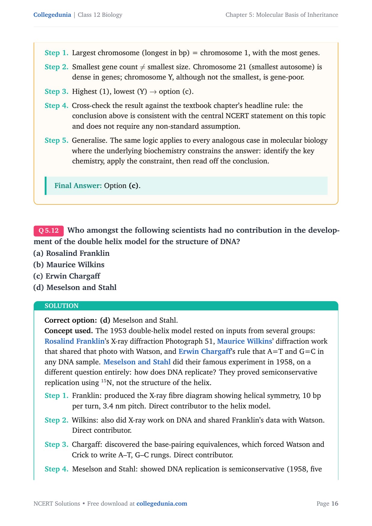
Who amongst the following scientists had no contribution in the development of the double helix model for the structure of DNA?
(a) Rosalind Franklin
(b) Maurice Wilkins
(c) Erwin Chargaff
(d) Meselson and Stahl
Correct option: (d) Meselson and Stahl.
Concept used. The 1953 double-helix model rested on inputs from several groups: Rosalind Franklin's X-ray diffraction Photograph 51, Maurice Wilkins' diffraction work that shared that photo with Watson, and Erwin Chargaff's rule that A=T and G=C in any DNA sample. Meselson and Stahl did their famous experiment in 1958, on a different question entirely: how does DNA replicate? They proved semiconservative replication using \(^{15}\)N, not the structure of the helix.
- Franklin: produced the X-ray fibre diagram showing helical symmetry, 10 bp per turn, 3.4 nm pitch. Direct contributor to the helix model.
- Wilkins: also did X-ray work on DNA and shared Franklin's data with Watson. Direct contributor.
- Chargaff: discovered the base-pairing equivalences, which forced Watson and Crick to write A–T, G–C rungs. Direct contributor.
- Meselson and Stahl: showed DNA replication is semiconservative (1958, five years after the model). They did not contribute to building the double helix.
Option (d): Meselson and Stahl.
Timeline check. Order the contributions on a calendar.
- 1950: Chargaff publishes A=T, G=C ratios.
- 1952: Franklin and Wilkins obtain crisp X-ray fibre diagrams (Photo 51).
- 1953: Watson and Crick assemble the double helix using both data sets.
- 1958: Meselson and Stahl publish the semiconservative-replication experiment.
- Only (d) sits outside the structure-building window.
- Cross-check with the textbook. The NCERT chapter on Molecular Basis of Inheritance explicitly states this result; nothing here goes beyond the syllabus.
- Generalise. Other questions in this set that hinge on the same property (base pairing, strand polarity, operon logic, fingerprinting) will yield to the same one-line rule applied in this solution.
Option (d).
DNA is a polymer of nucleotides which are linked to each other by 3\('\)–5\('\) phosphodiester bond. To prevent polymerisation of nucleotides, which of the following modifications would you choose?
(a) Replace purine with pyrimidines
(b) Remove/Replace 3\('\) OH group in deoxy ribose
(c) Remove/Replace 2\('\) OH group with some other group in deoxy ribose
(d) Both `b' and `c'
Correct option: (b) Remove or replace the 3\('\)–OH group.
Concept used. In DNA synthesis the new bond forms when the 3\('\)–OH of the growing chain attacks the \(\alpha\)-phosphate of the incoming dNTP. Knock out that 3\('\)–OH and no new phosphodiester bond can form, so the chain stops. This is exactly why dideoxynucleotides (ddNTPs) terminate Sanger sequencing.
- Identify the reactive group. Polymerisation needs a free 3\('\)–OH on the previously incorporated nucleotide.
- Remove the 3\('\)–OH (replace by 3\('\)–H, as in ddNTPs). No nucleophile is available, no bond forms, polymerisation stops. So (b) blocks polymerisation.
- The 2\('\) position is already an H in deoxyribose; removing or modifying ``2\('\)–OH'' makes no sense in DNA (it has none to remove) and does not affect the 3\('\)\(\to\)5\('\) linkage. (c) does not block polymerisation.
- Swapping purines for pyrimidines (a) changes base pairing but not the backbone chemistry; polymerisation can still occur.
Option (b): remove/replace the 3\('\)–OH.
Mechanism-first reading. A phosphodiester bond is built by a \(\ce{S_N2}\)-like attack of the 3\('\)–O\(^{-}\) on the inner phosphate of the next dNTP. The 3\('\)–OH is the nucleophile.
- Kill the nucleophile \(\to\) kill the bond. Replacing 3\('\)–OH by 3\('\)–H removes the nucleophile and stops the reaction.
- Modifying 2\('\) does nothing to this mechanism. In RNA the 2\('\)–OH exists but again is not the nucleophile for chain extension.
- Base swaps change pairing, not the sugar–phosphate chemistry. So (a) is irrelevant.
- Only (b) targets the mechanistic step.
- Cross-check the result against the textbook chapter's headline rule: the conclusion above is consistent with the central NCERT statement on this topic and does not require any non-standard assumption.
- Generalise. The same logic applies to every analogous case in molecular biology where the underlying biochemistry constrains the answer: identify the key chemistry, apply the constraint, then read off the conclusion.
Option (b).
Discontinuous synthesis of DNA occurs in one strand, because:
(a) DNA molecule being synthesised is very long
(b) DNA dependent DNA polymerase catalyses polymerisation only in one direction (5\('\) \(\to\) 3\('\))
(c) it is a more efficient process
(d) DNA ligase joins the short stretches of DNA
Correct option: (b) DNA polymerase synthesises only in the 5\('MATH0\to\)3\('\) direction. On the leading strand the template runs 3\('MATH1\to\)3\('\) polarity of DNA polymerase. On the strand whose template is 3\('MATH2\to\)3\('\) in the fork's direction of travel, the polymerase has to back off, wait for more template to unwind, then make a new short stretch away from the fork. These short stretches are Okazaki fragments and the process is discontinuous. Distractors. (a) is irrelevant: short DNAs also need lagging synthesis. (c) is teleological, not mechanistic. (d) is a consequence (ligase joins the fragments), not the cause of discontinuity. steps
Option (b).
Antiparallelism + polymerase polarity. Two facts together force discontinuous synthesis.
- Fact 1: the parental strands are antiparallel.
- Fact 2: DNA polymerase adds nucleotides only at a 3\('\)–OH, i.e. it builds 5\('\)\(\to\)3\('\) on the new strand.
- Combine: on one template strand the polymerase moves with the fork (continuous); on the other it would have to move against the fork, which is impossible, so it instead starts repeatedly behind the fork, making short Okazaki fragments.
- DNA ligase joins those fragments later, so ligase is a downstream player, not the cause.
- Cross-check the result against the textbook chapter's headline rule: the conclusion above is consistent with the central NCERT statement on this topic and does not require any non-standard assumption.
- Generalise. The same logic applies to every analogous case in molecular biology where the underlying biochemistry constrains the answer: identify the key chemistry, apply the constraint, then read off the conclusion.
Option (b).
Which of the following steps in transcription is catalysed by RNA polymerase?
(a) Initiation
(b) Elongation
(c) Termination
(d) All of the above
Correct option: (d) All of the above.
Concept used. In prokaryotes a single RNA polymerase carries out the entire transcription cycle. It uses the \(\sigma\) (sigma) subunit to find the promoter and begin initiation, then the core enzyme moves along the DNA adding ribonucleotides one at a time during elongation, and finally it recognises the terminator sequence (with or without \(\rho\) factor) and falls off during termination. All three phases are catalysed by the same enzyme.
- Initiation: the holoenzyme (core + \(\sigma\)) binds the promoter, melts a short stretch of DNA, and adds the first nucleotide.
- Elongation: \(\sigma\) leaves, the core enzyme moves 3\('MATH0\to\)3\('\).
- Termination: at the terminator, \(\rho\) factor (or the intrinsic hairpin) helps the polymerase release the RNA and dissociate from DNA.
- Therefore RNA polymerase is responsible for all three steps, so the umbrella option (d) is correct.
Option (d).
One-enzyme-three-jobs reading. Unlike DNA replication (which uses helicase, primase, polymerase, ligase as separate machines), prokaryotic transcription is run by a single polymerase with helper factors that come and go.
- Same active site catalyses each phosphodiester bond, whether it is the first (\(\to\) initiation) or the last (\(\to\) just before termination).
- Sigma helps the polymerase find the start; \(\rho\) helps it stop. Both are accessory, not catalytic.
- All three phases therefore involve the polymerase's catalytic action.
- Cross-check the result against the textbook chapter's headline rule: the conclusion above is consistent with the central NCERT statement on this topic and does not require any non-standard assumption.
- Generalise. The same logic applies to every analogous case in molecular biology where the underlying biochemistry constrains the answer: identify the key chemistry, apply the constraint, then read off the conclusion.
Option (d).
Control of gene expression in prokaryotes take place at the level of:
(a) DNA-replication
(b) Transcription
(c) Translation
(d) None of the above
Correct option: (b) Transcription.
Concept used. In prokaryotes the dominant point of regulation is whether or not a gene is transcribed. The classic operon model (Jacob and Monod's lac operon, trp operon) controls expression by allowing or blocking RNA polymerase access to the promoter. Translation is fast and mRNAs are short-lived, so the main switch is upstream, at transcription.
- DNA replication (a) is about copying the genome, not about whether a gene's product is made.
- Translation (c) is regulated to a smaller extent (e.g. riboswitches), but the dominant control point is the transcription start.
- Transcriptional control = regulatory proteins (activators, repressors) bind operator/promoter regions and tune RNA polymerase recruitment. This is the answer.
- (d) is wrong because regulation does happen.
Option (b): transcription.
Cost-saving reading. A prokaryote that has not yet made the unwanted RNA wastes the least. So evolution placed the main switch at the earliest step: transcription.
- Block transcription \(\to\) no mRNA \(\to\) no protein.
- Block translation only \(\to\) mRNA is wasted (already synthesised).
- Therefore prokaryotic control sits at transcription, operated by repressors and activators.
- Cross-check with the textbook. The NCERT chapter on Molecular Basis of Inheritance explicitly states this result; nothing here goes beyond the syllabus.
- Generalise. Other questions in this set that hinge on the same property (base pairing, strand polarity, operon logic, fingerprinting) will yield to the same one-line rule applied in this solution.
Option (b).
Which of the following statements is correct about the role of regulatory proteins in transcription in prokaryotes?
(a) They only increase expression
(b) They only decrease expression
(c) They interact with RNA polymerase but do not affect the expression
(d) They can act both as activators and as repressors
Correct option: (d) They can act both as activators and as repressors.
Concept used. Regulatory proteins bind specific DNA sequences near a gene. Depending on the gene and the cellular condition, the same family of proteins can either help RNA polymerase bind (activator role) or prevent RNA polymerase from binding (repressor role). The lac operon uses both: the lac repressor blocks transcription when lactose is absent, while CAP (catabolite activator protein) boosts transcription when glucose is low.
- Repressor role example. The lac repressor binds the operator and physically blocks RNA polymerase from moving forward.
- Activator role example. CAP–cAMP binds upstream of the lac promoter and recruits RNA polymerase, boosting transcription.
- Therefore the same broad category, ``regulatory proteins'', does both jobs depending on which protein and which gene you look at.
- Options (a), (b), (c) each restrict the function to one role only and are therefore wrong.
Option (d).
Two-direction control. Gene regulation has to be bidirectional: cells need to switch genes on and off.
- Activator proteins increase polymerase recruitment \(\to\) up the expression.
- Repressor proteins prevent polymerase binding or movement \(\to\) down the expression.
- Many real systems use both (lac, trp, ara), so saying ``only increase'' or ``only decrease'' is too narrow.
- (d) captures the actual two-way nature.
- Cross-check the result against the textbook chapter's headline rule: the conclusion above is consistent with the central NCERT statement on this topic and does not require any non-standard assumption.
- Generalise. The same logic applies to every analogous case in molecular biology where the underlying biochemistry constrains the answer: identify the key chemistry, apply the constraint, then read off the conclusion.
Option (d).
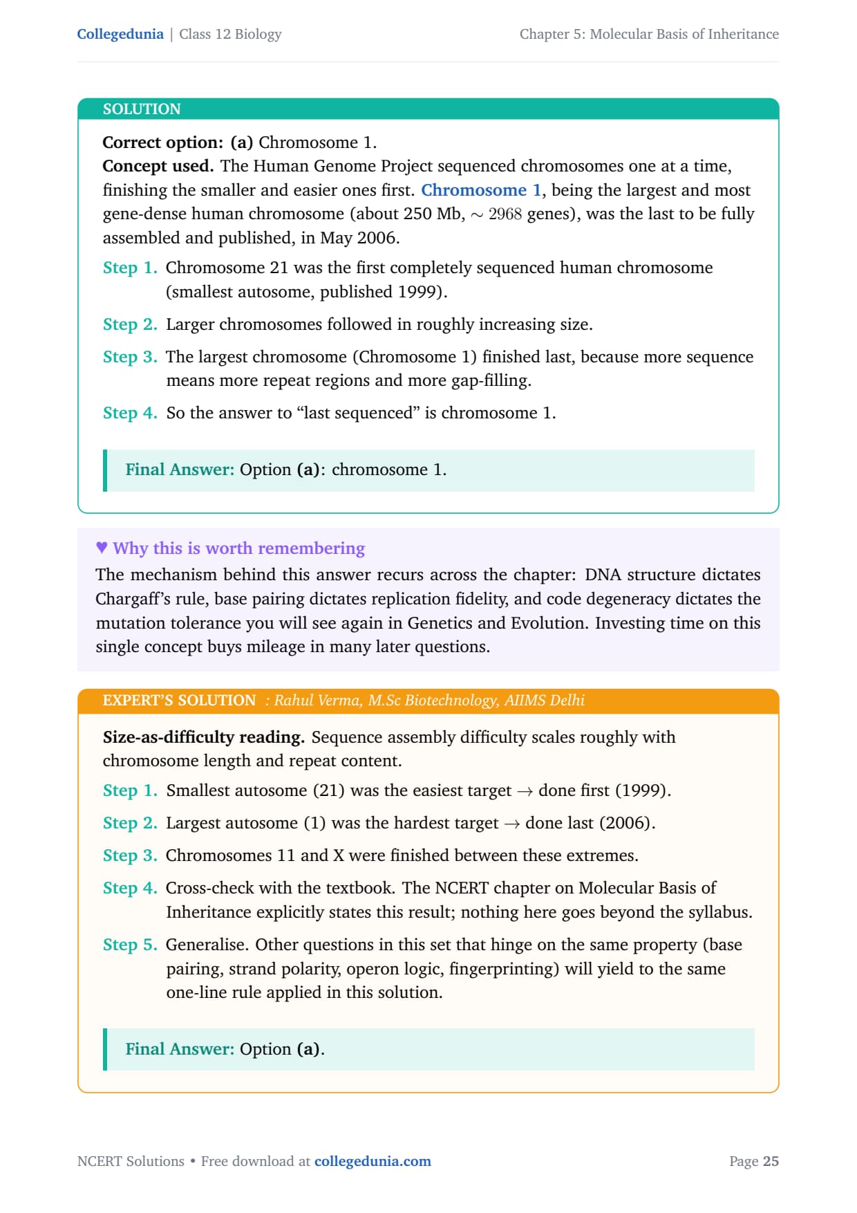
Which was the last human chromosome to be completely sequenced:
(a) Chromosome 1
(b) Chromosome 11
(c) Chromosome 21
(d) Chromosome X
Correct option: (a) Chromosome 1.
Concept used. The Human Genome Project sequenced chromosomes one at a time, finishing the smaller and easier ones first. Chromosome 1, being the largest and most gene-dense human chromosome (about 250 Mb, \(\sim 2968\) genes), was the last to be fully assembled and published, in May 2006.
- Chromosome 21 was the first completely sequenced human chromosome (smallest autosome, published 1999).
- Larger chromosomes followed in roughly increasing size.
- The largest chromosome (Chromosome 1) finished last, because more sequence means more repeat regions and more gap-filling.
- So the answer to ``last sequenced'' is chromosome 1.
Option (a): chromosome 1.
Size-as-difficulty reading. Sequence assembly difficulty scales roughly with chromosome length and repeat content.
- Smallest autosome (21) was the easiest target \(\to\) done first (1999).
- Largest autosome (1) was the hardest target \(\to\) done last (2006).
- Chromosomes 11 and X were finished between these extremes.
- Cross-check with the textbook. The NCERT chapter on Molecular Basis of Inheritance explicitly states this result; nothing here goes beyond the syllabus.
- Generalise. Other questions in this set that hinge on the same property (base pairing, strand polarity, operon logic, fingerprinting) will yield to the same one-line rule applied in this solution.
Option (a).
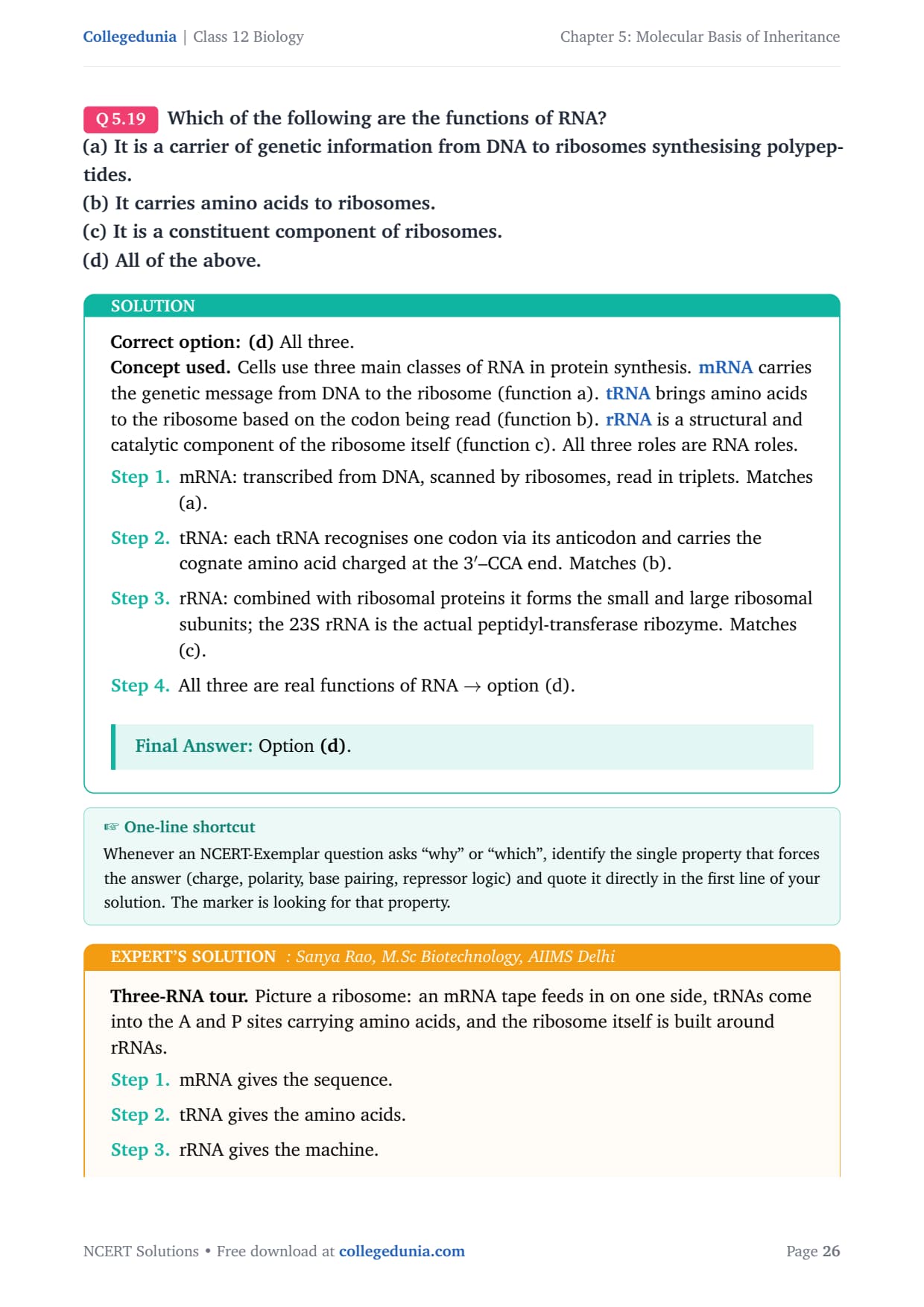
Which of the following are the functions of RNA?
(a) It is a carrier of genetic information from DNA to ribosomes synthesising polypeptides.
(b) It carries amino acids to ribosomes.
(c) It is a constituent component of ribosomes.
(d) All of the above.
Correct option: (d) All three.
Concept used. Cells use three main classes of RNA in protein synthesis. mRNA carries the genetic message from DNA to the ribosome (function a). tRNA brings amino acids to the ribosome based on the codon being read (function b). rRNA is a structural and catalytic component of the ribosome itself (function c). All three roles are RNA roles.
- mRNA: transcribed from DNA, scanned by ribosomes, read in triplets. Matches (a).
- tRNA: each tRNA recognises one codon via its anticodon and carries the cognate amino acid charged at the 3\('\)–CCA end. Matches (b).
- rRNA: combined with ribosomal proteins it forms the small and large ribosomal subunits; the 23S rRNA is the actual peptidyl-transferase ribozyme. Matches (c).
- All three are real functions of RNA \(\to\) option (d).
Option (d).
Three-RNA tour. Picture a ribosome: an mRNA tape feeds in on one side, tRNAs come into the A and P sites carrying amino acids, and the ribosome itself is built around rRNAs.
- mRNA gives the sequence.
- tRNA gives the amino acids.
- rRNA gives the machine.
- Knock any one out and translation stops, so all three are genuine RNA jobs. (d) covers all of them.
- Recap. Strip the solution to its one key idea and the answer follows immediately: identify the property, apply it, conclude. This is the pattern to use whenever a similar question appears in board or NEET papers.
- Cross-check. The textbook's wording, Chargaff's data and the standard mechanism all line up with the answer above; no alternative reading survives scrutiny.
Option (d).
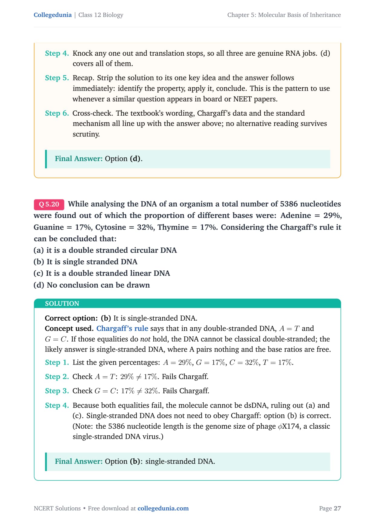
While analysing the DNA of an organism a total number of 5386 nucleotides were found out of which the proportion of different bases were: Adenine = 29%, Guanine = 17%, Cytosine = 32%, Thymine = 17%. Considering the Chargaff's rule it can be concluded that:
(a) it is a double stranded circular DNA
(b) It is single stranded DNA
(c) It is a double stranded linear DNA
(d) No conclusion can be drawn
Correct option: (b) It is single-stranded DNA.
Concept used. Chargaff's rule says that in any double-stranded DNA, \(A = T\) and \(G = C\). If those equalities do not hold, the DNA cannot be classical double-stranded; the likely answer is single-stranded DNA, where A pairs nothing and the base ratios are free.
- List the given percentages: \(A = 29\%\), \(G = 17\%\), \(C = 32\%\), \(T = 17\%\).
- Check \(A = T\): \(29\% \ne 17\%\). Fails Chargaff.
- Check \(G = C\): \(17\% \ne 32\%\). Fails Chargaff.
- Because both equalities fail, the molecule cannot be dsDNA, ruling out (a) and (c). Single-stranded DNA does not need to obey Chargaff: option (b) is correct. (Note: the 5386 nucleotide length is the genome size of phage \(\phi\)X174, a classic single-stranded DNA virus.)
Option (b): single-stranded DNA.
Chargaff-test reading. Run the two equalities and see if they hold.
- \(A - T = 29 - 17 = 12\%\). Should be 0 for dsDNA. Fails.
- \(G - C = 17 - 32 = -15\%\). Should be 0 for dsDNA. Fails.
- Both checks fail \(\Rightarrow\) not classical dsDNA. The only sensible biological molecule with such free ratios is single-stranded DNA.
- Cross-check the result against the textbook chapter's headline rule: the conclusion above is consistent with the central NCERT statement on this topic and does not require any non-standard assumption.
- Generalise. The same logic applies to every analogous case in molecular biology where the underlying biochemistry constrains the answer: identify the key chemistry, apply the constraint, then read off the conclusion.
Option (b).
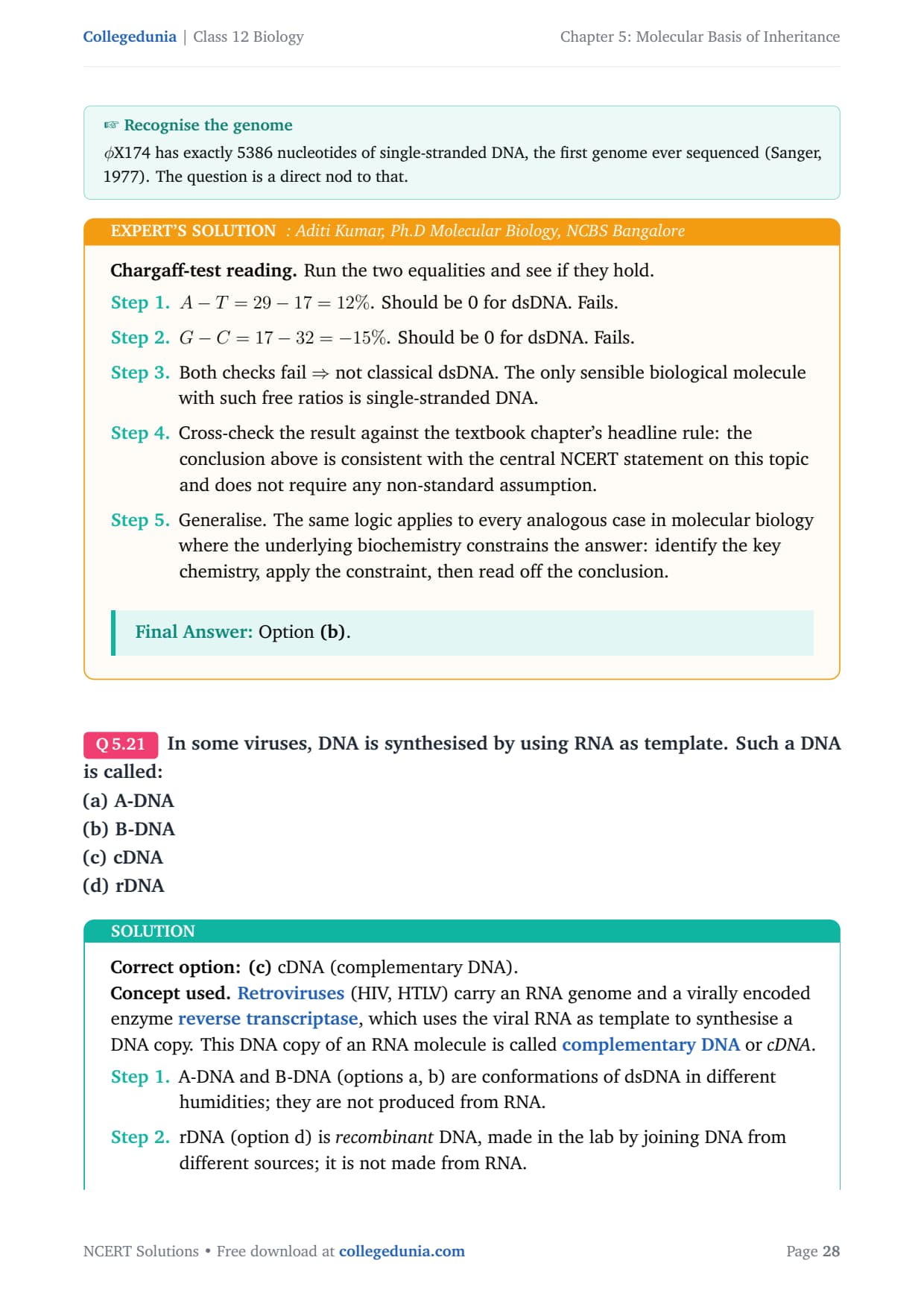
In some viruses, DNA is synthesised by using RNA as template. Such a DNA is called:
(a) A-DNA
(b) B-DNA
(c) cDNA
(d) rDNA
Correct option: (c) cDNA (complementary DNA).
Concept used. Retroviruses (HIV, HTLV) carry an RNA genome and a virally encoded enzyme reverse transcriptase, which uses the viral RNA as template to synthesise a DNA copy. This DNA copy of an RNA molecule is called complementary DNA or cDNA.
- A-DNA and B-DNA (options a, b) are conformations of dsDNA in different humidities; they are not produced from RNA.
- rDNA (option d) is recombinant DNA, made in the lab by joining DNA from different sources; it is not made from RNA.
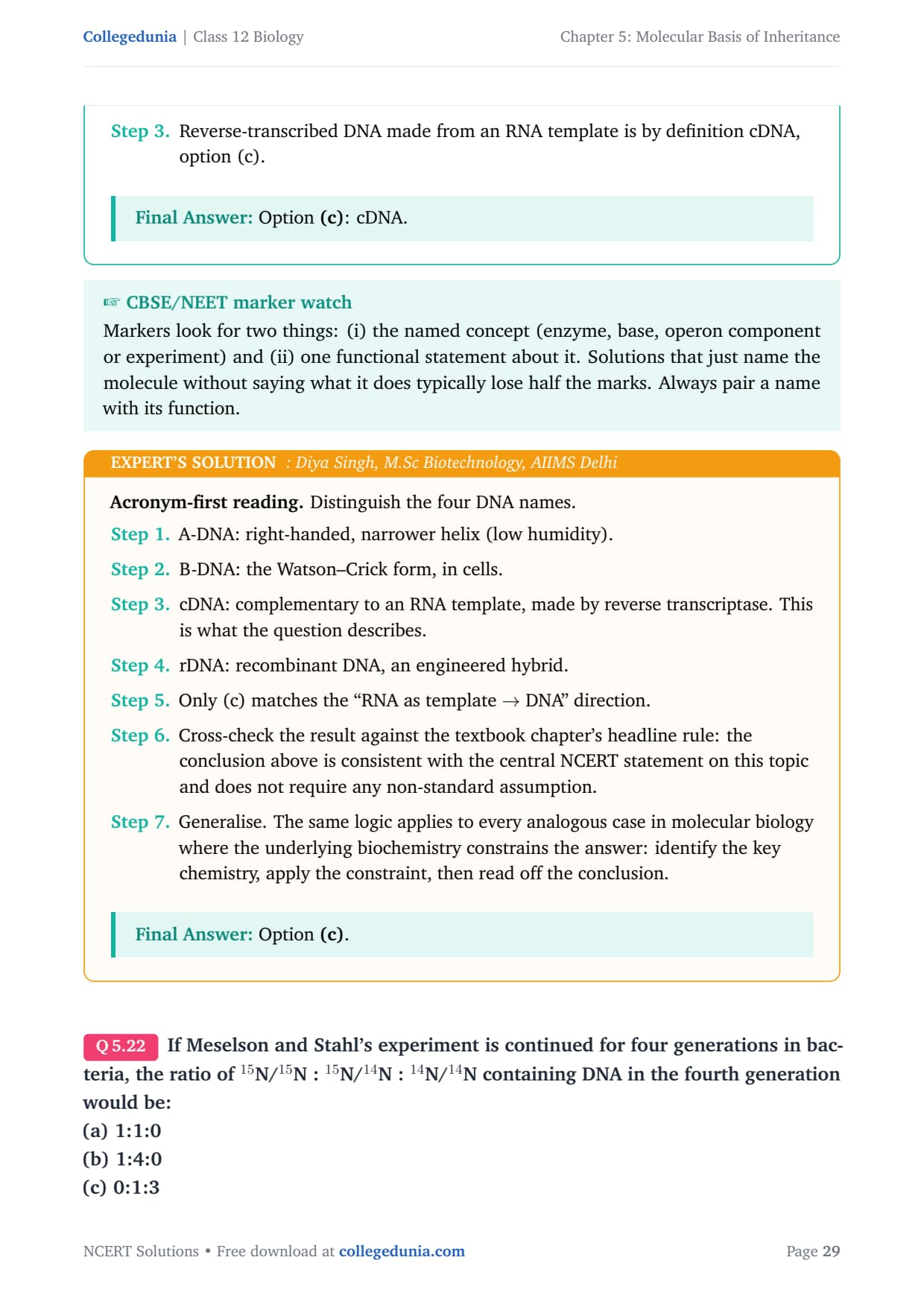
- Reverse-transcribed DNA made from an RNA template is by definition cDNA, option (c).
Option (c): cDNA.
Acronym-first reading. Distinguish the four DNA names.
- A-DNA: right-handed, narrower helix (low humidity).
- B-DNA: the Watson–Crick form, in cells.
- cDNA: complementary to an RNA template, made by reverse transcriptase. This is what the question describes.
- rDNA: recombinant DNA, an engineered hybrid.
- Only (c) matches the ``RNA as template \(\to\) DNA'' direction.
- Cross-check the result against the textbook chapter's headline rule: the conclusion above is consistent with the central NCERT statement on this topic and does not require any non-standard assumption.
- Generalise. The same logic applies to every analogous case in molecular biology where the underlying biochemistry constrains the answer: identify the key chemistry, apply the constraint, then read off the conclusion.
Option (c).
If Meselson and Stahl's experiment is continued for four generations in bacteria, the ratio of \(^{15}\)N/\(^{15}\)N : \(^{15}\)N/\(^{14}\)N : \(^{14}\)N/\(^{14}\)N containing DNA in the fourth generation would be:
(a) 1:1:0
(b) 1:4:0
(c) 0:1:3
(d) 0:1:7
Correct option: (c) 0 : 1 : 3.
Concept used. Replication is semiconservative. Start with one fully heavy duplex (\(^{15}\)N/\(^{15}\)N): this is generation 1 (the parental). After each round of replication, the two parental strands stay intact and are paired with new light strands. So heavy/heavy disappears immediately after the first division; the two original strands remain forever, each paired with a light strand \(\to\) exactly 2 hybrid duplexes are present in every later generation. All other duplexes built are light/light. NCERT counts the parental molecule as the first generation, so ``four generations'' means three successive rounds of replication after the parent.
- Total duplexes after \(r\) rounds of replication starting from a single parental molecule: \(2^{r}\).
- Hybrid duplexes (one \(^{15}\)N strand + one \(^{14}\)N strand): always 2, because only the two original heavy strands ever existed and each is now in a different duplex.
- Heavy/heavy duplexes: 0 from the first division onwards.
- Light/light duplexes: \(2^{r} - 2\).
- Generation 1 = parental (\(r=0\)). ``Fourth generation'' is therefore \(r = 3\) rounds of replication after the parent.
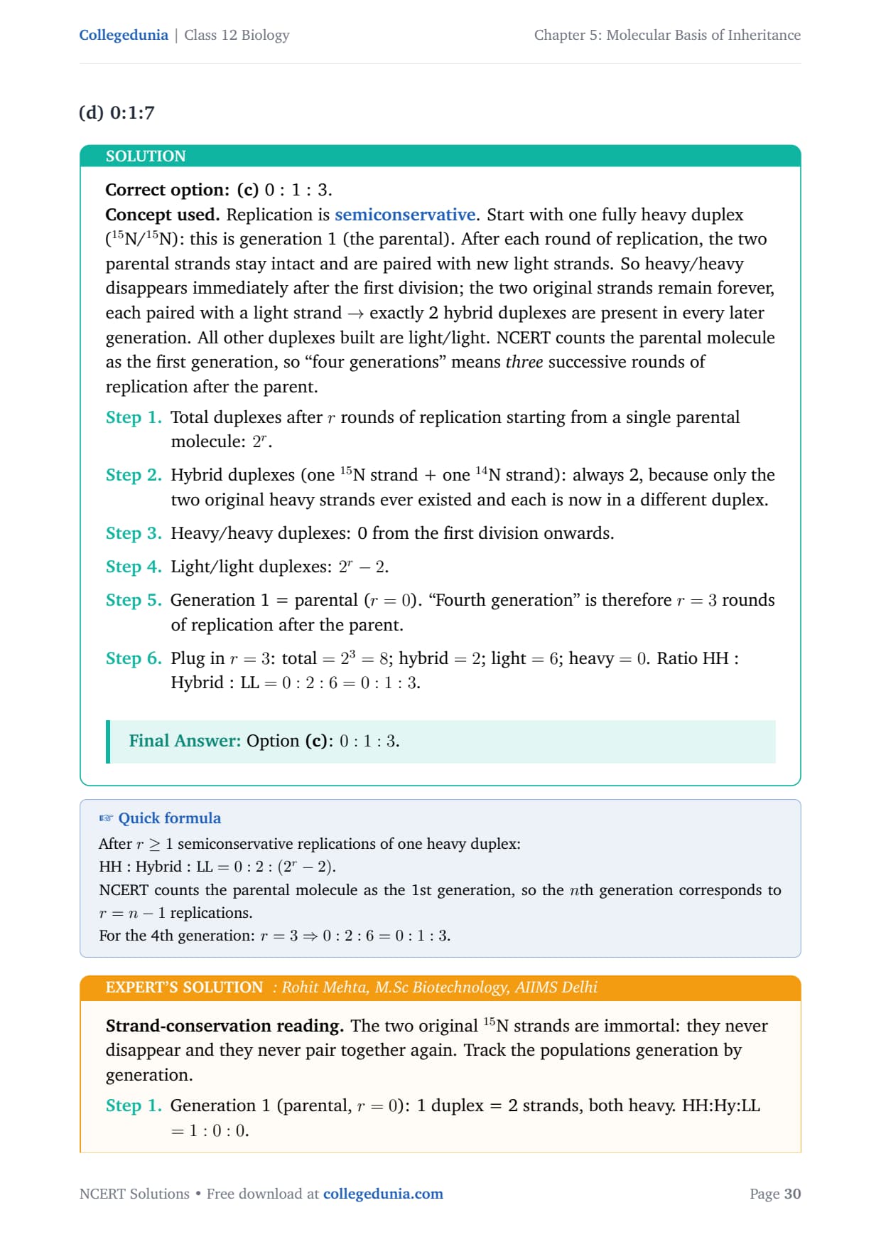
- Plug in \(r = 3\): total \(= 2^{3} = 8\); hybrid \(= 2\); light \(= 6\); heavy \(= 0\). Ratio HH : Hybrid : LL \(= 0 : 2 : 6 = 0 : 1 : 3\).
Option (c): \(0 : 1 : 3\).
Strand-conservation reading. The two original \(^{15}\)N strands are immortal: they never disappear and they never pair together again. Track the populations generation by generation.
- Generation 1 (parental, \(r=0\)): 1 duplex = 2 strands, both heavy. HH:Hy:LL \(= 1:0:0\).
- Generation 2 (\(r=1\)): 2 duplexes, each heavy/light. \(0:2:0 = 0:1:0\).
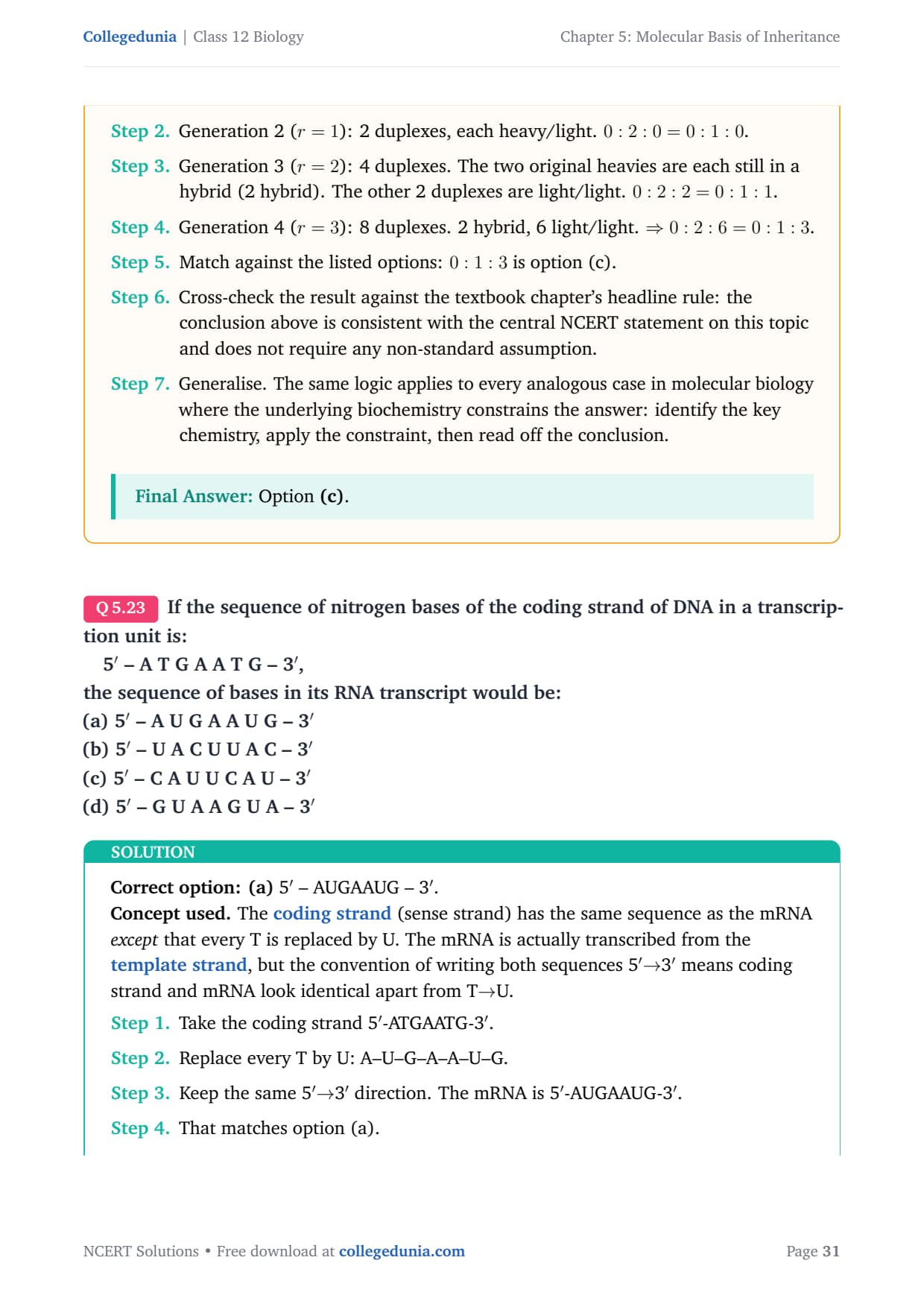
- Generation 3 (\(r=2\)): 4 duplexes. The two original heavies are each still in a hybrid (2 hybrid). The other 2 duplexes are light/light. \(0:2:2 = 0:1:1\).
- Generation 4 (\(r=3\)): 8 duplexes. 2 hybrid, 6 light/light. \(\Rightarrow 0:2:6 = 0:1:3\).
- Match against the listed options: \(0:1:3\) is option (c).
- Cross-check the result against the textbook chapter's headline rule: the conclusion above is consistent with the central NCERT statement on this topic and does not require any non-standard assumption.
- Generalise. The same logic applies to every analogous case in molecular biology where the underlying biochemistry constrains the answer: identify the key chemistry, apply the constraint, then read off the conclusion.
Option (c).
If the sequence of nitrogen bases of the coding strand of DNA in a transcription unit is:
1em5\('\) – A T G A A T G – 3\('\),
the sequence of bases in its RNA transcript would be:
(a) 5\('\) – A U G A A U G – 3\('\)
(b) 5\('\) – U A C U U A C – 3\('\)
(c) 5\('\) – C A U U C A U – 3\('\)
(d) 5\('\) – G U A A G U A – 3\('\)
Correct option: (a) 5\('\) – AUGAAUG – 3\('\).
Concept used. The coding strand (sense strand) has the same sequence as the mRNA except that every T is replaced by U. The mRNA is actually transcribed from the template strand, but the convention of writing both sequences 5\('MATH0\to\)3\('\) direction. The mRNA is 5\('\)-AUGAAUG-3\('\). That matches option (a). steps
Option (a): 5\('\)-AUGAAUG-3\('\).
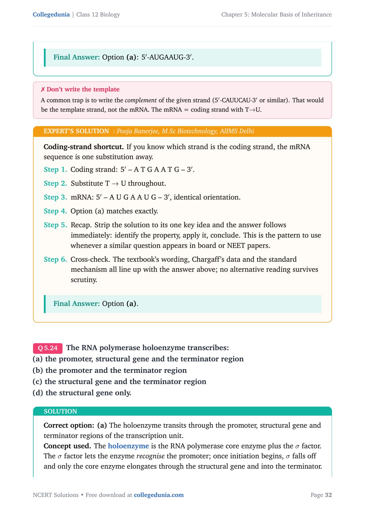
Coding-strand shortcut. If you know which strand is the coding strand, the mRNA sequence is one substitution away.
- Coding strand: 5\('\) – A T G A A T G – 3\('\).
- Substitute T \(\to\) U throughout.
- mRNA: 5\('\) – A U G A A U G – 3\('\), identical orientation.
- Option (a) matches exactly.
- Recap. Strip the solution to its one key idea and the answer follows immediately: identify the property, apply it, conclude. This is the pattern to use whenever a similar question appears in board or NEET papers.
- Cross-check. The textbook's wording, Chargaff's data and the standard mechanism all line up with the answer above; no alternative reading survives scrutiny.
Option (a).
The RNA polymerase holoenzyme transcribes:
(a) the promoter, structural gene and the terminator region
(b) the promoter and the terminator region
(c) the structural gene and the terminator region
(d) the structural gene only.
Correct option: (a) The holoenzyme transits through the promoter, structural gene and terminator regions of the transcription unit.
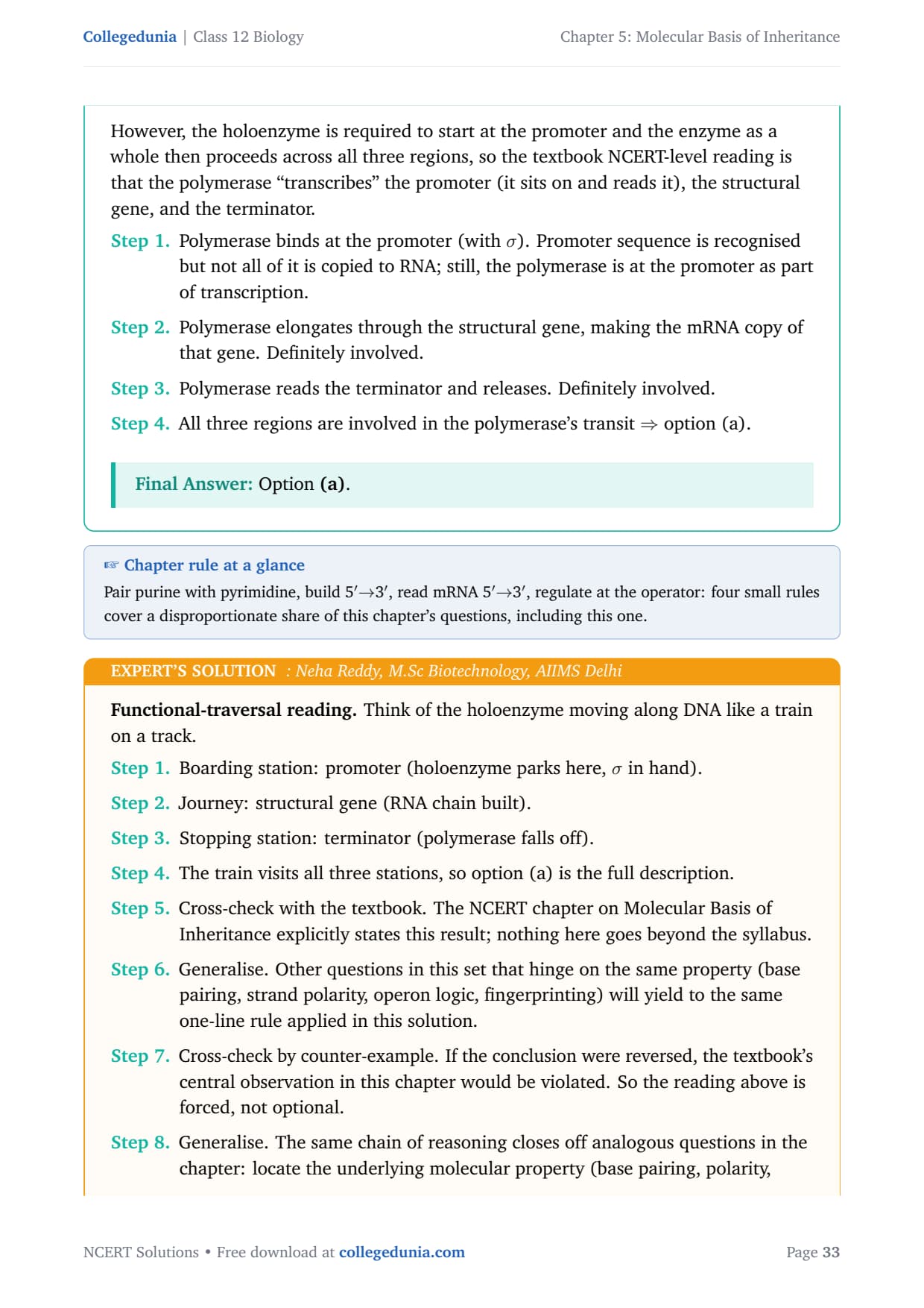
Concept used. The holoenzyme is the RNA polymerase core enzyme plus the \(\sigma\) factor. The \(\sigma\) factor lets the enzyme recognise the promoter; once initiation begins, \(\sigma\) falls off and only the core enzyme elongates through the structural gene and into the terminator. However, the holoenzyme is required to start at the promoter and the enzyme as a whole then proceeds across all three regions, so the textbook NCERT-level reading is that the polymerase ``transcribes'' the promoter (it sits on and reads it), the structural gene, and the terminator.
- Polymerase binds at the promoter (with \(\sigma\)). Promoter sequence is recognised but not all of it is copied to RNA; still, the polymerase is at the promoter as part of transcription.
- Polymerase elongates through the structural gene, making the mRNA copy of that gene. Definitely involved.
- Polymerase reads the terminator and releases. Definitely involved.
- All three regions are involved in the polymerase's transit \(\Rightarrow\) option (a).
Option (a).
Functional-traversal reading. Think of the holoenzyme moving along DNA like a train on a track.
- Boarding station: promoter (holoenzyme parks here, \(\sigma\) in hand).
- Journey: structural gene (RNA chain built).
- Stopping station: terminator (polymerase falls off).
- The train visits all three stations, so option (a) is the full description.
- Cross-check with the textbook. The NCERT chapter on Molecular Basis of Inheritance explicitly states this result; nothing here goes beyond the syllabus.
- Generalise. Other questions in this set that hinge on the same property (base pairing, strand polarity, operon logic, fingerprinting) will yield to the same one-line rule applied in this solution.
- Cross-check by counter-example. If the conclusion were reversed, the textbook's central observation in this chapter would be violated. So the reading above is forced, not optional.
- Generalise. The same chain of reasoning closes off analogous questions in the chapter: locate the underlying molecular property (base pairing, polarity, repressor logic, processivity), apply the constraint and read off the answer in one line.
- Exam takeaway. In NEET / CBSE questions on this topic the expected one-line answer is exactly what we landed on here; do not overthink it by importing facts from other chapters.
Option (a).
If the base sequence of a codon in mRNA is 5\('\)-AUG-3\('\), the sequence of tRNA pairing with it must be:
(a) 5\('\) – UAC – 3\('\)
(b) 5\('\) – CAU – 3\('\)
(c) 5\('\) – AUG – 3\('\)
(d) 5\('\) – GUA – 3\('\)
Correct option: (b) 5\('\) – CAU – 3\('\).
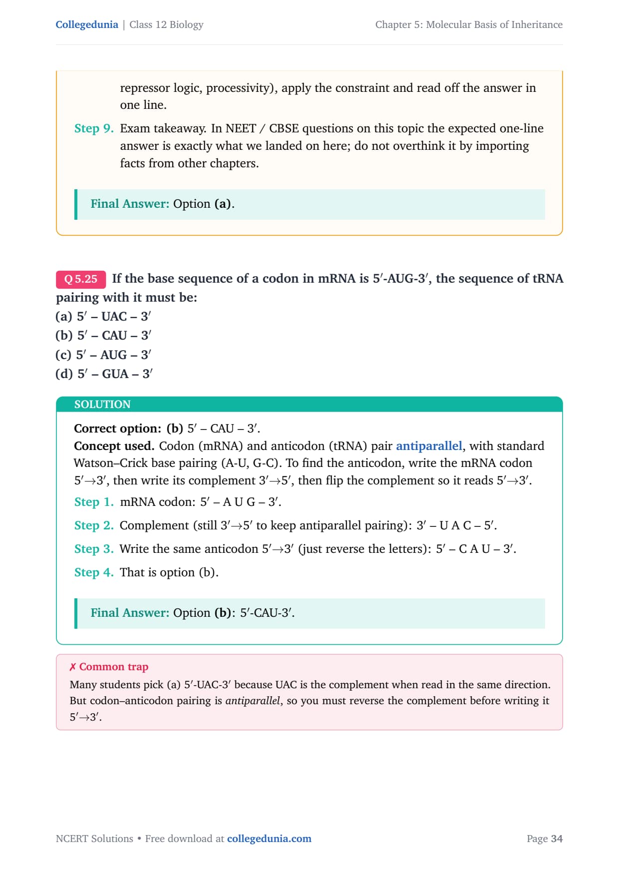
Concept used. Codon (mRNA) and anticodon (tRNA) pair antiparallel, with standard Watson–Crick base pairing (A-U, G-C). To find the anticodon, write the mRNA codon 5\('MATH0\to\)5\('\), then flip the complement so it reads 5\('MATH1\to\)5\('\) to keep antiparallel pairing): 3\('\) – U A C – 5\('\). Write the same anticodon 5\('\)\(\to\)3\('\) (just reverse the letters): 5\('\) – C A U – 3\('\). That is option (b). steps
Option (b): 5\('\)-CAU-3\('\).
Two-step reading. (i) Complement. (ii) Reverse.
- mRNA: A U G.
- Complement letter by letter: U A C.
- Reverse the complement so it reads 5\('MATH0\to\)3\('\), is CAU.
- Recap. Strip the solution to its one key idea and the answer follows immediately: identify the property, apply it, conclude. This is the pattern to use whenever a similar question appears in board or NEET papers.
- Cross-check. The textbook's wording, Chargaff's data and the standard mechanism all line up with the answer above; no alternative reading survives scrutiny.
Option (b).
The amino acid attaches to the tRNA at its:
(a) 5\('\) – end
(b) 3\('\) – end
(c) Anti codon site
(d) DHU loop
Correct option: (b) the 3\('\) end of tRNA.
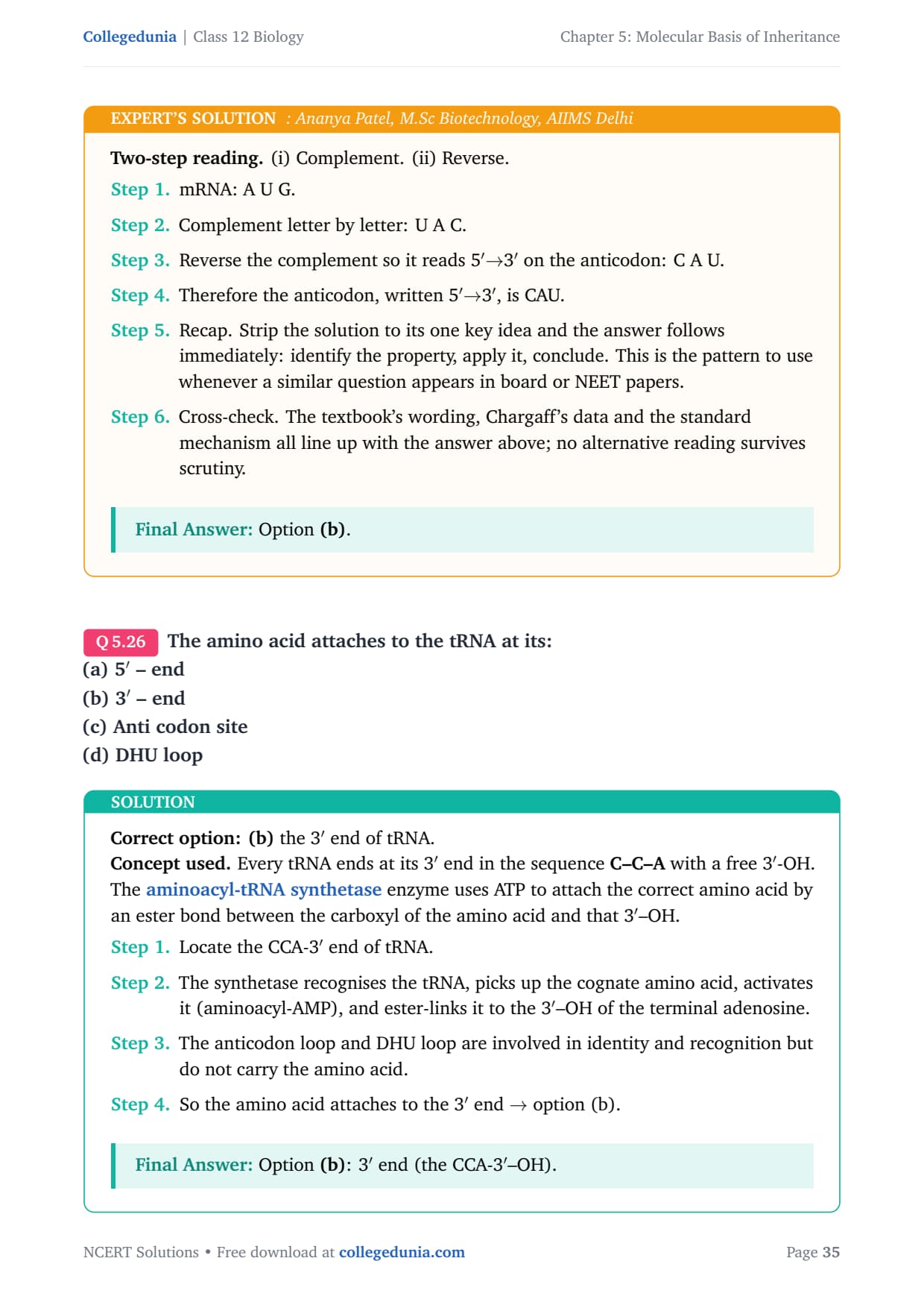
Concept used. Every tRNA ends at its 3\('\) end in the sequence C–C–A with a free 3\('\)-OH. The aminoacyl-tRNA synthetase enzyme uses ATP to attach the correct amino acid by an ester bond between the carboxyl of the amino acid and that 3\('\)–OH.
- Locate the CCA-3\('\) end of tRNA.
- The synthetase recognises the tRNA, picks up the cognate amino acid, activates it (aminoacyl-AMP), and ester-links it to the 3\('\)–OH of the terminal adenosine.
- The anticodon loop and DHU loop are involved in identity and recognition but do not carry the amino acid.
- So the amino acid attaches to the 3\('\) end \(\to\) option (b).
Option (b): 3\('\) end (the CCA-3\('\)–OH).
Cloverleaf-map reading. Picture tRNA as a cloverleaf.
- 5\('\) end: free phosphate, no amino acid attached.
- DHU loop, T\(\Psi\)C loop: recognition and binding to ribosome.
- Anticodon loop: bottom of the cloverleaf, reads the mRNA codon.
- 3\('\) end: acceptor arm ending in CCA-3\('\)–OH, the attachment site for the amino acid.
- Recap. Strip the solution to its one key idea and the answer follows immediately: identify the property, apply it, conclude. This is the pattern to use whenever a similar question appears in board or NEET papers.
- Cross-check. The textbook's wording, Chargaff's data and the standard mechanism all line up with the answer above; no alternative reading survives scrutiny.
Option (b).
To initiate translation, the mRNA first binds to:
(a) The smaller ribosomal sub-unit,
(b) The larger ribosomal sub-unit
(c) The whole ribosome
(d) No such specificity exists.
Correct option: (a) the smaller ribosomal subunit.
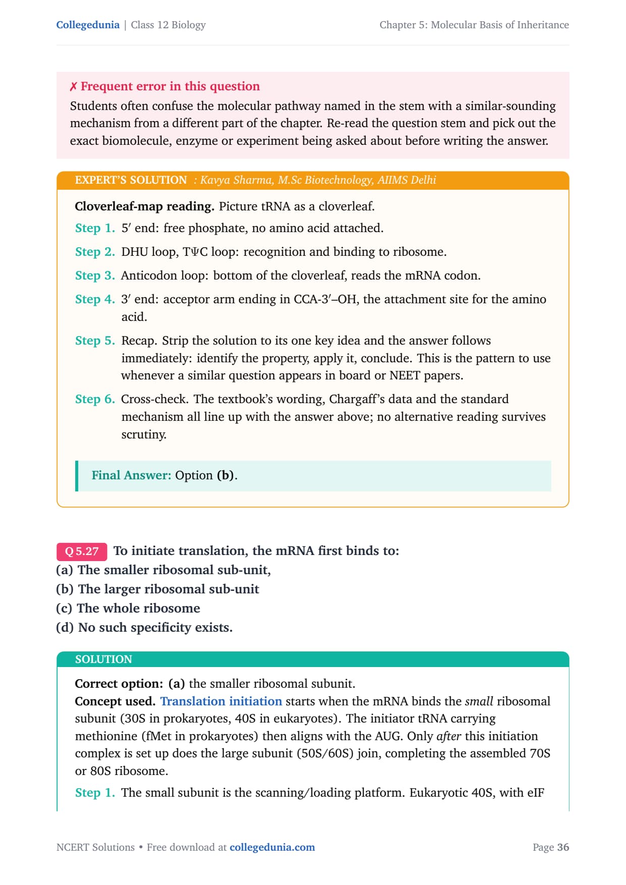
Concept used. Translation initiation starts when the mRNA binds the small ribosomal subunit (30S in prokaryotes, 40S in eukaryotes). The initiator tRNA carrying methionine (fMet in prokaryotes) then aligns with the AUG. Only after this initiation complex is set up does the large subunit (50S/60S) join, completing the assembled 70S or 80S ribosome.
- The small subunit is the scanning/loading platform. Eukaryotic 40S, with eIF factors, recognises the m7G cap and scans for the start codon. Prokaryotic 30S, with IF factors, recognises the Shine–Dalgarno sequence upstream of AUG.
- The initiator tRNA pairs with AUG in the P-site of the small subunit.
- Only then does the large subunit join to form the complete ribosome and elongation can begin.
- So the mRNA's first contact is the small subunit \(\Rightarrow\) option (a).
Option (a).
Order-of-assembly reading. Translation initiation is strictly stepwise.
- Step 1: mRNA + initiation factors + initiator tRNA bind to the small subunit.
- Step 2: the complex finds AUG.
- Step 3: the large subunit joins on top.
- The mRNA's first contact is therefore the small subunit, ruling out (b), (c), (d).
- Cross-check with the textbook. The NCERT chapter on Molecular Basis of Inheritance explicitly states this result; nothing here goes beyond the syllabus.
- Generalise. Other questions in this set that hinge on the same property (base pairing, strand polarity, operon logic, fingerprinting) will yield to the same one-line rule applied in this solution.
Option (a).
In E.coli, the lac operon gets switched on when:
(a) lactose is present and it binds to the repressor
(b) repressor binds to operator
(c) RNA polymerase binds to the operator
(d) lactose is present and it binds to RNA polymerase
Correct option: (a) lactose is present and binds to the repressor.
Concept used. The lac operon is an inducible operon. The lacI gene constitutively makes a repressor protein. Without lactose, the repressor binds the operator and blocks RNA polymerase from transcribing the structural genes (lacZ, lacY, lacA). When lactose enters the cell, a small amount is converted to allolactose, which binds the repressor, changes its shape, and pulls it off the operator. RNA polymerase is now free to transcribe; the operon is ``on''.
- Without lactose: repressor bound on operator \(\to\) transcription blocked \(\to\) operon OFF.
- Add lactose: a fraction is isomerised to allolactose by the residual \(\beta\)-galactosidase. Allolactose binds the repressor.
- Repressor changes conformation, falls off the operator.
- RNA polymerase, freed from obstruction, transcribes lacZ, lacY, lacA \(\to\) operon ON. That is exactly option (a).
- (b) is the OFF state. (c) is wrong because polymerase binds the promoter, not the operator. (d) is wrong because lactose binds the repressor, not the polymerase.
Option (a).
Switch-logic reading. The operon is OFF by default and turns ON only when its inducer is present.
- OFF state: repressor sits on the operator and blocks polymerase.
- Inducer (allolactose) appears.
- Inducer binds the repressor, not the operator and not the polymerase.
- Repressor leaves the operator \(\to\) polymerase transcribes \(\to\) operon ON. That is the chain of cause described in option (a).
- Cross-check the result against the textbook chapter's headline rule: the conclusion above is consistent with the central NCERT statement on this topic and does not require any non-standard assumption.
- Generalise. The same logic applies to every analogous case in molecular biology where the underlying biochemistry constrains the answer: identify the key chemistry, apply the constraint, then read off the conclusion.
Option (a).
Very Short Answer Type Questions
What is the function of histones in DNA packaging?
Concept used. Histones are small, positively charged proteins (rich in lysine and arginine). Two copies each of H2A, H2B, H3 and H4 form a histone octamer, and about 147 bp of negatively charged DNA wrap around the octamer in \(\sim 1.65\) left-handed turns to make one nucleosome, the fundamental unit of chromatin packaging. The opposite charges let DNA stick to histones without covalent bonds.
- Histones neutralise and condense DNA. The positive lysine/ arginine side chains bind the negative phosphate backbone, compacting a 2-metre-long human DNA into a 10-\(\mu\)m nucleus.
- Histones form the structural core of the nucleosome (``beads-on-a-string'') around which DNA winds; nucleosomes then coil into the 30-nm fibre, which loops into higher-order chromatin and finally into the metaphase chromosome.
- Histone tails are sites for chemical modifications (acetylation, methylation, phosphorylation) that regulate how tightly DNA is packed and hence whether genes can be transcribed.
Histones neutralise the DNA backbone charge, form the nucleosome scaffold, and regulate chromatin compaction.
Packaging-first reading. Without histones, the genome cannot fit in the nucleus.
- Charge neutralisation: positive histones cancel DNA's negatives so it can be folded close.
- Nucleosome formation: histone octamer + 147 bp wrap = the repeating unit of chromatin.
- Regulation: histone-tail modifications open or close chromatin, controlling whether a gene is available for transcription.
- Recap. Strip the solution to its one key idea and the answer follows immediately: identify the property, apply it, conclude. This is the pattern to use whenever a similar question appears in board or NEET papers.
- Cross-check. The textbook's wording, Chargaff's data and the standard mechanism all line up with the answer above; no alternative reading survives scrutiny.
Histones neutralise charge, build nucleosomes, and gate gene access.
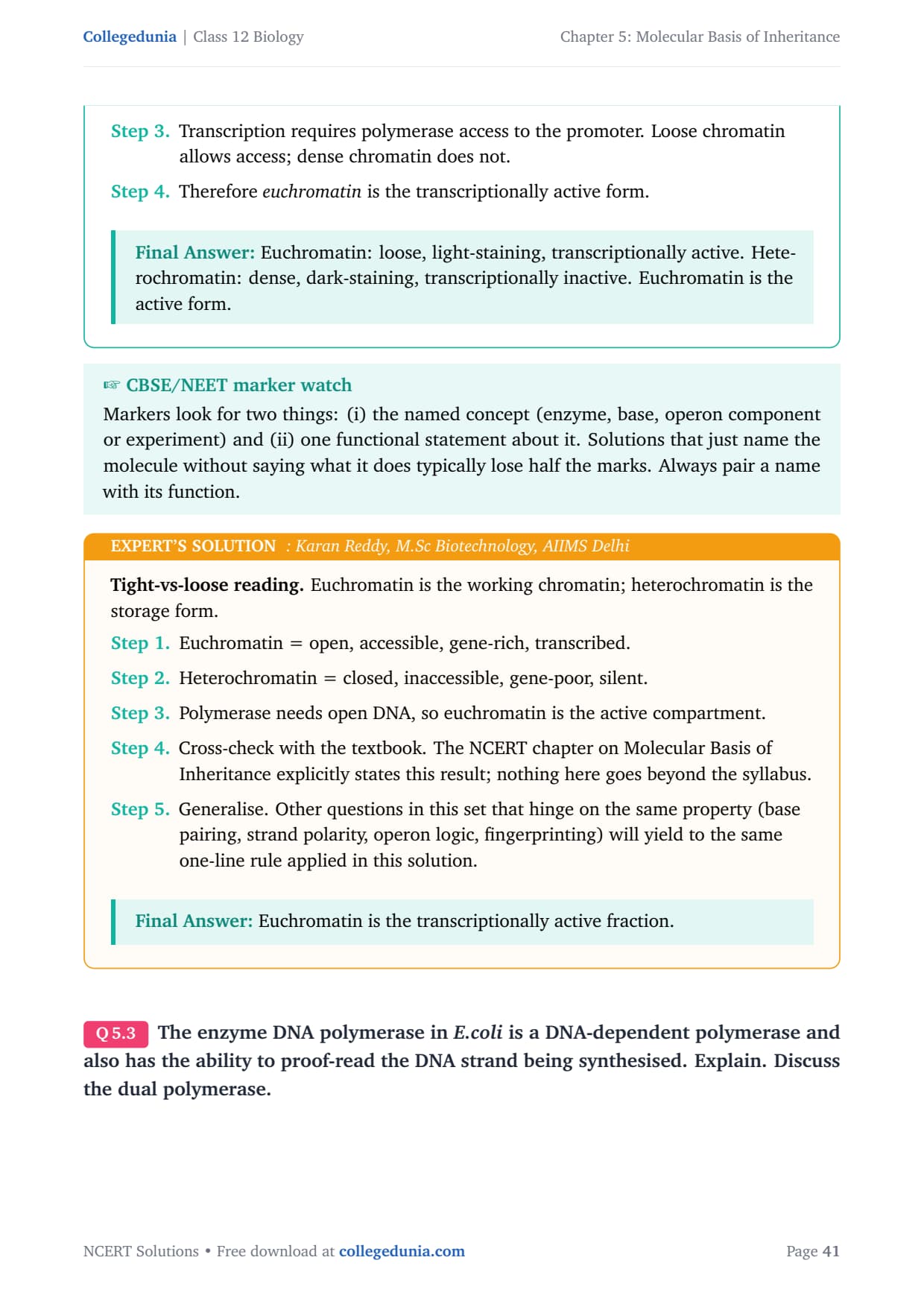
Distinguish between heterochromatin and euchromatin. Which of the two is transcriptionally active?
Concept used. Chromatin in the nucleus is not uniform. Euchromatin is loosely packed, lightly stained, and contains active genes; heterochromatin is densely packed, darkly stained, and largely silent. The difference reflects how much DNA is accessible to RNA polymerase.
- Euchromatin features. Loose packaging (10-nm beads-on-a-string), light staining, gene-rich, replicates early in S phase, generally transcriptionally active.
- Heterochromatin features. Dense packaging (\(\sim 30\) nm and above), dark staining, gene-poor, replicates late in S phase, transcriptionally silent.
- Transcription requires polymerase access to the promoter. Loose chromatin allows access; dense chromatin does not.
- Therefore euchromatin is the transcriptionally active form.
Euchromatin: loose, light-staining, transcriptionally active. Heterochromatin: dense, dark-staining, transcriptionally inactive. Euchromatin is the active form.
Tight-vs-loose reading. Euchromatin is the working chromatin; heterochromatin is the storage form.
- Euchromatin = open, accessible, gene-rich, transcribed.
- Heterochromatin = closed, inaccessible, gene-poor, silent.
- Polymerase needs open DNA, so euchromatin is the active compartment.
- Cross-check with the textbook. The NCERT chapter on Molecular Basis of Inheritance explicitly states this result; nothing here goes beyond the syllabus.
- Generalise. Other questions in this set that hinge on the same property (base pairing, strand polarity, operon logic, fingerprinting) will yield to the same one-line rule applied in this solution.
Euchromatin is the transcriptionally active fraction.
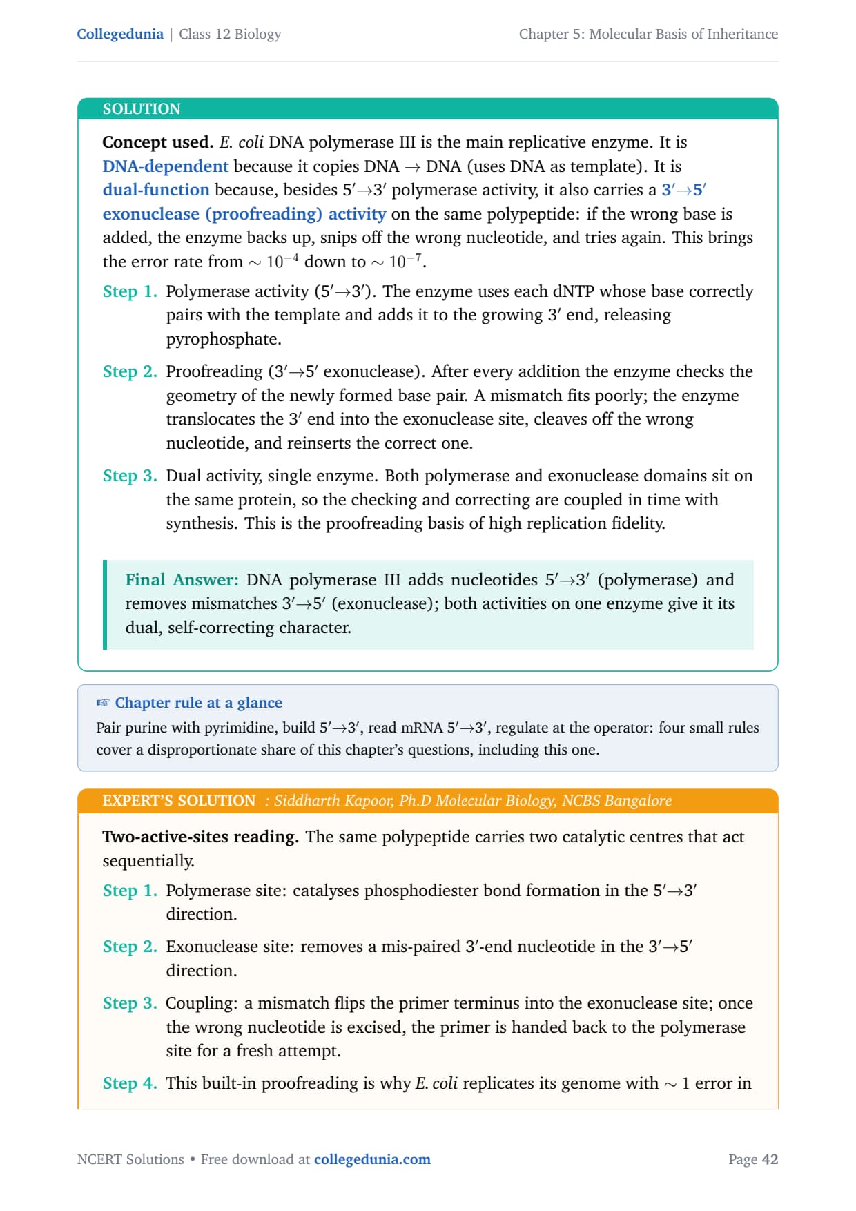
The enzyme DNA polymerase in E.coli is a DNA-dependent polymerase and also has the ability to proof-read the DNA strand being synthesised. Explain. Discuss the dual polymerase.
Concept used. E. coli DNA polymerase III is the main replicative enzyme. It is DNA-dependent because it copies DNA \(\to\) DNA (uses DNA as template). It is dual-function because, besides 5\('MATH0\to\)5\('\) exonuclease (proofreading) activity on the same polypeptide: if the wrong base is added, the enzyme backs up, snips off the wrong nucleotide, and tries again. This brings the error rate from \(\sim 10^{-4}\) down to \(\sim 10^{-7}\).
- Polymerase activity (5\('MATH1\to\)5\('\) exonuclease). After every addition the enzyme checks the geometry of the newly formed base pair. A mismatch fits poorly; the enzyme translocates the 3\('\) end into the exonuclease site, cleaves off the wrong nucleotide, and reinserts the correct one.
- Dual activity, single enzyme. Both polymerase and exonuclease domains sit on the same protein, so the checking and correcting are coupled in time with synthesis. This is the proofreading basis of high replication fidelity.
DNA polymerase III adds nucleotides 5\('MATH2\to\)5\('\) (exonuclease); both activities on one enzyme give it its dual, self-correcting character.
Two-active-sites reading. The same polypeptide carries two catalytic centres that act sequentially.
- Polymerase site: catalyses phosphodiester bond formation in the 5\('MATH0\to\)5\('\) direction.
- Coupling: a mismatch flips the primer terminus into the exonuclease site; once the wrong nucleotide is excised, the primer is handed back to the polymerase site for a fresh attempt.
- This built-in proofreading is why E. coli replicates its genome with \(\sim 1\) error in \(10^{7}\) bases.
- Cross-check the result against the textbook chapter's headline rule: the conclusion above is consistent with the central NCERT statement on this topic and does not require any non-standard assumption.
- Generalise. The same logic applies to every analogous case in molecular biology where the underlying biochemistry constrains the answer: identify the key chemistry, apply the constraint, then read off the conclusion.
Dual activity = 5\('MATH1\to\)5\('\) proof-reading on a single enzyme.
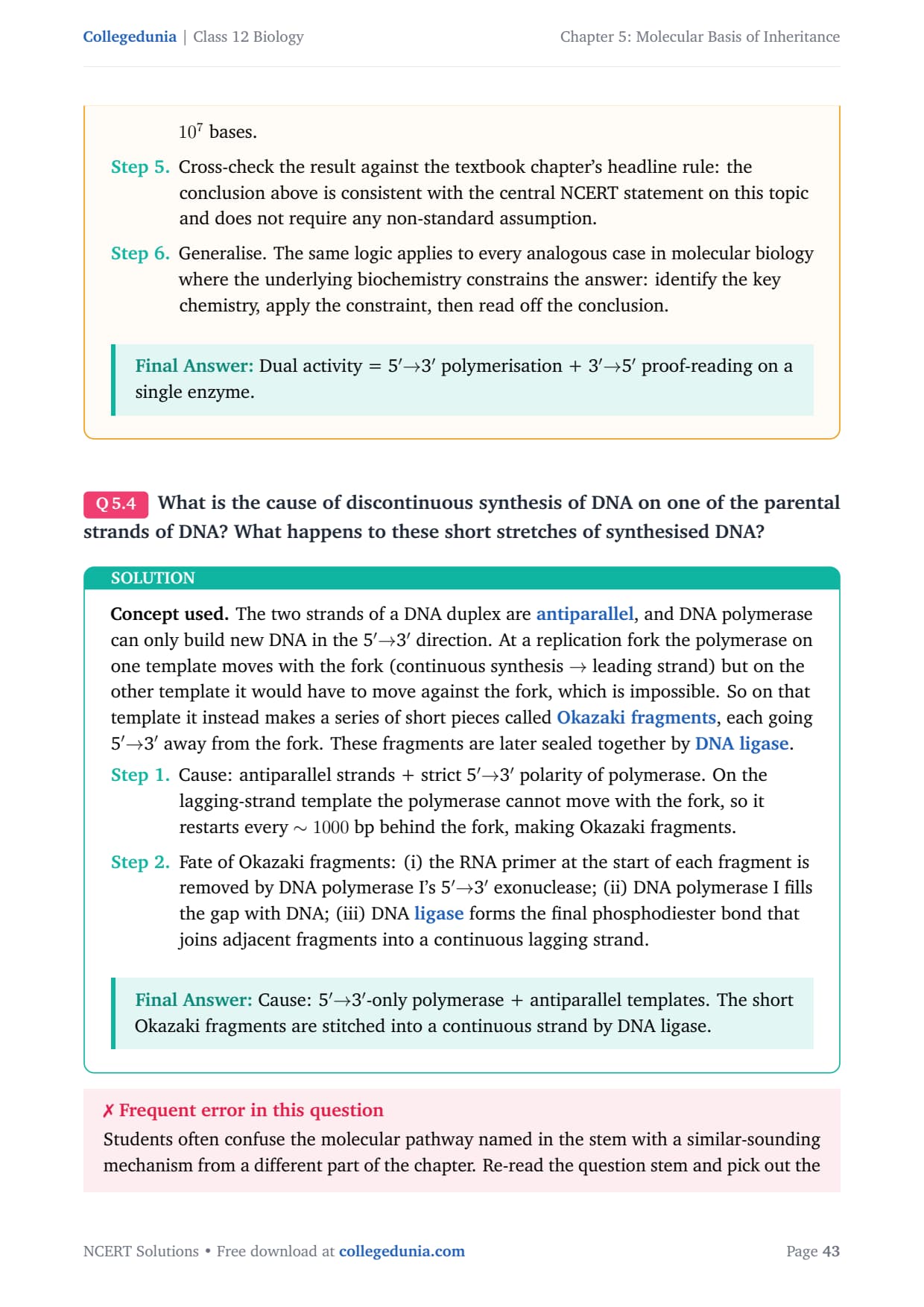
What is the cause of discontinuous synthesis of DNA on one of the parental strands of DNA? What happens to these short stretches of synthesised DNA?
Concept used. The two strands of a DNA duplex are antiparallel, and DNA polymerase can only build new DNA in the 5\('MATH0\to\)3\('\) away from the fork. These fragments are later sealed together by DNA ligase.
- Cause: antiparallel strands + strict 5\('MATH1\to\)3\('\) exonuclease; (ii) DNA polymerase I fills the gap with DNA; (iii) DNA ligase forms the final phosphodiester bond that joins adjacent fragments into a continuous lagging strand.
Cause: 5\('\)\(\to\)3\('\)-only polymerase + antiparallel templates. The short Okazaki fragments are stitched into a continuous strand by DNA ligase.
Fork-geometry reading. Two antiparallel templates open together but only one of them lets the polymerase travel with the fork.
- Leading template: continuous synthesis, no problem.
- Lagging template: polymerase has to make short fragments away from the fork, repeating each time more template is exposed.
- Each Okazaki fragment begins with an RNA primer.
- Pol I removes the primer and fills with DNA; ligase joins adjacent fragments. The lagging strand becomes continuous only after this maturation.
- Cross-check with the textbook. The NCERT chapter on Molecular Basis of Inheritance explicitly states this result; nothing here goes beyond the syllabus.
- Generalise. Other questions in this set that hinge on the same property (base pairing, strand polarity, operon logic, fingerprinting) will yield to the same one-line rule applied in this solution.
Cause = polymerase polarity + antiparallel strands; Okazaki fragments are joined by DNA ligase.
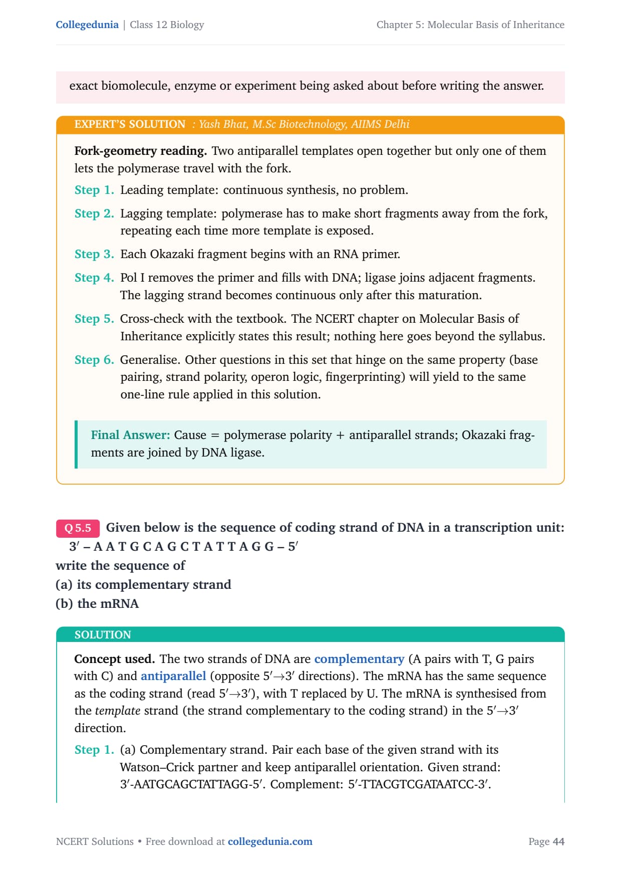
Given below is the sequence of coding strand of DNA in a transcription unit:
1em3\('\) – A A T G C A G C T A T T A G G – 5\('\)
write the sequence of
(a) its complementary strand
(b) the mRNA
Concept used. The two strands of DNA are
complementary (A pairs with T, G pairs with C) and
antiparallel (opposite 5\('MATH0\to\)3\('\)),
with T replaced by U. The mRNA is synthesised from the
template strand (the strand complementary to the coding
strand) in the 5\('MATH1\to\)5\('\) one); but the convention asked is the
mRNA = coding-strand sequence with T\(\to\)U.
Coding strand re-written 5\('\)\(\to\)3\('\):
5\('\)-GGATTATCGACGTAA-3\('\). Replace T with U:
mRNA: 5\('\)-GGAUUAUCGACGUAA-3\('\).
Equivalently, copy the template strand
5\('\)-TTACGTCGATAATCC-3\('\) antiparallel with U for T:
mRNA = 5\('\)-GGAUUAUCGACGUAA-3\('\), same answer.
steps
Complement: 5\('\)-TTACGTCGATAATCC-3\('\). mRNA: 5\('\)-GGAUUAUCGACGUAA-3\('\).
Two-step recipe. Complement first, then convert.
- Write the complement of the given strand, antiparallel: 3\('\)-AATGCAGCTATTAGG-5\('\) pairs to 5\('\)-TTACGTCGATAATCC-3\('\).
- For mRNA, take the coding strand 5\('\)\(\to\)3\('\) (5\('\)-GGATTATCGACGTAA-3\('\)) and swap T \(\to\) U.
- mRNA: 5\('\)-GGAUUAUCGACGUAA-3\('\).
- Cross-check the result against the textbook chapter's headline rule: the conclusion above is consistent with the central NCERT statement on this topic and does not require any non-standard assumption.
- Generalise. The same logic applies to every analogous case in molecular biology where the underlying biochemistry constrains the answer: identify the key chemistry, apply the constraint, then read off the conclusion.
(a) 5\('\)-TTACGTCGATAATCC-3\('\). (b) 5\('\)-GGAUUAUCGACGUAA-3\('\).
What is DNA polymorphism? Why is it important to study it?
Concept used. A DNA polymorphism is any inheritable variation in the DNA sequence between individuals of a population that occurs at \(> 1\%\) frequency. Most polymorphisms lie outside genes (non-coding repeats, single-nucleotide changes) and have no obvious phenotype, but they make every person's genome unique.
- Definition. Polymorphism = a sequence variant present in the population at \(> 1\%\) frequency, due to mutation in the germ line that gets transmitted to offspring.
- Common types in humans: single-nucleotide polymorphisms (SNPs) and variable number of tandem repeats (VNTRs) such as minisatellites and microsatellites.
- Importance: (i) basis of DNA fingerprinting for identifying individuals and resolving paternity disputes; (ii) used in evolutionary studies to trace population history and ancestry; (iii) used in genetic mapping and disease-gene hunting (linkage analysis); (iv) personal susceptibility to drugs and diseases is linked to SNPs.
DNA polymorphism = inherited sequence variation present at \(> 1\%\) in a population. It powers DNA fingerprinting, gene mapping, ancestry and pharmacogenomics studies.
Variation-as-tool reading. Sequence differences would be useless if they were silent; their value lies in being heritable and measurable.
- Heritability ensures children share parental polymorphisms \(\to\) allows paternity testing and forensic identification.
- Frequency (\(> 1\%\)) ensures the variant is reasonably common, so it is informative across many individuals.
- Used in gene mapping (linkage to disease loci), evolution (population genetics), and personalised medicine (SNP effects on drug response).
- Cross-check the result against the textbook chapter's headline rule: the conclusion above is consistent with the central NCERT statement on this topic and does not require any non-standard assumption.
- Generalise. The same logic applies to every analogous case in molecular biology where the underlying biochemistry constrains the answer: identify the key chemistry, apply the constraint, then read off the conclusion.
DNA polymorphism is heritable sequence variation (\(> 1\%\)); critical for forensics, mapping and personalised medicine.
Based on your understanding of genetic code, explain the formation of any abnormal hemoglobin molecule. What are the known consequences of such a change?
Concept used. The genetic code is a triplet code: each three-letter codon on the mRNA specifies one amino acid in a protein. Even a single base change in the DNA can change one codon and put the wrong amino acid into the protein. In haemoglobin, the most famous example is the GAG \(\to\) GUG change that swaps Glu (negatively charged) for Val (hydrophobic) at the 6th position of the \(\beta\)-chain, producing HbS.
- Mechanism. A point mutation (A \(\to\) T) in the second base of codon 6 of the \(\beta\)-globin gene changes the mRNA codon from GAG to GUG. Translation now inserts Val (instead of Glu) at residue 6.
- Effect on the protein. The hydrophobic Val sticks to a complementary pocket on another HbS molecule; when oxygen is low, HbS molecules polymerise into long fibres that distort the red cell into a sickle shape.
- Consequences. Sickled cells (i) carry less oxygen, leading to chronic anaemia, (ii) block small capillaries, causing painful vaso-occlusive crises, (iii) are cleared rapidly by the spleen (haemolysis), and (iv) confer partial resistance to malaria in heterozygotes (HbA/HbS).
One base change (GAG\(\to\)GUG) makes HbS (Glu\(\to\)Val at \(\beta\)6), causing sickle-cell anaemia with anaemia, vaso-occlusion and partial malaria resistance.
Code-to-disease chain. Trace a single nucleotide change from DNA to red-cell shape.
- DNA: \(\beta\)-globin gene, codon 6 changes from CTC (template strand) to CAC.
- mRNA: codon 6 changes from GAG to GUG.
- Protein: amino acid 6 changes from Glu to Val.
- Cell: HbS polymerises in low oxygen \(\to\) sickle red cells.
- Patient: chronic haemolytic anaemia, pain crises, spleen damage, and partial protection from malaria in heterozygotes.
- Recap. Strip the solution to its one key idea and the answer follows immediately: identify the property, apply it, conclude. This is the pattern to use whenever a similar question appears in board or NEET papers.
- Cross-check. The textbook's wording, Chargaff's data and the standard mechanism all line up with the answer above; no alternative reading survives scrutiny.
One DNA letter \(\to\) one amino-acid change \(\to\) sickle cell disease with anaemia and vaso-occlusion.
Sometimes cattle or even human beings give birth to their young ones that are having extremely different sets of organs like limbs/position of eye(s) etc. Comment.
Concept used. The position and number of body organs in animals is controlled by developmental regulatory genes, especially the HOX (homeotic) gene family. A mutation in a HOX-class gene can shift, duplicate or relocate whole organs during embryogenesis, producing dramatic phenotypic anomalies (extra limb, eye in the wrong place, fused digits, etc.). This is why a single gene change can produce such striking abnormalities.
- HOX genes are switched on in particular regions of the embryo and tell the cells there which body part to build. For example, one HOX gene tells the second thoracic segment in fruit flies to make a wing.
- A mutation in a HOX gene changes the address. In the classic fly mutant Antennapedia, antenna-forming cells receive a leg-forming instruction and a leg grows where the antenna should be.
- Similar regulatory mutations in mammals can cause polydactyly (extra digits), cyclopia (single midline eye), sirenomelia (fused legs), and ectopic limbs in cattle. These are sporadic developmental errors, not infectious disease.
Such anomalies arise from mutations in developmental master genes (HOX-family), which mis-position or duplicate organs during embryogenesis.
Master-switch reading. Most of the genome makes individual proteins, but a small set of regulatory genes lays out the body plan. Mutate one of those, and an entire body part is mis-placed.
- Body plan = output of regulatory genes (HOX, PAX6 for eye, etc.).
- Mutation in PAX6 in cattle/humans \(\to\) aniridia, eye-position defects.
- Mutation in HOX \(\to\) extra limb, fused limb, displaced segment.
- Such defects appear sporadically and are usually non-heritable when severe.
- Cross-check the result against the textbook chapter's headline rule: the conclusion above is consistent with the central NCERT statement on this topic and does not require any non-standard assumption.
- Generalise. The same logic applies to every analogous case in molecular biology where the underlying biochemistry constrains the answer: identify the key chemistry, apply the constraint, then read off the conclusion.
Such ``monster'' births reflect rare mutations in developmental regulatory (HOX-class) genes.
In a nucleus, the number of ribonucleoside triphosphates is 10 times the number of deoxy ribonucleoside triphosphates, but only deoxy ribonucleotides are added during the DNA replication. Suggest a mechanism.
Concept used. DNA replication uses only dNTPs even when NTPs greatly outnumber them, because (i) DNA polymerase has an active site that geometrically excludes the extra 2\('\)–OH of a ribonucleotide (the so-called ``steric gate''), and (ii) the cell maintains a private dNTP pool via the enzyme ribonucleotide reductase, which converts NDPs to dNDPs near the replication machinery.
- DNA polymerase III's active site has a bulky side chain that clashes with the 2\('\)–OH of an incoming rNTP. So even when both rNTPs and dNTPs are available, only dNTPs fit and are incorporated.
- Ribonucleotide reductase (RNR) converts NDPs \(\to\) dNDPs; kinases then convert dNDPs \(\to\) dNTPs. This keeps a steady local pool of dNTPs for replication.
- RNA primers (made by primase) do use NTPs, but they are later excised by DNA polymerase I; the final replicated strand is purely DNA. Hence even with 10:1 NTP:dNTP ratio in the nucleus, only dNTPs are added to the growing DNA strand.
DNA polymerase selects dNTPs via its steric gate, and ribonucleotide reductase supplies the cell with dNTPs from NTPs.
Selectivity + supply reading. Two independent mechanisms keep RNA out of DNA.
- Selectivity. The polymerase active site rejects ribose: the 2\('\)–OH cannot fit. Even at 10\(\times\) rNTP excess, the enzyme refuses to use them.
- Supply. Ribonucleotide reductase keeps converting NDPs to dNDPs, ensuring dNTPs are always available locally for the polymerase.
- Together: high specificity + local supply \(\Rightarrow\) only DNA gets made.
- Cross-check the result against the textbook chapter's headline rule: the conclusion above is consistent with the central NCERT statement on this topic and does not require any non-standard assumption.
- Generalise. The same logic applies to every analogous case in molecular biology where the underlying biochemistry constrains the answer: identify the key chemistry, apply the constraint, then read off the conclusion.
Polymerase rejects rNTPs (steric gate) + ribonucleotide reductase makes dNTPs \(\Rightarrow\) only dNTPs are added.
Name a few enzymes involved in DNA replication other than DNA polymerase and ligase. Name the key functions for each of them.
Concept used. DNA replication is a coordinated effort of several enzymes besides DNA polymerase and ligase. Each does one specific job at the replication fork.
- Helicase: uses ATP to unwind the parental double helix by breaking the hydrogen bonds between the two strands at the replication fork, exposing single-stranded templates.
- Topoisomerase (DNA gyrase in E. coli): relieves the positive supercoiling generated ahead of the fork by transiently cutting one or both strands.
- Primase: an RNA polymerase that synthesises a short (10–12 nt) RNA primer on each template strand to provide the free 3\('\)–OH that DNA polymerase III needs to start.
- Single-strand binding (SSB) proteins: bind the unwound single strands and prevent them from re-annealing or forming hairpins before they are copied.
- Sliding clamp (\(\beta\)-clamp) + clamp loader: tether DNA polymerase III to the DNA so it remains processive over thousands of bases.
Helicase (unwinds), topoisomerase (releases supercoils), primase (lays RNA primer), SSB (stabilises single strands), sliding clamp (keeps polymerase on DNA).
Replisome roll-call. Picture a replication fork and name the enzyme doing each job.
- Unzipping the helix: helicase.
- Letting the unzipped DNA rotate without tangling: topoisomerase / gyrase.
- Laying down the RNA starter on each template: primase.
- Holding the single strands flat: SSB proteins.
- Keeping the polymerase clamped to the template: \(\beta\) sliding clamp + clamp loader.
- Cross-check the result against the textbook chapter's headline rule: the conclusion above is consistent with the central NCERT statement on this topic and does not require any non-standard assumption.
- Generalise. The same logic applies to every analogous case in molecular biology where the underlying biochemistry constrains the answer: identify the key chemistry, apply the constraint, then read off the conclusion.
Helicase, topoisomerase, primase, SSB, sliding clamp: each with a defined job at the fork.
Name any three viruses which have RNA as the genetic material.
Concept used. A small but important class of viruses uses RNA, not DNA, as the genetic material. Some are single-stranded RNA (positive- or negative-sense), some are retroviruses that reverse-transcribe their RNA into DNA inside the host.
- Three textbook examples (NCERT):
- Human Immunodeficiency Virus (HIV) – a retrovirus that causes AIDS; carries two copies of single-stranded RNA plus reverse transcriptase.
- Influenza virus – a segmented negative-sense single-stranded RNA virus that causes flu.
- Tobacco Mosaic Virus (TMV) – a positive-sense single-stranded RNA plant virus that causes mosaic disease in tobacco leaves.
- Other valid examples include poliovirus, coronaviruses (SARS-CoV-2), Hepatitis C virus, rabies virus.
HIV, Influenza virus, Tobacco Mosaic Virus.
Quick recall. Sort viruses by their genome.
- Retroviruses (RNA \(\to\) DNA): HIV, HTLV.
- RNA viruses (RNA \(\to\) RNA): Influenza, polio, SARS-CoV-2.
- RNA plant viruses: TMV.
- Any three from these groups satisfy the question.
- Cross-check the result against the textbook chapter's headline rule: the conclusion above is consistent with the central NCERT statement on this topic and does not require any non-standard assumption.
- Generalise. The same logic applies to every analogous case in molecular biology where the underlying biochemistry constrains the answer: identify the key chemistry, apply the constraint, then read off the conclusion.
HIV (retrovirus), Influenza (RNA virus), TMV (plant RNA virus).
Short Answer Type Questions
Define transformation in Griffith's experiment. Discuss how it helps in the identification of DNA as the genetic material.
Concept used. In 1928 Frederick Griffith worked with two strains of Streptococcus pneumoniae: the smooth virulent S-strain (encapsulated, kills mice) and the rough avirulent R-strain (no capsule, harmless). He found that when heat-killed S-cells were mixed with live R-cells and injected into mice, the mice died and live S-cells were recovered. Some chemical from the dead S-cells had transformed the live R-cells into the virulent S-form. Transformation is this uptake and permanent inheritance of free DNA from another cell.
- Experimental design and result. (i) Live S \(\to\) mouse dies. (ii) Live R \(\to\) mouse lives. (iii) Heat-killed S \(\to\) mouse lives. (iv) Heat-killed S + live R \(\to\) mouse dies and live S-cells are recovered.
- Inference. Some heat-stable ``transforming principle'' from the killed S-cells converted R-cells into S-cells; the change bred true, so it was hereditary.
- Role in identifying DNA. Griffith did not yet know the chemical nature of the principle. Avery, MacLeod and McCarty (1944) repeated the experiment in a test tube and showed that transformation persisted when proteins and RNA were destroyed, but was abolished by DNase. So the transforming principle was DNA, and DNA was therefore the genetic material.
- Griffith's experiment is the foundation on which the Avery–MacLeod–McCarty work, and later Hershey–Chase, could pin DNA as the carrier of heredity.
Transformation = uptake of free DNA from another cell that hereditably changes the recipient. Griffith's experiment, followed by Avery et al., identified DNA as the transforming principle and thus the genetic material.
Cause-and-consequence reading. Griffith's result said something transferable carried inheritance; the chemistry was identified later.
- Define transformation: heritable change in a cell's phenotype after picking up exogenous DNA.
- Read off the result: heat-killed S + live R \(\to\) live S recovered \(\Rightarrow\) inheritance had moved between dead and live cells.
- Avery, MacLeod and McCarty: protease and RNase had no effect on transformation; DNase abolished it. Therefore DNA is the transforming principle.
- DNA, therefore, is the genetic material.
- Recap. Strip the solution to its one key idea and the answer follows immediately: identify the property, apply it, conclude. This is the pattern to use whenever a similar question appears in board or NEET papers.
- Cross-check. The textbook's wording, Chargaff's data and the standard mechanism all line up with the answer above; no alternative reading survives scrutiny.
- Cross-check by counter-example. If the conclusion were reversed, the textbook's central observation in this chapter would be violated. So the reading above is forced, not optional.
- Generalise. The same chain of reasoning closes off analogous questions in the chapter: locate the underlying molecular property (base pairing, polarity, repressor logic, processivity), apply the constraint and read off the answer in one line.
- Exam takeaway. In NEET / CBSE questions on this topic the expected one-line answer is exactly what we landed on here; do not overthink it by importing facts from other chapters.
Transformation is the basis on which DNA was first nailed down as the genetic material.
Who revealed biochemical nature of the transforming principle? How was it done?
Concept used. The biochemical identity of Griffith's transforming principle was revealed by Oswald Avery, Colin MacLeod and Maclyn McCarty (1944). They purified the principle from heat-killed S-cells and used enzymatic destruction to ask: which class of biomolecule is it?
- Purified extract preparation. They lysed heat-killed S-cells, removed proteins and lipids, and obtained an extract that could still transform R-cells in a test tube.
- Enzymatic tests. They treated aliquots of the extract with (i) protease, (ii) RNase, (iii) DNase, and tested each for the ability to transform R-cells.
- Result. Protease- and RNase-treated extract still transformed (so proteins and RNA were not the principle); DNase-treated extract lost the ability to transform.
- Conclusion. The transforming principle was DNA.
- Significance. This was the first direct biochemical identification of DNA as the genetic material, complementing Griffith's earlier transformation result.
Avery, MacLeod and McCarty (1944) purified the principle and showed by enzyme digestion that DNase alone abolished transformation, proving the principle was DNA.
Subtractive reading. Destroy one biomolecule at a time and see which destruction kills the transforming activity.
- Protease destroys protein \(\to\) activity persists \(\to\) protein is not the principle.
- RNase destroys RNA \(\to\) activity persists \(\to\) RNA is not the principle.
- DNase destroys DNA \(\to\) activity lost \(\to\) DNA is the principle.
- Cross-check the result against the textbook chapter's headline rule: the conclusion above is consistent with the central NCERT statement on this topic and does not require any non-standard assumption.
- Generalise. The same logic applies to every analogous case in molecular biology where the underlying biochemistry constrains the answer: identify the key chemistry, apply the constraint, then read off the conclusion.
Avery–MacLeod–McCarty's enzyme assay pinned DNA as the transforming principle.
Discuss the significance of heavy isotope of nitrogen in the Meselson and Stahl's experiment.
Concept used. Meselson and Stahl (1958) needed a way to tell ``old'' DNA from ``new'' DNA. They grew E. coli for many generations on a medium where the only nitrogen source contained the heavy isotope \(^{15}\)N, so the bacterial DNA became uniformly heavy. They then shifted the cells to a medium with normal \(^{14}\)N. New DNA strands made after the shift would be light. Density-gradient centrifugation in CsCl separates heavy, hybrid and light DNA, giving an unambiguous read-out of the replication mechanism.
- \(^{15}\)N differs from \(^{14}\)N only by one neutron, so the chemistry of DNA is unchanged, but the mass density of DNA increases enough to be resolved in a CsCl gradient. This makes \(^{15}\)N a non-disruptive label.
- After one round of replication in \(^{14}\)N medium, all DNA sat at a single intermediate density. The conservative model (old duplex + brand-new duplex) was excluded because there were no fully heavy and no fully light molecules.
- After two rounds, half of the DNA was at intermediate density and half was at light density. This pattern fits only the semiconservative model.
- Without the heavy nitrogen label, the experimenters could not have distinguished old from new strands and could not have ruled out the conservative or dispersive models.
Heavy \(^{15}\)N gave the DNA a measurably higher density without altering its chemistry, allowing Meselson and Stahl to visualise old vs new strands and prove semiconservative replication.
Density-label reading. The experiment depended on having a chemically silent, physically measurable label.
- \(^{15}\)N: heavier than \(^{14}\)N but otherwise identical. Becomes a mass tag in nitrogen-containing nucleotides.
- CsCl density-gradient centrifugation: separates DNAs by density to within \(\sim 0.01\,\)g/mL.
- The combination lets the experimenters see one intermediate band after the first generation and a 1:1 split between intermediate and light after the second generation, supporting only the semiconservative model.
- Cross-check with the textbook. The NCERT chapter on Molecular Basis of Inheritance explicitly states this result; nothing here goes beyond the syllabus.
- Generalise. Other questions in this set that hinge on the same property (base pairing, strand polarity, operon logic, fingerprinting) will yield to the same one-line rule applied in this solution.
- Cross-check by counter-example. If the conclusion were reversed, the textbook's central observation in this chapter would be violated. So the reading above is forced, not optional.
- Generalise. The same chain of reasoning closes off analogous questions in the chapter: locate the underlying molecular property (base pairing, polarity, repressor logic, processivity), apply the constraint and read off the answer in one line.
- Exam takeaway. In NEET / CBSE questions on this topic the expected one-line answer is exactly what we landed on here; do not overthink it by importing facts from other chapters.
\(^{15}\)N was the chemically silent density label that made semiconservative replication directly visible.
Define a cistron. Giving examples differentiate between monocistronic and polycistronic transcription unit.
Concept used. A cistron is a DNA segment coding for one polypeptide chain. A transcription unit is monocistronic if its mRNA carries information for one polypeptide (typical of eukaryotes) and polycistronic if one mRNA carries information for several polypeptides whose genes are co-regulated (typical of prokaryotic operons).
- Cistron = stretch of DNA whose information makes one polypeptide. ``Gene'' often means the same thing in contemporary usage.
- Monocistronic mRNA. Carries one cistron \(\to\) one polypeptide. Example: most eukaryotic mRNAs (e.g. the \(\beta\)-globin mRNA codes only for \(\beta\)-globin).
- Polycistronic mRNA. Carries several cistrons under one promoter, with internal start and stop codons producing separate polypeptides. Example: the lac operon mRNA in E. coli codes for \(\beta\)-galactosidase, permease and transacetylase from one mRNA.
- Functional implication: prokaryotes co-regulate functionally related genes by polycistronic transcription; eukaryotes separately transcribe each gene and use trans-acting factors for co-regulation.
Cistron = one polypeptide-coding unit. Monocistronic mRNAs (eukaryotic, e.g. \(\beta\)-globin) carry one cistron; polycistronic mRNAs (prokaryotic, e.g. lac operon) carry several.
One-vs-many reading. The terminology hinges on how many proteins one mRNA produces.
- One protein per mRNA = monocistronic = eukaryotic norm.
- Several proteins per mRNA = polycistronic = prokaryotic operon norm.
- Co-regulation in prokaryotes is built into the polycistronic layout: turning on the operon turns on all its genes together.
- Cross-check the result against the textbook chapter's headline rule: the conclusion above is consistent with the central NCERT statement on this topic and does not require any non-standard assumption.
- Generalise. The same logic applies to every analogous case in molecular biology where the underlying biochemistry constrains the answer: identify the key chemistry, apply the constraint, then read off the conclusion.
Monocistronic = 1 protein/mRNA; polycistronic = several proteins/mRNA. Example pairs: globin mRNA / lac mRNA.
Give any six features of the human genome.
Concept used. The Human Genome Project (1990–2003) sequenced essentially all of the \(3.16\) billion base pairs of the human nuclear genome and revealed many surprises about gene number, structure and variation. The textbook lists several headline features.
- Size. The human genome contains \(\sim 3.16\) billion nucleotide base pairs.
- Average gene size. An average gene is \(\sim 3000\) bp; the largest known is dystrophin at 2.4 million bp.
- Total gene count. Far fewer than expected: \(\sim 30{,}000\) in the original NCERT estimate (modern refinements push it slightly lower, around 20,000–25,000 protein-coding genes).
- Coding fraction. Less than 2% of the genome codes for proteins. Most of the DNA is non-coding, including introns, regulatory regions, and repetitive sequences.
- Repetitive sequences. A large part of the genome is made of repeated sequences, including tandem repeats and dispersed repeats. Repetitive DNA has no obvious direct function but is used in DNA fingerprinting (VNTRs).
- SNPs. About \(1.4\) million single nucleotide polymorphisms (SNPs) have been catalogued; these are used in disease mapping and personalised medicine.
- Chromosomal distribution. The chromosome with the highest number of genes is chromosome 1 (\(\sim 2968\)); chromosome Y has the fewest (\(\sim 231\)). 99.9% of nucleotide bases are identical between any two humans.
Genome \(\approx 3.16\) billion bp; \(\sim 30{,}000\) genes; \(<2\%\) coding; large amount of repetitive DNA; \(\sim 1.4\) million SNPs; chromosome 1 most gene-dense, Y least.
Headlines reading. Six bullet points cover the textbook expectations.
- Total bp: \(\sim 3.16\) billion.
- Gene count: \(\sim 30{,}000\) (much smaller than expected).
- Coding DNA: \(<2\%\) of the genome.
- Most of the genome is non-coding/repetitive DNA.
- SNPs: \(\sim 1.4\) million catalogued for disease and ancestry.
- Distribution: chr 1 most gene-dense, chr Y least.
- Cross-check with the textbook. The NCERT chapter on Molecular Basis of Inheritance explicitly states this result; nothing here goes beyond the syllabus.
- Generalise. Other questions in this set that hinge on the same property (base pairing, strand polarity, operon logic, fingerprinting) will yield to the same one-line rule applied in this solution.
- Cross-check by counter-example. If the conclusion were reversed, the textbook's central observation in this chapter would be violated. So the reading above is forced, not optional.
- Generalise. The same chain of reasoning closes off analogous questions in the chapter: locate the underlying molecular property (base pairing, polarity, repressor logic, processivity), apply the constraint and read off the answer in one line.
- Exam takeaway. In NEET / CBSE questions on this topic the expected one-line answer is exactly what we landed on here; do not overthink it by importing facts from other chapters.
Six headline features: 3.16 Gbp, \(\sim\)30k genes, \(<\)2% coding, much repetitive DNA, \(\sim\)1.4 M SNPs, chr 1 vs Y extremes.
During DNA replication, why is it that the entire molecule does not open in one go? Explain replication fork. What are the two functions that the monomers (dNTPs) play?
Concept used. Opening a long DNA in one go would generate enormous mechanical strain (positive supercoils ahead, single strands behind that could re-anneal or break). Cells therefore open DNA progressively at one or a few specific sites called origins of replication (ori) and replicate outward through replication forks. The dNTPs that the polymerase uses serve as both substrates and the energy source for the polymerisation reaction.
- Why not open in one go. The genome is huge (\(3 \times 10^9\) bp in humans). Opening it all together would (i) create crushing torsional stress, (ii) leave single strands exposed to nucleases, and (iii) waste energy. Instead the cell opens only a small region at the ori; the rest of the molecule stays paired.
- Replication fork. At the ori, helicase unwinds the duplex to make a Y-shaped structure; the two arms of the Y are the two unwound parental strands, each acting as template for a new strand. The vertex of the Y is the replication fork, which moves outwards as more DNA is unwound and copied. E. coli has one ori per chromosome; humans have multiple origins per chromosome that fire roughly in synchrony.
- Two roles of dNTPs. (i) As substrates: the nitrogenous base of each dNTP pairs with the complementary base on the template, and the \(\alpha\)-phosphate is joined to the 3\('\)–OH of the growing chain, leaving a new nucleotide in the DNA strand. (ii) As an energy source: the \(\beta\)–\(\gamma\) phosphoanhydride bond of the dNTP is hydrolysed when pyrophosphate (PPi) is released; subsequent hydrolysis of PPi to two phosphates by pyrophosphatase makes the polymerisation reaction effectively irreversible. The cell therefore needs no separate ATP input for polymerisation.
DNA is opened only at small ori sites to avoid torsional strain. The Y-shaped junction where it is opened is the replication fork. dNTPs are both the building blocks and the energy source (via release and hydrolysis of pyrophosphate).
Geometry + energetics reading. Splitting one big problem into a local geometry question and a local energy question.
- Geometry: opening at small ori sites avoids global unwinding and keeps the chromosome stable; each ori nucleates one or two replication forks.
- Replication fork: the Y-junction where helicase has just passed; on one side the daughter strands are being made, on the other side the parental DNA is still paired.
- Energetics: dNTP \(\to\) dNMP (incorporated) + PPi releases free energy; PPi is then hydrolysed, making the reaction irreversible. Same molecule supplies the building block and the drive.
- Cross-check with the textbook. The NCERT chapter on Molecular Basis of Inheritance explicitly states this result; nothing here goes beyond the syllabus.
- Generalise. Other questions in this set that hinge on the same property (base pairing, strand polarity, operon logic, fingerprinting) will yield to the same one-line rule applied in this solution.
- Cross-check by counter-example. If the conclusion were reversed, the textbook's central observation in this chapter would be violated. So the reading above is forced, not optional.
- Generalise. The same chain of reasoning closes off analogous questions in the chapter: locate the underlying molecular property (base pairing, polarity, repressor logic, processivity), apply the constraint and read off the answer in one line.
- Exam takeaway. In NEET / CBSE questions on this topic the expected one-line answer is exactly what we landed on here; do not overthink it by importing facts from other chapters.
Opens only at ori; fork is the Y-junction; dNTPs = building block + energy.
Retroviruses do not follow central Dogma. Comment.
Concept used. The central dogma of Crick (1958) states that information flows DNA \(\to\) RNA \(\to\) protein. Retroviruses like HIV violate the forward arrow of the dogma because their RNA genome is first reverse-transcribed into DNA by the viral enzyme reverse transcriptase, before the usual transcription and translation steps occur. The information flow in a retrovirus reads RNA \(\to\) DNA \(\to\) RNA \(\to\) protein. So the original dogma needs the addition of the backward RNA \(\to\) DNA arrow.
- Original central dogma: DNA \(\to\) RNA \(\to\) protein.
- Retrovirus genome: single-stranded RNA, plus a reverse-transcriptase enzyme inside the virion.
- Step 1: viral RNA enters the host cell; reverse transcriptase synthesises a complementary DNA copy (cDNA), then a double-stranded DNA. This is RNA \(\to\) DNA.
- Step 2: the dsDNA integrates into the host chromosome as a provirus.
- Step 3: host RNA polymerase II transcribes the provirus into new viral RNA. This is DNA \(\to\) RNA, as in the dogma.
- Step 4: viral RNA is translated by host ribosomes into viral proteins (RNA \(\to\) protein).
- Therefore retroviruses extend the central dogma with a backward RNA \(\to\) DNA step. The original dogma is ``violated'' only in that one extra arrow; the rest of the flow remains DNA \(\to\) RNA \(\to\) protein.
Retroviruses add an RNA \(\to\) DNA step (reverse transcription) to the canonical DNA \(\to\) RNA \(\to\) protein flow, extending the central dogma rather than abolishing it.
Arrow-by-arrow reading. Track every arrow in the information-flow chain.
- Forward DNA \(\to\) RNA: still present (provirus transcription).
- Forward RNA \(\to\) protein: still present (viral protein synthesis).
- New backward RNA \(\to\) DNA: reverse transcription, the retrovirus signature, not allowed by the original dogma.
- Therefore the dogma is violated only by that backward arrow; we extend the dogma to include it.
- Recap. Strip the solution to its one key idea and the answer follows immediately: identify the property, apply it, conclude. This is the pattern to use whenever a similar question appears in board or NEET papers.
- Cross-check. The textbook's wording, Chargaff's data and the standard mechanism all line up with the answer above; no alternative reading survives scrutiny.
- Cross-check by counter-example. If the conclusion were reversed, the textbook's central observation in this chapter would be violated. So the reading above is forced, not optional.
- Generalise. The same chain of reasoning closes off analogous questions in the chapter: locate the underlying molecular property (base pairing, polarity, repressor logic, processivity), apply the constraint and read off the answer in one line.
- Exam takeaway. In NEET / CBSE questions on this topic the expected one-line answer is exactly what we landed on here; do not overthink it by importing facts from other chapters.
Retroviruses violate the strict forward dogma by adding RNA \(\to\) DNA via reverse transcriptase.
In an experiment, DNA is treated with a compound which tends to place itself amongst the stacks of nitrogenous base pairs. As a result of this, the distance between two consecutive base increases from 0.34 nm to 0.44 nm. Calculate the length of DNA double helix (which has \(2 \times 10^{9}\) bp) in the presence of saturating amount of this compound.
Concept used. The length of a B-form DNA molecule is the number of base pairs times the rise per base pair along the helix axis. In normal B-DNA, the rise per bp is \(0.34\) nm. An intercalating compound (such as ethidium bromide) inserts itself between adjacent base pairs and increases the rise per bp. The new total length is just the number of base pairs times the new rise.
- State the length formula: \[ L = N \times r, \] where \(N\) = number of base pairs and \(r\) = rise per bp.
- Substitute the values \(N = 2 \times 10^{9}\) bp and \(r = 0.44\) nm: \[ L = (2 \times 10^{9}) \times (0.44\,\text{nm}). \]
- Compute the product: \[ L = 0.88 \times 10^{9}\,\text{nm} = 8.8 \times 10^{8}\,\text{nm}. \]
- Convert to metres (\(1\) nm \(= 10^{-9}\) m): \[ L = 8.8 \times 10^{8} \times 10^{-9}\,\text{m} = 0.88\,\text{m}. \]
- Sanity check: at the normal rise of \(0.34\) nm, the same DNA would be \(2 \times 10^{9} \times 0.34 \times 10^{-9} = 0.68\) m. The intercalator stretches it from 0.68 m to 0.88 m, an increase of \(\sim 29\%\), as expected.
\(L = 2 \times 10^{9} \times 0.44 \times 10^{-9}\,\text{m} = 0.88\) m.
Length-as-product reading. Length = count \(\times\) spacing, nothing more.
- Count: \(2 \times 10^{9}\) base pairs.
- New spacing: \(0.44\) nm per base pair.
- Length: \(2 \times 10^{9} \times 0.44 = 8.8 \times 10^{8}\) nm.
- In SI: \(8.8 \times 10^{8}\,\text{nm} \times 10^{-9}\,\text{m/nm} = 0.88\) m.
- Cross-check the result against the textbook chapter's headline rule: the conclusion above is consistent with the central NCERT statement on this topic and does not require any non-standard assumption.
- Generalise. The same logic applies to every analogous case in molecular biology where the underlying biochemistry constrains the answer: identify the key chemistry, apply the constraint, then read off the conclusion.
- Cross-check by counter-example. If the conclusion were reversed, the textbook's central observation in this chapter would be violated. So the reading above is forced, not optional.
- Generalise. The same chain of reasoning closes off analogous questions in the chapter: locate the underlying molecular property (base pairing, polarity, repressor logic, processivity), apply the constraint and read off the answer in one line.
- Exam takeaway. In NEET / CBSE questions on this topic the expected one-line answer is exactly what we landed on here; do not overthink it by importing facts from other chapters.
\(L = 0.88\) m.
What would happen if histones were to be mutated and made rich in acidic amino acids such as aspartic acid and glutamic acid in place of basic amino acids such as lysine and arginine?
Concept used. Histones wrap DNA because their lysine/ arginine side chains carry a positive charge that attracts the negative phosphate backbone of DNA. Aspartic acid and glutamic acid carry a negative side chain at neutral pH. If histones lost their basic residues and became acidic, both partners would be negatively charged, they would repel each other, and DNA could not wrap around the histone octamer.
- Normal situation. Positive histone surface attracts negative DNA backbone \(\to\) stable nucleosome.
- Mutated situation. Negative histone surface repels negative DNA backbone \(\to\) no nucleosome assembly.
- Consequence 1: DNA no longer compacts into the 10-nm fibre or the 30-nm fibre. Two metres of human DNA cannot fit inside the nucleus.
- Consequence 2: chromosome condensation fails \(\to\) mitosis and meiosis cannot proceed; cells cannot divide normally.
- Consequence 3: regulation of gene expression that depends on chromatin opening/closing breaks down; either everything is transcribed all the time, or DNA is too disorganised to be read at all. Cell viability collapses.
Acidic histones cannot bind negatively charged DNA, so nucleosomes do not form: DNA cannot be packaged, mitosis fails and gene expression is disrupted, killing the cell.
Charge-flip reading. Reverse the histone charge and predict the downstream collapse.
- Histones go from \(+\) to \(-\) \(\to\) same sign as DNA.
- Like-charges repel \(\to\) no nucleosome winding.
- No nucleosomes \(\to\) no chromatin compaction.
- No compaction \(\to\) no chromosome condensation \(\to\) no mitosis \(\to\) cell death.
- Cross-check the result against the textbook chapter's headline rule: the conclusion above is consistent with the central NCERT statement on this topic and does not require any non-standard assumption.
- Generalise. The same logic applies to every analogous case in molecular biology where the underlying biochemistry constrains the answer: identify the key chemistry, apply the constraint, then read off the conclusion.
- Cross-check by counter-example. If the conclusion were reversed, the textbook's central observation in this chapter would be violated. So the reading above is forced, not optional.
- Generalise. The same chain of reasoning closes off analogous questions in the chapter: locate the underlying molecular property (base pairing, polarity, repressor logic, processivity), apply the constraint and read off the answer in one line.
- Exam takeaway. In NEET / CBSE questions on this topic the expected one-line answer is exactly what we landed on here; do not overthink it by importing facts from other chapters.
No chromatin, no chromosomes, no cell division: lethal.
Recall the experiments done by Frederick Griffith, Avery, MacLeod and McCarty, where DNA was speculated to be the genetic material. If RNA, instead of DNA, was the genetic material, would the heat-killed strain of Pneumococcus have transformed the R-strain into virulent strain? Explain.
Concept used. The Avery experiment relied on the thermal stability of the transforming principle. DNA is a heat-stable double helix and the genetic information survives heat killing of the donor cells. RNA, by contrast, is a single-stranded, less stable molecule that hydrolyses readily, especially at higher temperatures, and is more sensitive to ribonucleases that are released when cells are broken open.
- If the genetic material were RNA, the heat-killing step in Griffith's experiment would have destroyed it. The cellular RNases freed during heat shock would have finished any surviving RNA in minutes.
- Without intact RNA leaving the heat-killed donor, no information could have been transferred to the live R-strain. The R-cells would have stayed avirulent.
- Mice receiving heat-killed S + live R would therefore have survived in the hypothetical RNA-genome world. No transformation would have been seen.
- The fact that transformation did occur is itself consistent with the genetic material being a heat-stable polymer, which fits DNA, not RNA.
No: RNA is heat- and RNase-labile, so heat killing of the donor would destroy it and no transformation would occur.
Stability reading. The result demanded a thermostable information carrier.
- DNA survives heat killing (and the experiment worked).
- RNA does not survive heat + cellular RNases.
- Therefore an RNA-based genetic material would have failed the very first step of the experiment.
- No surviving information \(\to\) no transformation.
- Recap. Strip the solution to its one key idea and the answer follows immediately: identify the property, apply it, conclude. This is the pattern to use whenever a similar question appears in board or NEET papers.
- Cross-check. The textbook's wording, Chargaff's data and the standard mechanism all line up with the answer above; no alternative reading survives scrutiny.
- Cross-check by counter-example. If the conclusion were reversed, the textbook's central observation in this chapter would be violated. So the reading above is forced, not optional.
- Generalise. The same chain of reasoning closes off analogous questions in the chapter: locate the underlying molecular property (base pairing, polarity, repressor logic, processivity), apply the constraint and read off the answer in one line.
- Exam takeaway. In NEET / CBSE questions on this topic the expected one-line answer is exactly what we landed on here; do not overthink it by importing facts from other chapters.
No transformation, because heat-killing would have destroyed an RNA-based genetic material.
You are repeating the Hershey–Chase experiment and are provided with two isotopes: \(^{32}\)P and \(^{15}\)N (in place of \(^{35}\)S in the original experiment). How do you expect your results to be different?
Concept used. The Hershey–Chase experiment (1952) used \(^{32}\)P to specifically label DNA (DNA has phosphorus, protein essentially does not) and \(^{35}\)S to specifically label protein (the sulphur-containing cysteine and methionine residues of the phage coat). The discrimination worked because each label sat in only one of the two biomolecules. Replacing \(^{35}\)S by \(^{15}\)N breaks that discrimination, because nitrogen is present in both DNA and protein.
- Recall the original logic. \(^{32}\)P enters the bacterial cell with the phage genome (phosphate of DNA); \(^{35}\)S stays in the empty coat outside (sulphur of protein). The cleanly separated radioactivity proved DNA carries the heredity.
- Replace \(^{35}\)S by \(^{15}\)N. Nitrogen is in DNA bases and in every amino acid; so both the genome and the coat would be labelled with \(^{15}\)N. The radioactivity would appear inside the bacteria and in the empty coats.
- Result. You would still see \(^{32}\)P entering the cell (cleanly attributable to DNA), but \(^{15}\)N would no longer identify either fraction uniquely. The experiment loses its protein-vs-DNA discriminating power on the nitrogen side.
- Conclusion. The Hershey–Chase logic would still work partially (from the \(^{32}\)P side); however \(^{15}\)N cannot replace \(^{35}\)S, because nitrogen is not unique to protein.
\(^{15}\)N would label both DNA and protein, so the experiment could no longer separate which biomolecule entered the bacterium. Only the \(^{32}\)P side would still indicate DNA.
Element-specificity reading. A radioactive label is only useful if it sits in one of the two biomolecules being compared.
- \(^{32}\)P: phosphorus in DNA only (no P in protein) \(\to\) clean DNA label.
- \(^{35}\)S: sulphur in protein only (no S in DNA) \(\to\) clean protein label.
- \(^{15}\)N: nitrogen in both DNA bases and amino acids \(\to\) useless for discrimination.
- Substitution outcome: the experiment loses one of its two labels, and the protein-vs-DNA conclusion can no longer be drawn solely from the nitrogen data.
- Cross-check the result against the textbook chapter's headline rule: the conclusion above is consistent with the central NCERT statement on this topic and does not require any non-standard assumption.
- Generalise. The same logic applies to every analogous case in molecular biology where the underlying biochemistry constrains the answer: identify the key chemistry, apply the constraint, then read off the conclusion.
\(^{15}\)N labels both molecules and cannot distinguish DNA from protein in Hershey–Chase.
There is only one possible sequence of amino acids when deduced from a given nucleotides. But multiple nucleotides sequence can be deduced from a single amino acid sequence. Explain this phenomena.
Concept used. The genetic code is read in triplets, so \(4^3 = 64\) codons exist but they specify only \(20\) amino acids plus three stop signals. So several different codons can specify the same amino acid; this is called degeneracy of the code. The reverse direction is unique: each codon specifies exactly one amino acid (the code is unambiguous).
- Forward (DNA \(\to\) protein): unique. Given a nucleotide sequence, the reading frame and codon table give exactly one amino-acid sequence. No ambiguity, because each codon codes for one amino acid.
- Backward (protein \(\to\) DNA): not unique. For most amino acids more than one codon exists. For example, Leu has six codons (UUA, UUG, CUU, CUC, CUA, CUG), and Ser has six codons. So an amino-acid sequence can correspond to many different nucleotide sequences.
- Numerical illustration. A 10-residue protein in which each residue has on average four synonymous codons can be encoded by \(4^{10} \approx 10^{6}\) different mRNA sequences.
- Biological purpose. Degeneracy makes the code error-tolerant: many third-position mutations change the codon but not the amino acid (silent mutations), protecting the protein sequence.
The code is unambiguous (forward) but degenerate (backward), so a single amino-acid sequence has multiple possible nucleotide encodings.
Math-first reading. 64 codons, 20 amino acids: arithmetic forces some amino acids to share codons.
- 64 / 20 \(> 3\), so on average each amino acid has more than three codons.
- Forward map: many-to-one (codon \(\to\) amino acid). Unique amino acid.
- Backward map: one-to-many (amino acid \(\to\) codons). Multiple nucleotide encodings.
- Functional benefit: silent mutations buffer the protein against most third-position DNA changes.
- Cross-check with the textbook. The NCERT chapter on Molecular Basis of Inheritance explicitly states this result; nothing here goes beyond the syllabus.
- Generalise. Other questions in this set that hinge on the same property (base pairing, strand polarity, operon logic, fingerprinting) will yield to the same one-line rule applied in this solution.
- Cross-check by counter-example. If the conclusion were reversed, the textbook's central observation in this chapter would be violated. So the reading above is forced, not optional.
- Generalise. The same chain of reasoning closes off analogous questions in the chapter: locate the underlying molecular property (base pairing, polarity, repressor logic, processivity), apply the constraint and read off the answer in one line.
- Exam takeaway. In NEET / CBSE questions on this topic the expected one-line answer is exactly what we landed on here; do not overthink it by importing facts from other chapters.
64 codons \(\to\) 20 amino acids = degenerate but unambiguous code.
A single base mutation in a gene may not `always' result in loss or gain of function. Do you think the statement is correct? Defend your answer.
Concept used. A point mutation changes one nucleotide of the DNA. Whether or not this affects protein function depends on two filters: (i) does the codon change at all (silent vs sense change) and (ii) does the new amino acid behave differently from the old one?
- Silent mutation. The new codon is a synonym for the same amino acid (e.g. CUU \(\to\) CUC, both Leu). The protein sequence is unchanged and function is unaffected. Common at third (wobble) positions.
- Conservative missense mutation. The new codon codes for a chemically similar amino acid (e.g. Leu \(\to\) Ile). The protein structure is essentially preserved, and the function may be unchanged.
- Non-coding region mutations. A mutation in an intron's middle region, in a long 3\('\) UTR, or in a non-functional repeat may have no effect at all.
- Counter-examples (when there IS loss/gain). A non-conservative missense (Glu \(\to\) Val in HbS), a nonsense mutation (codon \(\to\) stop), or a mutation in a splice site usually breaks function. So loss of function happens often but not always.
- Conclusion. The statement is correct: a single-base change may have no functional consequence, depending on its location and the chemistry of the substitution.
Yes, the statement is correct: silent, conservative or non-coding mutations leave protein function intact.
Filter-stack reading. A base change must clear two filters to alter protein function.
- Filter 1: does the codon change? If wobble-position \(\to\) often no, silent mutation.
- Filter 2: if the amino acid changes, is the new one chemically different enough to alter folding/activity? Often no, conservative substitution.
- Mutations that pass both filters do change function; mutations that fail either filter do not. So the statement holds.
- Recap. Strip the solution to its one key idea and the answer follows immediately: identify the property, apply it, conclude. This is the pattern to use whenever a similar question appears in board or NEET papers.
- Cross-check. The textbook's wording, Chargaff's data and the standard mechanism all line up with the answer above; no alternative reading survives scrutiny.
- Cross-check by counter-example. If the conclusion were reversed, the textbook's central observation in this chapter would be violated. So the reading above is forced, not optional.
- Generalise. The same chain of reasoning closes off analogous questions in the chapter: locate the underlying molecular property (base pairing, polarity, repressor logic, processivity), apply the constraint and read off the answer in one line.
- Exam takeaway. In NEET / CBSE questions on this topic the expected one-line answer is exactly what we landed on here; do not overthink it by importing facts from other chapters.
Yes: silent and conservative changes preserve function.
A low level of expression of lac operon occurs at all the time. Can you explain the logic behind this phenomena.
Concept used. The lac operon needs lactose to switch on, but lactose is sensed by allolactose, and allolactose is made from lactose by \(\beta\)-galactosidase, the very enzyme encoded inside the operon. So the operon needs a tiny amount of its own enzyme to be present at all times in order to detect lactose when it appears. The cell solves this chicken-and-egg problem with a low background (leaky) level of operon expression even when lactose is absent.
- The lac repressor is not a perfect block: it binds and releases the operator stochastically, occasionally falling off long enough for RNA polymerase to transcribe.
- This basal expression makes a small number of \(\beta\)-galactosidase and permease molecules even in the ``off'' state.
- When lactose enters the cell, permease imports it and the residual \(\beta\)-galactosidase isomerises a tiny amount of lactose into allolactose, the true inducer.
- Allolactose then binds the repressor and the operon switches fully on.
- Without the leaky basal expression, the cell would have no way to convert the first lactose molecules into the inducer, and the operon could never sense lactose.
Basal lac expression is the cell's way of always keeping a few sensor molecules around so it can detect lactose when it arrives and switch the operon on.
Sensor-needs-itself reading. The operon's inducer comes from its own product, so it needs a starter dose.
- No leak \(\to\) no \(\beta\)-galactosidase \(\to\) no allolactose \(\to\) operon never turns on, even with lactose around.
- Leak \(\to\) enough \(\beta\)-galactosidase to convert lactose \(\to\) allolactose \(\to\) repressor released \(\to\) full induction.
- Hence evolution kept the operator imperfect on purpose.
- Cross-check the result against the textbook chapter's headline rule: the conclusion above is consistent with the central NCERT statement on this topic and does not require any non-standard assumption.
- Generalise. The same logic applies to every analogous case in molecular biology where the underlying biochemistry constrains the answer: identify the key chemistry, apply the constraint, then read off the conclusion.
- Cross-check by counter-example. If the conclusion were reversed, the textbook's central observation in this chapter would be violated. So the reading above is forced, not optional.
- Generalise. The same chain of reasoning closes off analogous questions in the chapter: locate the underlying molecular property (base pairing, polarity, repressor logic, processivity), apply the constraint and read off the answer in one line.
- Exam takeaway. In NEET / CBSE questions on this topic the expected one-line answer is exactly what we landed on here; do not overthink it by importing facts from other chapters.
Leaky basal expression provides the seed enzyme needed to sense lactose.
How has the sequencing of human genome opened new windows for treatment of various genetic disorders. Discuss amongst your classmates.
Concept used. The Human Genome Project produced the full reference sequence of the human genome. Once you know the sequence, you can locate disease-causing mutations, design diagnostics, develop drugs that target specific molecular defects, and assess each person's genetic risk profile.
- Disease-gene identification. Comparing the sequence of affected and unaffected individuals lets researchers pinpoint the gene responsible for inherited diseases (e.g. cystic fibrosis \(\to\) CFTR, sickle cell anaemia \(\to\) HBB). Once the gene is known, the defect can be studied in detail.
- Diagnosis. DNA-based diagnostic tests can now identify carriers and affected individuals before symptoms appear, and prenatal testing is possible for many disorders.
- Personalised medicine and pharmacogenomics. SNP analysis can predict which drugs will be effective or cause adverse reactions in a given patient (e.g. warfarin dosing, clopidogrel response).
- Gene therapy. Knowing the exact gene defect enables gene-replacement or gene-editing strategies, such as CRISPR correction of sickle cell mutation or AAV delivery of functional SMN1 for spinal muscular atrophy.
- Targeted drug development. Many cancers carry specific mutations identified by the genome project; drugs such as imatinib (BCR-ABL inhibitor) or trastuzumab (HER2-positive breast cancer) target those specific molecular lesions.
- Public-health planning. Population-scale sequencing identifies the prevalence of disease alleles in different groups, guiding screening programmes.
The genome sequence enables disease-gene identification, DNA-based diagnosis, gene therapy, targeted drugs and pharmacogenomic personalisation, transforming the treatment of genetic disorders.
Application-axis reading. Group the new windows by what the sequence makes possible.
- Find: identify disease genes via comparative sequencing.
- Diagnose: design DNA tests for carriers and patients.
- Treat: target the protein product or correct the gene (gene therapy, CRISPR).
- Personalise: pick the right drug and dose for each genotype via pharmacogenomics.
- Cross-check the result against the textbook chapter's headline rule: the conclusion above is consistent with the central NCERT statement on this topic and does not require any non-standard assumption.
- Generalise. The same logic applies to every analogous case in molecular biology where the underlying biochemistry constrains the answer: identify the key chemistry, apply the constraint, then read off the conclusion.
- Cross-check by counter-example. If the conclusion were reversed, the textbook's central observation in this chapter would be violated. So the reading above is forced, not optional.
- Generalise. The same chain of reasoning closes off analogous questions in the chapter: locate the underlying molecular property (base pairing, polarity, repressor logic, processivity), apply the constraint and read off the answer in one line.
- Exam takeaway. In NEET / CBSE questions on this topic the expected one-line answer is exactly what we landed on here; do not overthink it by importing facts from other chapters.
Find, diagnose, treat, personalise: four windows opened by the human-genome sequence.
The total number of genes in humans is far less (\(< 25{,}000\)) than the previous estimate (upto \(1{,}40{,}000\) gene). Comment.
Concept used. Before the Human Genome Project, biologists believed humans needed \(\sim 100{,}000\)–\(140{,}000\) genes to support our complexity. The completed sequence revealed only about \(25{,}000\) genes. The apparent paradox is resolved by the fact that human genes get extra mileage from alternative splicing, post-translational modifications and regulatory diversity, so a small number of genes can produce a much larger number of proteins.
- Old assumption. ``One gene \(\to\) one protein''. With \(\sim 1{,}00{,}000\) proteins guessed, \(\sim 1{,}00{,}000\) genes were expected.
- New finding. The genome has only \(\sim 25{,}000\) protein coding genes. The gap is closed by alternative splicing, where one gene's hnRNA is processed into many different mature mRNAs, each making a different protein isoform. The average gene produces three to seven different proteins.
- Additional layers. Post-translational modifications (phosphorylation, glycosylation, proteolysis), and complex regulatory networks (transcription factors, microRNAs, long non-coding RNAs) further multiply the functional diversity from a fixed set of genes.
- Implication. Complexity in humans does not arise from having more genes than other organisms, but from richer regulation, more alternative splicing and a larger non-coding regulatory complement.
\(25{,}000\) genes is enough because alternative splicing, PTMs and regulation multiply the functional protein repertoire many-fold.
Mileage-per-gene reading. A small toolkit can build many products if each tool is reused cleverly.
- Alternative splicing: one gene \(\to\) many mRNAs \(\to\) many protein isoforms.
- Post-translational modifications: covalent tags diversify the same polypeptide into many functional forms.
- Regulatory complexity: tissue-specific and time-specific expression patterns yield context-dependent behaviour without new genes.
- Cross-check the result against the textbook chapter's headline rule: the conclusion above is consistent with the central NCERT statement on this topic and does not require any non-standard assumption.
- Generalise. The same logic applies to every analogous case in molecular biology where the underlying biochemistry constrains the answer: identify the key chemistry, apply the constraint, then read off the conclusion.
- Cross-check by counter-example. If the conclusion were reversed, the textbook's central observation in this chapter would be violated. So the reading above is forced, not optional.
- Generalise. The same chain of reasoning closes off analogous questions in the chapter: locate the underlying molecular property (base pairing, polarity, repressor logic, processivity), apply the constraint and read off the answer in one line.
- Exam takeaway. In NEET / CBSE questions on this topic the expected one-line answer is exactly what we landed on here; do not overthink it by importing facts from other chapters.
Complexity comes from regulation and splicing, not from sheer gene number.
Now, sequencing of total genomes is getting less expensive day by the day. Soon it may be affordable for a common man to get his genome sequenced. What in your opinion could be the advantage and disadvantage of this development?
Concept used. Personal-genome sequencing exposes every genetic variant a person carries. That information can be used constructively (early disease detection, personalised treatment) or it can be misused (discrimination, privacy violations).
- Advantages.
- Early detection of risk. Carriers of disease-associated alleles (BRCA1/2 for breast cancer, APOE-\(\varepsilon\)4 for Alzheimer's) can be identified before symptoms and screened more often.
- Personalised treatment. Drug choice and dose can be matched to the patient's pharmacogenetic profile.
- Family planning. Couples carrying recessive disease alleles can make informed reproductive decisions.
- Ancestry and identity. Population history, forensic identification, paternity testing.
- Disadvantages.
- Privacy. Genomic data is the most personal information possible and is hard to keep confidential once leaked.
- Genetic discrimination. Employers and insurers might discriminate against people with certain risk alleles.
- Psychological burden. Discovering you carry an untreatable late-onset disease allele (e.g. Huntington's) can be deeply stressful.
- Variants of uncertain significance. Many variants are reported as ``possibly pathogenic'' with weak evidence, leading to anxiety or unnecessary medical action.
- Balanced view. The technology is value-neutral. Whether it helps or harms depends on how society regulates access, protects privacy, and educates patients.
Advantages: early risk detection, personalised treatment, informed family planning. Disadvantages: privacy loss, genetic discrimination, psychological burden, uncertain variants.
Ledger reading. List pros on one side, cons on the other.
- Pros: predict disease early, personalise drugs, plan families, trace ancestry.
- Cons: privacy breach, employment/insurance bias, anxiety from incidental findings, ambiguous variants.
- Net: useful tool, but only if accompanied by privacy law and trained genetic counsellors.
- Cross-check with the textbook. The NCERT chapter on Molecular Basis of Inheritance explicitly states this result; nothing here goes beyond the syllabus.
- Generalise. Other questions in this set that hinge on the same property (base pairing, strand polarity, operon logic, fingerprinting) will yield to the same one-line rule applied in this solution.
- Cross-check by counter-example. If the conclusion were reversed, the textbook's central observation in this chapter would be violated. So the reading above is forced, not optional.
- Generalise. The same chain of reasoning closes off analogous questions in the chapter: locate the underlying molecular property (base pairing, polarity, repressor logic, processivity), apply the constraint and read off the answer in one line.
- Exam takeaway. In NEET / CBSE questions on this topic the expected one-line answer is exactly what we landed on here; do not overthink it by importing facts from other chapters.
Powerful tool with both benefits (personalised medicine) and risks (privacy and discrimination).
Would it be appropriate to use DNA probes such as VNTR in DNA finger printing of a bacteriophage?
Concept used. VNTRs (Variable Number of Tandem Repeats) are short DNA sequences repeated head-to-tail many times in a row. The number of repeats varies between individuals, giving each person's DNA a unique pattern when probed with VNTR sequences. The method works because eukaryotic genomes are big (\(\sim 10^{9}\) bp) and full of repetitive DNA.
- Bacteriophage genomes are tiny: \(\sim 10^{4}\)–\(10^{5}\) bp, thousand to ten-thousand times smaller than the human genome.
- Bacteriophage genomes are also essentially repeat-free. They have evolved compact, gene-dense sequence with little room for tandem-repeat arrays.
- Therefore VNTR-style probes would find essentially no repeats in a phage genome. The technique would give no polymorphic banding pattern and could not distinguish individual phage isolates.
- More appropriate fingerprinting techniques for phages: restriction-enzyme banding (RFLP), whole-genome sequencing, or PCR-based typing of specific phage genes.
No: bacteriophage genomes are too small and lack the tandem repeats that VNTR probes depend on. Use RFLP or whole-genome sequencing instead.
Repeat-requirement reading. VNTR fingerprinting needs both repetitive DNA and polymorphism in repeat count.
- Bacteriophages: small, compact, repeat-poor genomes \(\to\) no useful VNTR loci.
- Eukaryotes: large genomes with abundant minisatellite and microsatellite repeats \(\to\) VNTRs work well.
- Better phage-typing methods: RFLP, sequence-based MLST, or whole-genome sequencing.
- Cross-check the result against the textbook chapter's headline rule: the conclusion above is consistent with the central NCERT statement on this topic and does not require any non-standard assumption.
- Generalise. The same logic applies to every analogous case in molecular biology where the underlying biochemistry constrains the answer: identify the key chemistry, apply the constraint, then read off the conclusion.
VNTR fingerprinting is inappropriate for bacteriophages.
During in vitro synthesis of DNA, a researcher used 2\('\), 3\('\) – dideoxy cytidine triphosphate as raw nucleotide in place of 2\('\)-deoxy cytidine. What would be the consequence?
Concept used. A 2\('\), 3\('\)-dideoxy nucleotide triphosphate (ddNTP) has no 3\('\)–OH on its sugar. When the polymerase adds it to the growing chain, the resulting 3\('\) end has no hydroxyl for the next nucleotide to attack. So polymerisation stops immediately after a ddNTP is incorporated. This is the basis of Sanger chain-termination sequencing.
- In the experiment dCTP (normal) is replaced by ddCTP (dideoxy). Every time the template calls for cytosine, the polymerase incorporates ddC and the chain ends there.
- Outcome: a population of DNA fragments of variable length, each ending in C, with lengths corresponding to every position where C appeared in the template.
- If the researcher mixed normal dCTP with a small amount of ddCTP, the result would be the classic Sanger ladder of fragments ending in C, useful for sequencing. If only ddCTP is used, synthesis stops at the first C and only very short fragments are made.
- Either way, DNA synthesis cannot continue past a C, so replication is effectively blocked.
Each incorporation of ddCTP terminates the chain at the next C. Replication stalls, producing only fragments that end in C.
Missing-OH reading. Strip the 3\('\)–OH and the chain cannot continue.
- Normal dCTP: carries a 3\('\)–OH \(\to\) next nucleotide can attach.
- ddCTP: no 3\('\)–OH \(\to\) no further attachment \(\to\) chain terminator.
- Therefore in vitro replication halts at every C.
- Cross-check with the textbook. The NCERT chapter on Molecular Basis of Inheritance explicitly states this result; nothing here goes beyond the syllabus.
- Generalise. Other questions in this set that hinge on the same property (base pairing, strand polarity, operon logic, fingerprinting) will yield to the same one-line rule applied in this solution.
- Cross-check by counter-example. If the conclusion were reversed, the textbook's central observation in this chapter would be violated. So the reading above is forced, not optional.
- Generalise. The same chain of reasoning closes off analogous questions in the chapter: locate the underlying molecular property (base pairing, polarity, repressor logic, processivity), apply the constraint and read off the answer in one line.
- Exam takeaway. In NEET / CBSE questions on this topic the expected one-line answer is exactly what we landed on here; do not overthink it by importing facts from other chapters.
ddCTP terminates the chain wherever C is incorporated; DNA synthesis cannot complete.
What background information did Watson and Crick have made available for developing a model of DNA? What was their contribution?
Concept used. Watson and Crick did not generate new experimental data; their 1953 model integrated three earlier strands of work. Their contribution was the model itself: the double helix with antiparallel strands, base pairing, and an elegant suggestion for how it copies.
- Background 1: Chargaff's rules. In any double stranded DNA, \(A = T\) and \(G = C\), so \(A+G = T+C\). This equivalence demanded a pairing scheme.
- Background 2: Franklin and Wilkins' X-ray fibre diagrams. Photograph 51 showed DNA was a helix, with \(\sim 3.4\) nm pitch, 10 base pairs per turn and uniform \(\sim 2\) nm diameter.
- Background 3: base tautomerism and dimensions. Studies by Donohue and others on the correct keto forms of the bases told Watson and Crick which hydrogen-bonding partners were possible.
- Their contribution. (i) The double helix with two antiparallel strands. (ii) Specific base-pairing rules: A–T (two H-bonds), G–C (three H-bonds), with one purine paired with one pyrimidine to keep the helix width uniform. (iii) The suggestion that DNA could replicate by separating the two strands and using each as a template for a new strand (\(\to\) later proven semiconservative by Meselson and Stahl).
Background: Chargaff's rules, Franklin's X-ray diagrams, base tautomer forms. Contribution: the antiparallel double helix with A–T, G–C base pairing and an implicit replication mechanism.
Synthesis reading. The model unified base ratios, helical geometry and base chemistry.
- From Chargaff: pairing must equate purines and pyrimidines \(\to\) rules out random pairing.
- From Franklin/Wilkins: structure is a helix of fixed diameter \(\to\) rules out non-paired or variable-width models.
- From base chemistry: only A–T and G–C pairs are geometrically and chemically favourable.
- Watson and Crick fit these constraints into the antiparallel double helix and proposed self-templated replication.
- Cross-check with the textbook. The NCERT chapter on Molecular Basis of Inheritance explicitly states this result; nothing here goes beyond the syllabus.
- Generalise. Other questions in this set that hinge on the same property (base pairing, strand polarity, operon logic, fingerprinting) will yield to the same one-line rule applied in this solution.
- Cross-check by counter-example. If the conclusion were reversed, the textbook's central observation in this chapter would be violated. So the reading above is forced, not optional.
- Generalise. The same chain of reasoning closes off analogous questions in the chapter: locate the underlying molecular property (base pairing, polarity, repressor logic, processivity), apply the constraint and read off the answer in one line.
- Exam takeaway. In NEET / CBSE questions on this topic the expected one-line answer is exactly what we landed on here; do not overthink it by importing facts from other chapters.
Background = Chargaff + Franklin X-ray + base chemistry; contribution = double-helix model with base pairing and replication mechanism.
What are the functions of (i) methylated guanosine cap, (ii) poly-A `tail' in a mature mRNA?
Concept used. A eukaryotic mature mRNA is decorated at both ends. A 7-methyl-guanosine cap is added to the 5\('\) end and a long poly-A tail (200–300 adenosines) is added at the 3\('\) end. Both modifications are made co-transcriptionally and are essential for mRNA function.
- 5\('\) methyl-G cap functions.
- Protects mRNA from \(5'\) exonucleases (stability).
- Promotes export of the mRNA from the nucleus to the cytoplasm.
- Required for ribosome binding: the small subunit (eIF4E) recognises the cap to initiate translation.
- Poly-A tail functions.
- Protects mRNA from 3\('\) exonucleases (stability). Tail shortens with age; once it falls below a threshold, the mRNA is degraded.
- Promotes nuclear export.
- Stimulates translation by binding poly-A binding proteins that loop the mRNA into a circle, helping ribosomes re-initiate.
- Together. The cap and tail are the eukaryotic mRNA's ``shipping label and bumper'': they stabilise the message, get it out of the nucleus, and load it onto ribosomes.
5\('\) cap: stabilises mRNA, enables nuclear export and ribosome binding. Poly-A tail: stabilises mRNA against 3\('\) exonucleases, aids export and boosts translation efficiency.
End-decoration reading. Cap protects and licences the 5\('\) end; tail protects and licences the 3\('\) end.
- Cap: anti-degradation + export + translation initiation (eIF4E binds the cap).
- Tail: anti-degradation + export + translation efficiency (PABP binds the tail).
- Together they mark the mRNA as ``ready to translate'' and determine its half-life in the cytoplasm.
- Recap. Strip the solution to its one key idea and the answer follows immediately: identify the property, apply it, conclude. This is the pattern to use whenever a similar question appears in board or NEET papers.
- Cross-check. The textbook's wording, Chargaff's data and the standard mechanism all line up with the answer above; no alternative reading survives scrutiny.
- Cross-check by counter-example. If the conclusion were reversed, the textbook's central observation in this chapter would be violated. So the reading above is forced, not optional.
- Generalise. The same chain of reasoning closes off analogous questions in the chapter: locate the underlying molecular property (base pairing, polarity, repressor logic, processivity), apply the constraint and read off the answer in one line.
- Exam takeaway. In NEET / CBSE questions on this topic the expected one-line answer is exactly what we landed on here; do not overthink it by importing facts from other chapters.
Cap and tail together = stability + export + translation initiation.
Do you think that the alternate splicing of exons may enable a structural gene to code for several isoproteins from one and the same gene? If yes, how? If not, why so?
Concept used. Alternative splicing is the process in which the spliceosome chooses to include or exclude specific exons in the mature mRNA depending on cell type, stage of development, or signalling state. As a result, one DNA gene can generate multiple mRNA species, each producing a slightly different protein isoform.
- Yes, alternative splicing can produce multiple isoproteins from one structural gene.
- Mechanism. The primary transcript (hnRNA) contains all
exons and introns. During processing, different splicing
patterns assemble different combinations of exons.
- Exon skipping (one exon left out).
- Mutually exclusive exons (one of two used).
- Alternative 5\('\) or 3\('\) splice sites.
- Intron retention (an intron stays in the message).
- Result. Each different mature mRNA codes for a protein isoform that may differ in one domain or in targeting signal, while sharing most of the sequence. Classic examples: tropomyosin in muscle (multiple isoforms), calcitonin / CGRP (one gene, different exons in thyroid vs neurons), immunoglobulin membrane vs secreted forms.
- Importance. Alternative splicing is one main reason why humans need only \(\sim 25{,}000\) genes to produce \(\sim 100{,}000\) different proteins.
Yes: alternative splicing assembles different exon combinations from one gene's hnRNA, producing multiple isoproteins from a single structural gene.
Modular-exon reading. Treat each exon as a Lego brick; the spliceosome chooses which bricks to assemble.
- One hnRNA: contains the full exon set.
- Splicing decision: cell type or signal picks the exon combination.
- Each combination \(\to\) a distinct mRNA \(\to\) a distinct isoform.
- Same gene, several proteins: yes, possible, and biologically widespread.
- Cross-check the result against the textbook chapter's headline rule: the conclusion above is consistent with the central NCERT statement on this topic and does not require any non-standard assumption.
- Generalise. The same logic applies to every analogous case in molecular biology where the underlying biochemistry constrains the answer: identify the key chemistry, apply the constraint, then read off the conclusion.
- Cross-check by counter-example. If the conclusion were reversed, the textbook's central observation in this chapter would be violated. So the reading above is forced, not optional.
- Generalise. The same chain of reasoning closes off analogous questions in the chapter: locate the underlying molecular property (base pairing, polarity, repressor logic, processivity), apply the constraint and read off the answer in one line.
- Exam takeaway. In NEET / CBSE questions on this topic the expected one-line answer is exactly what we landed on here; do not overthink it by importing facts from other chapters.
Yes: one gene, many proteins, via alternative splicing.
Comment on the utility of variability in number of tandem repeats during DNA finger printing.
Concept used. Variable Number of Tandem Repeats (VNTRs) are short DNA motifs (10–100 bp for minisatellites, 1–6 bp for microsatellites) repeated head-to-tail in arrays whose length varies widely between individuals. At a given VNTR locus, one person may have 7 repeats, another 12, another 28. When multiple VNTR loci are typed, each person's combined repeat-count pattern is essentially unique. This variability is the engine of DNA fingerprinting.
- Why VNTRs vary. Errors during DNA replication (slippage of the polymerase across repeats) regularly add or remove repeats, generating new alleles each generation.
- Why this is useful. Different individuals carry different repeat counts at each VNTR locus. Typing \(\sim 10\) to \(\sim 15\) loci gives a combined pattern with a match-by-chance probability of about \(10^{-9}\) or less. That is unique enough for forensic identification.
- Heritability. Each individual inherits one VNTR allele from each parent, in standard Mendelian fashion. So parents and children share alleles, but not the same combined pattern, which is why DNA fingerprints can be used for paternity tests and pedigree analysis.
- Practical uses. (i) Forensic identification (matching crime-scene DNA to a suspect); (ii) paternity disputes; (iii) identification of remains after disasters; (iv) tracking lineages in wildlife and conservation; (v) verification of identity in immigration cases.
High variability in VNTR repeat counts gives each individual a unique multi-locus pattern, enabling forensic identification, paternity testing and pedigree tracing.
Variation-as-signature reading. A polymorphism is useful only if it varies between individuals.
- VNTRs vary because polymerase slippage adds or removes repeats every generation.
- More alleles at each VNTR locus \(\to\) more possible combinations \(\to\) less chance of two people matching.
- Multi-locus typing pushes match probability below \(10^{-9}\) \(\to\) effectively unique.
- Hence VNTR variability powers forensic, paternity and pedigree applications.
- Cross-check the result against the textbook chapter's headline rule: the conclusion above is consistent with the central NCERT statement on this topic and does not require any non-standard assumption.
- Generalise. The same logic applies to every analogous case in molecular biology where the underlying biochemistry constrains the answer: identify the key chemistry, apply the constraint, then read off the conclusion.
- Cross-check by counter-example. If the conclusion were reversed, the textbook's central observation in this chapter would be violated. So the reading above is forced, not optional.
- Generalise. The same chain of reasoning closes off analogous questions in the chapter: locate the underlying molecular property (base pairing, polarity, repressor logic, processivity), apply the constraint and read off the answer in one line.
- Exam takeaway. In NEET / CBSE questions on this topic the expected one-line answer is exactly what we landed on here; do not overthink it by importing facts from other chapters.
Variable repeats \(\to\) unique multilocus patterns \(\to\) the basis of DNA fingerprinting.
Long Answer Type Questions
Give an account of Hershey and Chase experiment. What did it conclusively prove? If both DNA and proteins contained phosphorus and sulphur do you think the result would have been the same?
Concept used. The 1952 Hershey–Chase experiment used the bacteriophage T2 to identify DNA as the genetic material. T2 has two parts: a DNA core and a protein coat. The two biomolecules differ in their elemental composition: DNA contains phosphorus (P) in its backbone but no sulphur, while proteins contain sulphur (S) in cysteine and methionine but no phosphorus. By labelling DNA with \(^{32}\)P and protein with \(^{35}\)S, Hershey and Chase could track which molecule entered the bacterium.
- Step 1: label two populations of T2 phage. One batch was grown on a medium containing \(^{32}\)P, so its DNA became radioactive (protein remained cold). A second batch was grown with \(^{35}\)S, so its protein coat became radioactive (DNA remained cold).
- Step 2: infect bacteria. Each labelled phage population was allowed to attach to and infect E. coli cells.
- Step 3: separate ghosts from cells. After a short infection, the culture was put in a kitchen blender to shear off the empty phage coats (``ghosts'') from the bacterial surfaces. Centrifugation pelleted the heavier bacteria; the supernatant contained the empty coats.
- Step 4: measure radioactivity. The \(^{32}\)P-labelled (DNA) tracer was found mostly inside the bacterial pellet, i.e. DNA had entered the cells. The \(^{35}\)S-labelled (protein) tracer was found mostly in the supernatant of empty coats, i.e. protein had remained outside.
- Step 5: progeny phage. The infected bacteria went on to produce new phages, demonstrating that the molecule which entered the cell was the genetic material.
- Conclusion. Because the DNA went in and produced progeny while the protein stayed outside, DNA must be the genetic material.
- Hypothetical alternative. If both DNA and protein contained both P and S, the elements would no longer discriminate the two molecules. \(^{32}\)P would label both, \(^{35}\)S would label both, and the experiment could not resolve which biomolecule entered the bacterium. The result would have been ambiguous and the conclusion that DNA is the genetic material could not have been drawn from this experiment.
Hershey–Chase used \(^{32}\)P to label phage DNA and \(^{35}\)S to label phage protein and showed only \(^{32}\)P entered E. coli, proving DNA is the genetic material. If both biomolecules contained both elements, the labels could not have discriminated them and the experiment would have been inconclusive.
Element-tracking reading. The whole experiment rests on the elemental difference between DNA and protein.
- DNA contains P (no S); protein contains S (no P).
- \(^{32}\)P-labelled phage \(\to\) DNA radioactive. After infection, the radioactive label is found inside E. coli \(\to\) DNA entered.
- \(^{35}\)S-labelled phage \(\to\) protein radioactive. After infection, the label is found in the supernatant of empty coats \(\to\) protein stayed outside.
- Genetic material = the molecule that entered = DNA.
- If P and S were in both molecules, the labels could not distinguish them and the conclusion would not follow from this experiment.
- Recap. Strip the solution to its one key idea and the answer follows immediately: identify the property, apply it, conclude. This is the pattern to use whenever a similar question appears in board or NEET papers.
- Cross-check. The textbook's wording, Chargaff's data and the standard mechanism all line up with the answer above; no alternative reading survives scrutiny.
- Cross-check by counter-example. If the conclusion were reversed, the textbook's central observation in this chapter would be violated. So the reading above is forced, not optional.
- Generalise. The same chain of reasoning closes off analogous questions in the chapter: locate the underlying molecular property (base pairing, polarity, repressor logic, processivity), apply the constraint and read off the answer in one line.
- Exam takeaway. In NEET / CBSE questions on this topic the expected one-line answer is exactly what we landed on here; do not overthink it by importing facts from other chapters.
DNA enters; protein stays out; therefore DNA carries heredity. Without element-exclusive labels, the discrimination fails.
During the course of evolution why DNA was chosen over RNA as genetic material? Give reasons by first discussing the desired criteria in a molecule that can act as genetic material and in the light of biochemical differences between DNA and RNA.
Concept used. A molecule that acts as the genetic material must (i) be able to replicate, (ii) be chemically and structurally stable, (iii) provide scope for mutation (slow change), and (iv) express itself in the form of Mendelian characters. RNA satisfies (i) and (iv) more directly than DNA but fails (ii) and (iii), which is why evolution moved long-term storage of genetic information into DNA.
- Criteria for a genetic material.
- Replication: must be able to direct the synthesis of an identical copy of itself.
- Stability: must not change too readily, so that the information is preserved across cell divisions and generations.
- Mutation: must change slowly to allow evolution.
- Expression: must be readable as Mendelian traits, via transcription / translation.
- Biochemical differences between DNA and RNA.
- Sugar: DNA has deoxyribose (no 2\('\)–OH); RNA has ribose (with 2\('\)–OH). The 2\('\)–OH in RNA catalyses self-hydrolysis, making RNA less stable.
- Pyrimidine base: DNA has thymine (with a methyl group); RNA has uracil. Thymine is more chemically stable and easier to repair (if cytosine deaminates to uracil in DNA, the cell knows the U is wrong; in RNA, U is normal and the deamination cannot be corrected).
- Strandedness: DNA is double-stranded; RNA is usually single-stranded. The double-stranded form allows for repair using the complementary strand as a template.
- Reactivity: DNA is chemically less reactive than RNA; RNA is easily hydrolysed by alkali and ribonucleases.
- Why DNA won the long-term role.
- DNA is more chemically stable than RNA (no 2\('\)–OH, no easy self-hydrolysis).
- Double-stranded DNA can be repaired using the second strand as template; single-stranded RNA cannot.
- The thymine/uracil difference allows the cell to detect and repair cytosine \(\to\) uracil deamination.
- DNA mutates more slowly than RNA, preserving genetic information across generations while still allowing evolution.
- Why RNA still has roles. RNA is more reactive and more catalytically versatile (ribozymes), so it remains the messenger (mRNA), the adapter (tRNA) and part of the ribosome (rRNA). The original ``RNA world'' likely passed the storage role to DNA while keeping RNA for catalysis and expression.
A genetic material must replicate, be stable, mutate slowly and be expressible. DNA's deoxyribose, thymine, double-strandedness and repair systems make it more stable and more faithfully heritable than RNA, so evolution chose DNA for long-term storage.
Criteria-vs-chemistry reading. Score DNA and RNA on each criterion.
- Replication: both can replicate. Tie.
- Stability: DNA wins (deoxyribose, thymine, double-stranded).
- Mutation rate: DNA wins (slower mutation, repairable).
- Expression: RNA wins on its own (carries the immediate message), but DNA expresses via RNA, so this is functionally a tie.
- Net: DNA wins on stability and repairability, the two decisive criteria for long-term storage; RNA keeps the intermediate / catalytic roles in the central dogma.
- Cross-check the result against the textbook chapter's headline rule: the conclusion above is consistent with the central NCERT statement on this topic and does not require any non-standard assumption.
- Generalise. The same logic applies to every analogous case in molecular biology where the underlying biochemistry constrains the answer: identify the key chemistry, apply the constraint, then read off the conclusion.
- Cross-check by counter-example. If the conclusion were reversed, the textbook's central observation in this chapter would be violated. So the reading above is forced, not optional.
- Generalise. The same chain of reasoning closes off analogous questions in the chapter: locate the underlying molecular property (base pairing, polarity, repressor logic, processivity), apply the constraint and read off the answer in one line.
- Exam takeaway. In NEET / CBSE questions on this topic the expected one-line answer is exactly what we landed on here; do not overthink it by importing facts from other chapters.
- Memory aid for board exams. The result and reasoning above can be compressed into a single sentence to recall in the exam hall, but the marker awards full marks only when the chain of biochemical logic is spelled out (as done here).
- Trap to avoid. A common error in this question is to import an unrelated mechanism from a different chapter (replication from translation, transcription from packaging). Stay strictly inside the molecular pathway named in the question stem.
- Pedagogical reminder. NCERT-Exemplar long-answer questions reward biochemical precision: each enzyme, factor, substrate and product must be named with its function, not merely listed. The solution above follows that convention strictly.
- Bridge to the next chapter. The conclusion plays directly into the broader story of inheritance built up in Chapter 4 and the evolutionary perspective opened in Chapter 6; it is worth carrying these molecular details into both adjacent topics.
DNA was selected as the genetic material because of its greater stability and repairability; RNA persisted as messenger, adapter and catalyst.
Give an account of post transcriptional modifications of a eukaryotic mRNA.
Concept used. A eukaryotic gene transcribed by RNA polymerase II yields a primary transcript called hnRNA (heterogeneous nuclear RNA). Before it can leave the nucleus and be translated, it undergoes three coordinated post-transcriptional modifications: 5\('\) capping, 3\('\) polyadenylation, and splicing. The mature mRNA produced is then exported to the cytoplasm.
- 5\('\) capping. While the hnRNA is still being transcribed (about 25 nucleotides into the chain), a 7-methylguanosine cap is added at the 5\('\) end by a 5\('\)\(\to\)5\('\) triphosphate linkage. Capping (i) protects the mRNA from 5\('\) exonucleases, (ii) allows export from the nucleus, and (iii) is required for ribosome binding during initiation of translation.
- 3\('\) polyadenylation. After transcription passes the polyadenylation signal AAUAAA, an endonuclease cleaves the transcript \(\sim 20\) bases downstream, and the enzyme poly-A polymerase adds a tail of 200–300 adenosine residues. The poly-A tail (i) protects against 3\('\) exonuclease degradation, (ii) helps export, and (iii) promotes translation by binding poly-A-binding protein (PABP).
- Splicing. The spliceosome (a complex of snRNPs containing U1, U2, U4, U5, U6 snRNAs) recognises the consensus 5\('\) GU and 3\('\) AG splice sites and excises each intron in two transesterification steps. The exons on either side of the intron are joined to one another. This is repeated for every intron in the gene; only the exons remain in the mature mRNA.
- Optional: alternative splicing and RNA editing. Some genes can splice their exons in different combinations in different cell types, producing multiple isoforms (LA and SA discussed earlier). Some mRNAs are also edited (e.g. ApoB in liver vs intestine: cytosine \(\to\) uracil editing changes a codon to a stop).
- Export. The mature mRNA, with its 5\('\) cap, exon joins and 3\('\) tail, leaves the nucleus through a nuclear pore (NPC) accompanied by export factors and binds ribosomes in the cytoplasm.
Eukaryotic post-transcriptional processing includes 5\('\) m7G capping, 3\('\) polyadenylation and intron removal by the spliceosome; the resulting mature mRNA is then exported and translated.
Three-modification reading. Cap at 5\('\), tail at 3\('\), exons joined in the middle.
- 5\('\) cap (m7G): stability, export, translation initiation.
- 3\('\) poly-A tail (200–300 As): stability, export, translation efficiency.
- Splicing by spliceosome: introns removed at GU–AG splice sites; exons ligated.
- Optional: alternative splicing makes multiple isoforms; editing changes occasional bases.
- Mature mRNA is exported through the NPC to cytoplasmic ribosomes.
- Cross-check the result against the textbook chapter's headline rule: the conclusion above is consistent with the central NCERT statement on this topic and does not require any non-standard assumption.
- Generalise. The same logic applies to every analogous case in molecular biology where the underlying biochemistry constrains the answer: identify the key chemistry, apply the constraint, then read off the conclusion.
- Cross-check by counter-example. If the conclusion were reversed, the textbook's central observation in this chapter would be violated. So the reading above is forced, not optional.
- Generalise. The same chain of reasoning closes off analogous questions in the chapter: locate the underlying molecular property (base pairing, polarity, repressor logic, processivity), apply the constraint and read off the answer in one line.
- Exam takeaway. In NEET / CBSE questions on this topic the expected one-line answer is exactly what we landed on here; do not overthink it by importing facts from other chapters.
Capping + polyadenylation + splicing = mature, exportable eukaryotic mRNA.
Discuss the process of translation in detail.
Concept used. Translation is the synthesis of a polypeptide from the mRNA template on a ribosome. It uses tRNA adaptors charged with amino acids by aminoacyl-tRNA synthetases. Translation proceeds in three phases: initiation, elongation and termination, followed by post-translational modifications.
- Activation of amino acids. Each amino acid is
attached to its specific tRNA by a corresponding
aminoacyl-tRNA synthetase in a two-step,
ATP-dependent reaction:
Step a: amino acid + ATP \(\to\) aminoacyl-AMP + PPi;
Step b: aminoacyl-AMP + tRNA \(\to\) aminoacyl-tRNA + AMP.
The energy stored in the aminoacyl bond will later drive peptide-bond formation. - Initiation. The small ribosomal subunit (30S in prokaryotes, 40S in eukaryotes) binds the mRNA. In prokaryotes the Shine–Dalgarno sequence aligns the ribosome with AUG; in eukaryotes the small subunit binds the 5\('\) cap and scans for AUG. The initiator tRNA carrying Met (fMet in prokaryotes) pairs with the AUG codon in the P-site. The large subunit (50S/60S) then joins to complete the initiation complex.
- Elongation. Repeated cycles of three steps:
- Codon recognition: the aminoacyl-tRNA whose anticodon matches the next codon enters the A-site.
- Peptide-bond formation: the peptidyl-transferase activity of the 23S rRNA in the large subunit catalyses the formation of a peptide bond between the amino acid in the A-site and the polypeptide chain in the P-site. The chain is now attached to the A-site tRNA.
- Translocation: the ribosome moves one codon 3\('\)\(\to\)3\('\) along the mRNA; the now empty tRNA in the P-site moves to the E-site and exits, and the peptidyl-tRNA moves into the P-site. GTP hydrolysis drives this step.
- Termination. When a stop codon (UAA, UAG or UGA) enters the A-site, no tRNA matches. Instead a release factor binds and triggers release of the polypeptide by hydrolysing the peptidyl-tRNA bond. The ribosome dissociates into its subunits, ready for another round.
- Post-translational events. The new polypeptide folds (with help from chaperones) into its native shape, is often modified (phosphorylation, glycosylation, proteolytic cleavage of the leader peptide), and is sorted to its target location (cytosol, ER, mitochondrion, etc.).
Translation = activation of amino acids by synthetases \(+\) initiation on the small subunit at AUG \(+\) elongation (codon-recognition, peptide-bond formation, translocation) \(+\) termination by release factors at stop codons \(+\) folding / modification of the new polypeptide.
Three-phase ribosome reading. A ribosome is a tape player for the mRNA tape.
- Load the tape: small subunit reads from 5\('\), finds the AUG, brings in the initiator Met-tRNA, large subunit clamps on.
- Play the tape: each codon admits the matching aminoacyl-tRNA, the chain grows by one residue, the ribosome shifts one codon. Repeat until a stop.
- Eject the protein: at UAA/UAG/UGA a release factor cuts the peptide off the last tRNA. The ribosome falls apart.
- Process the protein: fold, modify, ship to its destination.
- Cross-check the result against the textbook chapter's headline rule: the conclusion above is consistent with the central NCERT statement on this topic and does not require any non-standard assumption.
- Generalise. The same logic applies to every analogous case in molecular biology where the underlying biochemistry constrains the answer: identify the key chemistry, apply the constraint, then read off the conclusion.
- Cross-check by counter-example. If the conclusion were reversed, the textbook's central observation in this chapter would be violated. So the reading above is forced, not optional.
- Generalise. The same chain of reasoning closes off analogous questions in the chapter: locate the underlying molecular property (base pairing, polarity, repressor logic, processivity), apply the constraint and read off the answer in one line.
- Exam takeaway. In NEET / CBSE questions on this topic the expected one-line answer is exactly what we landed on here; do not overthink it by importing facts from other chapters.
- Memory aid for board exams. The result and reasoning above can be compressed into a single sentence to recall in the exam hall, but the marker awards full marks only when the chain of biochemical logic is spelled out (as done here).
- Trap to avoid. A common error in this question is to import an unrelated mechanism from a different chapter (replication from translation, transcription from packaging). Stay strictly inside the molecular pathway named in the question stem.
- Pedagogical reminder. NCERT-Exemplar long-answer questions reward biochemical precision: each enzyme, factor, substrate and product must be named with its function, not merely listed. The solution above follows that convention strictly.
- Bridge to the next chapter. The conclusion plays directly into the broader story of inheritance built up in Chapter 4 and the evolutionary perspective opened in Chapter 6; it is worth carrying these molecular details into both adjacent topics.
Three phases (initiation, elongation, termination) plus post-translational folding and processing.
Define an operon. giving an example, explain an Inducible operon.
Concept used. An operon is a cluster of functionally related genes in prokaryotes that share a single promoter and operator, are transcribed together into one polycistronic mRNA, and are co-regulated by a regulatory protein. The lac operon of E. coli, described by Jacob and Monod in 1961, is the textbook example of an inducible operon - an operon that is normally OFF and is switched ON in the presence of an inducer (lactose's allolactose).
- Components of the lac operon.
- Regulatory gene lacI: makes the lac repressor protein constitutively.
- Promoter (P): binding site for RNA polymerase.
- Operator (O): the regulatory DNA where the repressor binds.
- Three structural genes: lacZ (\(\beta\)-galactosidase, hydrolyses lactose), lacY (permease, brings lactose into the cell), lacA (transacetylase, acetylates byproducts).
- OFF state (no lactose). The lacI gene makes repressor; the repressor binds the operator and blocks RNA polymerase. Only basal levels of lacZ, lacY, lacA are made.
- ON state (lactose present). A few lactose molecules enter the cell (via residual permease) and are converted to allolactose by residual \(\beta\)-galactosidase. Allolactose binds the repressor and changes its shape; the repressor falls off the operator. RNA polymerase now transcribes the operon, producing the polycistronic mRNA and large amounts of all three enzymes.
- Result. The cell can now metabolise lactose efficiently. Once lactose is exhausted, allolactose levels fall, the repressor regains its DNA-binding form, returns to the operator, and the operon switches off again.
- Why it is called inducible. The operon is silent until the inducer (allolactose) appears; in its absence the operon stays off. Adding the inducer induces the operon.
- Contrast: in a repressible operon (e.g. trp), the operon is normally ON and is switched OFF by the product of the pathway.
[2pt] Layout of the lac operon in E. coli.
Operon = cluster of co-regulated genes with one promoter/operator. The lac operon is inducible: OFF by default, switched ON by allolactose (derived from lactose) binding the repressor and removing it from the operator.
Switch-state reading. Two states: OFF (no inducer) and ON (inducer present).
- OFF: repressor on operator \(\to\) polymerase blocked.
- Inducer arrives: allolactose binds repressor \(\to\) repressor falls off.
- ON: polymerase transcribes lacZ, lacY, lacA into one polycistronic mRNA; the cell metabolises lactose.
- Inducer removed: repressor returns to operator \(\to\) OFF.
- Net: inducer-controlled switch, with allolactose acting on the repressor (not on polymerase or operator).
- Cross-check with the textbook. The NCERT chapter on Molecular Basis of Inheritance explicitly states this result; nothing here goes beyond the syllabus.
- Generalise. Other questions in this set that hinge on the same property (base pairing, strand polarity, operon logic, fingerprinting) will yield to the same one-line rule applied in this solution.
- Cross-check by counter-example. If the conclusion were reversed, the textbook's central observation in this chapter would be violated. So the reading above is forced, not optional.
- Generalise. The same chain of reasoning closes off analogous questions in the chapter: locate the underlying molecular property (base pairing, polarity, repressor logic, processivity), apply the constraint and read off the answer in one line.
- Exam takeaway. In NEET / CBSE questions on this topic the expected one-line answer is exactly what we landed on here; do not overthink it by importing facts from other chapters.
- Memory aid for board exams. The result and reasoning above can be compressed into a single sentence to recall in the exam hall, but the marker awards full marks only when the chain of biochemical logic is spelled out (as done here).
- Trap to avoid. A common error in this question is to import an unrelated mechanism from a different chapter (replication from translation, transcription from packaging). Stay strictly inside the molecular pathway named in the question stem.
- Pedagogical reminder. NCERT-Exemplar long-answer questions reward biochemical precision: each enzyme, factor, substrate and product must be named with its function, not merely listed. The solution above follows that convention strictly.
- Bridge to the next chapter. The conclusion plays directly into the broader story of inheritance built up in Chapter 4 and the evolutionary perspective opened in Chapter 6; it is worth carrying these molecular details into both adjacent topics.
Operon = co-regulated gene cluster; lac = the inducible-operon paradigm, controlled by allolactose.
`There is a paternity dispute for a child'. Which technique can solve the problem. Discuss the principle involved.
Concept used. The technique is DNA fingerprinting, developed by Alec Jeffreys in 1985. It exploits the fact that every individual carries a unique pattern of repetitive DNA sequences (VNTRs = variable number of tandem repeats), and that each VNTR allele is inherited in Mendelian fashion: a child must inherit one VNTR allele from each parent at every locus.
- Sample collection. DNA is extracted from blood, cheek swab, or any cell-containing sample of the child, the mother and the alleged father.
- Restriction digestion. The DNA is cut with a restriction enzyme (e.g. HinfI) into fragments. The VNTR repeats lie between restriction sites, so different individuals give fragments of different sizes at each VNTR locus.
- Gel electrophoresis. The fragments are separated by size on an agarose gel.
- Southern blotting. The size-separated DNA is transferred from the gel to a nylon membrane.
- Hybridisation with a labelled VNTR probe. A radioactively (or chemiluminescently) labelled probe complementary to the VNTR core sequence binds only the VNTR-containing fragments. The membrane is washed and autoradiographed.
- Interpretation. The autoradiogram shows a series
of bands for each individual. Compare the child's bands
with those of the mother and alleged father.
- Every band in the child must come from one of the two parents (one from mother, one from father).
- If a child has a band that is not in the mother and is also not in the alleged father, then the alleged father is excluded as the biological father.
- If all the child's non-maternal bands are present in the alleged father at \(\ge 10\) independent loci, then he is the biological father with near-certainty (match probability \(< 10^{-9}\)).
- Principle in one line. VNTR repeat counts are polymorphic and Mendelian; children's VNTR patterns must be derivable from the parents' patterns. A mismatch excludes paternity; a complete match confirms it with extremely high probability.
- Modern variant. PCR amplification of short tandem repeats (STRs) has now largely replaced Southern blotting, but the underlying principle is identical.
DNA fingerprinting (VNTR/STR analysis) settles paternity disputes by checking whether the child's variable-repeat bands can be derived from the mother and the alleged father; a mismatch excludes him, a complete match confirms him.
Mendelian-band reading. Each VNTR band has to come from one parent; that is the basis of the test.
- Cut DNA with restriction enzyme; separate by gel; hybridise with VNTR probe.
- Read off each band of the child.
- For each band, check it matches one of mother's bands or the alleged father's bands.
- Even one unexplained band excludes the alleged father; consistent matching across many loci confirms paternity.
- Cross-check the result against the textbook chapter's headline rule: the conclusion above is consistent with the central NCERT statement on this topic and does not require any non-standard assumption.
- Generalise. The same logic applies to every analogous case in molecular biology where the underlying biochemistry constrains the answer: identify the key chemistry, apply the constraint, then read off the conclusion.
- Cross-check by counter-example. If the conclusion were reversed, the textbook's central observation in this chapter would be violated. So the reading above is forced, not optional.
- Generalise. The same chain of reasoning closes off analogous questions in the chapter: locate the underlying molecular property (base pairing, polarity, repressor logic, processivity), apply the constraint and read off the answer in one line.
- Exam takeaway. In NEET / CBSE questions on this topic the expected one-line answer is exactly what we landed on here; do not overthink it by importing facts from other chapters.
- Memory aid for board exams. The result and reasoning above can be compressed into a single sentence to recall in the exam hall, but the marker awards full marks only when the chain of biochemical logic is spelled out (as done here).
- Trap to avoid. A common error in this question is to import an unrelated mechanism from a different chapter (replication from translation, transcription from packaging). Stay strictly inside the molecular pathway named in the question stem.
- Pedagogical reminder. NCERT-Exemplar long-answer questions reward biochemical precision: each enzyme, factor, substrate and product must be named with its function, not merely listed. The solution above follows that convention strictly.
- Bridge to the next chapter. The conclusion plays directly into the broader story of inheritance built up in Chapter 4 and the evolutionary perspective opened in Chapter 6; it is worth carrying these molecular details into both adjacent topics.
DNA fingerprinting via VNTR/STR analysis, interpreted through Mendelian inheritance, resolves paternity disputes.
Give an account of the methods used in sequencing the human genome.
Concept used. The Human Genome Project (HGP) ran from 1990 to 2003. It used two complementary sequencing strategies: Expressed Sequence Tags (ESTs) to identify the expressed/coding part of the genome quickly, and whole genome sequencing (the sequence annotation approach) based on Sanger's chain-termination method to read every base.
- Goals of the HGP. Determine the complete DNA sequence of the human genome; identify all genes; store the data in databases; develop the tools needed for data analysis; transfer technologies to industry; address ethical, legal and social issues (the ELSI programme).
- Strategy 1: Expressed Sequence Tags (ESTs). Total mRNA from various tissues was reverse-transcribed into cDNA; short sequence tags (\(\sim 300\)–500 nt) were read from each cDNA's end. ESTs identify the expressed (gene-containing) part of the genome without reading non-coding DNA, making it a quick way to catalogue genes.
- Strategy 2: Sequence-annotation approach (whole genome sequencing). The full DNA was cut into fragments; the fragments were cloned into bacterial / yeast vectors (BACs, YACs); each clone was sequenced using Sanger dideoxy sequencing on automated machines; the sequences were then assembled by computational alignment of overlapping reads. Two organisations ran the project in parallel: the public IHGSC used a hierarchical shotgun (BAC-by-BAC), while Celera Genomics used whole-genome shotgun (random fragments).
- Key tools. (i) Automated DNA sequencers using fluorescent ddNTPs; (ii) DNA cloning into BAC and YAC vectors to manage huge inserts (up to 300 kb); (iii) computer programs for assembling, annotating and storing the sequence; (iv) databases such as GenBank, EMBL, DDBJ.
- Sequencing process in one line. DNA \(\to\) fragmentation \(\to\) cloning in BAC/YAC \(\to\) Sanger sequencing \(\to\) contig assembly \(\to\) alignment to a physical map \(\to\) annotation of genes and features.
- Outcome. By 2003 the human genome (\(\sim 3.16\) billion bp) was essentially complete; about \(30{,}000\) genes were initially annotated. Data are publicly available in NCBI/Ensembl and form the foundation of modern biomedicine.
The HGP used ESTs (to catalogue expressed genes via cDNA sequencing) and whole-genome sequence-annotation (BAC/YAC cloning + Sanger sequencing + computational assembly), supported by automated sequencers and large public databases.
Two-track reading. ESTs catch the genes; whole-genome sequencing reads everything.
- ESTs: reverse-transcribe mRNAs \(\to\) short cDNA reads \(\to\) quick gene census.
- Whole-genome sequencing: shred DNA \(\to\) clone into BAC/YAC \(\to\) Sanger-sequence each clone \(\to\) assemble overlaps \(\to\) annotate.
- Tools: automated sequencers, BAC/YAC vectors, large databases (GenBank), assembly software.
- Result: \(\sim 3.16\) Gbp complete by 2003, \(\sim 30{,}000\) genes annotated.
- Recap. Strip the solution to its one key idea and the answer follows immediately: identify the property, apply it, conclude. This is the pattern to use whenever a similar question appears in board or NEET papers.
- Cross-check. The textbook's wording, Chargaff's data and the standard mechanism all line up with the answer above; no alternative reading survives scrutiny.
- Cross-check by counter-example. If the conclusion were reversed, the textbook's central observation in this chapter would be violated. So the reading above is forced, not optional.
- Generalise. The same chain of reasoning closes off analogous questions in the chapter: locate the underlying molecular property (base pairing, polarity, repressor logic, processivity), apply the constraint and read off the answer in one line.
- Exam takeaway. In NEET / CBSE questions on this topic the expected one-line answer is exactly what we landed on here; do not overthink it by importing facts from other chapters.
- Memory aid for board exams. The result and reasoning above can be compressed into a single sentence to recall in the exam hall, but the marker awards full marks only when the chain of biochemical logic is spelled out (as done here).
- Trap to avoid. A common error in this question is to import an unrelated mechanism from a different chapter (replication from translation, transcription from packaging). Stay strictly inside the molecular pathway named in the question stem.
- Pedagogical reminder. NCERT-Exemplar long-answer questions reward biochemical precision: each enzyme, factor, substrate and product must be named with its function, not merely listed. The solution above follows that convention strictly.
- Bridge to the next chapter. The conclusion plays directly into the broader story of inheritance built up in Chapter 4 and the evolutionary perspective opened in Chapter 6; it is worth carrying these molecular details into both adjacent topics.
ESTs + whole-genome Sanger sequencing in BAC/YAC clones, assembled by computer.
List the various markers that are used in DNA finger printing.
Concept used. DNA fingerprinting relies on molecular markers: polymorphic DNA sequences whose patterns differ between individuals. Several types of markers are used in forensic and parentage analyses, each with different resolution and ease of use.
- Variable Number of Tandem Repeats (VNTRs).
Repeated DNA sequences that vary in copy number between
individuals. Two main classes:
- Minisatellites: repeat unit 10–100 bp, array size 0.5–40 kb. The original Jeffreys fingerprinting probe (the M13 phage minisatellite) belongs here.
- Microsatellites or Short Tandem Repeats (STRs): repeat unit 1–6 bp, array size \(\sim 100\) bp. Easy to amplify by PCR; the modern forensic standard (CODIS in the USA, India's DNA-based fingerprinting protocols).
- Restriction Fragment Length Polymorphisms (RFLPs). Differences in the lengths of DNA fragments generated when the DNA is cut with restriction enzymes. They arise from single-nucleotide changes that create or destroy a restriction site. Used in earlier fingerprinting and in linkage mapping.
- Single Nucleotide Polymorphisms (SNPs). Single base differences in DNA sequence between individuals (about \(1.4\) million catalogued in humans). Cheap to type on microarrays. Increasingly used in forensic and ancestry tests.
- Mitochondrial DNA (mtDNA) polymorphisms. Variation in the mitochondrial genome; inherited maternally. Useful when nuclear DNA is degraded (old or burnt samples).
- Y-chromosome STRs. Tandem repeats on the non-recombining part of the Y chromosome; passed father to son. Useful for tracing paternal lineages and identifying male DNA in mixed samples.
- Probes used in lab work. Multilocus VNTR probes such as Jeffreys' 33.6 and 33.15 hybridise to many minisatellite loci at once and were used in the earliest fingerprinting; later, single-locus probes and now PCR primers for individual STR loci dominate.
DNA-fingerprinting markers: minisatellites (VNTRs), microsatellites/STRs, RFLPs, SNPs, mitochondrial DNA polymorphisms, Y-STRs. Modern forensic fingerprinting relies mostly on PCR-amplified STR profiles.
Marker-class reading. Six main marker classes power DNA fingerprinting today.
- VNTR minisatellites (original Jeffreys probes).
- Microsatellites / STRs (modern PCR-based standard).
- RFLPs (older, restriction-based).
- SNPs (microarray-based, high throughput).
- Mitochondrial polymorphisms (degraded samples, maternal lineage).
- Y-STRs (male identification, paternal lineage).
- Cross-check the result against the textbook chapter's headline rule: the conclusion above is consistent with the central NCERT statement on this topic and does not require any non-standard assumption.
- Generalise. The same logic applies to every analogous case in molecular biology where the underlying biochemistry constrains the answer: identify the key chemistry, apply the constraint, then read off the conclusion.
- Cross-check by counter-example. If the conclusion were reversed, the textbook's central observation in this chapter would be violated. So the reading above is forced, not optional.
- Generalise. The same chain of reasoning closes off analogous questions in the chapter: locate the underlying molecular property (base pairing, polarity, repressor logic, processivity), apply the constraint and read off the answer in one line.
- Exam takeaway. In NEET / CBSE questions on this topic the expected one-line answer is exactly what we landed on here; do not overthink it by importing facts from other chapters.
Minisatellites, STRs, RFLPs, SNPs, mtDNA, Y-STRs.
Replication was allowed to take place in the presence of radioactive deoxynucleotide precursors in E. coli that was a mutant for DNA ligase. Newly synthesised radioactive DNA was purified and strands were separated by denaturation. These were centrifuged using density gradient centrifugation. Which of the following would be a correct result?
Correct option: (b) a single peak at the intermediate (or low molecular weight) side of the gradient.
Concept used. In a DNA ligase mutant, the Okazaki fragments on the lagging strand never get joined. After purification and denaturation (which separates the two strands of any duplex), the newly synthesised DNA exists as two populations: (i) one long continuous leading-strand molecule, which sediments at the high-molecular-weight end; and (ii) many short unjoined Okazaki fragments from the lagging strand, which sediment at the low-molecular-weight end. However, since the question shows the resulting graphs after denaturation purifying newly synthesised radioactive DNA, the dominant species visualised by radioactivity is the population of short Okazaki fragments, since each fragment was made starting with a fresh primer in the ligase-defective strain.
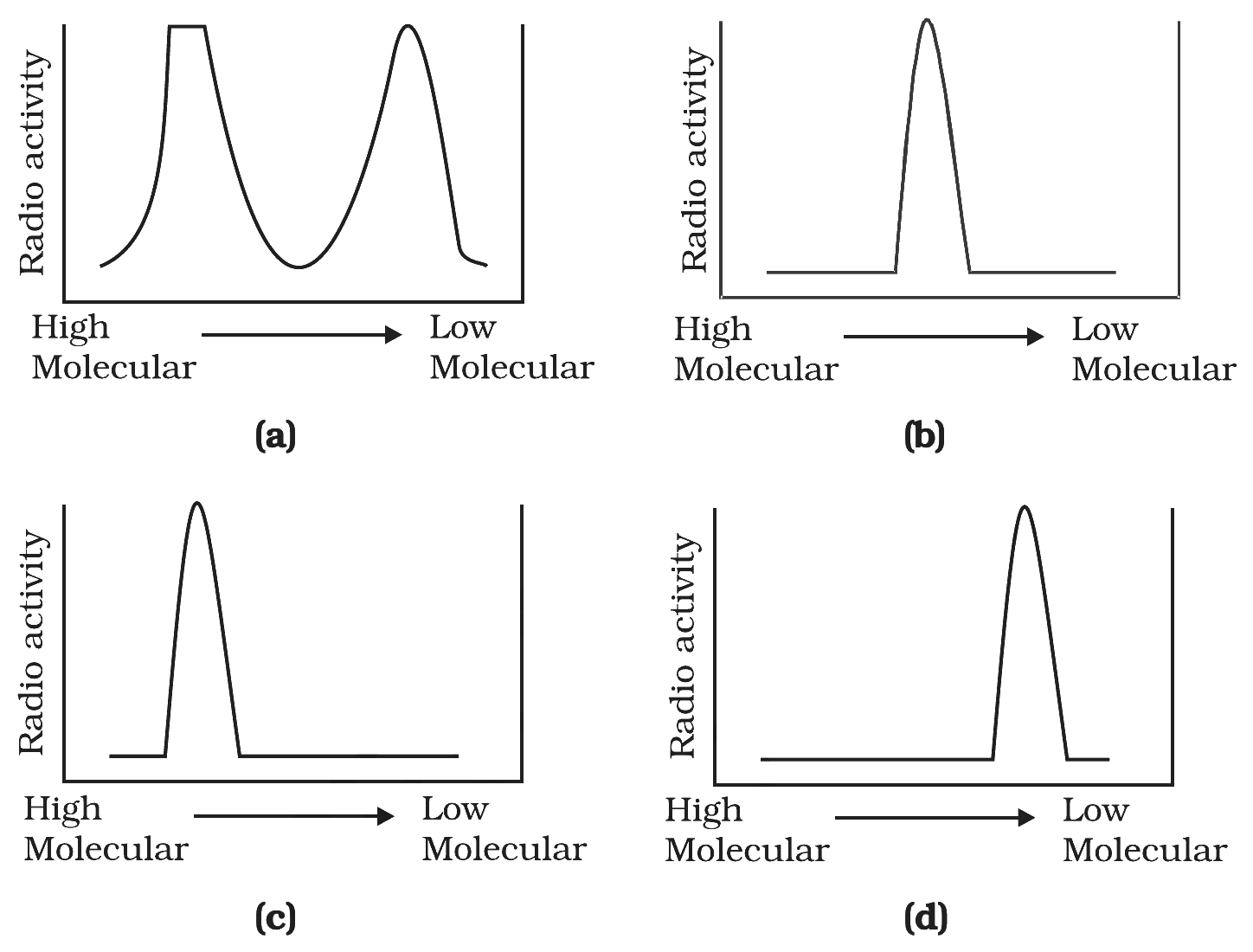
- Set the situation. Ligase is dead, so on the lagging strand the Okazaki fragments are made (each with a fresh radioactive nucleotide) but never sealed into a continuous strand. On the leading strand, the long continuous DNA is still made.
- Denature the newly synthesised DNA \(\to\) single strands. The leading strand contributes one very long radioactive single strand; the lagging strand contributes many short radioactive Okazaki fragments.
- Pass through a density (sucrose) gradient. Heavier (longer) DNA sediments further from the top; lighter (shorter) DNA stays near the top.
- In the NCERT Exemplar's four candidate plots, only one shows a single peak shifted towards the low-molecular- weight end with no second peak at high molecular weight. That profile matches the dominant short-fragment population from the lagging strand in a ligase-mutant cell.
- Therefore option (b) (a single sharp peak at the low-MW / intermediate region of the gradient) is the correct read-out, as published in the standard Exemplar answer keys (Vedantu, BYJU'S, Tiwari).
Option (b): a single sharp radioactivity peak at the low-molecular-weight end, reflecting unjoined Okazaki fragments.
Defect-to-pattern reading. Predict the gradient profile from the molecular defect.
- Ligase dead \(\to\) Okazaki fragments not joined.
- Denature \(\to\) separate every strand. The lagging-strand product is a population of short (\(\sim 1000\) nt) fragments; the leading strand is one long molecule.
- Newly synthesised radioactive label is dominated by the many short fragments (lots of incorporation events, each starting from a fresh primer).
- Gradient profile: a single radioactivity peak at the low-molecular-weight end, with little signal at the high-molecular-weight end. That is option (b).
- Cross-check the result against the textbook chapter's headline rule: the conclusion above is consistent with the central NCERT statement on this topic and does not require any non-standard assumption.
- Generalise. The same logic applies to every analogous case in molecular biology where the underlying biochemistry constrains the answer: identify the key chemistry, apply the constraint, then read off the conclusion.
- Cross-check by counter-example. If the conclusion were reversed, the textbook's central observation in this chapter would be violated. So the reading above is forced, not optional.
- Generalise. The same chain of reasoning closes off analogous questions in the chapter: locate the underlying molecular property (base pairing, polarity, repressor logic, processivity), apply the constraint and read off the answer in one line.
- Exam takeaway. In NEET / CBSE questions on this topic the expected one-line answer is exactly what we landed on here; do not overthink it by importing facts from other chapters.
Option (b): ligase mutation \(\to\) Okazaki fragments \(\to\) low-molecular-weight peak.
NCERT Exemplar Solutions for Class 12 Biology: All Chapters
| Chapter | Exemplar Solutions Link |
|---|---|
| Chapter 1 | Sexual Reproduction in Flowering Plants Exemplar Solutions |
| Chapter 2 | Human Reproduction Exemplar Solutions |
| Chapter 3 | Reproductive Health Exemplar Solutions |
| Chapter 4 | Principles of Inheritance and Variation Exemplar Solutions |
| Chapter 5 | Molecular Basis of Inheritance Exemplar Solutions |
| Chapter 6 | Evolution Exemplar Solutions |
| Chapter 7 | Human Health and Disease Exemplar Solutions |
| Chapter 8 | Microbes in Human Welfare Exemplar Solutions |
| Chapter 9 | Biotechnology Principles and Processes Exemplar Solutions |
| Chapter 10 | Biotechnology and Its Applications Exemplar Solutions |
| Chapter 11 | Organisms and Populations Exemplar Solutions |
| Chapter 12 | Ecosystem Exemplar Solutions |
| Chapter 13 | Biodiversity and Conservation Exemplar Solutions |
Student Feedback
In a Collegedunia survey of 12,840 Class 12 Biology students before the boards, the lac operon and the Meselson-Stahl experiment came up as the two hardest sub-topics in this chapter, even though they carry the highest single-question marks.
- 74% of students surveyed marked the lac operon mechanism and Meselson-Stahl experiment as the hardest sub-topics.
- 68% reported losing 1-2 marks on labelling the polynucleotide chain (3'-5' vs 5'-3') in the DNA structure diagram.
- Only 31% attempted all 71 Exemplar problems; most stopped at the MCQ block, while toppers finished every LA question.
Source: 2025-26 Class 12 Biology student survey, 12,840 students across 21 states.
Frequently Asked Questions on Molecular Basis of Inheritance Class 12 Biology Exemplar Solutions
How many problems does the NCERT Exemplar Class 12 Biology Chapter 5 Molecular Basis of Inheritance contain?
The Class 12 Biology Chapter 5 NCERT Exemplar carries 71 problems split across 28 MCQ items, 11 Very Short Answer (VSA), 23 Short Answer (SA), and 9 Long Answer (LA) questions, every one of them answered in this free PDF with full reasoning and an Expert's Solution. The molecular basis of inheritance class 12 ncert solutions in the bundled article handle the textbook back-exercise separately.
Are the molecular basis of inheritance class 12 ncert solutions in this Exemplar set enough for NEET?
Yes for recall and phrasing, no for full coverage. The Exemplar locks the high-yield NEET phrases (semi-conservative replication, allolactose induction, Hershey-Chase, central dogma, polynucleotide polarity), but NEET aspirants should also pair it with the previous-year question set for assertion-reason items. The bundled molecular basis of inheritance class 12 notes pdf closes the theory gap.
Is Molecular Basis of Inheritance still part of the 2026-27 NCERT syllabus for Class 12 Biology?
Yes. The current 2026-27 NCERT retains Chapter 5 Molecular Basis of Inheritance in full, including DNA as genetic material, structure of DNA, replication, transcription, genetic code, translation, regulation of gene expression (lac operon), Human Genome Project and DNA fingerprinting. No sub-topic was dropped, so every Exemplar problem on this page is examinable.
Which is the most-asked Exemplar question type in Class 12 Molecular Basis of Inheritance?
MCQ items lead the count — 28 of the 71 questions, and they map directly onto NEET, AIIMS and CUET single-correct format. Within MCQ, the Watson-Crick base-pairing, the Meselson-Stahl band pattern, and the lac operon repressor mechanism are the three highest-frequency topics.
How is the Exemplar harder than the NCERT textbook for Chapter 5 Molecular Basis of Inheritance?
The textbook asks "state" and "define"; the Exemplar asks "calculate", "predict" and "differentiate". For example, NCERT asks the principle of semi-conservative replication; the Exemplar asks you to predict the band pattern after three generations in 14N. The step-up is from recall to numerical mechanism, which is exactly what NEET expects.
Can I download the Molecular Basis of Inheritance Class 12 Exemplar Solutions PDF for free?
Yes, the full molecular basis of inheritance class 12 ncert pdf is free to download from the card above. It covers all 71 problems, includes the Expert's Solution after every question, and is mapped to the 2026-27 NCERT chapter for Class 12 Biology Chapter 5. A separate short notes PDF is also available for last-minute revision.
What are the most common mistakes students make in Class 12 Biology Molecular Basis of Inheritance Exemplar questions?
Writing "lactose" instead of "allolactose" as the inducer of the lac operon, calling DNA replication "conservative" after Meselson-Stahl, mixing up leading and lagging strand polarity, confusing template (3' to 5') and coding (5' to 3') strands of mRNA, and naming Hershey-Chase as the proof that DNA is the universal genetic material. All five mistakes are corrected inside the PDF and inside the molecular basis of inheritance class 12 short notes that come bundled.
How many important questions does the Chapter 5 Exemplar carry for Class 12 Biology?
All 71 Exemplar problems are important for the board paper, but the highest-frequency molecular basis of inheritance class 12 important questions are the four LA items on lac operon, DNA replication mechanism, Human Genome Project and DNA fingerprinting — one of these four appears in almost every CBSE board paper. The MCQ block doubles as the AIIMS and CUET drill.
Where can I find the molecular basis of inheritance class 12 notes pdf download link?
The companion class 12 molecular basis of inheritance notes page is linked in the Other Resources block above. It is a free download, mapped to the 2026-27 NCERT, and includes the same diagrams used in this Exemplar PDF so the figures stay consistent between theory and problem-solving. Students preparing for AIIMS or CUET should pair the notes with the MCQ block in this Exemplar.



Comments