The NCERT Solutions PDF for Chapter 10 Biomolecules helps students preparing for CBSE Boards, NEET and JEE Main in 2027. This chapter carries 3 to 5 marks in the CBSE Class 12 Chemistry paper.
It is also an NTA favourite for 'Assertion-Reason' questions in NEET, and this page carries the full solutions PDF and PYQs.
CBSE Weightage: 3-5 marks (Unit 8, combined with Amines).
JEE Main Weightage: 1-2%, usually one question on disaccharide linkage or vitamin classification.
NEET Weightage: 1-2 questions per paper, mostly carbohydrate classification and DNA-RNA differences.
These NCERT Solutions are mapped to the 2026-27 syllabus and refined against the last five years of CBSE, JEE Main and NEET papers. Biomolecules bridges organic Chemistry and Class 12 Biology, so each answer names the functional group, draws the glycosidic linkage and writes the assertion-reason logic in full.
Why Biomolecules is the Easiest 5-Mark Block of Class 12 Chemistry
Biomolecules is largely memory-driven: no mechanisms, no arrow-pushing, and only three named reactions of glucose (acetylation, oxidation with HNO3, reduction with HI). Learn the open-chain and cyclic structures of glucose plus the four levels of protein structure and you recover almost the whole chapter in two days. CBSE has repeated "differentiate starch and cellulose" in 2019, 2022, 2023 and 2024.
Watch Out: The 2026-27 NCERT dropped the detailed hormone classification. Skip hormone questions in pre-2023 papers; they will not appear in CBSE 2027.
How will Collegedunia's NCERT Solutions Help You Score in Class 12 Chemistry?
The solutions target the chapter's two trap zones: open-chain versus cyclic conversions and protein secondary-structure terminology. Each answer names the anomeric centre and the peptide bond and gives every "differentiate" question as a two-column table with the exact points CBSE rewards.
Quick Tip: Write the "ADEK" fat-soluble vitamin reminder (A, D, E, K) at the top of your answer sheet before the Biomolecules question. It saves the recall doubt that catches most students on vitamins.
Section 10.1 gives 8 of the 24 solutions, fixing the aldohexose vs ketohexose and pyranose vs furanose points where most students drop marks.
Classification and glucose: monosaccharides (glucose, fructose) do not hydrolyse; oligosaccharides (sucrose, maltose, lactose) give 2-10 units; polysaccharides (starch, cellulose, glycogen) give more than 10. Glucose is an aldohexose whose C5-OH closes onto C1 to form a pyranose ring with a new anomeric carbon. Alpha has -OH below the ring at C1, beta above.
Mutarotation and fructose: a pure anomer equilibrates in water (alpha +112, beta +19, mix +52.5 degrees). Fructose is a ketohexose that closes to a furanose ring and is laevorotatory, so "D" does not mean dextrorotatory; do not confuse anomers with epimers.
Sucrose, Maltose, Lactose and Polysaccharide Linkages - Section 10.1.3 to 10.1.4 Solutions
Six solutions cover disaccharide and polysaccharide structure. The two recycled concepts are sucrose hydrolysis / invert sugar and the alpha-1,4 vs beta-1,4 starch-cellulose linkage swap.
Sucrose hydrolysis (invert sugar): sucrose + H2O gives glucose + fructose and the rotation flips from +66.5 to -39.9, hence "invert sugar", used in honey and jams because it is sweeter than sucrose and resists crystallisation.
Amino Acids, Zwitterions and the Four Levels of Protein Structure - Section 10.2 Solutions
Five solutions cover proteins; the key points are the zwitterion, the peptide bond and the primary to quaternary classification.
Amino acids and zwitterion: general formula R-CH(NH2)-COOH; only glycine is achiral and the body cannot make the 10 essential ones. In water they form the dipolar zwitterion (net charge zero); the pH of zero charge is the isoelectric point.
Peptide bond and structure levels: a planar -CO-NH- amide links residues. Primary = sequence, secondary = alpha-helix (intra-chain H-bonds) or beta-sheet (inter-chain), tertiary = 3-D fold, quaternary = multiple chains (haemoglobin). Heat or urea denatures 2°-4° but leaves the peptide bonds intact.
Enzymes, Vitamins and Nucleic Acids - Sections 10.3 to 10.5 Solutions
Six solutions cover enzymes, vitamins and nucleic acids, the highest-NEET-density block here.
Enzymes and vitamins: enzymes are globular protein catalysts (usually "-ase") that lower activation energy with lock-and-key specificity, optimum near pH 7.4 and 37 degrees C. Fat-soluble A, D, E, K are stored; water-soluble B-complex and C need regular supply. Key deficiencies: A (night blindness), C (scurvy), B12 (pernicious anaemia).
Nucleic acids: a nucleoside is base + sugar, a nucleotide adds phosphate and links through 5' to 3' phosphodiester bonds. DNA has deoxyribose, thymine and a double helix (A=T, G≡C, Chargaff's rule); RNA has ribose, uracil and is single-stranded.
NCERT Class 12 Chemistry Chapter 10 Exercise-wise Question Map
The chapter has one main exercise of 24 questions plus 10 intext questions. The split below tags each by sub-topic so students can target one block at a time.
Disaccharides, polysaccharides, starch and cellulose linkages
Main Exercise Q11-Q15
5
Amino acids, peptide bonds, protein structure
Main Exercise Q16-Q20
5
Enzymes, enzyme action, vitamin classification
Main Exercise Q21-Q24
4
Nucleic acids, DNA double helix
Each five-question block targets one biomolecule family, and the PDF keeps this ordering for direct cross-reference.
Biomolecules Class 12 Previous Year Questions Weightage (2021-2026)
The year-wise pattern below shows Biomolecules as a steady 3-to-5-mark contributor.
Year
CBSE Board
JEE Main
NEET
2026
Structure and function of proteins (2 marks)
Vitamins classification, 1 Q
Nucleic acids: DNA versus RNA (1 Q)
2025
DNA structure, 3 marks; glucose reactions, 2 marks
Disaccharide linkage, 1 Q
Protein structure, 1 Q
2024
Starch vs cellulose, 3 marks
Amino acid classification, 1 Q
Vitamins, 1 Q
2023
Peptide bond + denaturation, 3 marks
-
Nucleic acids, 2 Qs
2022
Glucose open vs cyclic, 2 marks; enzymes, 3 marks
Carbohydrate classification, 1 Q
DNA-RNA difference, 1 Q
2021
Vitamins and deficiency, 2 marks
-
Anomeric carbon, 1 Q
No CBSE paper in the last 6 years has skipped Biomolecules, so plan for at least 3 guaranteed marks if the differentiation tables and glucose reactions are memorised.
Common Mistakes Students Make in Biomolecules Answers
Most mark losses here come from terminology slips, not concept gaps.
Dropping the alpha/beta on glycosidic linkages: writing "starch has 1,4 linkage" and stopping loses the alpha qualifier worth 1 of the 3 marks.
Calling RNA a double helix: only DNA is double-helical, a throwaway 1-mark loss.
Mixing vitamin solubility: Vitamin K is fat-soluble, B12 is water-soluble; swapping them is a common 1-mark loss.
Remember: Use the phrase "alpha-D-glucose" in full whenever the answer requires the glucose structure. Dropping the "alpha" or the "D" loses precision marks even when the drawing is correct.
All NCERT Solutions for Biomolecules with Step-by-Step Working
Every NCERT textbook question for Class 12 Chemistry Chapter 10 Biomolecules is listed below with its full Solution and Expert Solution inside collapsible tabs.
Questions
Q 10.1
What are monosaccharides?
Concept used.Carbohydrates are optically active polyhydroxy aldehydes or polyhydroxy ketones (or substances that yield these on hydrolysis). They are classified on the basis of their behaviour towards hydrolysis: the chemical breaking of a molecule by addition of water in the presence of an acid or an enzyme. On hydrolysis, a carbohydrate either splits into smaller sugar units or does not split at all.
The three families
Carbohydrates are grouped as: monosaccharides (do not hydrolyse further), oligosaccharides (give 2 to 10 monosaccharide units on hydrolysis) and polysaccharides (give many monosaccharide units on hydrolysis).
Define the term. Monosaccharides are those carbohydrates that cannot be hydrolysed further into smaller simpler sugar units. They are the simplest carbohydrates and the fundamental building blocks of all larger carbohydrates.
Recognise the general molecular formula. The general formula of a monosaccharide is CnH2nOn where n is usually 3, 4, 5, 6 or 7. Each carbon (except the carbonyl carbon) bears a hydroxyl (-OH) group; one carbon carries a carbonyl group (-CHO or >C=O).
Sub-classify. Monosaccharides containing an aldehyde group are called aldoses; those containing a ketone group are called ketoses. They are further classified by the number of carbon atoms: triose (3 C), tetrose (4 C), pentose (5 C), hexose (6 C), heptose (7 C).
Name common examples.
Aldohexose: glucose, C6H12O6.
Ketohexose: fructose, C6H12O6.
Aldopentose: ribose, C5H10O5.
Aldotriose: glyceraldehyde, C3H6O3.
[See diagram in the PDF version]
Monosaccharides are the simplest carbohydrates that cannot be hydrolysed further into smaller sugars. About 20 naturally occurring monosaccharides are known. Examples: glucose (C6H12O6), fructose (C6H12O6) and ribose (C5H10O5).
PS
Pranav Sharma
M.Sc Biochemistry, IIT Bombay
Verified Expert
Definition-first angle. The cleanest way to fix the idea of a monosaccharide in the mind is to anchor it against the two families that can be broken down: oligosaccharides and polysaccharides. A monosaccharide is the end-product of carbohydrate hydrolysis: once you reach it, water can no longer split it further.
Alternative approach: counting carbons before naming. A neat mental trick is to read the molecular formula first and the structure second. Any molecule of the form CnH2nOn where n ∈ 3,4,5,6,7 is automatically a monosaccharide of n carbons. So C3H6O3 is a triose, C5H10O5 a pentose, C6H12O6 a hexose; whether it is aldo or keto is then decided by which carbon carries the C=O. This carbon-count-first habit avoids the common mistake of guessing structure before reading the formula.
Hydrolysis test as a definition. If a carbohydrate X is boiled with dilute acid (or treated with an enzyme) and the product is still a carbohydrate of smaller mass, X is not a monosaccharide. If the same treatment leaves the molecule unchanged in carbohydrate skeleton, X is a monosaccharide.
General formula CnH2nOn with n = 3 to 7. Plug in n = 6: C6H12O6 describes glucose, fructose and galactose, three different monosaccharides with the same formula but different structures (structural isomers). Plug in n = 5: C5H10O5 describes ribose (RNA sugar); the deoxy form C5H10O4 is 2-deoxyribose (DNA sugar).
Reading the family name: aldo- or keto- fixes the carbonyl type; -triose, -tetrose, -pentose, -hexose fixes the carbon count. Numerical check: an aldohexose has 4 chiral carbons (C-2, C-3, C-4, C-5), so 24 = 16 possible stereoisomers; glucose is just one of them.
State examples by category:
Glucose: aldohexose;
Fructose: ketohexose;
Ribose: aldopentose (the sugar in RNA);
2-Deoxyribose: aldopentose with no -OH at C-2 (the sugar in DNA);
Galactose: aldohexose differing from glucose only at C-4 (a C-4 epimer).
Concept linkage. Monosaccharides are not isolated objects: the glucose you eat is the same glucose that builds the starch in a potato, the cellulose in cotton and the lactose in milk. The family connection is via the glycosidic linkage, a C-O-C bond joining two monosaccharides into a larger carbohydrate. Hold this thread loosely now - it returns in Q5 and Q8.
Why this matters. Every disaccharide and polysaccharide we will meet (sucrose, lactose, starch, cellulose, glycogen) is built from monosaccharide ``Lego bricks'' joined by glycosidic linkages. Knowing the bricks is the first step to reading the polymer. In NEET and JEE Main this single line, ``carbohydrates that cannot be hydrolysed further'', is the most-asked one-mark fact from biomolecules.
Exam-relevance flag. CBSE has asked the definition of a monosaccharide directly in 2014, 2017 and 2021 (1 mark each), and asked for ``examples of an aldopentose and a ketohexose'' as part of a 2-mark question in 2019. State the formula and at least one named example per category.
A monosaccharide is a carbohydrate that cannot be hydrolysed into a smaller carbohydrate. Formula CnH2nOn (n = 3–7). Examples: glucose, fructose, ribose.
Q 10.2
What are reducing sugars?
Concept used. A reducing sugar is any carbohydrate that can reduce a mild oxidising agent. The chemistry behind the name is simple: the sugar contains a free aldehyde (-CHO) group, or a free ketone (>C=O) that can tautomerise to an aldehyde, and this group gets oxidised to a carboxylic acid while it reduces metal ions like Cu2+ or Ag+ to Cu+ or Ag0.
Two classic tests
Fehling's test: blue Cu2+ in alkaline tartrate solution → brick-red Cu2O precipitate. Tollens' test: ammoniacal AgNO3→ silver mirror on the inside of the test tube. A positive result with either reagent confirms a reducing sugar.
Identify the chemical feature. The functional group that does the reducing is a free anomeric hydroxyl (which exists in equilibrium with the open-chain -CHO). In a free monosaccharide, this -OH is always present at C-1 (or C-2 of fructose), so all free monosaccharides are reducing.
Apply to each class.
All monosaccharides (glucose, fructose, ribose, galactose) are reducing sugars, because their anomeric -OH is always free.
Disaccharides are reducing only if at least one of the two anomeric -OH groups is free. Maltose and lactose are reducing; sucrose is not (both anomeric carbons are tied up in the glycosidic bond).
Polysaccharides are essentially non-reducing because the very few free anomeric ends are negligible compared to the size of the molecule.
State the redox equation in symbols. With Fehling's reagent: R-CHO + 2 Cu2+ + 5 OH- -> R-COO- + Cu2O v + 3 H2O. The red precipitate of Cu2O is the visible sign of a reducing sugar.
Reducing sugars are carbohydrates that reduce Fehling's solution to red Cu2O or Tollens' reagent to silver mirror, by virtue of a free aldehyde / ketone (anomeric -OH) group. All monosaccharides and most disaccharides (except sucrose) are reducing sugars.
AI
Aanya Iyer
Ph.D Organic Chemistry, IISc Bangalore
Verified Expert
Structural angle. Think of a sugar's ``reducing power'' as a question about its anomeric carbon: is it free or is it engaged?
Alternative approach: a flow-chart in three questions. (i) Is the sugar a free monosaccharide? If yes, it is reducing. (ii) If it is a disaccharide, ask: are BOTH anomeric centres tied up in the glycosidic bond? If yes, non-reducing (sucrose, trehalose). If only one is tied up, reducing (maltose, lactose, cellobiose). (iii) If it is a polysaccharide, the one or two free ends are negligible compared to the molecular mass, so the polysaccharide is operationally non-reducing.
Locate the anomeric carbon. In an aldose, it is C-1 (the one with the -CHO in the open chain or with the hemiacetal -OH in the ring). In a ketose like fructose, it is C-2. The anomeric carbon is the only one that carries two oxygens after cyclisation - a ring-O and a hemiacetal -OH.
``Free'' anomeric centre ⇒ sugar can open into the chain form ⇒ free aldehyde ⇒ reduces Cu(II)/Ag(I). ``Locked'' anomeric centre (engaged in a glycosidic bond) ⇒ no open chain ⇒ no reduction. The hemiacetal open-chain aldehyde equilibrium is what supplies the trace of -CHO that the test reagent oxidises.
Quick verdict table.
Glucose, fructose, galactose, mannose: free ⇒ reducing. (Fructose, though a ketose, tautomerises in alkaline Fehling's medium to an aldose via the Lobry-de-Bruyn-van-Ekenstein rearrangement, hence it tests positive.)
Maltose, lactose: only ONE anomeric centre locked, the other free ⇒ reducing.
Sucrose: BOTH anomeric centres locked ⇒ non-reducing. The glycosidic bond between C-1 of glucose and C-2 of fructose is a head-to-head linkage that uses up both reducing groups.
Concept linkage to amino acids and proteins. ``Reducing groups'' in biomolecules is a recurring theme: a free -CHO in a sugar, a free -NH2 in an amino acid, a free 5'-end in a nucleic acid. Each ``free'' end is a chemical handle. As soon as biology wants to make a polymer, it spends those free ends in covalent linkages.
Why this matters. The reducing/non-reducing distinction is the first chemical fingerprint of an unknown sugar in the lab and the basis of Benedict's test for glucose in urine (a common diabetes screen). It is also a standard 2-mark CBSE prompt: ``Why is sucrose non-reducing whereas maltose is reducing?''
Exam-relevance flag. The most common trap is to mark fructose as non-reducing because it is a ketone - wrong. Fructose IS reducing because alkaline Fehling's medium tautomerises it. Equally, do not mark starch as ``reducing because it has glucose units'' - wrong, its many anomeric carbons are all locked in glycosidic bonds.
Reducing sugars contain a free aldehyde or hemiacetal that gets oxidised by Fehling's or Tollens' reagent; all monosaccharides and disaccharides except sucrose are reducing.
Q 10.3
Write two main functions of carbohydrates in plants.
Concept used. Carbohydrates serve plants in two broad ways: as energy stores and as structural materials. Each role is played by a different polysaccharide built from glucose monomers.
Function 1: Storage of chemical energy. Starch is the main carbohydrate used by plants to store the chemical energy captured in photosynthesis. Starch is deposited as granules in seeds, tubers and roots. When the plant needs energy, enzymes hydrolyse starch to glucose, which is oxidised in respiration to release ATP.
Function 2: Structural support of cell walls. Cellulose is the principal structural polysaccharide of plants. It is a linear polymer of β-D-glucose units joined by C1→C4 glycosidic linkages. The straight chains align side-by-side and hydrogen-bond into rigid microfibrils that give the cell wall its tensile strength. Wood, cotton and flax are largely cellulose.
[See diagram in the PDF version]
Two main functions of carbohydrates in plants: (i) storage of food energy as starch, and (ii) structural support of cell walls as cellulose.
AV
Aditya Verma
M.Sc Biochemistry, IIT Kanpur
Verified Expert
Picture-first angle. Two polymers, same monomer, two completely different jobs. The difference is one stereochemical detail at C-1.
Alternative approach: think of the plant as a bank. Sugars made by photosynthesis are like cash flowing in. The plant has to park some of that cash in the vault (energy reserve → starch granules) and use some of it to build the walls of the bank (cell-wall material → cellulose microfibrils). One polymer is liquid (easy to hydrolyse back to glucose), the other is fixed (hard to hydrolyse). This ``bank-vs-bricks'' analogy makes the dual function memorable.
Energy stores need to release glucose on demand. Starch is α-D-glucose polymerised through α-1,4 (and α-1,6 in amylopectin) glycosidic bonds. The α geometry produces a helical, easily-hydrolysable chain that amylase enzymes can quickly digest. Starch granules in the endosperm of wheat and rice are exactly this kind of mobile energy reserve.
Structural roles need a rigid, water-resistant fibre. Cellulose is β-D-glucose polymerised through β-1,4 links. The β geometry forces each successive glucose to flip 180∘, producing a straight ribbon. Hundreds of ribbons hydrogen-bond into microfibrils that are insoluble in water and resistant to attack by most digestive enzymes. Cotton fibre is more than 90% pure cellulose; wood is about 50%.
Concept linkage to proteins and nucleic acids. The same ``storage vs structure'' duality reappears later: globular proteins (storage of catalytic identity) vs fibrous proteins (collagen, structural); DNA (storage of information) vs the ribosome and microtubules (structural scaffolding). The ``two-jobs'' design pattern is universal in biology.
Why this matters. Cows can graze grass because their gut bacteria produce cellulase; humans cannot, so cellulose passes through us as dietary fibre. The same molecule (glucose), assembled with two different stereochemistries, gives two utterly different biological roles. This single fact powers most of the world's textile industry (cotton, linen, viscose) and most of the world's food calories (rice, wheat, maize starch).
Exam-relevance flag. CBSE has asked ``State two functions of carbohydrates in plants'' as a 2-mark VSA in 2015, 2018 and 2022. The expected answer is the simple pair ``energy storage (starch)'' + ``structural support (cellulose)''. One extra mark is sometimes given for naming a specific tissue (endosperm/cell wall).
In plants, carbohydrates (i) store chemical energy (starch) and (ii) build the cell wall (cellulose).
Q 10.4
Classify the following into monosaccharides and disaccharides:
Ribose, 2-deoxyribose, maltose, galactose, fructose and lactose.
Concept used. Apply the hydrolysis test from Q1. A monosaccharide cannot be hydrolysed into a smaller sugar; a disaccharide hydrolyses into exactly two monosaccharide units.
Go through each name and recall whether it hydrolyses.
Ribose: aldopentose (C5H10O5); does not hydrolyse. Monosaccharide.
2-Deoxyribose: aldopentose with no -OH at C-2; does not hydrolyse. Monosaccharide.
Maltose: hydrolyses to two glucose units. Disaccharide.
Galactose: aldohexose (C6H12O6); does not hydrolyse. Monosaccharide.
Fructose: ketohexose (C6H12O6); does not hydrolyse. Monosaccharide.
Lactose: hydrolyses to glucose + galactose. Disaccharide.
Pattern-spotting angle. A useful mnemonic: most disaccharides end in ``-ose'' and contain the syllable indicating a pair (sucr-, malt-, lact-). All four other names here are simple sugar names you have seen separately in metabolism.
Alternative approach: molecular-formula sieve. Each monosaccharide on the list obeys CnH2nOn (ribose C5H10O5, fructose C6H12O6, etc.), while each disaccharide on the list obeys C12H22O11 (maltose, lactose). Counting atoms in the formula immediately classifies the sugar: total carbon ≤ 7⇒ monosaccharide; carbon = 12 with one water lost ⇒ disaccharide.
Carbon count check. Ribose and 2-deoxyribose are 5-carbon pentoses (C5H10O5 and C5H10O4 respectively). Galactose and fructose are 6-carbon hexoses (C6H12O6). All four are single-ring sugars: monosaccharides.
Hydrolysis check. Maltose is barley sugar (C12H22O11); hydrolysis gives 2 glucose. Lactose is milk sugar (C12H22O11); hydrolysis gives glucose + galactose. Both are double-ring sugars: disaccharides.
Stereochemical note. Galactose and glucose are C-4 epimers- they differ only at C-4. Fructose is a structural isomer of glucose, differing in functional group (ketone vs aldehyde) not in stereochemistry. Knowing these relations helps you read more advanced Exemplar questions.
Final grouping in two lists exactly as in the boxed answer.
Why this matters. Recognising the family of a sugar at sight is the foundation for understanding why milk gives babies energy (lactose → glucose + galactose) and why bread tastes sweeter as you chew (amylase breaks starch to maltose, then to glucose). It also prepares the ground for Q5, Q6 and Q7, where we look at the glycosidic bond that joins these monosaccharides into the actual disaccharides on the list.
Concept linkage. Of the four monosaccharides here, ribose and 2-deoxyribose reappear as the sugar backbones of RNA and DNA (Q21, Q24); galactose is one half of lactose; fructose is one half of sucrose. So today's classification is tomorrow's biopolymer chemistry.
What do you understand by the term glycosidic linkage?
Concept used. A glycosidic linkage is the covalent C-O-C bond that joins two monosaccharide units in a di- or poly-saccharide. It is formed by a condensation reaction in which the hemiacetal -OH at the anomeric carbon of one sugar reacts with an -OH of a second sugar, eliminating one molecule of water.
Anomeric carbon, hemiacetal
The anomeric carbon of a cyclic sugar is the carbon that was the carbonyl carbon in the open chain (C-1 in aldoses, C-2 in ketoses). It carries the hemiacetal-OH that condenses with another sugar to form a glycosidic bond.
General reaction (with loss of H2O): Sugar1-OH + HO-Sugar2 -> Sugar1-O-Sugar2. The new C-O-C bond is the glycosidic linkage.
In sucrose, C-1 of α-D-glucose is linked to C-2 of β-D-fructose: this is an α-1,2-glycosidic linkage.
In maltose, C-1 of one α-D-glucose is linked to C-4 of a second α-D-glucose: an α-1,4-glycosidic linkage.
In lactose, C-1 of β-D-galactose is linked to C-4 of β-D-glucose: a β-1,4-glycosidic linkage.
[See diagram in the PDF version]
A glycosidic linkage is the C-O-C bond formed when the hemiacetal -OH of one sugar condenses with an -OH of another sugar, with elimination of H2O. It is the bond that joins monosaccharide units in di- and poly-saccharides.
KJ
Karan Joshi
M.Sc Chemistry, IIT Madras
Verified Expert
Reaction-mechanism angle. A glycosidic bond is just an acetal formed from a hemiacetal: classical organic chemistry applied to sugars.
Alternative approach: Fischer vs Haworth notation. A Fischer projection draws the open chain vertically with -CHO at the top; the glycosidic bond cannot be shown easily because the chain has to close into a ring first. The Haworth projection draws the cyclic form as a flat hexagon (pyranose) or pentagon (furanose) with the glycosidic -O- shown as a clean bond between two ring carbons. Whenever a question mentions a glycosidic bond, switch to Haworth - the structure jumps off the page.
Open-chain glucose has -CHO at C-1. When it cyclises, the C-5 -OH attacks the -CHO carbon, generating a hemiacetal: a carbon bearing -OH and -OR on the same atom. This is the same hemiacetal step you learned for aldehyde + alcohol → hemiacetal in the carbonyl chemistry of Class 12 Unit 8.
The new -OH at C-1 (the anomeric hydroxyl) is reactive. A second alcohol R'-OH can displace water from it to give the corresponding acetal: a carbon bearing two -OR groups. This C-O-R' is the glycosidic linkage. The α or β label tells you which face of the ring the new -OR projects from.
Hence every disaccharide is, mechanistically, the acetal of a sugar with another sugar acting as the alcohol. The general notation α-1,4 says: α-anomer of the first sugar + its C-1 + bonded through O + to C-4 of the second sugar.
Numerical / structural assignment of D vs L. In a Haworth projection drawn with C-1 at the right and the ring oxygen at the top right, the -CH2OH at C-6 points up for D-sugars and down for L-sugars. In the Fischer projection, look at the highest-numbered chiral carbon (C-5 in a hexose): -OH on the right ⇒ D-sugar; on the left ⇒ L-sugar. All naturally occurring monosaccharides in food are D-sugars.
Why this matters. Acetals are stable to base but cleaved by acid or by specific enzymes (glycosidases). That is exactly why sugars survive in plant cells (largely neutral pH) and yet are digested in the small intestine, where amylase and maltase quickly hydrolyse the glycosidic bond back to monosaccharides. The same acetal stability is also why processed sugar (sucrose) keeps for years on the shelf.
Exam-relevance flag. CBSE often pairs ``Define glycosidic linkage'' with ``state the type of glycosidic bond in sucrose / maltose / lactose / cellulose''. Memorise the table: sucrose α-1,β-2; maltose α-1,4; lactose β-1,4; cellulose β-1,4; starch α-1,4 with α-1,6 branches; glycogen α-1,4 + α-1,6.
Glycosidic linkage = the C-O-C (acetal) bond joining two monosaccharide units, formed by condensation between the anomeric -OH of one sugar and an -OH of the next.
Q 10.6
What is glycogen? How is it different from starch?
Concept used.Glycogen is the storage polysaccharide of animals: the polymeric form in which the human and animal body stores glucose. It is a highly branched polymer of α-D-glucose linked by α-1,4 glycosidic bonds in the main chains and α-1,6 bonds at the branch points.
Where it is stored. Glycogen is stored mainly in the liver (large reserve, regulates blood glucose) and in skeletal muscle (used directly for muscle contraction). It is often called animal starch because of its role.
Compare with starch. Starch, the storage carbohydrate of plants, is a mixture of two polysaccharides: amylose (linear, α-1,4 linked, ∼15–20%) and amylopectin (branched, α-1,4 with α-1,6 branch points, ∼80–85%).
Therefore the structural difference is the degree of branching: glycogen is more highly branched than amylopectin, with branch points roughly every 8–12 glucose residues (vs every 24–30 in amylopectin).
[See diagram in the PDF version]
Glycogen is the animal storage polysaccharide of α-D-glucose, stored in liver and muscle. It is more highly branched than starch and contains only one type of polymer, while starch is a mixture of mostly-linear amylose and lightly-branched amylopectin.
AB
Ananya Banerjee
Ph.D Molecular Biology, NCBS Bangalore
Verified Expert
Comparison angle. Glycogen and starch are first cousins: same monomer (glucose), same family of bonds (α-1,4 and α-1,6), different geometries.
Alternative approach: count the chain ends. Each branch is a new chain end. The number of chain ends per molecule sets the maximum rate at which phosphorylase enzymes can release glucose phosphate. Glycogen, with the densest branching (every 8–12 residues), has the most ends per gram and so releases glucose fastest. Starch (amylopectin branched every 24–30 residues; amylose unbranched) is slower. Plants do not mind: their metabolic clock is days, not seconds.
Glycogen → branched at every 8–12 residues; appears as a bush-shaped molecule under the electron microscope. Stored in cytosolic granules in liver and muscle cells. Liver glycogen regulates blood glucose between meals; muscle glycogen powers sprinting and weight-lifting.
Amylose component of starch → unbranched, helical coil of α-1,4-linked glucose. Gives the characteristic deep-blue iodine-starch complex (iodine sits inside the helix). About 15–20% of typical plant starch.
Amylopectin component of starch → branched but less than glycogen. Branching every 24–30 residues. About 80–85% of typical plant starch.
Functional consequence: glycogen's many branches give animals rapid access to glucose; starch's fewer branches suit plants' slower energy demands.
Concept linkage to the chapter map. The four polysaccharides you see in this chapter - amylose, amylopectin, cellulose, glycogen - are all polymers of D-glucose. The only variables are (i) anomer (α vs β) and (ii) branching pattern. With this single insight all four polysaccharides compress into a single table.
Why this matters. The same chemistry can be tuned by branching density to give two very different physiological storage strategies. This is one of the cleanest examples in biochemistry of ``structure dictates rate''.
Exam-relevance flag. ``Why is glycogen more branched than starch?'' is a recurring 3-mark CBSE question (asked 2016, 2020). The expected line is the one above: ``Animals need rapid glucose release, so more branches ⇒ more chain ends ⇒ faster mobilisation.'' Memorise the two numbers 8--12 vs 24--30 to score the data mark.
Glycogen is the animal-body equivalent of starch, but more highly branched. Glycogen contains a single, heavily branched polymer of α-D-glucose; starch is a mixture of mostly-linear amylose and moderately branched amylopectin.
Q 10.7
What are the hydrolysis products of
(i) sucrose and (ii) lactose?
Concept used. A disaccharide is the condensation product of two monosaccharides; on hydrolysis (acid or enzyme catalysed), the glycosidic bond is cleaved by addition of water and the two original monosaccharides are recovered. Disaccharide + H2O H+ Monosaccharide1 + Monosaccharide2.
Sucrose (C12H22O11), common cane or table sugar, is joined through an α-1,β-2 glycosidic bond. On hydrolysis with dilute mineral acid or the enzyme invertase: C12H22O11 + H2O ->[H+/invertase] C6H12O6 + C6H12O6 i.e. D-(+)-glucose and D-(-)-fructose in equimolar amounts. This hydrolysis is called the inversion of sucrose because the optical rotation changes from +66.5∘ (sucrose, dextrorotatory) to -19.9∘ (the mixture, levorotatory, because the strongly levorotatory fructose dominates over the dextrorotatory glucose).
Lactose (C12H22O11), the natural sugar of mammalian milk, contains a β-1,4 glycosidic bond. On hydrolysis with dilute acid or the intestinal enzyme lactase: C12H22O11 + H2O ->[H+/lactase] C6H12O6 + C6H12O6 i.e. β-D-galactose and β-D-glucose.
[See diagram in the PDF version]
(i) Sucrose →D-glucose + D-fructose.
(ii) Lactose →D-galactose + D-glucose.
RM
Rohit Mehta
M.Sc Biochemistry, IIT Madras
Verified Expert
Quick reading. Two disaccharides, two hydrolyses, four monosaccharides in total. Glucose appears in both products.
Alternative approach: acid or enzyme. Both cleavages can be driven by either dilute mineral acid (general, slow, no specificity) or by a specific enzyme: invertase (also called sucrase) for sucrose; lactase for lactose; maltase for maltose. The enzyme route is fast and specific (one enzyme per substrate) and is the route used in your small intestine.
Sucrose: α-D-glucopyranosyl-β-D-fructofuranoside. Acidic hydrolysis cleaves the unique α,β-1,2 glycosidic bond to give glucose + fructose. The reaction is sometimes called invertase hydrolysis; the product mixture (equal moles of glucose and fructose) is invert sugar (Honey contains a lot of invert sugar).
Lactose: β-D-galactopyranosyl-(1→4)-D-glucose. Acid or the enzyme lactase cleaves the β-1,4 bond to give galactose + glucose. People with lactose intolerance lack the enzyme lactase, so undigested lactose passes to the colon and causes discomfort. Genetic ``lactase persistence'' into adulthood is concentrated in north-European and north-Indian populations.
Concept linkage to Q5. Both hydrolyses are the chemical reverse of the glycosidic-linkage formation discussed in Q5. Q5: condensation, loss of H2O; Q7: hydrolysis, addition of H2O. Same bond, two directions.
Why this matters. Hydrolysis turns a dietary disaccharide into absorbable monosaccharides. Without this single chemical step sucrose and lactose could not nourish us. The same hydrolysis chemistry powers fermentation in industry: sucrose → glucose + fructose → ethanol + CO2 (in yeast).
Exam-relevance flag. CBSE asks ``Write the hydrolysis products of sucrose / lactose / maltose'' as a 2-mark VSA almost every year. The answer is two reagents + two products + (optional) the enzyme name. Add the inversion-of-sucrose line for an extra mark.
What is the basic structural difference between starch and cellulose?
Concept used. Both starch and cellulose are polysaccharides of D-glucose, yet they differ in one stereochemical detail at C-1 of each monomer and one consequence in chain shape. That detail is the α vs βanomeric configuration.
Starch is built from α-D-glucose units. In each unit the -OH at C-1 points downward relative to the ring plane (α-anomer). Adjacent residues are joined by α-1,4 glycosidic linkages; amylopectin contains α-1,6 branch points. The α geometry produces a helical (coiled) chain.
Cellulose is built from β-D-glucose units. In each unit the -OH at C-1 points upward (β- anomer). Adjacent residues are joined exclusively by β- 1,4 glycosidic linkages. The β geometry forces each successive glucose to flip 180∘, producing a long, straight, ribbon-like chain.
Macroscopic consequence. Helical α-glucan chains pack loosely and trap iodine to give the deep-blue starch-iodine colour; straight β-glucan chains hydrogen-bond side-by-side into rigid microfibrils that build cell walls and cannot be digested by humans.
[See diagram in the PDF version]
Starch consists of α-D-glucose units linked by α-1,4 (and α-1,6 branch) glycosidic bonds and forms a coiled chain; cellulose consists of β-D-glucose units linked exclusively by β-1,4 glycosidic bonds and forms a straight, fibrous chain.
VN
Vivaan Nair
Ph.D Organic Chemistry, IISc Bangalore
Verified Expert
Structural angle. The whole difference rides on the chirality at C-1 of each monomer. Two letters (α, β), two completely different biological materials.
Alternative approach: Haworth projection comparison. Draw two glucose hexagons. For α-glucose the C-1 -OH points down (below the ring plane); for β-glucose it points up. In the polymer, the α linker holds successive rings on the same face, so the chain curls into a helix. The β linker forces every second ring to flip, so the chain straightens into a ribbon. Sketching two adjacent hexagons in each linkage type makes the geometric difference unforgettable.
Same monomer: C6H12O6 (D-glucose).
Two anomers at C-1: α (-OH down) and β (-OH up). Both exist in equilibrium with the open chain in solution (mutarotation), but once the monomer enters a glycosidic bond, its anomeric configuration is locked. This is the same mutarotation phenomenon discussed in Q10 below.
In starch every glycosidic bond is α; in cellulose every bond is β. The cumulative effect over thousands of residues converts a flexible coil (starch) into a rigid rod (cellulose). The ribbon-like cellulose chains then hydrogen-bond side-by-side into microfibrils that build the plant cell wall.
Enzymatic consequence: amylases that recognise α-1,4 do not bind β-1,4 (and vice versa). Humans secrete amylase but not cellulase, so we digest starch but not cellulose. Cows, sheep and termites host gut bacteria that secrete cellulase and so derive nutrition from grass and wood.
Concept linkage. The α vs β choice is exactly the same chemical handle that distinguishes maltose (α-1,4) from cellobiose (β-1,4), and is one of the few places in the chapter where pure stereochemistry has a macroscopic biological consequence.
Why this matters. The simplest possible change in stereochemistry - flipping one -OH from down to up - converts the body's main fuel into one of the strongest natural fibres. It is a clean reminder that biology cares about three-dimensional shape, not just composition.
Exam-relevance flag. ``State the basic structural difference between starch and cellulose'' is a stand-alone 2-mark CBSE question (asked 2014, 2017, 2023). Two crisp lines -α-1,4 vs β-1,4 + helical vs linear - get full marks.
Starch = α-1,4 (and α-1,6) linked α-D-glucose, helical and digestible; cellulose = β-1,4 linked β-D-glucose, linear and indigestible (to humans).
Q 10.9
What happens when D-glucose is treated with the following reagents?
(i) HI (ii) Bromine water (iii) HNO3.
Concept used. The reactions of glucose tell us about its functional groups. Open-chain D-glucose, CH2OH(CHOH)4CHO, contains five -OH groups and one -CHO group; the cyclic form (pyranose) contains a hemiacetal. Different reagents probe different features.
(i) With prolonged heating with excess HI. HI is a strong reducing agent that cleaves C-O bonds and replaces -OH groups with -H. Heating glucose with concentrated HI (red phosphorus, Δ) removes all five -OH groups and reduces the -CHO as well, giving a straight-chain alkane with the same carbon skeleton: CH2OH-(CHOH)4-CHO ->[HI, P, Δ] CH3-(CH2)4-CH3 i.e. n-hexane. This product confirms that glucose has a straight chain of six carbon atoms.
(ii) With bromine water.
Bromine water is a mild oxidising agent that oxidises an aldehyde to the corresponding carboxylic acid (but does not oxidise primary or secondary alcohols). With glucose: CH2OH-(CHOH)4-CHO ->[Br2/H2O] CH2OH-(CHOH)4-COOH The product is D-gluconic acid, a monocarboxylic acid. This proves the presence of an aldehyde group in glucose.
(iii) With nitric acid (concentrated, on heating).
HNO3 is a stronger oxidising agent. It oxidises both the -CHO at C-1 and the primary -CH2OH at C-6 to -COOH groups, but leaves the secondary -OH groups intact: CH2OH-(CHOH)4-CHO conc. HNO3, Δ HOOC-(CHOH)4-COOH The product is the dicarboxylic acid D-saccharic acid (also called D-glucaric acid). This shows that glucose has a primary alcoholic (-CH2OH) group along with an aldehyde.
[See diagram in the PDF version]
(i) HI →n-hexane; (ii) Bromine water →gluconic acid; (iii) HNO3 →saccharic acid (D-glucaric acid).
YK
Yash Kapoor
M.Sc Chemistry, IIT Kanpur
Verified Expert
Picture-first angle. Each reagent is a chemical ``stain'' that lights up one feature of glucose's open chain.
Alternative approach: read the carbon count of each product.n-Hexane has 6 carbons in a straight chain → glucose has 6 carbons in a straight chain. Gluconic acid has 6 carbons and one -COOH→ glucose has one -CHO at the end. Saccharic acid has 6 carbons and two -COOH groups → glucose has -CHO at one end and -CH2OH at the other end. Three products, three structural facts.
HI is the most aggressive: every oxygen leaves the molecule. What is left is the bare carbon skeleton, which proves straight chain of six carbons (the carbons cannot rearrange under HI conditions). Mechanism: HI reduces each C-OH via C-OH → C-I → C-H (the iodide is then displaced by HI acting as a hydride source).
Bromine water is selective: only the aldehyde is oxidised to -COOH. The fact that a monocarboxylic acid (gluconic acid) forms proves that there is exactly one aldehyde group. Bromine water does not touch the primary or secondary -OH groups under these conditions.
HNO3 is stronger still: it gets both the aldehyde and the primary alcohol. The fact that a dicarboxylic acid (saccharic acid) forms proves that there is also a primary -OH somewhere - at the opposite end of the chain (C-6), since secondary alcohols are not oxidised by HNO3 at the same temperature.
Concept linkage. These three reactions together prove the open-chain structure CH2OH-(CHOH)4-CHO. But Q10 will immediately show that this open chain is not the whole story and that a cyclic hemiacetal is also required. Q9 + Q10 together teach you the full ``story of D-glucose''.
Why this matters. These three classical reactions are the historical evidence behind the open-chain structure CH2OH-(CHOH)4-CHO for D-glucose. They are textbook examples of how organic chemists deduce the skeleton of an unknown by sequential ``probe'' reactions.
Exam-relevance flag. This is a 3-mark CBSE staple. Marks break-up: 1 mark per product (named correctly with formula). The diagram of glucose → three arrows → three products is worth a bonus mark when neatly drawn.
HI →n-hexane; bromine water → gluconic acid; HNO3 → saccharic acid.
Q 10.10
Enumerate the reactions of D-glucose which cannot be explained by its open chain structure.
Concept used. The open-chain (Fischer) structure of D-glucose CH2OH-(CHOH)4-CHO does not account for every experimental observation. Whenever a property points to a free aldehyde and another property denies it, we need a second, cyclic hemiacetal structure to reconcile both.
Glucose does not give the classical 2,4-dinitrophenylhydrazone with 2,4-DNP, nor does it form a bisulphite addition product with NaHSO3. Both reactions are characteristic of free aldehydes; their failure suggests the aldehyde group of glucose is not freely available.
Glucose does not respond to the Schiff's test for aldehydes (which gives the magenta colour with rosaniline / fuchsin). Again, a normal aldehyde would test positive.
Pentaacetate of glucose (obtained by treating glucose with acetic anhydride) does not react with hydroxylamine NH2OH. This shows that the -CHO group is no longer present in the acetate, meaning C-1 is not actually -CHO but is instead an -OH that gets acetylated. Six -OAc groups are obtained from glucose, not five - the extra one comes from the C-1 hemiacetal -OH.
Glucose exists in two crystalline forms with different specific rotations: α-D-glucose, [α]D = +111∘, and β-D-glucose, [α]D = +19.2∘. A fresh solution of either form slowly changes its rotation until both reach an equilibrium value of +52.7∘. This phenomenon is called mutarotation and is impossible to explain with a single open-chain structure. It is explained by the existence of two cyclic anomers in equilibrium with each other through the open chain.
[See diagram in the PDF version]
Failures of the open-chain structure: (i) no 2,4-DNP hydrazone; (ii) no bisulphite addition; (iii) no Schiff's test colour; (iv) glucose pentaacetate is unreactive to NH2OH; (v) mutarotation: existence of two crystalline anomers (α and β) with different specific rotations.
TD
Tara Desai
M.Sc Biochemistry, JNU
Verified Expert
Evidence-first angle. The cyclic structure of glucose was deduced from these anomalies, not assumed beforehand. Five clean experiments, one new structure.
Alternative approach: ask ``what would a real aldehyde do?'' For each test, picture what acetaldehyde or benzaldehyde would do: form a 2,4-DNP, give a bisulphite adduct, turn Schiff reagent pink, react with NH2OH. Now compare with glucose. Wherever glucose fails, it is hiding its -CHO inside a ring. The cyclic hemiacetal answer is just one structural change that explains all five experimental failures in one stroke.
No 2,4-DNP. A free -CHO would give an orange-yellow hydrazone with 2,4-dinitrophenylhydrazine. Glucose does not (or only very slowly through the trace amount of open chain at equilibrium).
No bisulphite addition. Aldehydes form crystalline R-CH(OH)-SO3Na with NaHSO3. Glucose does not.
No Schiff's colour. The Schiff reagent gives a magenta dye with aldehydes. Glucose is silent.
Glucose pentaacetate (5 -OAc on the open chain, or 6 -OAc on the cyclic form - in practice you can isolate only the cyclic acetate) does not react with NH2OH, showing C-1 is no longer -CHO.
Mutarotation. Two crystalline anomers, one rotation each, both drift to the same equilibrium value: the chain is opening and closing in solution. Numerically, α-D-glucose ([α]D = +111∘) and β-D-glucose ([α]D = +19.2∘) both relax to the equilibrium +52.7∘ in water; that single specific-rotation triple is a CBSE favourite.
Concept linkage. The same hemiacetal-acetal story you learned in Unit 8 (aldehydes + alcohols → hemiacetal → acetal) is the engine of glucose's cyclic form, of the glycosidic linkage (Q5), and of every disaccharide and polysaccharide in the chapter.
Why this matters. The pyranose ring is the foundational picture for everything that follows in biochemistry: starch, cellulose, glycogen, nucleic-acid sugars, glycoproteins. All of it rests on these five anomalies and their cyclic-form explanation.
Exam-relevance flag. CBSE has asked this 5-marker as recently as 2022. Score full marks by writing exactly 5 anomalies + 1 concluding line about the pyranose ring + a labelled diagram showing α open chain β.
Open-chain glucose cannot explain: lack of 2,4-DNP / bisulphite / Schiff colour, formation of glucose pentaacetate inert to NH2OH, and mutarotation. The cyclic hemiacetal (pyranose) form resolves all five.
Q 10.11
What are essential and non-essential amino acids? Give two examples of each type.
Concept used. The human body synthesises proteins from twenty standard α-amino acids. Ten of these we can make from other metabolites; the other ten must come from the diet. The dietary ones are called essential; the others are non-essential.
Essential amino acids are those amino acids that the human body cannot synthesise (or cannot synthesise in adequate amounts) and which therefore must be supplied through food. Their absence from the diet leads to protein deficiency disorders such as kwashiorkor.
Non-essential amino acids are those amino acids that the body can synthesise from other amino acids and metabolic intermediates, so they need not be supplied ready-made in food.
Essential amino acids must be supplied through diet (the body cannot make them): examples include valine and leucine. Non-essential amino acids can be made by the body itself: examples include glycine and alanine.
IB
Ishita Bhat
M.Sc Biotechnology, AIIMS Delhi
Verified Expert
Definition-first angle. ``Essential'' is a nutritional adjective, not a chemical one: it tells you about human metabolism, not about how the amino acid behaves in a peptide.
Alternative approach: how does the question set up the answer? The question asks for both the definition and two examples of each type. Write the definitions in parallel form: ``Essential = cannot be synthesised must come from diet''; ``Non-essential = can be synthesised need not come from diet''. Parallel phrasing helps the marker tick both halves. Add two examples per type, no more (extra examples don't add marks).
All twenty standard α-amino acids are chemically similar: each has -NH2, -COOH, -H and a side chain R on the central (α) carbon. The chemical identity of the amino acid is set by the side chain.
Whether we call one ``essential'' depends on whether human cells have the enzymes to synthesise its carbon skeleton. We lack such enzymes for lysine, methionine, tryptophan, threonine, valine, leucine, isoleucine, phenylalanine, histidine and (for children) arginine.
Two examples each, as required:
Essential: lysine, leucine.
Non-essential: glycine, alanine.
Numerical note for assignment. ``9 essential'' is the standard adult count (some textbooks say 10 by including arginine as conditionally essential for infants). Out of 20 standard amino acids, 9 essential + 11 non-essential is the safe count for Class-12 CBSE.
Concept linkage to proteins (Q12) and enzymes (Q17). Every peptide bond in every protein you eat is hydrolysed in the stomach back to amino acids; your cells then re-link them into your own proteins. So a dietary deficiency in any one essential amino acid limits the synthesis of every protein that contains it.
Why this matters. A vegetarian diet has to be designed so that lysine (low in cereals) and methionine (low in pulses) complement each other, ensuring all essentials are present. This is the chemical reason why the rice-dal (or roti-dal) combination is the nutritional backbone of Indian cuisine.
Exam-relevance flag. CBSE has asked this question almost verbatim in 2015, 2018, 2020 and 2023. The expected answer is exactly two definitions + two examples each + (optional) a line on dietary significance. Stick to that template; don't list all ten essentials.
Essential: must come from diet (e.g. lysine, leucine). Non-essential: synthesised by the body (e.g. glycine, alanine).
Q 10.12
Define the following as related to proteins:
(i) Peptide linkage (ii) Primary structure (iii) Denaturation.
Concept used. Proteins are polymers of α-amino acids linked by peptide bonds. Their three-dimensional shape is organised into four hierarchical levels (primary, secondary, tertiary, quaternary). Heat or chemical assault can destroy higher-order structure - a process called denaturation.
(i) Peptide linkage. The peptide bond (or peptide linkage) is the amide bond -CO-NH- formed by a condensation reaction between the α-carboxyl (-COOH) group of one amino acid and the α-amino (-NH2) group of the next amino acid, with loss of one molecule of water (loss of H2O): !$H2N-CHR1-COOH + H2N-CHR2-COOH -> H2N-CHR1-CO-NH-CHR2-COOH$. The product H2N-CHR1-CO-NH-CHR2-COOH is a dipeptide. Repeating this condensation builds tri-, tetra-,, polypeptides.
(ii) Primary structure. The primary structure of a protein is the specific linear sequence of amino acids joined by peptide bonds, read from the N-terminus (free -NH2) to the C-terminus (free -COOH). Even a single change in this sequence may produce a totally different protein (e.g. one amino-acid change converts normal haemoglobin into sickle-cell haemoglobin).
(iii) Denaturation. Denaturation is the loss of a protein's biological activity due to the destruction of its secondary, tertiary and quaternary structure, while the primary structure (the peptide bonds) remains intact. Causes: heat, change of pH (strong acid or base), heavy-metal ions, organic solvents (ethanol), detergents and ultraviolet radiation. Familiar examples are the coagulation of egg-white on heating and the curdling of milk by lemon juice.
[See diagram in the PDF version]
(i) Peptide linkage = amide bond -CO-NH- between two amino acids. (ii) Primary structure = exact sequence of amino acids in the polypeptide chain. (iii) Denaturation = loss of protein's native 3-D shape and biological activity, with primary structure preserved.
AP
Aarav Pillai
Ph.D Biochemistry, IISc Bangalore
Verified Expert
Structural angle. Each term sits at one rung of the protein ``structure ladder'': peptide bond (atomic), primary structure (sequence), denaturation (loss of higher-order folds).
Alternative approach: build a protein ladder in your mind. Imagine four floors of a building: atomic (peptide bond) → sequence (primary) → local fold (α-helix, β-sheet = secondary) → overall fold (tertiary) → assembly of chains (quaternary). Denaturation collapses floors 2, 3 and 4 but leaves floor 1 (the steel skeleton, i.e. the peptide bonds) intact. This analogy makes the three definitions easy to recall together.
Peptide bond. Mechanistically this is a nucleophilic acyl substitution: the amine -NH2 attacks the carboxyl -COOH, water leaves, and an amide remains. The carbonyl C=O and the N-H are coplanar (partial double-bond character), which fixes the backbone geometry and underlies α-helix and β-sheet formation.
Primary structure. Spell out the sequence with one-letter codes: e.g. the first few residues of human insulin's A chain are Gly-Ile-Val-Glu-Gln. Mutate one of these to a different amino acid and the protein may no longer fold or function. The famous sickle-cell mutation is one such single-residue change: Glu → Val at position 6 of β-haemoglobin.
Denaturation. Heating egg white from 20 ∘C to 70 ∘C converts the soluble globular ovalbumin into an opaque, insoluble coagulate; the peptide bonds are still there, but the hydrogen bonds and disulphide bridges that held the native fold have broken, so the protein no longer functions.
Concept linkage. Peptide bond - glycosidic bond - phosphodiester bond is a one-line summary of the three covalent condensation bonds in this whole chapter, between amino acids / sugars / nucleotides respectively. Every chapter question ultimately turns on these three bond families.
Why this matters. Cooking, pasteurisation, sterilisation by heat, and the action of acidic gastric juice all rely on the same chemistry: irreversible denaturation of protein structure without breaking peptide bonds. The Mediterranean ``ceviche'' (fish cooked in lemon juice, no heat) is the same chemistry driven by acid instead of temperature.
Exam-relevance flag. The three-part definition question (peptide / primary / denaturation) is a 3-mark CBSE staple. Score full marks by writing one definition per part + one concrete example for denaturation (egg white coagulation, milk curdling).
(i) Peptide bond -CO-NH-. (ii) Primary structure = amino-acid sequence. (iii) Denaturation = loss of 3-D fold and biological function while primary structure is preserved.
Q 10.13
What are the common types of secondary structure of proteins?
Concept used. The secondary structure of a protein is the regular, repeating spatial arrangement of the polypeptide backbone in space, stabilised by hydrogen bonding between the >C=O and the N-H groups of the peptide bonds. Two common patterns are observed in nature.
α-Helix. The polypeptide is coiled into a right-handed helix. The >C=O of every amino acid is hydrogen-bonded to the N-H of the amino acid four residues ahead in the sequence. Each turn of the helix contains 3.6 residues and is 0.54 nm in pitch. The side chains (R) project outward. The α-helix is the characteristic fold of α-keratin (in hair, wool and nails) and parts of haemoglobin and myoglobin.
β-Pleated sheet. Two or more nearly fully extended polypeptide chains lie side by side, held together by C=O ⋯ H-N hydrogen bonds between adjacent chains. The backbone takes a zig-zag pleated shape with side chains alternating above and below the plane. Adjacent strands may run in the same direction (parallel sheet) or in opposite directions (antiparallel sheet). The β-sheet is found in silk fibroin and many digestive enzymes.
[See diagram in the PDF version]
The two common secondary structures of proteins are the α-helix and the β-pleated sheet, both stabilised by hydrogen bonds between >C=O and N-H groups of the peptide backbone.
KR
Krishna Rao
M.Sc Biochemistry, JNU
Verified Expert
Picture-first angle. A protein's backbone has only a small number of stable repeating shapes: imagine taking a long chain and either coiling it into a spring (α-helix) or laying it out flat in zig-zag rows (β-sheet). All other arrangements are local turns or random coil.
Alternative approach: identify by length scale. If the hydrogen-bond distance is along the same chain and the period is 4 residues, you are looking at an α-helix. If the hydrogen bond links two different chains running side by side, you are looking at a β-sheet. Question paper diagrams sometimes hide the chain labels; checking the H-bond direction is the surest way to identify the motif.
α-Helix: 3.6 residues per turn; pitch 0.54 nm; C=O of residue i hydrogen-bonds to N-H of residue i+4. The side chains point outward from the cylindrical body of the helix. Each helix turn rises 0.54 nm; in a 36-residue helix that is exactly 10 turns and a height of 5.4 nm.
β-Pleated sheet: extended strands (∼0.35 nm per residue) run side by side. Hydrogen bonds run perpendicular to the chain direction. Side chains alternate above and below the sheet plane. Two flavours: parallel (both chains run 5' → 3' wait - peptides: both chains N → C in the same direction) and antiparallel (opposite directions). Antiparallel is more stable because the H-bonds are geometrically straight.
Each type forms whenever the local sequence has the right propensity (alanine, leucine, glutamate favour helix; valine, isoleucine, tyrosine favour sheet). Proline is a famous helix-breaker because its rigid ring cannot adopt the helical backbone angle.
Concept linkage. The pattern of regular intramolecular H-bonds is conceptually the same one you will meet in DNA (Q23), where the H-bonds form between bases on the two strands of the double helix. Hydrogen bonding is the universal stabiliser of biological secondary structure.
Why this matters. Almost every globular protein in the body is built by combining short stretches of α-helix and β-sheet, joined by turns. Understanding these two motifs is the foundation of structural biology and of the AlphaFold protein-folding revolution that won the 2024 Nobel Prize in Chemistry.
Exam-relevance flag. ``What are the common types of secondary structure'' is a 2-mark CBSE VSA. Bonus mark for a labelled sketch of either motif.
The two main secondary structures of proteins are the α-helix and the β-pleated sheet.
Q 10.14
What type of bonding helps in stabilising the α-helix structure of proteins?
Concept used. The α-helix is held together by intramolecular hydrogen bonds between peptide-bond atoms of the same polypeptide chain.
In the polypeptide backbone every peptide bond contributes a >C=O group (a hydrogen-bond acceptor) and an N-H group (a hydrogen-bond donor).
When the chain coils into the α-helix, the >C=O of residue i comes into line with the N-H of residue i+4. A hydrogen bond forms: >C=O ⋯ H-N<. Each such hydrogen bond is weak (∼20 kJ/mol), but several thousand of them along a helix add up to a strong cumulative stabilisation.
These hydrogen bonds run roughly parallel to the helix axis. The side chains R point outward from the axis and do not directly stabilise the helix in most cases.
The α-helix of a protein is stabilised by intramolecular hydrogen bonds between the >C=O of one peptide bond and the N-H of the peptide bond four amino acids ahead in the same chain.
SG
Sanya Gupta
M.Sc Biochemistry, IIT Delhi
Verified Expert
Quick reading. ``H-bond'' is the headline answer; everything else is detail about who donates and who accepts.
Alternative approach: count from the carbonyl. Imagine walking down the chain starting at the C=O of residue i. Step forward by four residues. Look at the N-H of residue i+4. That is the partner. The 4-residue spacing is what generates the 3.6-residue-per-turn helix; one turn brings the chain back to the same face of the cylinder.
Donor: amide N-H of residue i+4.
Acceptor: carbonyl >C=O of residue i.
Geometry: bond runs nearly parallel to the helix axis, with NO distance ∼0.29 nm. The N-H angle is almost linear (160∘ to 180∘), close to the ideal for hydrogen bonding.
Strength: ∼20 kJ/mol per bond; the helix carries thousands of them in tandem, so disruption (e.g. by heat or pH change) unfolds the helix even though each individual bond is weak. Compare with the ∼350 kJ/mol of a single C-C covalent bond: H-bonds are about 5% as strong but vastly more abundant in a folded protein.
Concept linkage. The same logic (∼20 kJ/mol H-bond, cooperative chains of them) explains the stability of the β-sheet, of double-stranded DNA, and of liquid water itself. Hydrogen bonding is the universal sub-covalent ``glue'' of biomolecules.
Why this matters. Hydrogen bonding is the universal stabilising interaction of every protein's secondary structure, the DNA double helix, and water itself. Recognising the pattern in the α-helix sets the template for all of biological hydrogen bonding.
Exam-relevance flag. This is a 1-mark CBSE VSA. Answer in a single sentence and move on - longer answers waste time.
Intramolecular H-bonds between >C=O (residue i) and N-H (residue i+4) stabilise the protein α-helix.
Q 10.15
Differentiate between globular and fibrous proteins.
Concept used. Proteins are classified by overall shape into two families. Globular proteins fold into roughly spherical shapes by extensive bending of the polypeptide; fibrous proteins are long, thread-like, often built from parallel chains held side by side.
Shape and solubility. Globular proteins are spherical and usually water-soluble; fibrous proteins are thread-like and insoluble in water.
Bonding pattern. Globular proteins are held by relatively weak hydrogen bonds plus disulphide bridges that pin folded regions together; fibrous proteins are stabilised mainly by strong hydrogen bonds between long parallel chains, sometimes cross-linked by disulphide bridges (as in keratin).
Biological role. Globular proteins serve dynamic functions: enzymes, transport, hormones, antibodies (e.g. insulin, haemoglobin, all enzymes). Fibrous proteins serve structural functions: connective tissue, hair, skin, nails (e.g. keratin in hair, collagen in tendons, fibroin in silk).
Comparison-first angle. Globular and fibrous are the two ``end members'' of protein architecture.
Alternative approach: starting from solubility. A quick way to classify an unknown protein is its behaviour in water. If it dissolves → globular; if it stays as fibres or sheets in water → fibrous. Why does this work? Globular proteins bury their hydrophobic side chains in a compact core and expose polar / charged side chains to water. Fibrous proteins, in contrast, expose hydrophobic side chains along the fibre's lateral surface, where they favour stacking with neighbouring fibres rather than wetting with water.
Look at the chain. If it folds tightly upon itself, hydrophobic residues inside, polar outside → globular, soluble. Compact spherical shape with a diameter of just a few nanometers.
If parallel chains stack and hydrogen-bond into long fibres with hydrophobic side chains buried in the fibre interior → fibrous, insoluble. These fibres can be metres long (- a single muscle protein in a giraffe's neck).
Match to function. Mobile (catalysis, signalling, transport, defence) → globular. Stationary (structure, tensile strength) → fibrous.
Concept linkage to denaturation (Q18). Heating both classes of protein destroys their higher-order folds, but the consequences are different: globular proteins become useless (the enzyme activity dies); fibrous proteins, often already biologically inactive, simply lose their mechanical strength (hair gets brittle, denatures and breaks).
Why this matters. Tendons (collagen, fibrous) and the enzymes of the blood (globular) both come out of the same twenty amino acids; only the folding differs. This sums up the elegance of protein architecture.
Exam-relevance flag. 3-mark CBSE staple; expected items: shape + solubility + bond type + function + examples. Five rows of parallel comparison.
How do you explain the amphoteric behaviour of amino acids?
Concept used. A species that can react both as an acid (donate H+) and as a base (accept H+) is called amphoteric. Every standard α-amino acid contains both an acidic carboxyl group (-COOH) and a basic amino group (-NH2) on the same carbon, so it is intrinsically amphoteric.
In aqueous solution the -COOH group loses a proton to the -NH2 group of the same molecule, producing a zwitterion (dipolar ion): H2N-CHR-COOH <=> +H3N-CHR-COO-. Because of the zwitterion, amino acids exist mostly as crystalline ionic solids with high melting points and good solubility in water.
In an acidic medium (low pH, excess H+) the -COO- accepts a proton and the amino acid becomes a cation: +H3N-CHR-COO- + H+ -> +H3N-CHR-COOH. Here the amino acid acts as a base.
In a basic medium (high pH, excess OH-) the +NH3 loses a proton and the amino acid becomes an anion: +H3N-CHR-COO- + OH- -> H2N-CHR-COO- + H2O. Here the amino acid acts as an acid.
The pH at which the amino acid carries zero net charge is its isoelectric point (pI). At this pH the molecule does not migrate in an electric field - a principle used in protein electrophoresis.
[See diagram in the PDF version]
Amino acids are amphoteric because each molecule carries both an acidic -COOH and a basic -NH2 group on the same α-carbon. They exist as zwitterions in neutral aqueous solution, form cations in acid and anions in base.
SS
Siddharth Singh
M.Sc Chemistry, IIT Bombay
Verified Expert
Structural angle. The amphoteric behaviour follows from the two functional groups; the zwitterion is what makes the textbook behaviour observable.
Alternative approach: isoelectric point as a numericalaverage. For a neutral amino acid like glycine with pKa1 = 2.34 (carboxyl) and pKa2 = 9.60 (amine), the isoelectric point is the average of the two: pI = 12(pKa1 + pKa2) = 12(2.34 + 9.60) = 5.97. At pH 5.97 the molecule carries zero net charge and does not migrate in an electric field - the principle behind electrophoresis. For alanine the pI is similar (∼ 6.0); for acidic amino acids (aspartate, pKa of side chain ∼ 3.9) the pI is much lower (∼ 2.8); for basic amino acids (lysine, pKa of side chain ∼ 10.5) the pI is much higher (∼ 9.7).
Identify the two groups: -COOH (pKa ∼2.3, acidic) and -NH2 (pKa ∼9.6 of +NH3, basic). At physiological pH (∼7) the carboxyl is fully deprotonated and the amine is fully protonated; the molecule carries both charges → zwitterion.
Add acid: -COO- picks up a proton; the amine stays protonated; net charge +1. The amino acid is a base.
Add base: the -NH3+ loses a proton; the carboxylate stays; net charge -1. The amino acid is an acid.
Concept linkage. The same two acidic / basic groups create the N-terminus and C-terminus of every protein; in a folded protein, the N-terminus is protonated and the C-terminus is deprotonated under physiological pH, mirroring the zwitterion story at the chain level.
Why this matters. Because amino acids buffer near pH 7 and near the pI, blood and intracellular fluid use amino-acid/protein zwitterions as part of their buffering system. The zwitterion is also why amino acids are crystalline solids with very high melting points - the crystal is held together by electrostatic attractions between + and - centres, like a tiny ionic lattice.
Exam-relevance flag. 3-mark CBSE asks ``explain amphoteric behaviour'' with the equation of zwitterion formation as the central chemistry. Include both equilibria (acid side, base side) and the sketch of cation zwitterion anion.
Two opposite functional groups on one carbon (acidic -COOH, basic -NH2) plus a zwitterionic form make every amino acid amphoteric.
Q 10.17
What are enzymes?
Concept used.Enzymes are the protein biocatalysts of living systems. They are essentially globular proteins (a few are catalytic RNA molecules called ribozymes, but the standard textbook definition covers proteins). Like any catalyst, an enzyme accelerates a reaction without being consumed and without changing the position of equilibrium.
Chemical identity. Each enzyme is a protein (or in some cases a protein associated with a small cofactor or coenzyme). The polypeptide chain folds into a precise 3-D shape with a small cavity, the active site, into which the substrate fits.
Catalytic property. Enzymes lower the activation energyEa of a biological reaction by many orders of magnitude, so the reaction proceeds many millions of times faster than the same reaction in the test tube at body temperature.
Specificity. Each enzyme catalyses essentially one reaction on one substrate (or one class of related substrates). The ``lock and key'' fit between the active site and the substrate explains this specificity.
Naming. Enzymes are usually named by adding the suffix -ase to the name of the substrate or reaction: maltase hydrolyses maltose; lipase hydrolyses lipids; lactate dehydrogenase dehydrogenates lactate. Some classical names persist: pepsin, trypsin.
[See diagram in the PDF version]
Enzymes are highly specific globular-protein biocatalysts that speed up biological reactions by lowering the activation energy, without being consumed and without changing the equilibrium.
NK
Neha Kumar
M.Sc Biochemistry, AIIMS Delhi
Verified Expert
Strategic angle. Three properties together pin down what an enzyme is: protein nature, catalytic action, and high specificity.
Alternative approach: lock-and-key vs induced-fit models. Two models describe how the substrate binds the active site. Lock and key (Fischer, 1894): a rigid active site whose shape exactly fits the substrate. Induced fit (Koshland, 1958): a flexible active site that re-shapes around the substrate as it binds, like a hand closing on a ball. The induced-fit model is closer to reality, but the lock-and-key picture is enough to explain enzyme specificity in Class 12.
Chemical: protein (almost always). Three-dimensional folding creates an active site with a precise size and chemical environment. A few enzymes (ribozymes) are RNA molecules rather than proteins, but those are an exception.
Functional: catalyst - lowers Ea, unchanged at the end of the reaction, does not shift equilibrium, but reaches it much faster. Enzymes can speed up a reaction by factors of 106 to 1017.
Discriminating: highly substrate- and reaction-specific. Maltase splits maltose but not sucrose; sucrase does the reverse. Two related but distinct sugars need two distinct enzymes.
Concept linkage. Enzymes themselves are proteins (Q12–Q15); proteins are polymers of amino acids (Q11–Q16); amino acids derive their reactivity from the zwitterionic form (Q16); nucleic acids encode the sequence of every enzyme (Q21–Q25). Q17 is a hub connecting the carbohydrate / protein / nucleic-acid threads of this chapter.
Why this matters. Without enzymes the chemistry of life would run too slowly to sustain growth and reproduction at 37 ∘C; every metabolic pathway - glycolysis, the citric acid cycle, DNA replication - depends entirely on enzyme catalysis.
Exam-relevance flag. ``What are enzymes? Give two examples'' is asked as a 2-mark CBSE VSA almost every year. State the nature-action-specificity triad + two examples (maltase, urease).
Enzymes are protein biocatalysts: they speed up specific biochemical reactions by lowering activation energy.
Q 10.18
What is the effect of denaturation on the structure of proteins?
Concept used.Denaturation of a protein is the process in which the native three-dimensional shape of the protein is destroyed by heat, change of pH, organic solvents, heavy-metal ions, detergents or ultraviolet radiation, leaving the polypeptide chain unable to perform its biological function. The peptide bonds (i.e. the primary structure) are not broken in denaturation.
Primary structure (the sequence of amino acids) remains unchanged because peptide bonds are covalent and are not affected by mild heat or pH changes.
Secondary structure (the regular α-helix or β-sheet hydrogen-bond pattern) is disrupted: heat or pH change breaks the weak hydrogen bonds.
Tertiary structure (the overall 3-D fold, held by hydrogen bonds, disulphide bridges, hydrophobic effects and electrostatic attractions) is destroyed.
Quaternary structure (the way different polypeptide subunits assemble) also disintegrates.
Consequence. The protein loses its biological activity: enzymes can no longer catalyse, antibodies can no longer bind antigen, haemoglobin can no longer carry oxygen. Familiar observations - coagulation of egg-white on boiling, curdling of milk by lemon juice - are everyday examples of denaturation.
[See diagram in the PDF version]
Denaturation destroys the secondary, tertiary and quaternary structures of a protein (i.e. the 3-D fold), while leaving the primary structure (the peptide bonds) intact. The protein loses its biological activity.
AP
Aditi Patel
Ph.D Biochemistry, IISc Bangalore
Verified Expert
Quick reading. Denaturation is the reversible (sometimes irreversible) unfolding of a protein. It is structural, not chemical in the peptide-bond sense.
Alternative approach: list of denaturing agents. Memorise the standard list: heat, strong acid or base, heavy metals (Hg2+, Pb2+, Ag+), organic solvents (ethanol, acetone), detergents (SDS), UV / ionising radiation, mechanical shaking. For each agent, name one everyday example (heat → boiling eggs, acid → curdling milk, heavy metal → Pb-poisoning of enzymes, organic solvent → surgical alcohol).
Heat. Boiling breaks H-bonds and disulphide bridges → ovalbumin in egg-white coagulates. The change is irreversible because the fold cannot find its way back to the native state once entangled.
Strong acid or base. Charged side chains repel or attract differently → the fold collapses (milk curdles when lemon juice lowers pH below the isoelectric point of casein, ∼4.6).
Heavy metals (Hg2+, Pb2+) and organic solvents bind to or strip away water from key residues → precipitation. Surgeons use 70% ethanol exactly because it denatures (and so destroys) the proteins of bacteria. Mercury and lead poisoning kill enzymes by binding cysteine -SH groups, breaking the disulphide bridges.
In every case the primary structure (peptide bonds) is intact; only the folded 3-D shape and biological activity are lost. Add urea or guanidinium chloride to a denatured protein solution and the chains will refold to the native state if the primary structure is short and simple (the Anfinsen experiment on ribonuclease, 1961 Nobel Prize).
Concept linkage. Denaturation is the structural counterpart of enzyme inhibition (Q17). An inhibitor blocks the active site without changing the fold; denaturation destroys the fold. Both end the enzyme's catalytic activity, but via different chemistries.
Why this matters. Cooking, sterilisation, gastric digestion and the action of antiseptics all rely on the same idea: change the environment, destroy the fold, kill the function.
Exam-relevance flag. 3-mark CBSE asks ``State the effect of denaturation on the structure of a protein''. Expected answer: secondary, tertiary, quaternary structures are destroyed; primary structure is preserved; activity is lost.
Denaturation: secondary, tertiary and quaternary structures collapse; primary structure (peptide bonds) survives; protein loses biological activity.
Q 10.19
How are vitamins classified? Name the vitamin responsible for the coagulation of blood.
Concept used.Vitamins are organic compounds required in small amounts by the body for normal growth and metabolic function; the body cannot synthesise most of them in adequate amounts, so they must come from the diet. They are classified by solubility.
Classification by solubility.
Fat-soluble vitamins: A, D, E and K. They dissolve in fat and oils, are stored in the liver and in fat depots, and need bile salts for absorption. Excessive intake can be toxic because the body stores rather than excretes them.
Water-soluble vitamins: the B group (B1 thiamine, B2 riboflavin, B3 niacin, B5 pantothenic acid, B6 pyridoxine, B7 biotin, B9 folic acid, B12 cobalamin) and vitamin C (ascorbic acid). They dissolve in water and are not stored to any large extent; the excess is excreted in urine, so they must be supplied regularly. Vitamin B12 is an exception that is stored in liver.
Coagulation of blood. The vitamin essential for normal blood clotting is Vitamin K (named from the German Koagulation). It is a fat-soluble vitamin and acts as a cofactor in the carboxylation of glutamate residues of certain clotting-factor proteins (II, VII, IX, X).
[See diagram in the PDF version]
Vitamins are classified by solubility as fat-soluble (A, D, E, K) and water-soluble (B-group, C). The vitamin responsible for blood coagulation is Vitamin K.
DJ
Dev Joshi
M.Sc Biotechnology, AIIMS Delhi
Verified Expert
Definition-first angle. Two questions in one: how to group vitamins and which one helps blood clot.
Alternative approach: classification by storage in the body. A second valid classification is ``stored'' vs ``not stored''. Fat-soluble vitamins are stored in liver and adipose tissue, so overdosing is possible (hypervitaminosis A or D). Water-soluble vitamins (except B12) are not stored and excess is excreted in urine, so daily intake is required and overdose is rare. This second classification matches the first almost perfectly and helps you justify why fat-soluble vitamins are toxic at high doses.
Group by solubility. Fat-soluble (A, D, E, K) dissolve in lipids and need bile salts for absorption. Water-soluble (B-complex, C) dissolve in water and travel freely in plasma.
Vitamin K is the clotting vitamin: it lets the liver carry out the post-translational γ-carboxylation of glutamic-acid residues on prothrombin, without which the clotting cascade cannot run.
Sources of vitamin K: green leafy vegetables, eggs, liver and bacterial synthesis in the gut.
Numerical / quantitative note. The recommended daily allowance (RDA) of vitamin K is around 90–120 μg per day for adults. A 100 g serving of spinach contains ∼500 μg, more than enough.
Concept linkage. Vitamins act as enzyme cofactors - they are the small molecules that turn an inactive apoenzyme into an active holoenzyme. So Q19 plugs directly into Q17 (enzymes).
Why this matters. Newborns receive an injection of vitamin K to avoid haemorrhagic disease of the newborn; people on blood thinners like warfarin must keep vitamin K intake steady because warfarin blocks vitamin-K recycling.
Exam-relevance flag. 2-mark CBSE asks ``How are vitamins classified? Name the vitamin for blood clotting.'' Two clean lines: classification by solubility + ``vitamin K''. Don't lose marks by forgetting the second part.
Fat-soluble (A, D, E, K) vs water-soluble (B-group, C). Vitamin K is essential for blood clotting.
Q 10.20
Why are vitamin A and vitamin C essential to us? Give their important sources.
Concept used. Vitamins serve very specific biochemical roles. Knowing the role of each vitamin makes it easy to remember the deficiency disease that results from its absence.
Vitamin A (retinol).
Function: it is essential for the synthesis of rhodopsin (the visual pigment of the rods of the retina) and for the maintenance of healthy epithelial tissue and skin.
Deficiency disease: night blindness (loss of ability to see in dim light) and xerophthalmia (drying of the cornea).
Function: it is required for the hydroxylation of proline and lysine residues during collagen synthesis, for the absorption of iron from the gut and as a water-soluble antioxidant.
Deficiency disease: scurvy (bleeding gums, loose teeth, slow wound healing, joint pain) because collagen cannot be properly cross-linked.
Sources: citrus fruits (lemon, orange, lime), amla (Indian gooseberry - very rich), guava, green chilli, tomato, fresh leafy vegetables.
Vitamin A is essential for vision and healthy epithelium; deficiency causes night blindness. Sources: carrots, spinach, milk, butter, egg yolk, fish-liver oil. Vitamin C is essential for collagen synthesis, iron absorption and antioxidant defence; deficiency causes scurvy. Sources: citrus fruits, amla, guava, tomato.
RC
Riya Chatterjee
M.Sc Biochemistry, JNU
Verified Expert
Function-first angle. Match each vitamin to the molecule it makes possible.
Alternative approach: pair each vitamin with its named disease. The shortest mnemonic in this chapter is ``A is for anti-night-blindness; C is for collagen / scurvy; D is for deficient bones (rickets); K is for klotting (coagulation); B1 is for the (b)beri-beri'' and so on. Going down this list of disease names is the quickest way to identify a vitamin from a question stem.
Vitamin A. Reacts (as 11-cis-retinal) with the protein opsin to give rhodopsin; light isomerises the chromophore and triggers the visual signal. Without vitamin A, rods cannot regenerate rhodopsin → night blindness. Chronic deficiency progresses to xerophthalmia (corneal dryness) and total blindness - still a leading cause of preventable childhood blindness in poor countries.
Vitamin C. Co-substrate for prolyl- and lysyl-hydroxylase enzymes that hydroxylate residues in pre-collagen. Without vitamin C, collagen helices do not cross-link → scurvy (weak blood-vessel walls, bleeding gums). Also acts as a water-soluble antioxidant and as a co-factor for iron absorption.
Sources, easy to remember: ``orange-coloured = A'' (carrot, papaya, mango) and ``sour citrus = C'' (lemon, amla, orange). Amla is by far the richest natural source of vitamin C in India (∼600 mg per 100 g) - ten times that of orange.
Concept linkage. Both vitamin A and vitamin C are involved in enzyme cofactor chemistry (Q17) and protein chemistry (Q12); A is part of rhodopsin (a protein-cofactor complex), and C helps modify collagen (a fibrous protein) post-translationally.
Why this matters. Two simple molecules sit at the heart of two major bodily systems (vision and connective tissue). Deficiency of either causes a disease named in textbooks for two hundred years.
Exam-relevance flag. 3-mark CBSE staple: ``Why are vitamins A and C essential to us? Give their important sources.'' Marker expects: function + deficiency + sources for each vitamin = 6 sub-items.
Vitamin A: needed for vision and epithelial health (sources: carrots, spinach, butter, fish-liver oil). Vitamin C: needed for collagen synthesis and iron absorption (sources: citrus fruits, amla, guava).
Q 10.21
What are nucleic acids? Mention their two important functions.
Concept used.Nucleic acids are long-chain biopolymers built from monomer units called nucleotides. There are two classes: deoxyribonucleic acid (DNA) and ribonucleic acid (RNA).
Building block. A nucleotide consists of three chemical pieces:
a pentose sugar (2-deoxyribose in DNA, ribose in RNA);
a nitrogenous base (adenine A, guanine G, cytosine C and either thymine T in DNA or uracil U in RNA);
a phosphate group -O-PO(OH)2.
Nucleotides are joined through phosphodiester bonds between the 3'-OH of one sugar and the 5'-phosphate of the next, producing a sugar-phosphate backbone with bases projecting sideways.
Functions of nucleic acids (two main ones).
Storage and transfer of genetic information. DNA stores the complete genetic blueprint of an organism; it is copied (replication) and passed to daughter cells, ensuring heredity.
Protein synthesis. The information stored in DNA is transcribed into messenger RNA (mRNA) and then translated by ribosomal RNA (rRNA) and transfer RNA (tRNA) into specific proteins. This is the central dogma of molecular biology: DNA → RNA → protein.
[See diagram in the PDF version]
Nucleic acids are biopolymers of nucleotides (sugar + base + phosphate). Two main functions: (i) storage and transmission of genetic information (DNA), and (ii) protein synthesis (mRNA, rRNA, tRNA).
AS
Ankit Sharma
Ph.D Molecular Biology, NCBS Bangalore
Verified Expert
Strategic angle. Define the polymer, then state what it does.
Alternative approach: information vs machinery. A nucleic acid is fundamentally an information molecule. Compare with proteins (machines), carbohydrates (fuel and scaffolding) and lipids (membranes and signals). Of all four biomolecule classes, only the nucleic acids carry sequence-encoded information. This single observation answers ``why two functions of nucleic acids'' in one line: store the information (DNA) and read out the information (RNA → protein synthesis).
Polymer: chain of nucleotides linked through 3',5'- phosphodiester bonds; the order of bases along the chain is the ``information''.
Each nucleotide carries three parts (pentose sugar + base + phosphate); the sequence of bases is the variable that encodes meaning. The four-letter alphabet (A, G, C, T/U) is enough because biology translates triplets of bases (codons) into the 20-letter alphabet of amino acids.
Two functions (in shortest form):
DNA holds the genetic blueprint and is duplicated at each cell division so the information is preserved. Semiconservative replication ensures that each daughter cell receives an identical copy.
RNA species (mRNA, tRNA, rRNA) take that information from the nucleus and translate it into the proteins that actually do the work in the cell.
Concept linkage to the chapter map. Carbohydrates → joined by glycosidic bonds; proteins → joined by peptide bonds; nucleic acids → joined by phosphodiester bonds. The whole chapter resolves into three condensation polymers each identified by its own characteristic bond.
Why this matters. Every inherited feature of an organism, and every protein it can make, is encoded in its nucleic acids. This is the basis of modern genetics, biotechnology and medicine - from PCR testing to gene therapy to mRNA vaccines.
Exam-relevance flag. 2-mark CBSE asks for ``two functions''. Don't list four or five; the marker wants exactly two. Storage (replication) and expression (transcription + translation) are the two to write.
Nucleic acids = polymers of nucleotides. Two functions: storage of genetic information (DNA) and protein synthesis (RNA).
Q 10.22
What is the difference between a nucleoside and a nucleotide?
Concept used. A nucleoside and a nucleotide are the fundamental building blocks of nucleic acids. The difference is the presence or absence of a phosphate group.
Nucleoside. A nucleoside is the compound formed by the attachment of a nitrogenous base to the 1'-carbon of a pentose sugar (ribose in RNA, 2-deoxyribose in DNA) through an N-glycosidic linkage. It contains only two components: base + sugar. Nucleoside = Base + Sugar. Examples: adenosine (adenine + ribose), guanosine (guanine + ribose), cytidine (cytosine + ribose), uridine (uracil + ribose), thymidine (thymine + 2-deoxyribose).
Nucleotide. A nucleotide is a nucleoside in which the 5'-OH of the sugar is esterified with phosphoric acid. It contains three components: base + sugar + phosphate. Nucleotide = Base + Sugar + Phosphate. Examples: adenosine monophosphate (AMP), guanosine monophosphate (GMP), cytidine monophosphate (CMP). Adding two more phosphates gives ADP and ATP, the energy currency of the cell.
Polymer relationship. Nucleotides are the actual monomers of DNA and RNA, joined through phosphodiester bonds between the 3'-OH of one sugar and the 5'-phosphate of the next. Nucleosides cannot polymerise on their own - they need the phosphate.
[See diagram in the PDF version]
Nucleoside = base + sugar.
Nucleotide = base + sugar + phosphate.
Nucleotides are the monomers of DNA and RNA.
KN
Kavya Nair
M.Sc Biotechnology, AIIMS Delhi
Verified Expert
Quick reading. The difference is the phosphate.
Alternative approach: trace the bond types. A nucleoside has exactly one covalent bond between its two pieces: the N-glycosidic bond between C-1' of the sugar and the nitrogen of the base. A nucleotide adds a second bond: a phosphoester bond between the 5'-OH of the sugar and the phosphate. Counting bonds is a quick structural way to tell the two apart.
Nucleoside: an N-glycoside of a base with a pentose sugar. Example: adenosine = adenine + ribose. The base is attached to C-1' of the sugar; no phosphate.
Nucleotide: the 5'-phosphate ester of a nucleoside. Example: AMP = adenine + ribose + phosphate. Nucleotides can carry one, two or three phosphate groups (AMP, ADP, ATP). The extra phosphates store metabolic energy in the P-O-P anhydride bonds.
Biological consequence. Free nucleotides (ATP, GTP, NADH, FADH2) act as energy carriers and cofactors. Polymerised nucleotides build DNA and RNA.
Concept linkage to Q21. Only nucleotides - not nucleosides - can polymerise into nucleic acids because the polymerisation is a phosphodiester condensation: each new monomer adds its own phosphate. Nucleosides need to be ``activated'' to the triphosphate before being added.
Why this matters. Every biochemistry exam asks for this distinction. Remembering ``phosphate makes the difference'' fixes the two definitions instantly.
Exam-relevance flag. 2-mark CBSE asks ``Differentiate between a nucleoside and a nucleotide / give one example of each''. Three lines suffice: the two definitions + one example pair (adenosine vs AMP).
Nucleoside = base + sugar; nucleotide = base + sugar + phosphate.
Q 10.23
The two strands in DNA are not identical but are complementary. Explain.
Concept used. A DNA molecule consists of two polynucleotide strands wound round each other in a right-handed double helix (Watson and Crick, 1953). The two strands are held together by hydrogen bonds between specific base pairs, and the rule that controls which base pairs with which is called Chargaff's base-pairing rule.
State the pairing rule.
Adenine (A, a purine) always pairs with Thymine (T, a pyrimidine) by two hydrogen bonds.
Guanine (G, a purine) always pairs with Cytosine (C, a pyrimidine) by three hydrogen bonds.
Symbolically: A = T (2 H-bonds), G ≡ C (3 H-bonds). A purine pairs only with a pyrimidine, so the diameter of the helix stays constant.
Therefore the two strands are not identical. If one strand reads 5'-A T G C C G T-3', the other strand must read 3'-T A C G G C A-5'. The sequences are different.
However, the two sequences are complementary: given one strand, the other is fully determined by the A-T and G-C rule. The strands also run antiparallel: one runs 5' → 3' while the other runs 3' → 5'.
Consequence. Because of complementarity, each strand carries all the information needed to reconstruct the other. This is the basis of the semiconservative replication of DNA: the strands separate, each acts as a template, and two identical daughter helices are produced.
[See diagram in the PDF version]
The two DNA strands are not identical in their base sequence, but each base on one strand has its specific complementary partner on the other (A with T, G with C). The strands are therefore complementary and run antiparallel, which is why one strand fully determines the other.
RV
Rahul Verma
Ph.D Molecular Biology, NCBS Bangalore
Verified Expert
Structural angle. Two strands, antiparallel, glued by specific H-bonds.
Alternative approach: think of the two strands as a key and its imprint. If one strand is the key, the other is a mould of the key. The two are not identical (a key and its mould are mirror opposites), but each carries all the information needed to reproduce the other - that is the definition of complementarity. This analogy carries over directly to PCR and to semiconservative DNA replication, where each parent strand templates a new daughter strand.
Strand orientation. One strand 5' → 3' paired with the other 3' → 5'. The sugar-phosphate backbones are on the outside; the bases project inward. The two backbones spiral round each other in a right-handed double helix of diameter 2 nm, with 10 base pairs per turn (3.4 nm per turn).
Specific pairing.
A (purine) with T (pyrimidine) via 2 H-bonds.
G (purine) with C (pyrimidine) via 3 H-bonds.
Always purine + pyrimidine - this keeps the helix diameter constant at 2 nm.
Complementarity in numbers. Chargaff's rule: % A = % T and % G = % C in every DNA, regardless of source. Numerical example: if human DNA has 30% A, it must have 30% T, leaving 40% between G and C (i.e. 20% G + 20% C). This is a classic 2-mark Exemplar numerical.
Replication uses complementarity: split, copy each strand, produce two identical daughter helices. The process is called semiconservative because each daughter helix retains one parental strand.
Concept linkage. The A-T and G-C H-bond pattern is just the nucleic-acid version of the same hydrogen-bond chemistry that stabilises the α-helix (Q13–Q14). Two H-bonds vs three H-bonds is also why GC-rich DNA has a higher melting temperature than AT-rich DNA.
Why this matters. Complementarity underlies PCR, gene sequencing, mRNA processing and the very idea of inheritance. It is the most important geometric idea in biology.
Exam-relevance flag. 3-mark CBSE asks ``Why are the two strands of DNA called complementary?'' Marker expects: A-T / G-C pairing rule + antiparallel orientation + Chargaff's rule statement.
DNA strands are different in sequence but each base pairs specifically with its partner (A–T, G–C); they are complementary and antiparallel.
Q 10.24
Write the important structural and functional differences between DNA and RNA.
Concept used. DNA and RNA differ in three structural features (sugar, bases, strandedness) and one main functional emphasis (storage vs expression of information).
Sugar. DNA contains 2-deoxyribose (no -OH at C-2). RNA contains ribose (-OH at C-2). The missing -OH makes DNA chemically more stable than RNA.
Pyrimidine bases. DNA contains the bases A, G, C and T (thymine). RNA contains A, G, C and U (uracil) instead of thymine. Thymine differs from uracil by an extra -CH3 group.
Strandedness and size. DNA is normally double-stranded, present as a long right-handed helix; the molecule can be millions of base pairs long. RNA is normally single-stranded, short to medium in length, and often folded into local secondary structure (loops, stems).
Location and stability. DNA lives almost entirely in the nucleus (and in mitochondria/chloroplasts); it is replicated but not normally degraded. RNA is synthesised in the nucleus but functions mostly in the cytoplasm; it is short-lived.
Functional roles.
DNA: stores hereditary information and transmits it from parent to daughter cells by replication.
RNA: expresses the information. mRNA carries the genetic message from DNA to the ribosome; tRNA brings the right amino acid; rRNA forms the catalytic core of the ribosome.
Comparison-first angle. Three structural differences plus one functional one.
Sugar: deoxyribose vs ribose.
Bases: T (DNA) vs U (RNA).
Strands: double vs single.
Function: master copy (DNA) vs working copy (RNA).
Why this matters. The double-stranded, deoxy form is built for safe long-term storage; the single-stranded ribo form is built for quick, disposable use. Evolution kept both for a reason.
What are the different types of RNA found in the cell?
Concept used. The cell contains three principal types of RNA, each devoted to a particular step in the synthesis of proteins from genetic information. All three are single-stranded polymers of ribose nucleotides containing the bases A, G, C and U.
Messenger RNA (mRNA). Synthesised in the nucleus by transcription of one strand of the DNA double helix. It carries the genetic message from the nucleus to the cytoplasm, where it is read three bases at a time (each triplet is a codon) by the ribosome. The order of codons in mRNA dictates the order of amino acids in the protein.
Ribosomal RNA (rRNA). rRNA is a structural and catalytic component of the ribosome, the molecular machine on which proteins are actually built. About 80% of the cell's total RNA is rRNA. It forms, together with ribosomal proteins, the large and small subunits of the ribosome.
Transfer RNA (tRNA). A small RNA (∼70–90 nucleotides) folded into a clover-leaf shape. Each tRNA carries one specific amino acid at one end and has a triplet anticodon loop at the other end. During translation it reads the codon on mRNA and delivers the corresponding amino acid to the growing polypeptide.
[See diagram in the PDF version]
Three principal types of RNA: messenger RNA (mRNA), ribosomal RNA (rRNA) and transfer RNA (tRNA); they work together to translate the genetic message stored in DNA into specific proteins.
AD
Arjun Desai
Ph.D Molecular Biology, NCBS Bangalore
Verified Expert
Role-first angle. Three RNAs, three jobs.
mRNA = the blueprint. Length varies with the gene; codons (groups of three bases) read in sequence.
rRNA = the factory. Combines with proteins to form ribosomes; catalyses peptide-bond formation through its peptidyl transferase activity (a ribozyme).
tRNA = the delivery van. One specific amino acid covalently attached at the 3' end; an anticodon at the loop that recognises the matching codon on mRNA.
Why this matters. The discovery that rRNA itself catalyses peptide-bond formation (rather than ribosomal proteins) was a major piece of evidence that life could have begun in an ``RNA world'' where RNA both stored information and catalysed reactions.
Three RNAs: mRNA (carries the genetic message), rRNA (structural component of ribosomes), tRNA (carries amino acids).
Student Feedback
In a Collegedunia poll of 1,150 Class 12 students, 82% said the two-column differentiation tables here were their most useful Biomolecules tool.
Other Resources for Biomolecules Class 12 Chemistry
Biomolecules Class 12 Chemistry NCERT Solutions FAQs
Ques. Where can I download Biomolecules Class 12 Chemistry NCERT Solutions PDF?
Ans. You can download the Biomolecules Class 12 Chemistry NCERT Solutions PDF directly from this page. Both the Normal and HD versions are available, and both are free.
Ques. How many questions are there in NCERT Class 12 Chemistry Chapter 10 Biomolecules?
Ans. The main exercise of Chapter 10 contains 24 questions, supplemented by 10 intext questions distributed across the chapter body. All 34 questions are solved step-by-step in the Collegedunia PDF on this page.
Ques. Is Biomolecules Chapter 10 part of the 2026-27 syllabus?
Ans. Yes. Chapter 10 Biomolecules is fully retained in the current 2026-27 NCERT print, with the four pillar topics (carbohydrates, proteins, nucleic acids, vitamins) intact. The detailed hormone classification sub-section has been trimmed in the new edition, but the rest of the chapter is unchanged.
Ques. How many pages is the Class 12th Chemistry Biomolecules Solutions PDF?
Ans. The Solutions PDF runs approximately 26 pages and covers all 24 exercise questions plus the 10 intext questions, with structural diagrams of glucose, sucrose, maltose, amino acids, and the DNA double helix drawn in full.
Ques. Why is sucrose a non-reducing sugar?
Ans. In sucrose, the C1 anomeric -OH of alpha-D-glucose and the C2 anomeric -OH of beta-D-fructose are both engaged in the alpha-1,beta-2 glycosidic linkage. With no free anomeric carbon available, sucrose cannot open into its open-chain aldehyde or ketone form and therefore cannot reduce Fehling's or Tollens' reagent.
Ques. What is the difference between DNA and RNA?
Ans. DNA contains deoxyribose sugar, the base thymine, and exists as a double helix. RNA contains ribose sugar, the base uracil (in place of thymine), and is normally single-stranded. DNA stores genetic information; RNA carries the information from DNA to the ribosome for protein synthesis.
Ques. What are essential and non-essential amino acids?
Ans. Essential amino acids cannot be synthesised by the human body and must be obtained from food. There are nine essential amino acids: valine, leucine, isoleucine, threonine, methionine, phenylalanine, tryptophan, lysine, and histidine. Non-essential amino acids can be synthesised by the body from other precursors.
Ques. How do I prepare Biomolecules in 2 days for CBSE Class 12 Chemistry?
Ans. Day 1: memorise the eight differentiation pairs (starch vs cellulose, DNA vs RNA, alpha-helix vs beta-pleated sheet, essential vs non-essential amino acids, glucose vs fructose, globular vs fibrous protein, Vitamin A vs Vitamin C, reducing vs non-reducing). Day 2: revise glucose reactions (acetylation, oxidation with HNO3, reduction with HI), the four protein structure levels, and the vitamin classification table. Then attempt the 24 exercise questions from the Collegedunia PDF.
Ques. What is mutarotation and how is it related to the alpha and beta anomers of glucose?
Ans. Mutarotation is the gradual change in optical rotation when a pure anomer of a reducing sugar is dissolved in water and equilibrates with the other anomer through the open-chain form. For glucose, pure alpha-D-glucopyranose has a specific rotation of +112 degrees and pure beta-D-glucopyranose has +19 degrees; both equilibrate via the open-chain aldehyde to give the equilibrium rotation of +52.5 degrees. Anomers are pairs of stereoisomers that differ only at the anomeric carbon (C1 in glucose), formed because cyclisation creates a new chiral centre. Do not confuse anomers with epimers (differ at one non-anomeric chiral carbon, e.g. glucose vs galactose at C4).
Ques. What is the difference between a nucleoside and a nucleotide?
Ans. A nucleoside is a nitrogen base joined to a pentose sugar (base + sugar; no phosphate) - the linkage is an N-glycosidic bond at the C1' of the sugar. A nucleotide is a nucleoside with a phosphate group added (base + sugar + phosphate); the phosphate is esterified to the C5' of the sugar. Nucleotides are the building blocks of nucleic acids - they polymerise through 5' to 3' phosphodiester linkages to give DNA or RNA. Mnemonic: nucleoside = NO-side (no phosphate); nucleotide = tide of phosphate. Examples - adenosine is a nucleoside; AMP (adenosine monophosphate) is a nucleotide.
Ques. What is the zwitterion form of an amino acid and what is the isoelectric point?
Ans. Amino acids carry both an acidic -COOH and a basic -NH2 on the same molecule. In aqueous solution the -COOH transfers its proton to the -NH2 on the same molecule to give the doubly-charged dipolar form +H3N-CHR-COO-, called the zwitterion. The net charge of the zwitterion is zero; the molecule is electrically neutral overall but carries internal positive and negative charges, which is why amino acids show unusually high melting points and high water solubility. The isoelectric point (pI) is the pH at which the molecule exists predominantly as the zwitterion and shows zero net charge; for a neutral amino acid pI = (pKa1 + pKa2)/2.
Ques. Why is haemoglobin a good example of quaternary protein structure?
Ans. Quaternary structure is the assembly of two or more polypeptide chains held together by non-covalent interactions and disulphide bridges. Haemoglobin is the canonical NCERT example because it is built from four polypeptide subunits - two alpha chains (141 residues each) and two beta chains (146 residues each) - each cradling a heme group with an iron(II) centre. The four subunits cooperate when binding O2: binding at one heme increases the affinity of the others (positive cooperativity), and this is what gives haemoglobin its sigmoidal oxygen-binding curve, which myoglobin (a tertiary-only protein) lacks. Total molecular mass is about 64,500 u.
Ques. Which vitamin causes scurvy and which causes pernicious anaemia?
Ans.Vitamin C (ascorbic acid), a water-soluble vitamin found in citrus fruits, amla and leafy vegetables, prevents scurvy - the deficiency disease that causes bleeding gums, loose teeth, slow wound healing and skin lesions. Vitamin B12 (cobalamin), another water-soluble vitamin (uniquely stored in the liver), prevents pernicious anaemia - a megaloblastic anaemia caused by impaired red blood cell maturation. Note the NCERT marking-scheme distinction: B1 (thiamine) deficiency causes beri-beri, not pernicious anaemia.




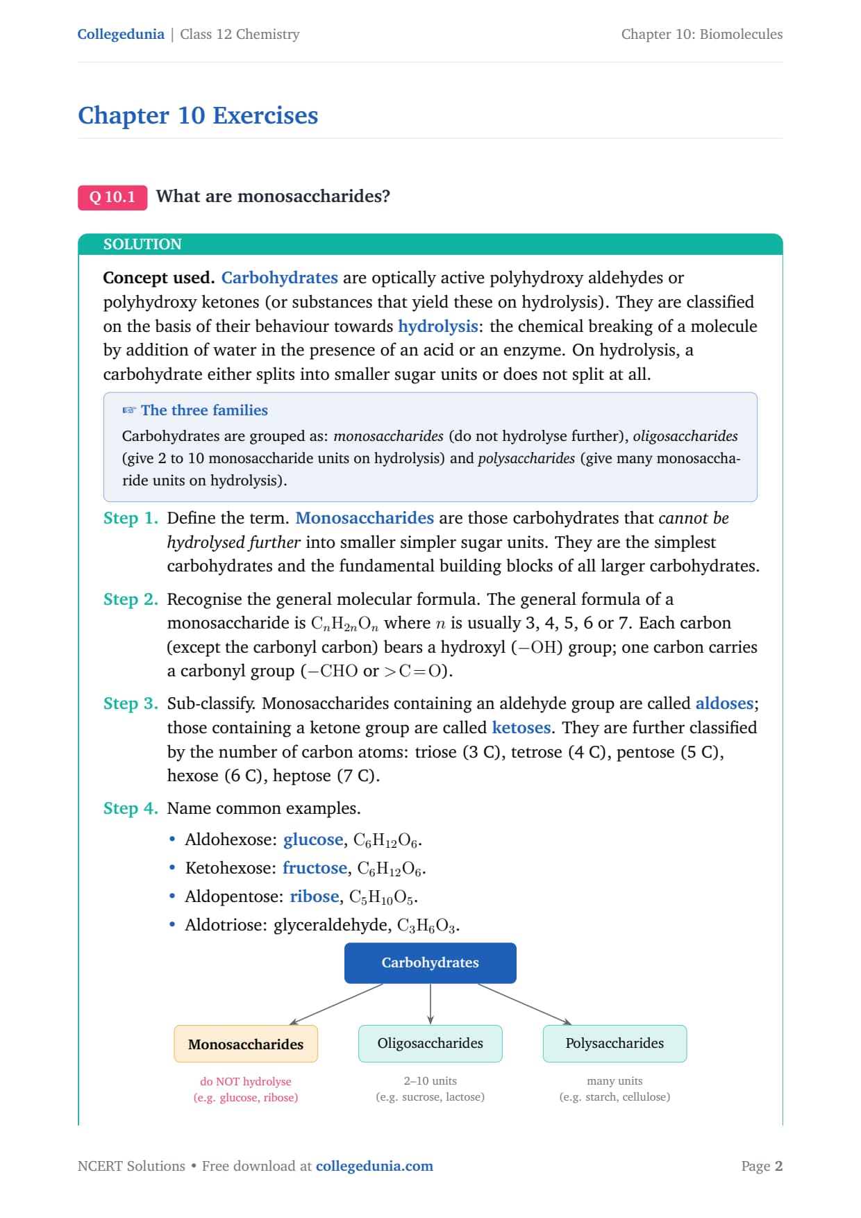
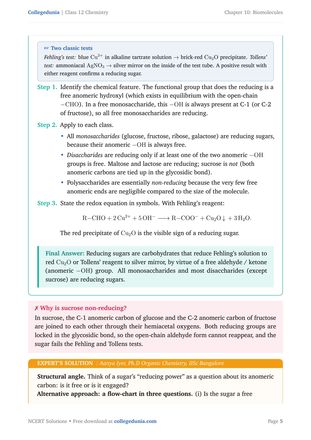
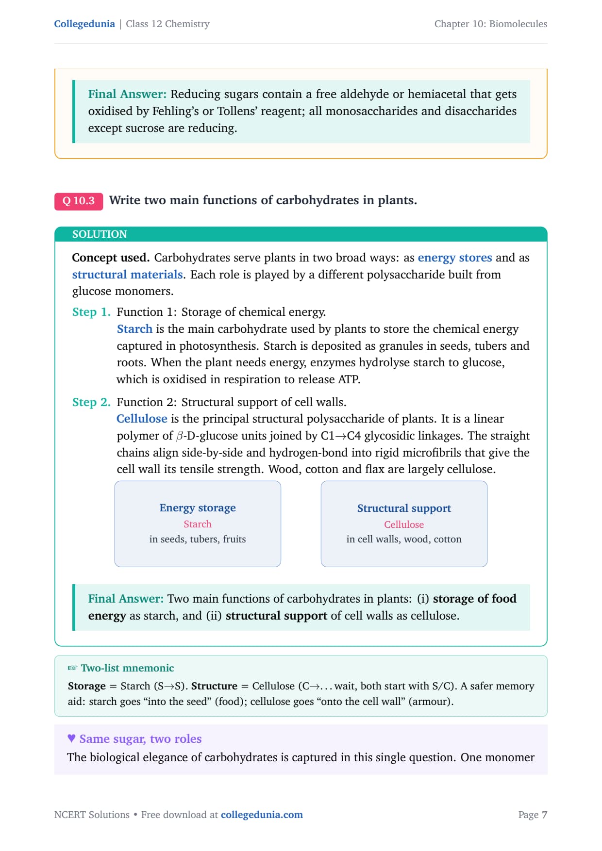
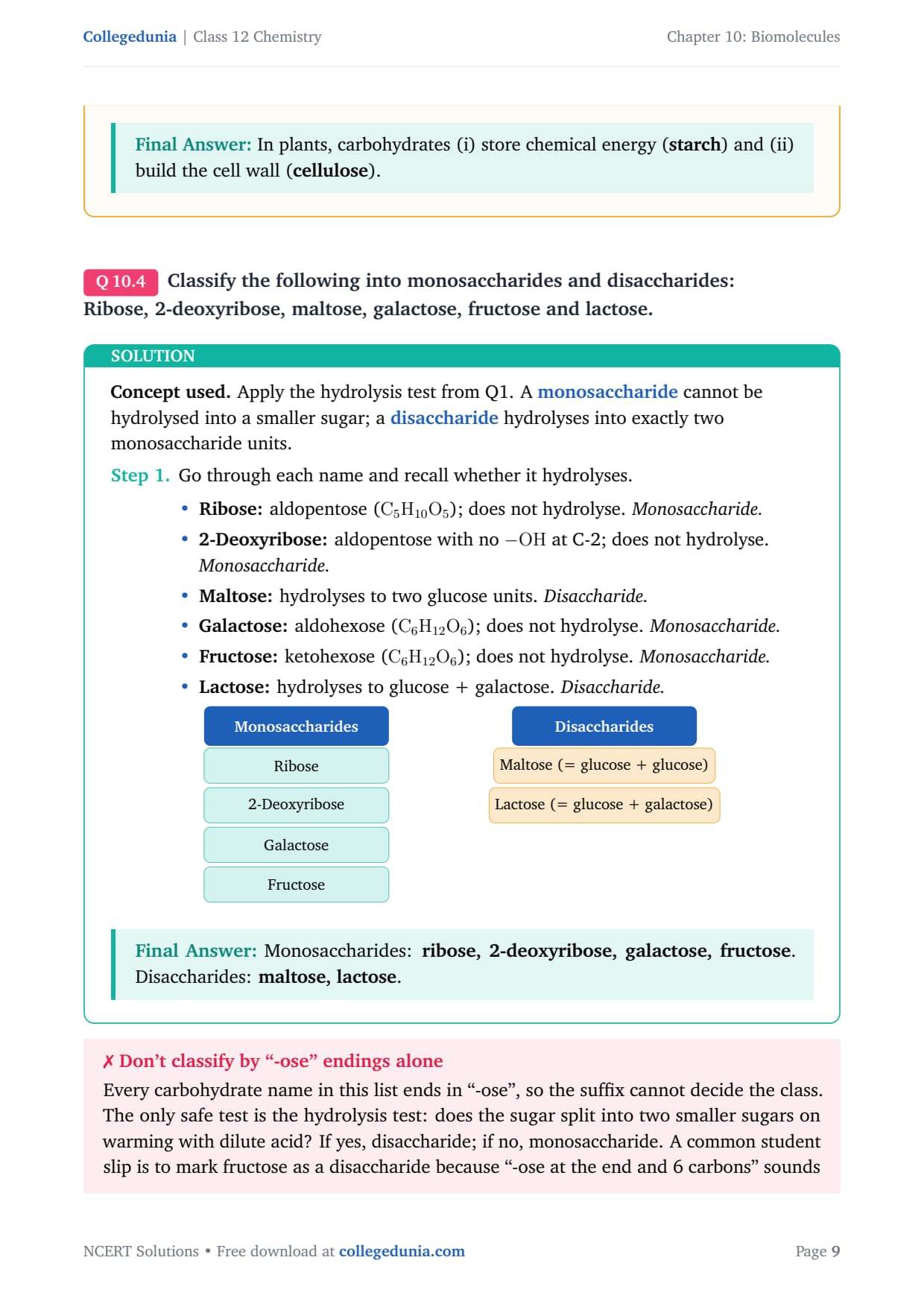
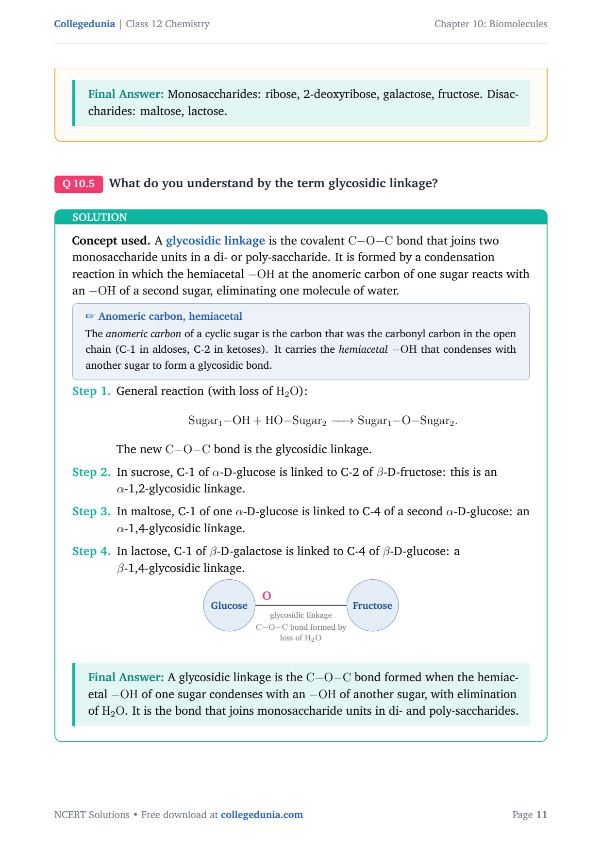
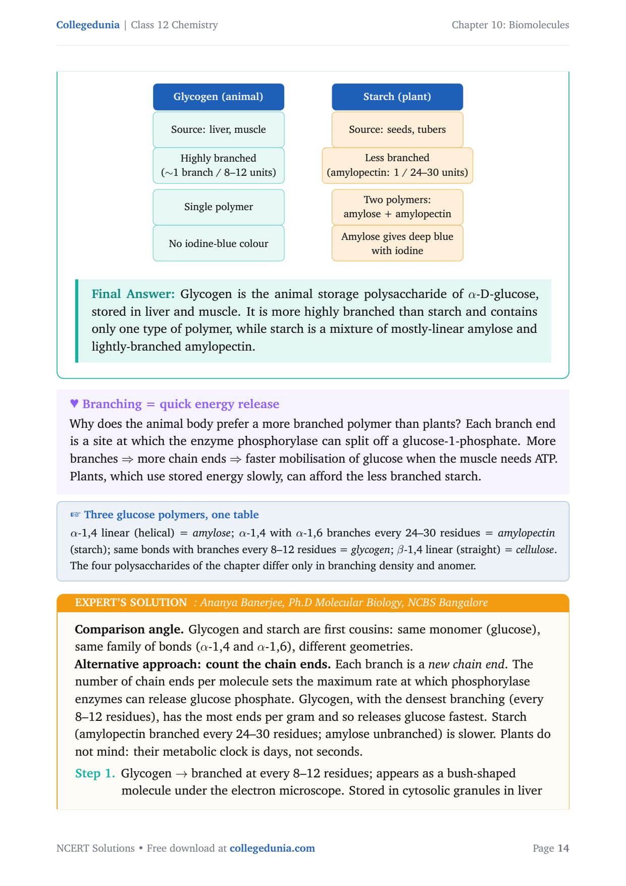
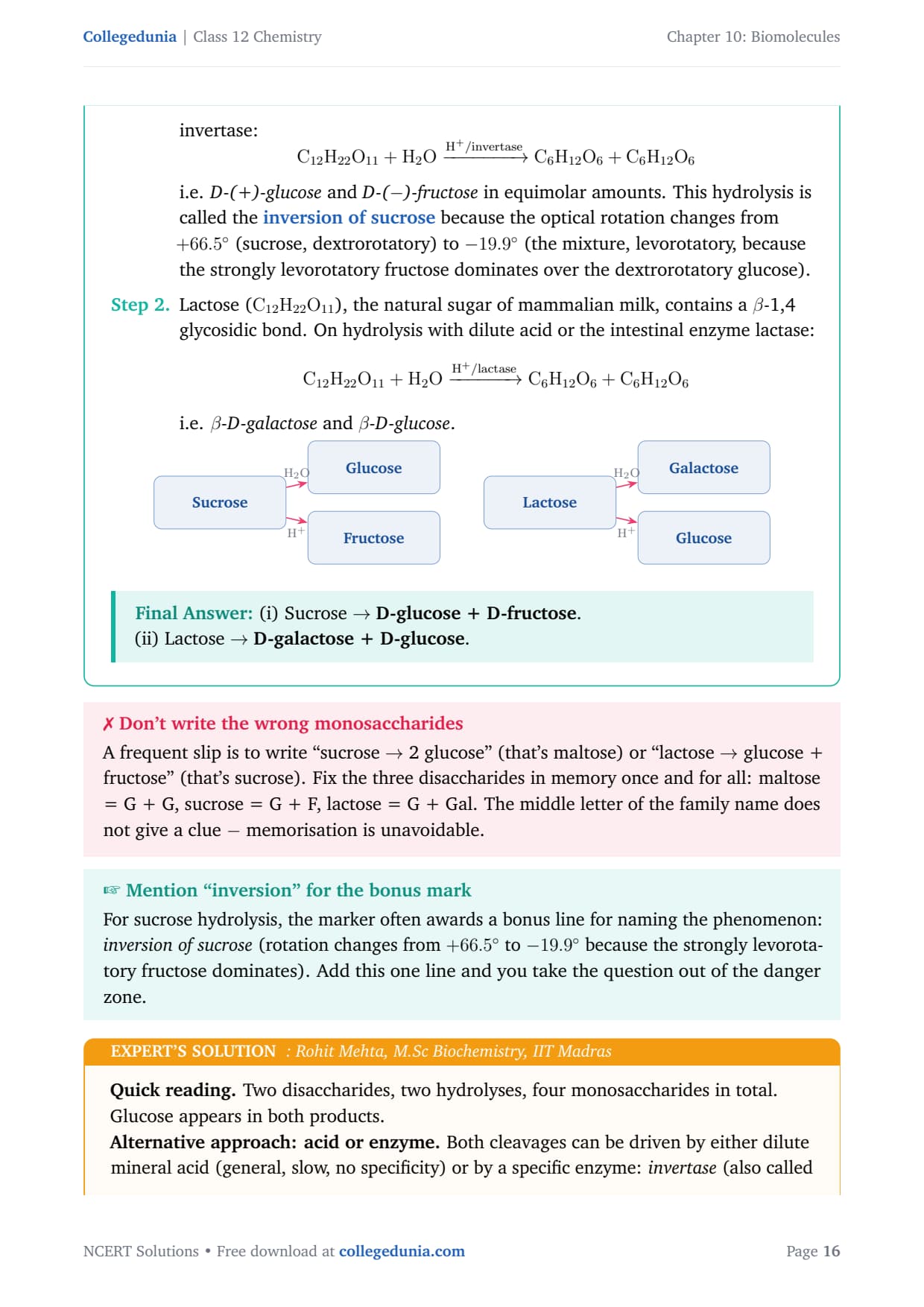
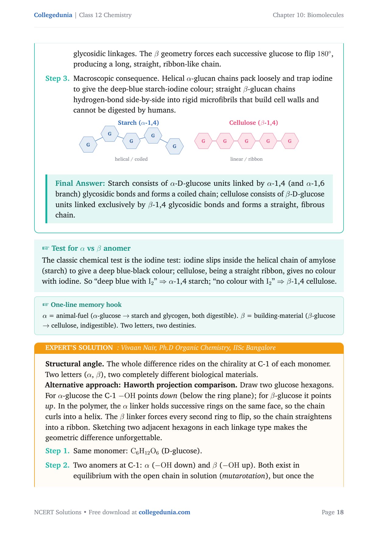
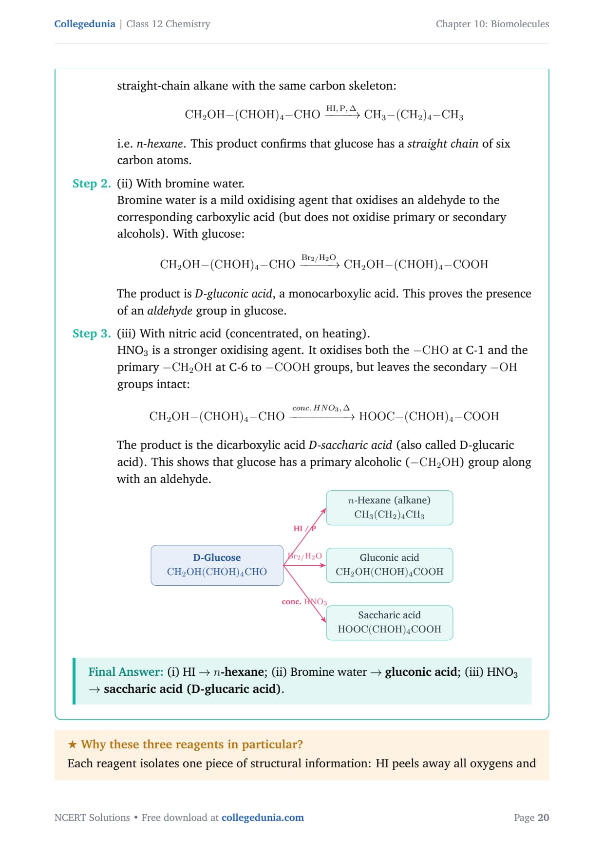
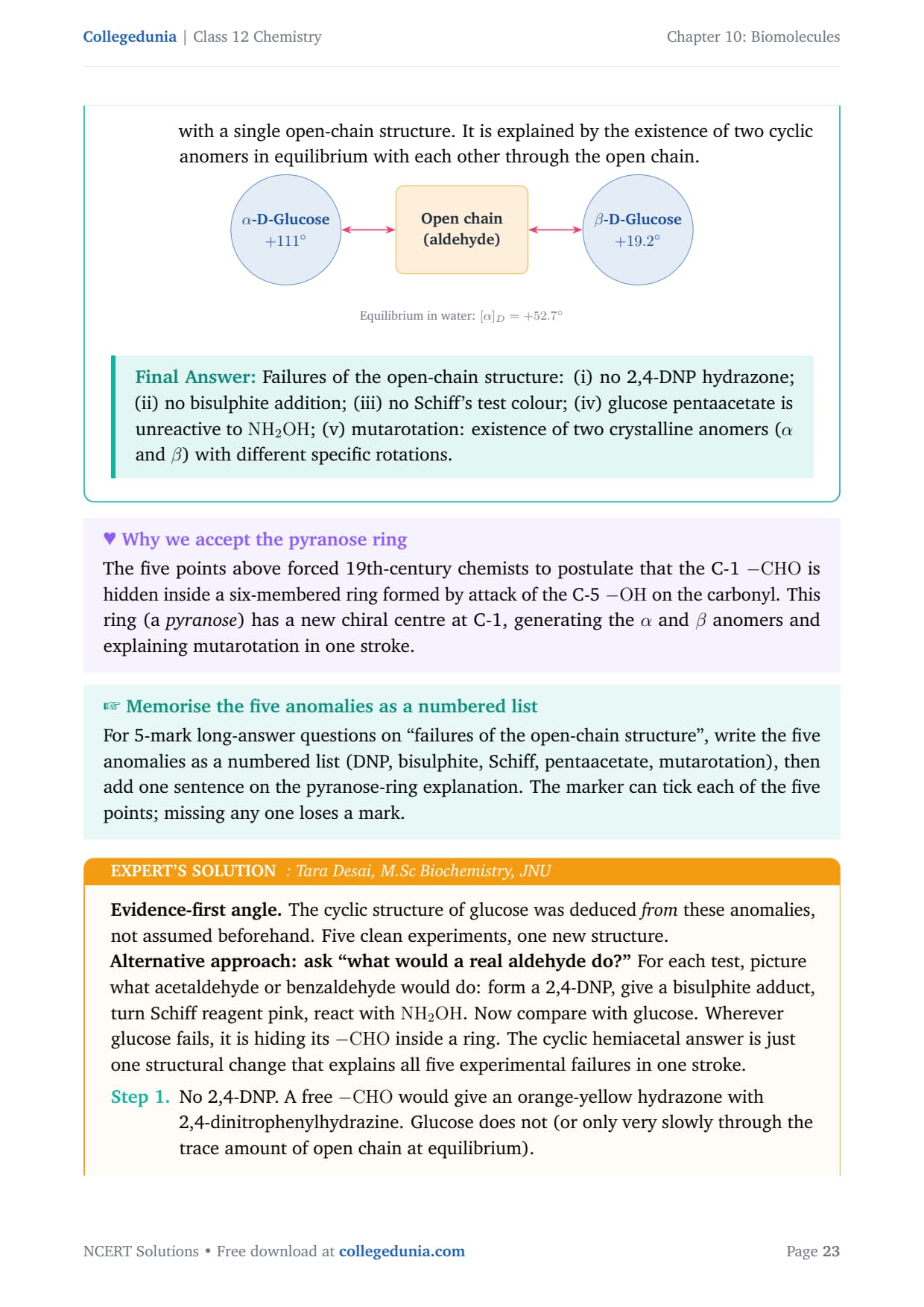
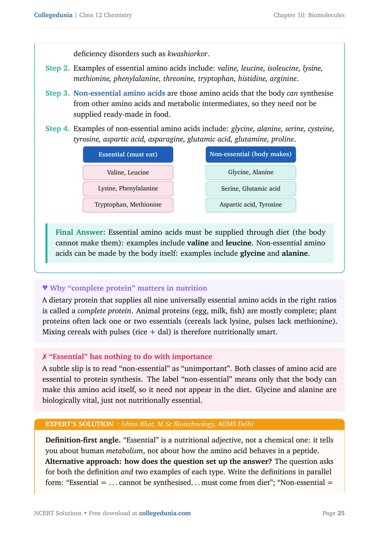
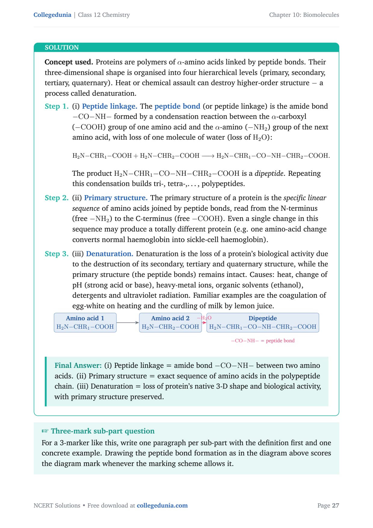
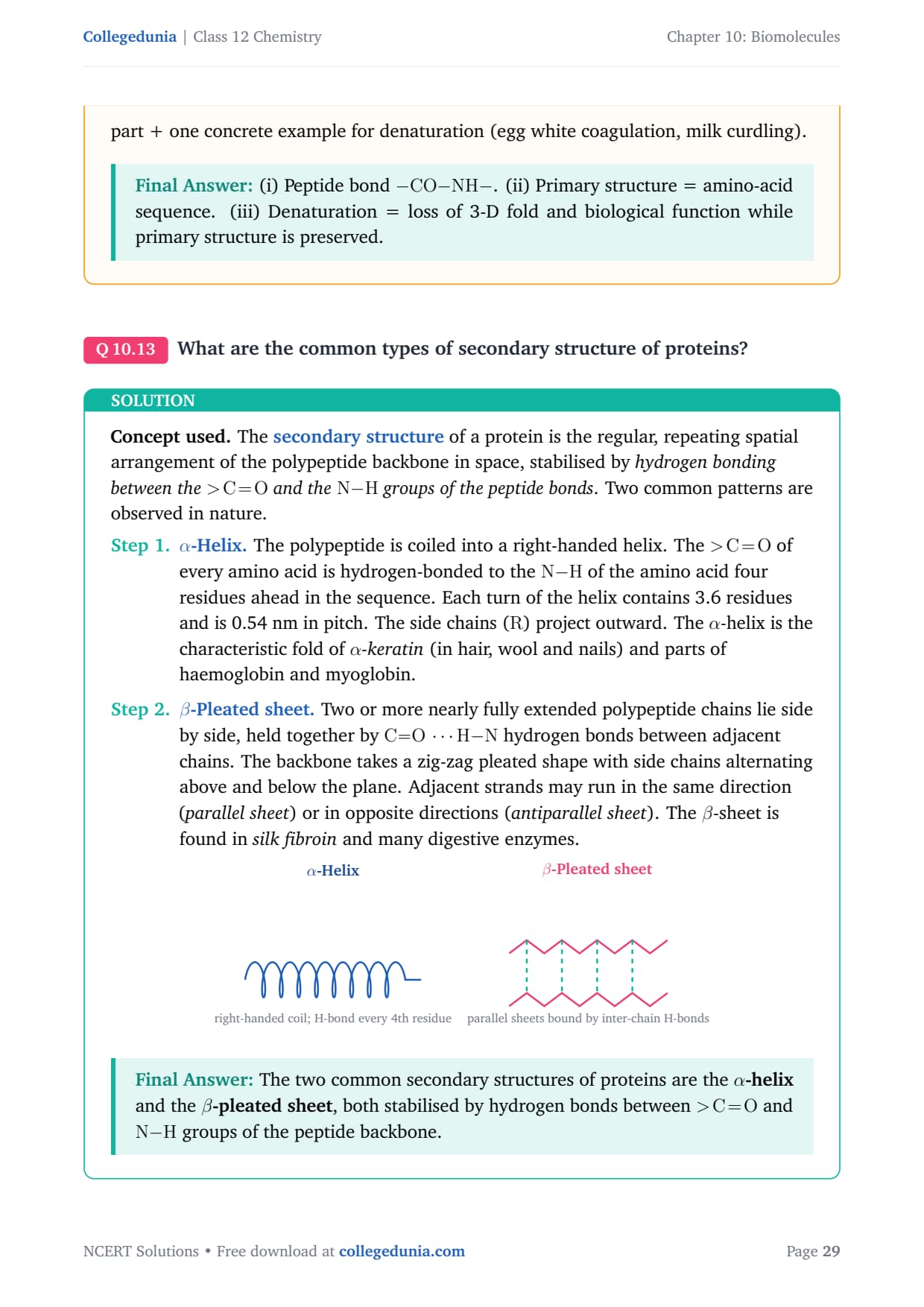
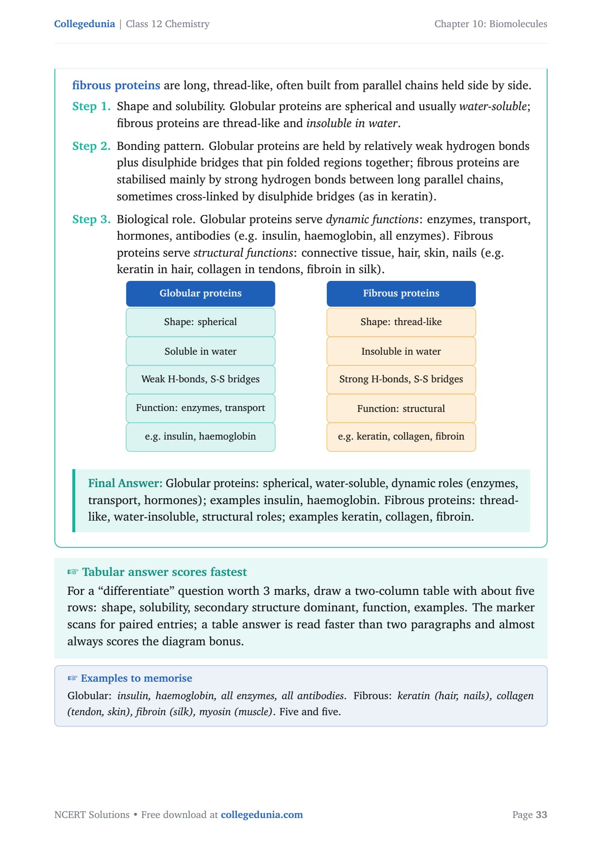
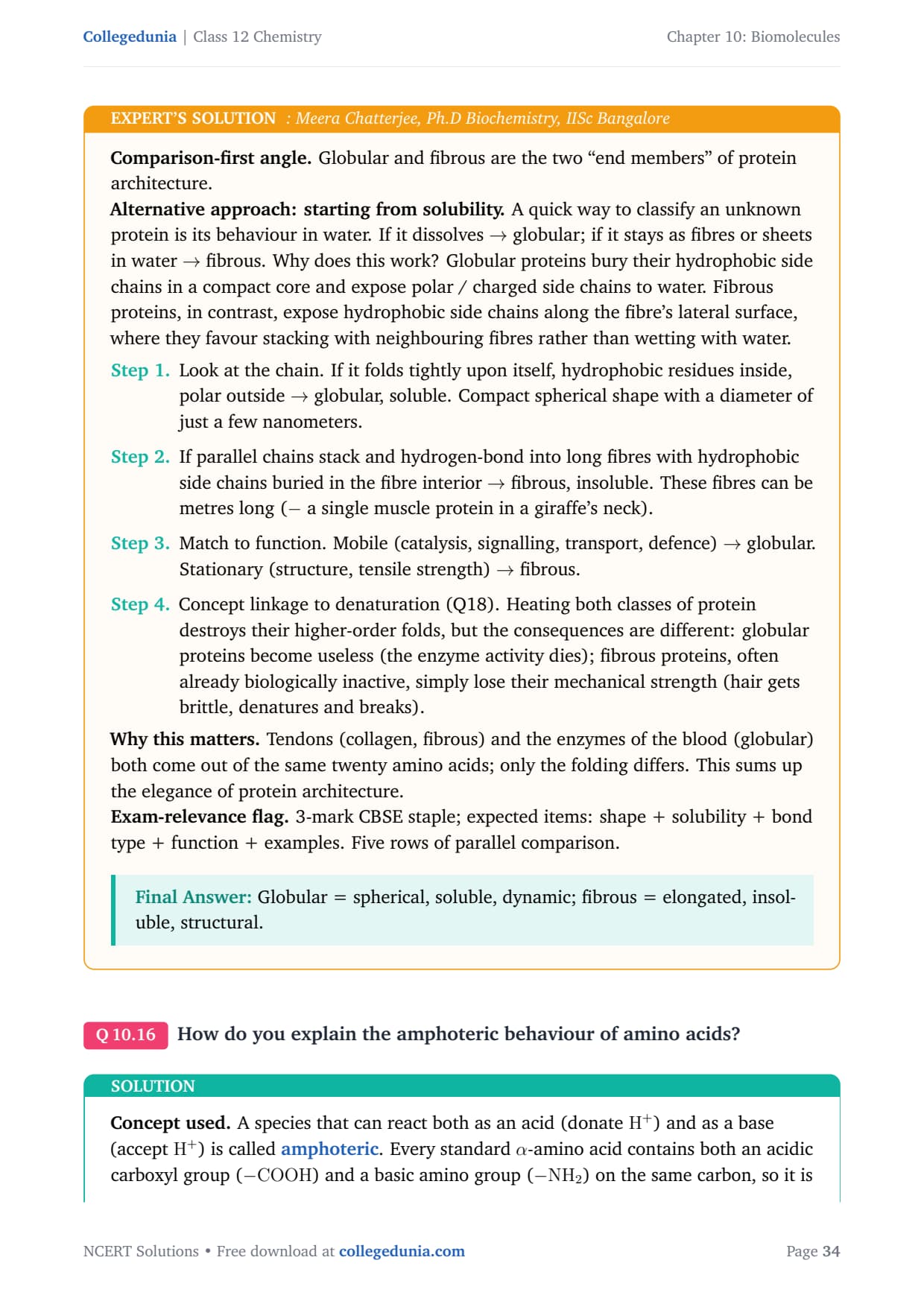
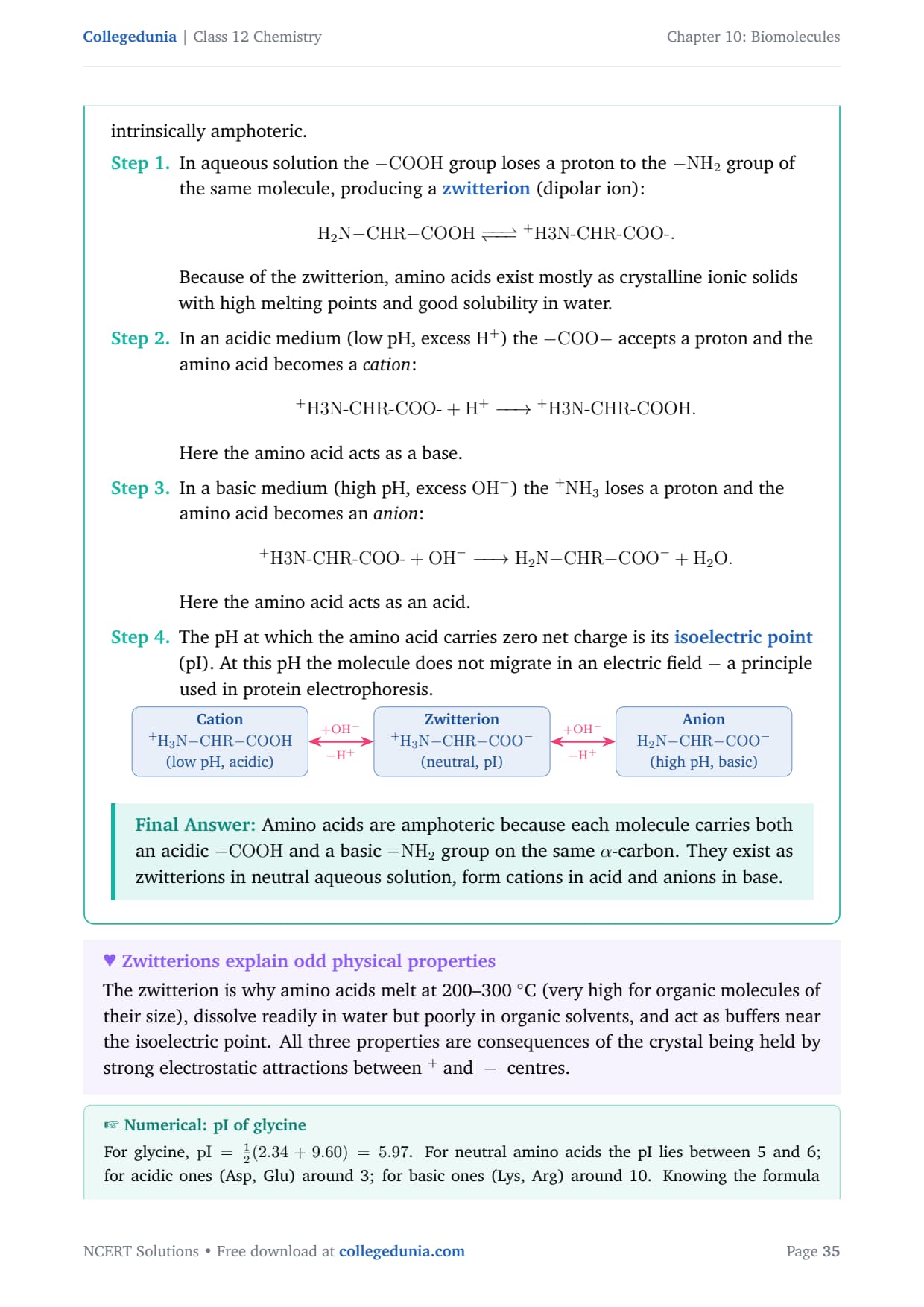
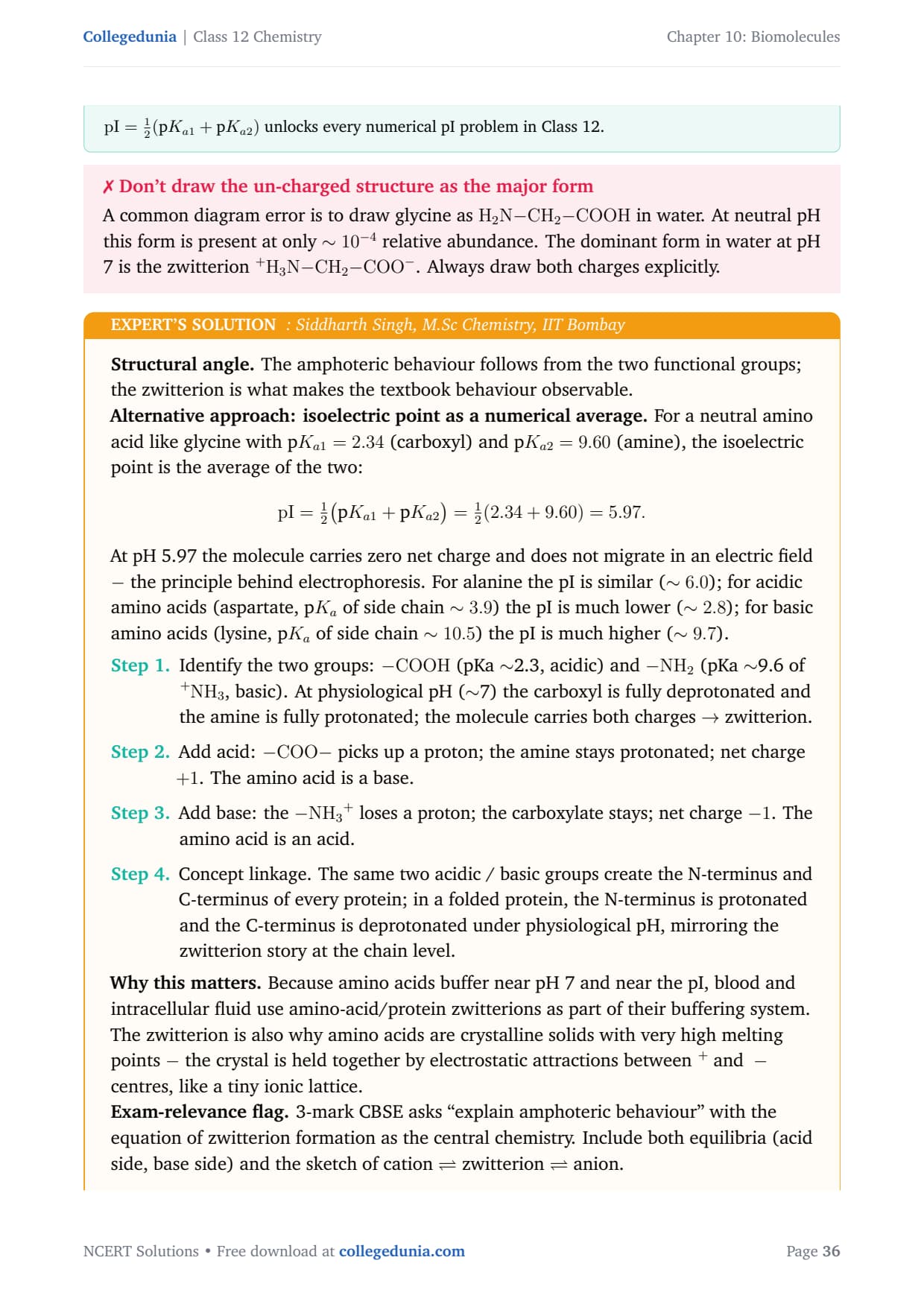
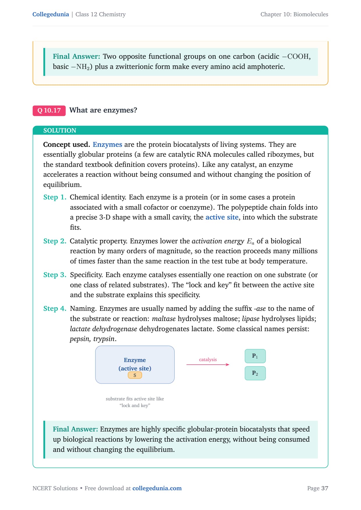
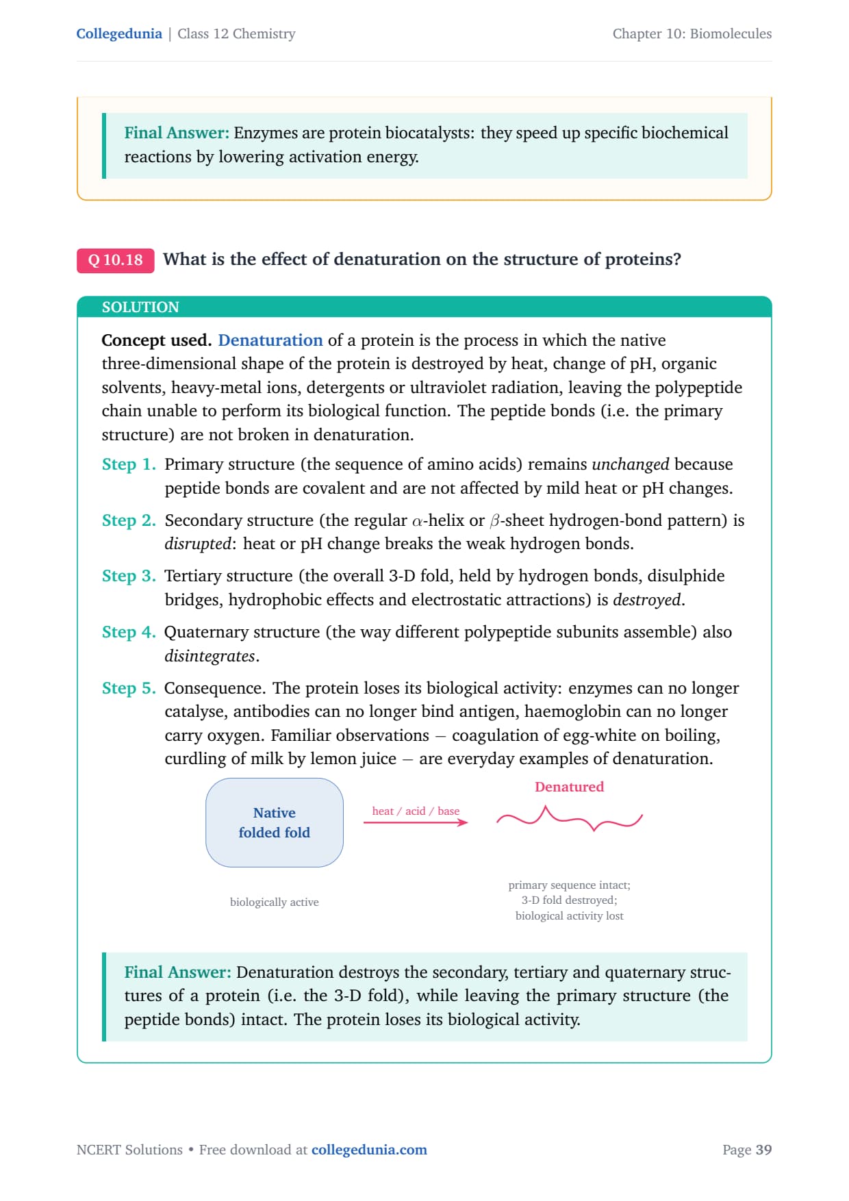
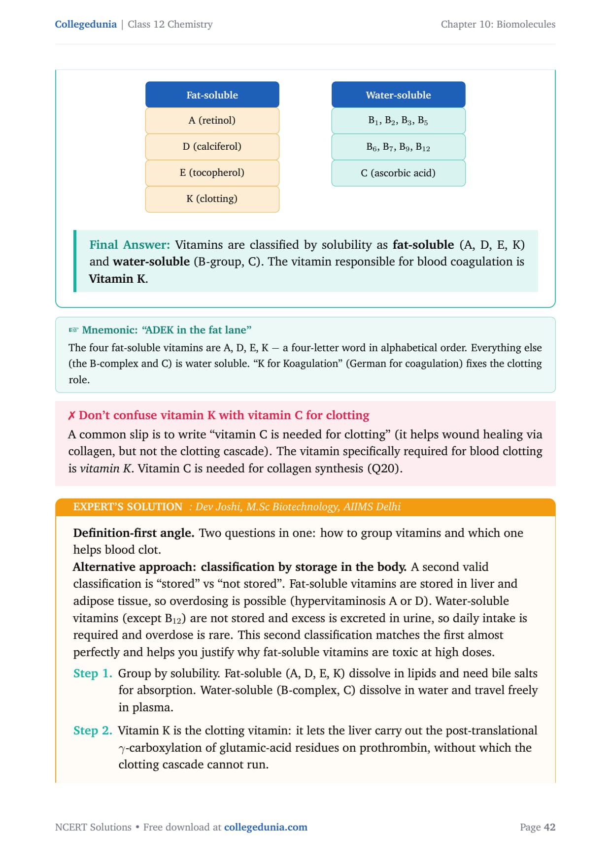
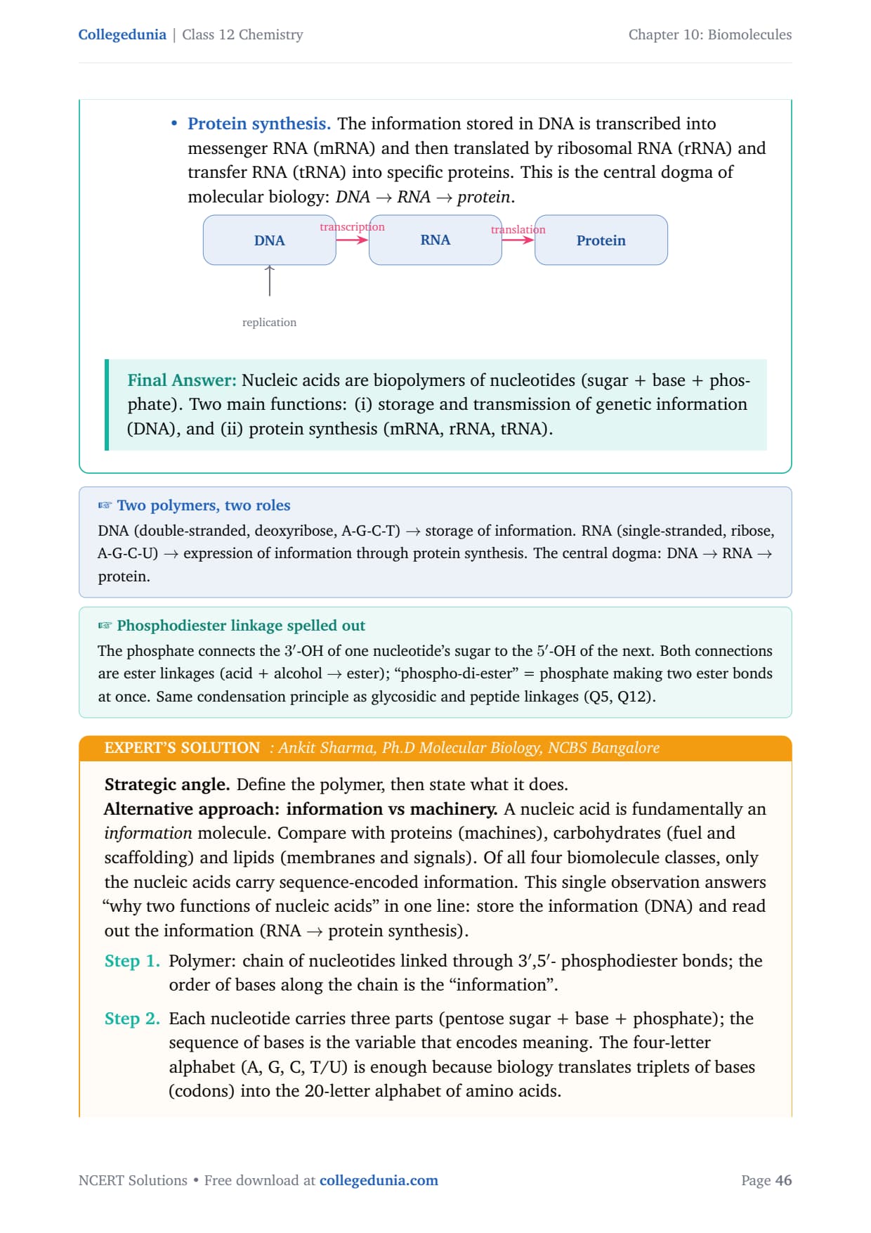
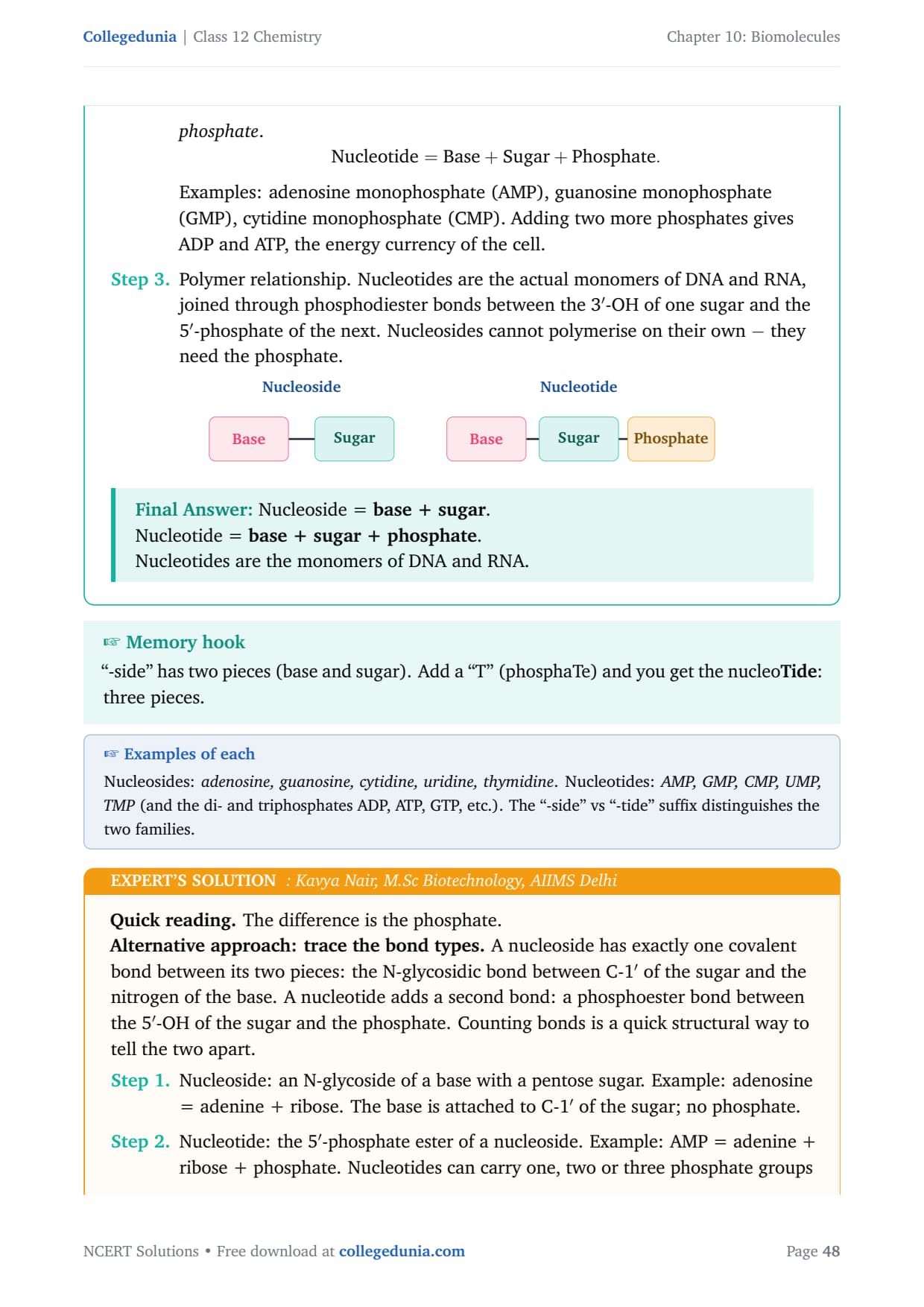
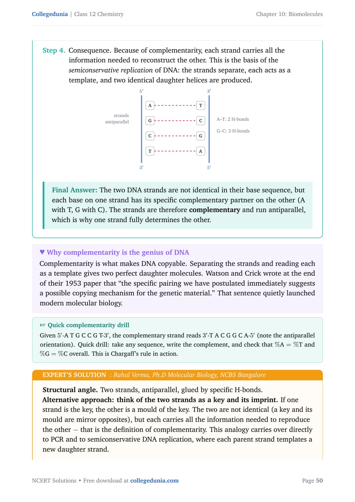
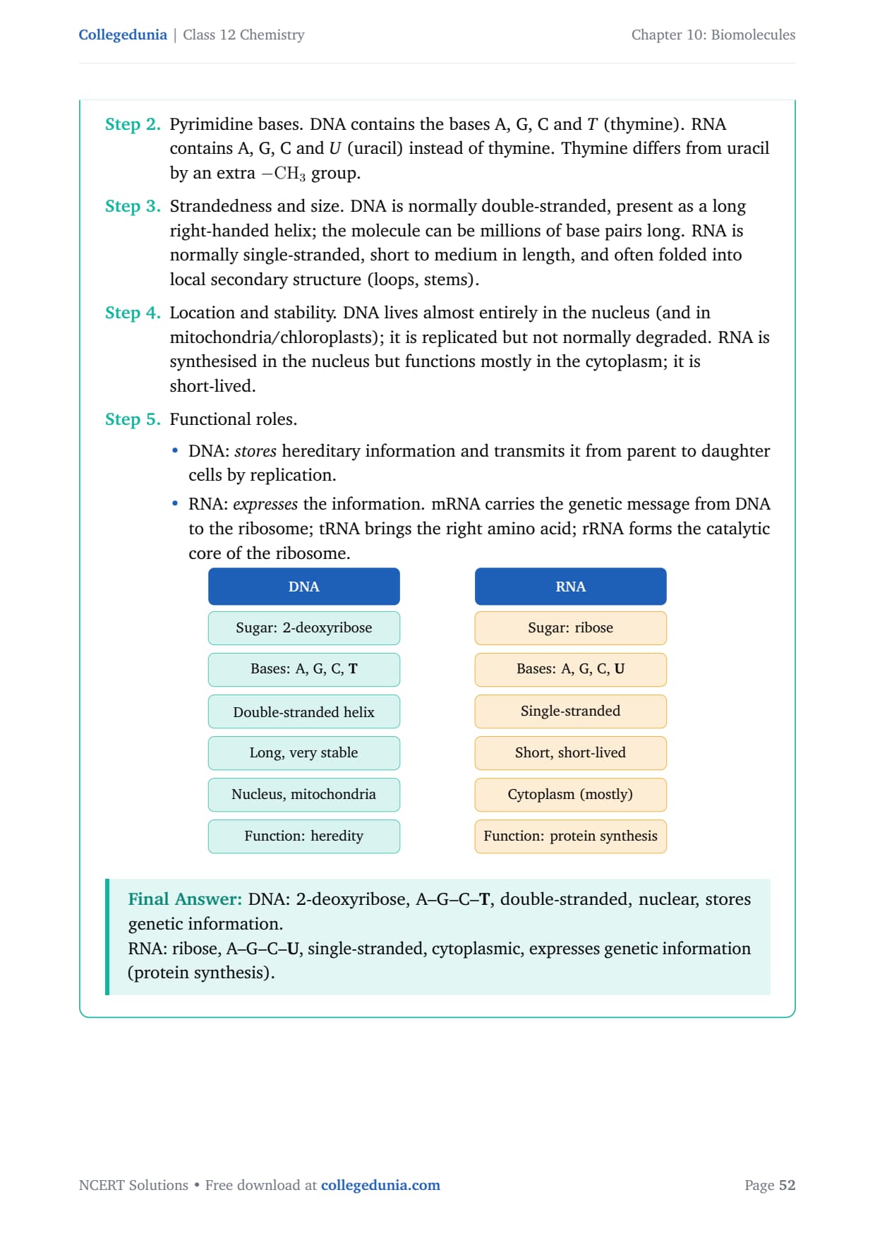
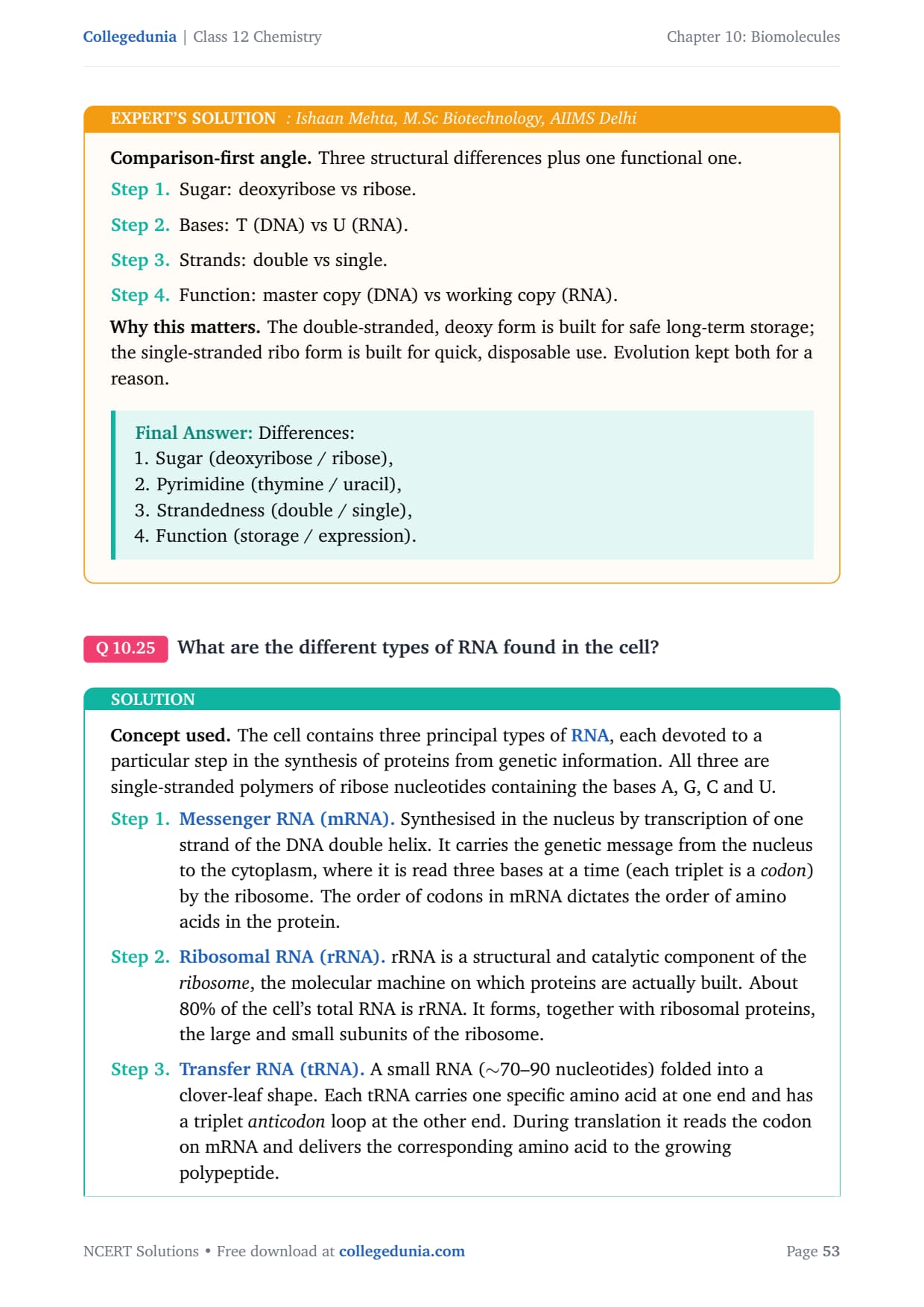
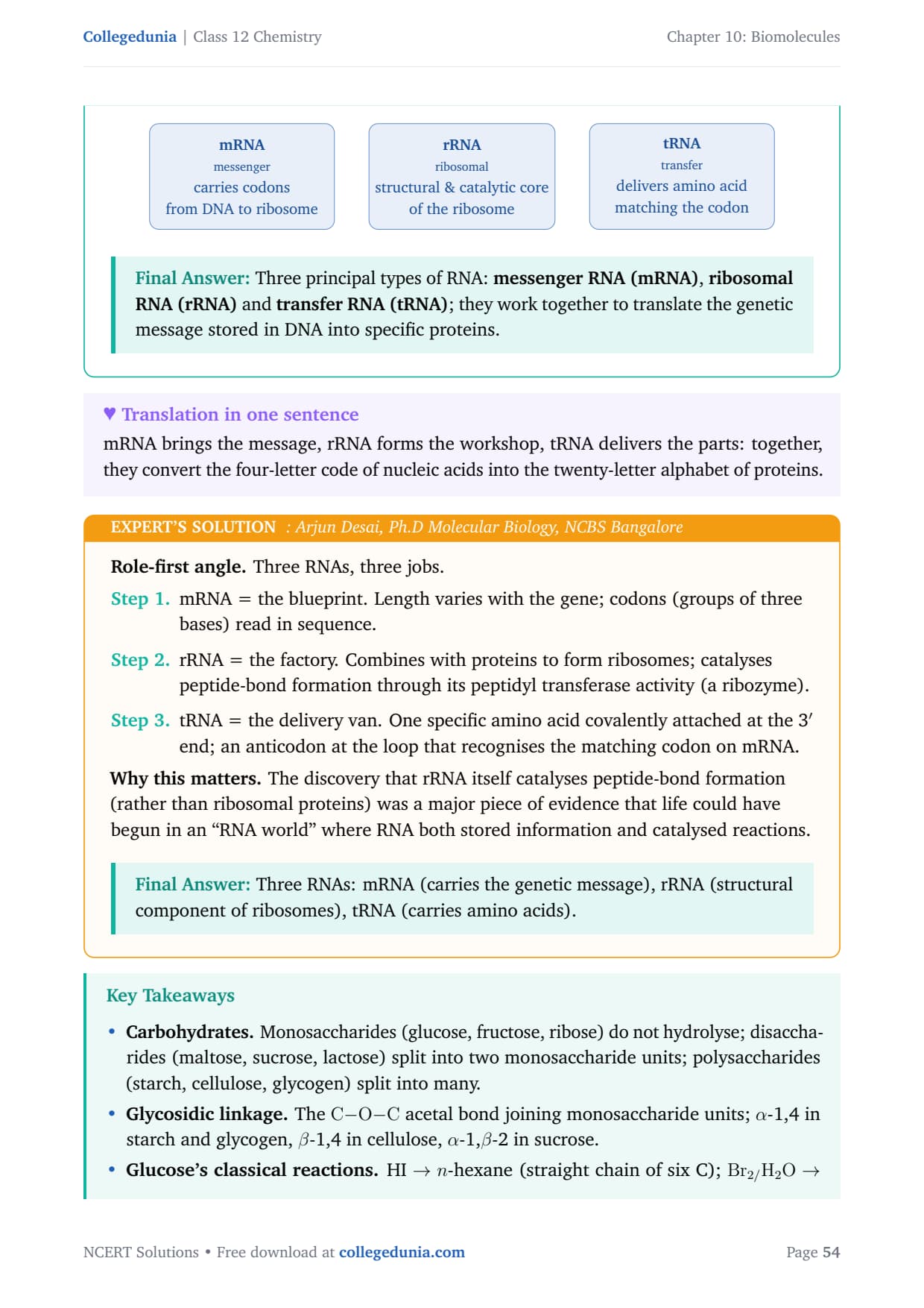
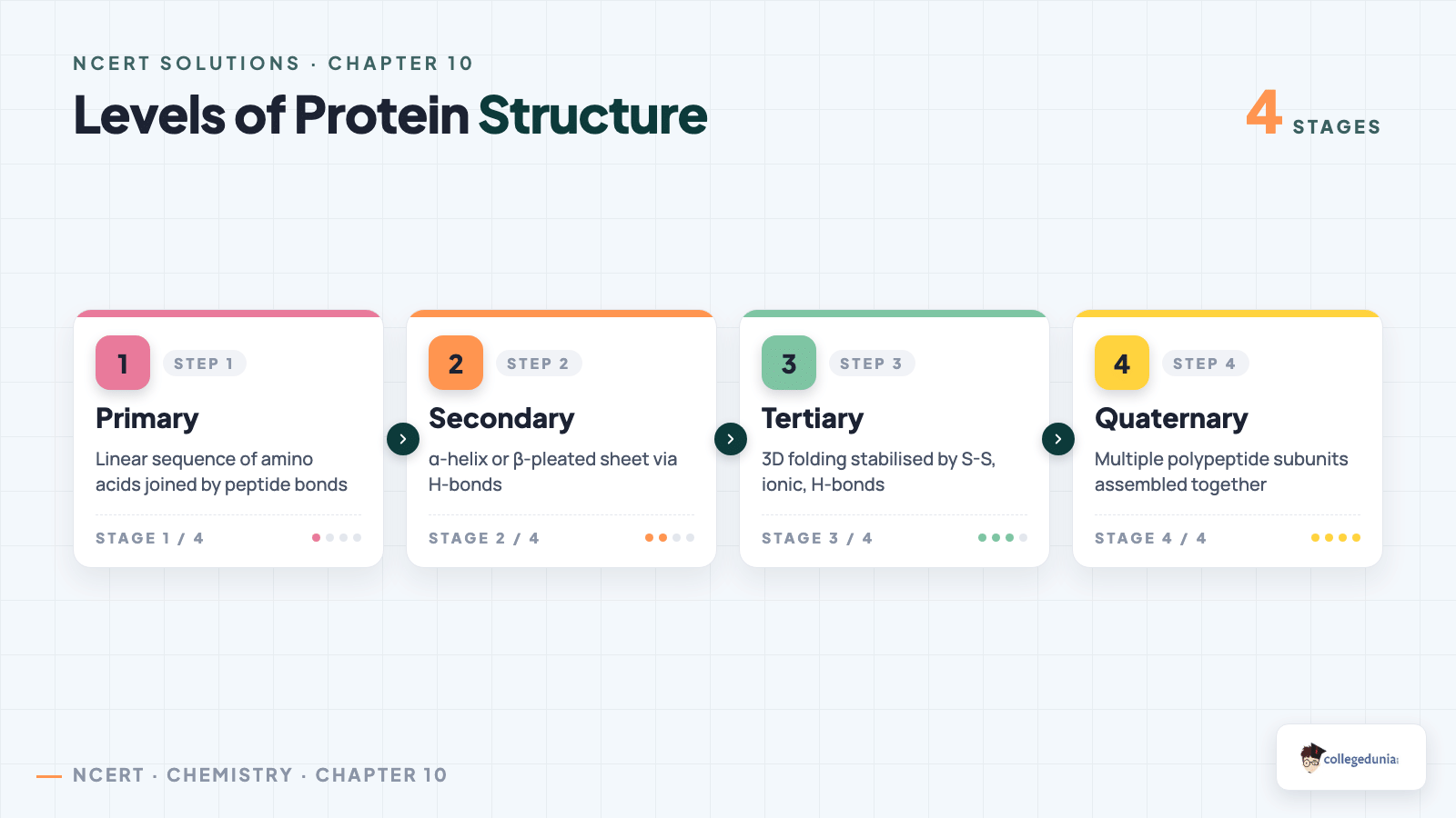



Comments