




The NCERT Formula Sheet Class 12 Chemistry Chapter 10 Biomolecules includes all important definitions, summary, and formulas. The sheet provided below will help you cover the entire chapter in 30-40 minutes during exam time.
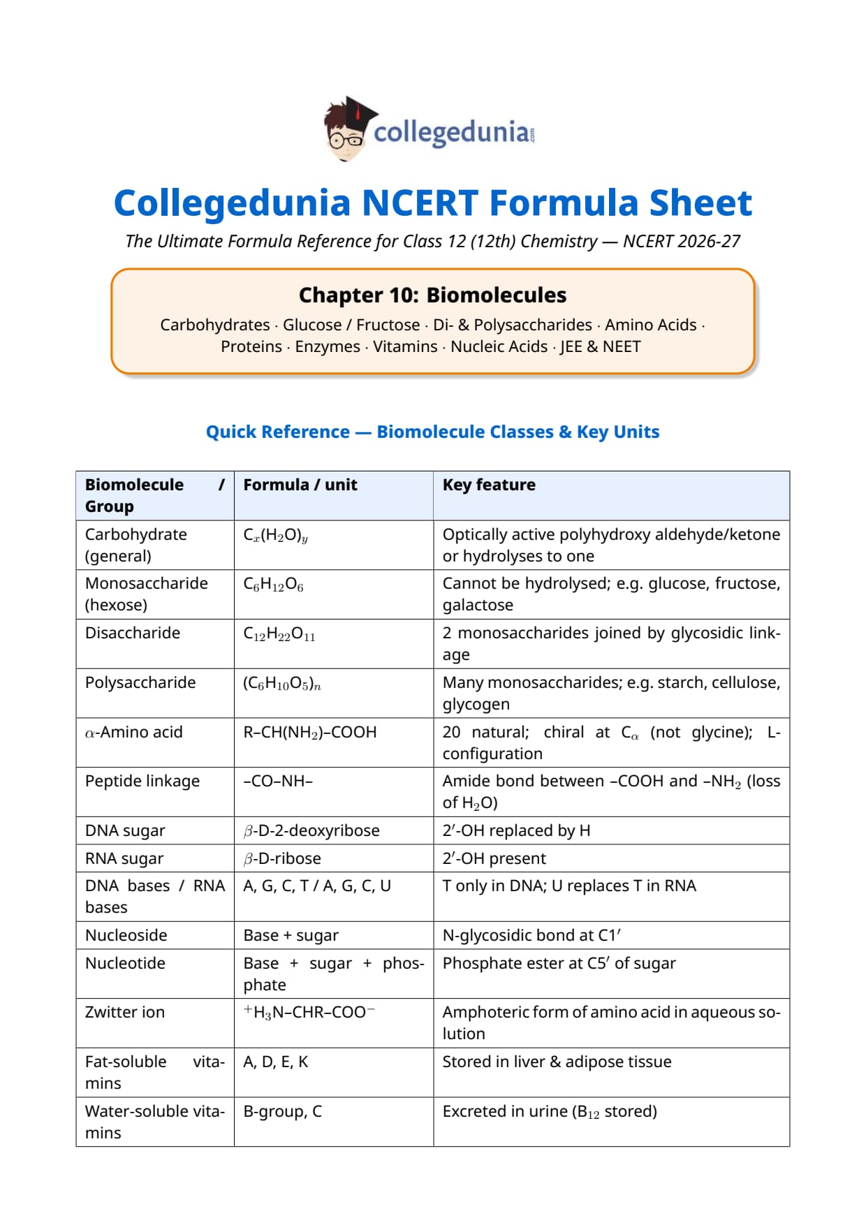
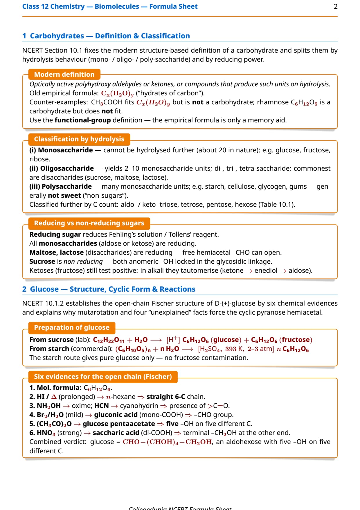
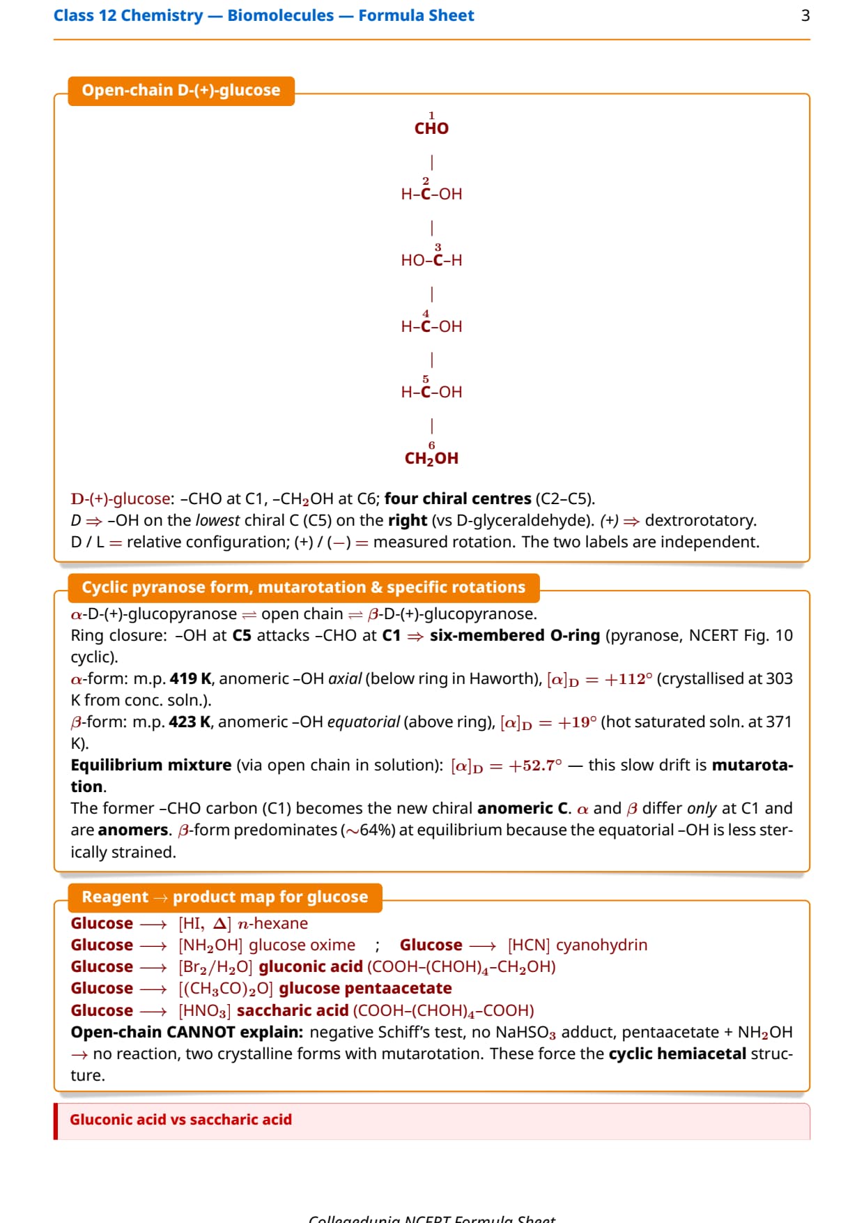
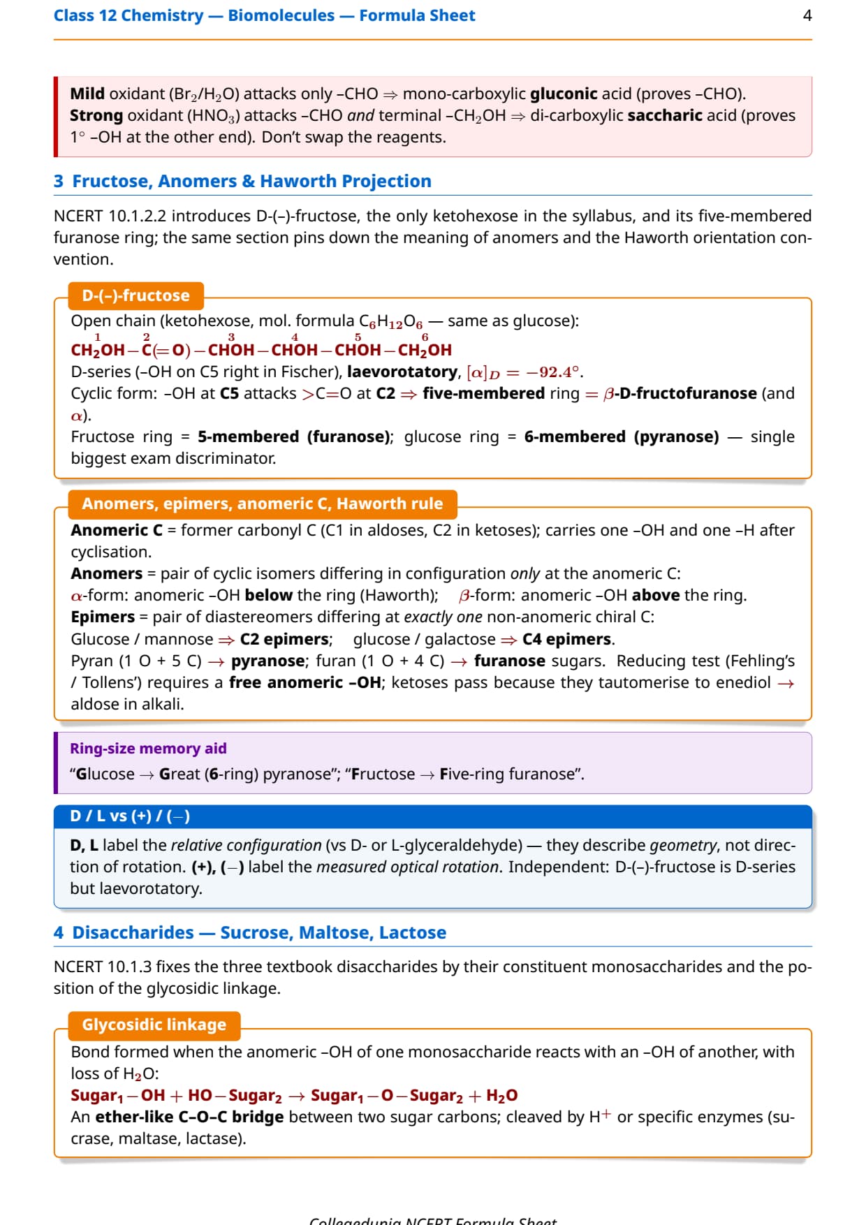
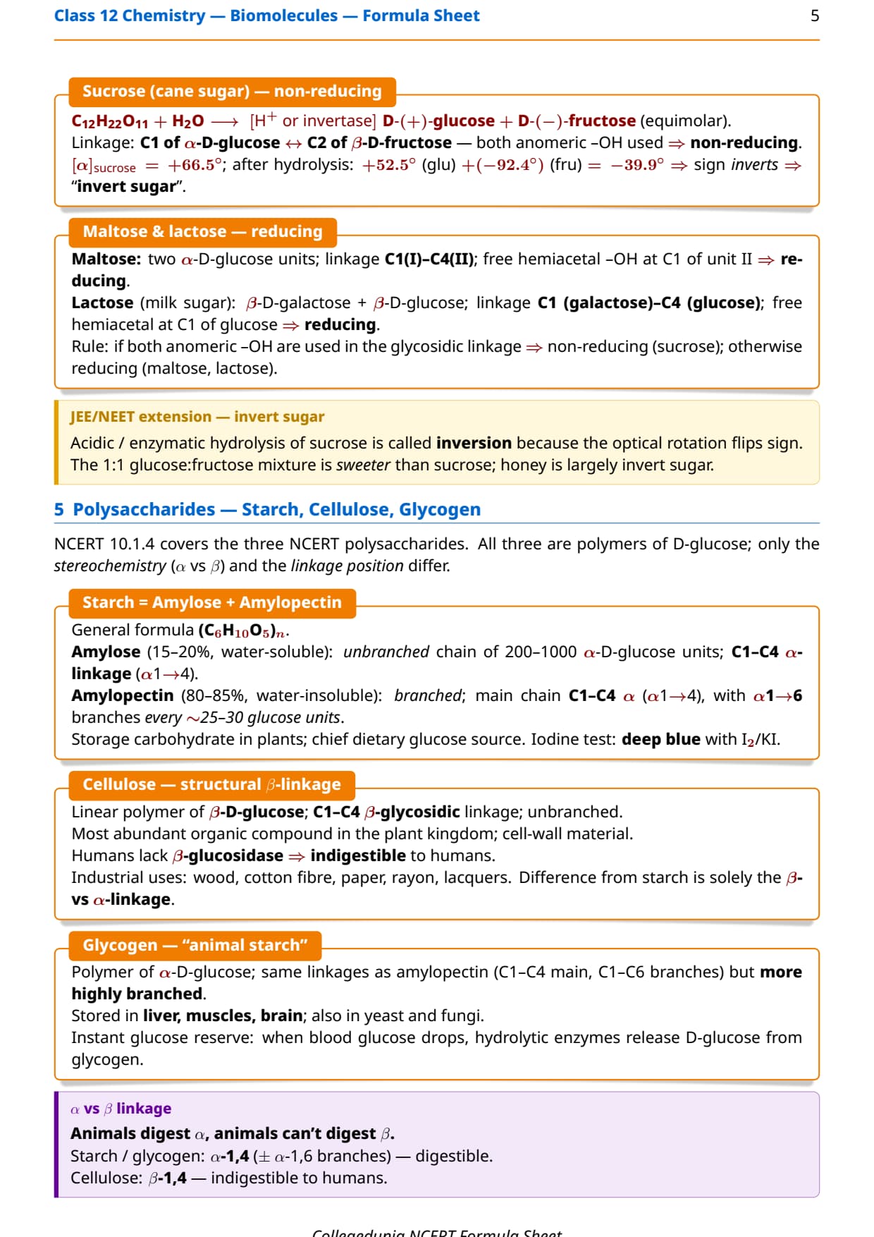
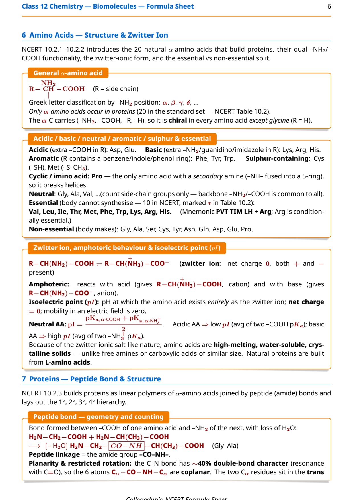
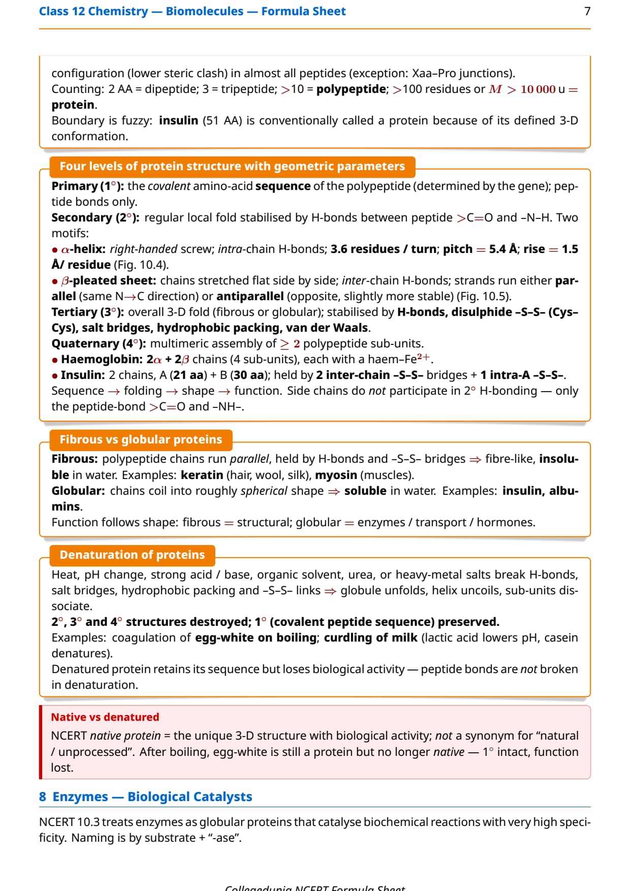
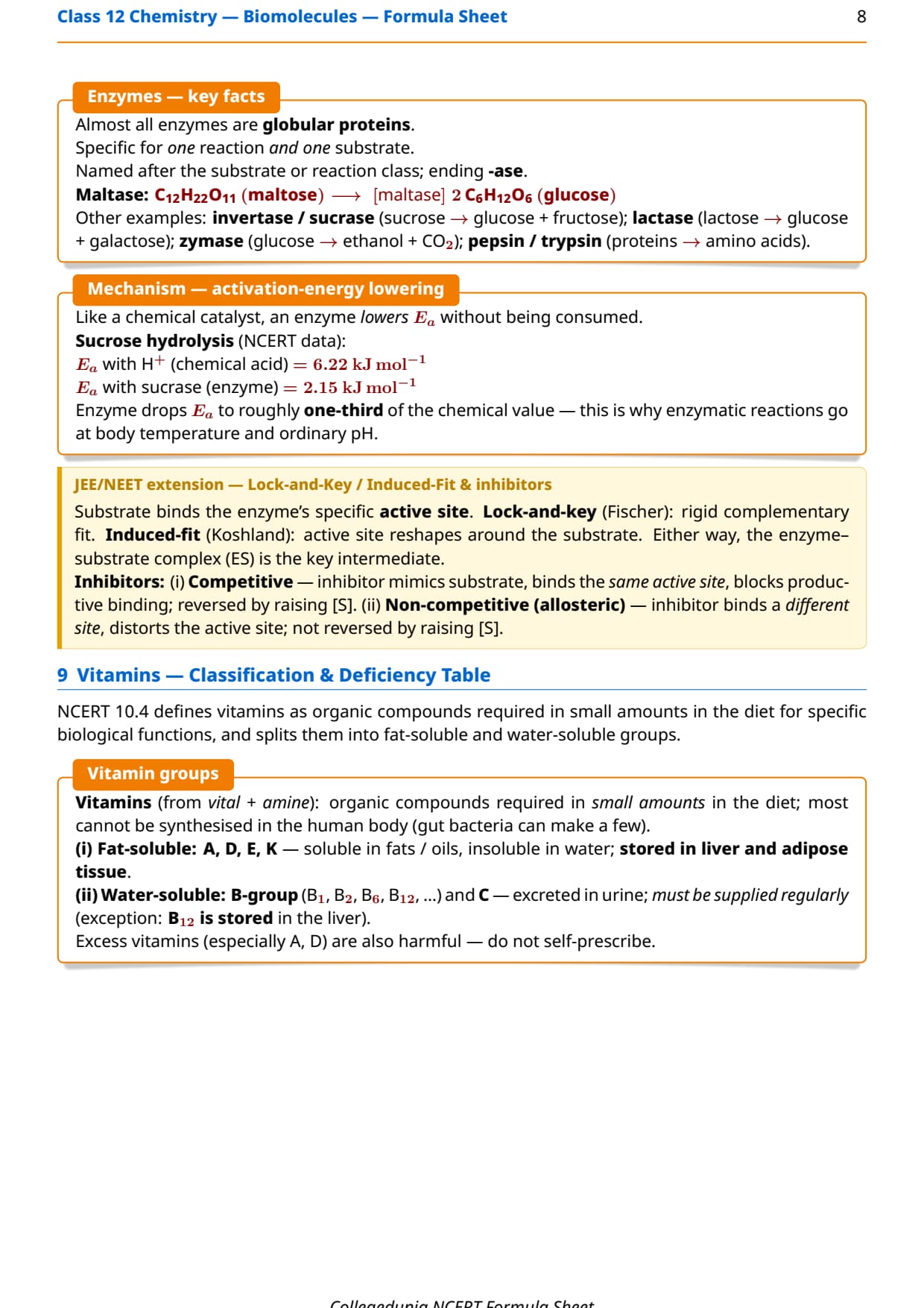
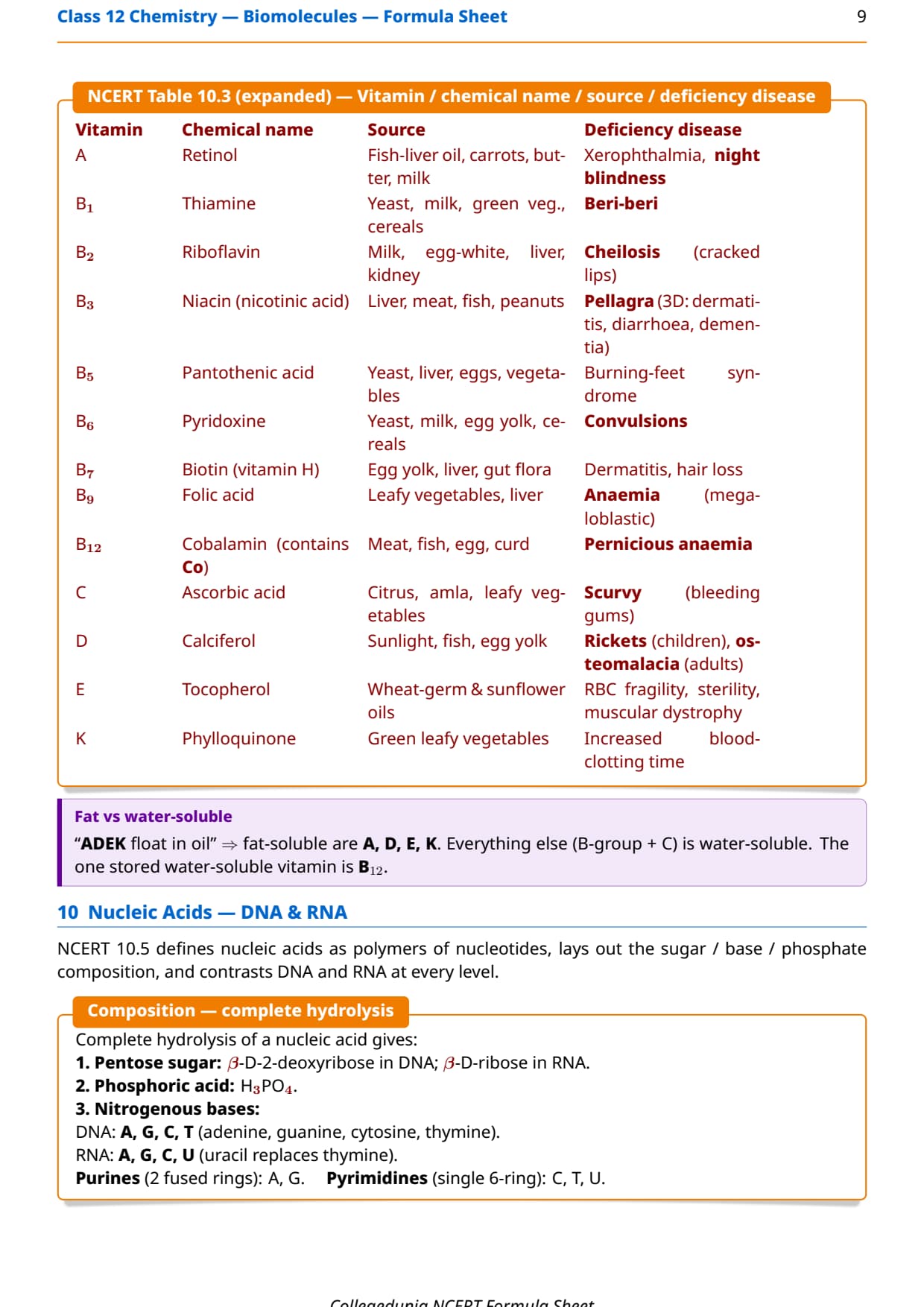
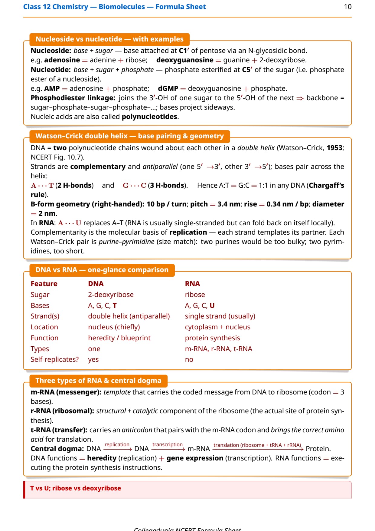

It puts every structural rule, reagent test, base-pair point, and vitamin-deficiency pair for carbohydrates, proteins, vitamins, and nucleic acids on one printable page.
- CBSE Weightage: 3 to 5 marks
- JEE Main Weightage: 1 to 2 per cent (1 to 2 questions per paper)
- NEET Weightage: 1 to 2 questions per year
Curated by Collegedunia subject experts and mapped to the 2026-27 NCERT edition.
Also Check:
- Biomolecules Class 12 Chemistry Notes
- Biomolecules Class 12 Chemistry NCERT Solutions
- CBSE Class 12 Chemistry Syllabus 2026-27
Why Biomolecules Matters in 12th Chemistry and Entrance Exams
Biomolecules is a high-return chapter. CBSE set a 2-mark DNA/RNA question in 4 of the last 5 papers, and NEET tests a vitamin-deficiency pair every year since 2021. Lock in the glycosidic, peptide, and phosphodiester linkages plus the fat-soluble ADEK rule for 3 to 4 near-certain marks.
Biomolecules Class 12 Chemistry Explained
Source: Magnet Brains on YouTube
How will Collegedunia's Biomolecules Formula Sheet Help You?
The sheet is built for a 20-minute final revision pass before the Chemistry paper.
- 2026-27 NCERT Alignment: matches Sections 10.1 to 10.6 of the current syllabus.
- One-Page Printability: the master table fits one A4 landscape sheet.
- Sub-System Tagging: each fact is tagged by biomolecule type for quick lookup.
- Expert Verification: cross-checked against NCERT and the last five JEE Main and NEET papers.
Biomolecules Symbol and Notation Glossary for 12th Chemistry
This glossary decodes every notation used in the master table.
| Symbol | Meaning | Typical Unit / Note |
|---|---|---|
| Cx(H2O)y | Empirical carbohydrate formula | Memory aid only; rhamnose breaks it |
| α, β | Anomeric configurations | α-OH below ring (Haworth); β-OH above ring |
| R−CH(NH2)−COOH | α-amino acid general formula | 20 natural; only α in proteins |
| +H3N−CHR−COO− | Zwitter ion | Net charge 0; amphoteric in water |
| −CO−NH− | Peptide / amide linkage | Between −COOH and −NH2 of next AA |
| 1°, 2°, 3°, 4° | Four levels of protein structure | Sequence; helix/sheet; 3-D fold; sub-unit assembly |
| Ea | Activation energy | Sucrose hydrolysis: 6.22 (H+) → 2.15 (sucrase) kJ mol−1 |
| A, T, G, C, U | Nitrogenous bases | A/G purines; C/T/U pyrimidines |
| 3′, 5′ | Sugar-ring carbons (nucleic acid) | 3′ of one sugar to 5′ of next |
| ADEK | Fat-soluble vitamins | A, D, E, K; stored in liver / adipose |
Biomolecules All Important Formulae and Reactions for Class 12 Chemistry
The master table lists every structural rule, reagent test, and linkage in Chapter 10 with conditions and NCERT reference. All entries are retained in the 2026-27 syllabus.
| Concept / Reaction | Formula / Structure | Conditions / Notes | NCERT Ref |
|---|---|---|---|
| Carbohydrate (modern definition) | Polyhydroxy aldehyde / ketone, optically active | Empirical Cx(H2O)y is only a memory aid | 10.1 |
| Monosaccharide (hexose) | C6H12O6 | Cannot be hydrolysed further | 10.1 |
| Disaccharide (sucrose) | C12H22O11 | 2 monosaccharide units; glycosidic linkage | 10.1.3 |
| Polysaccharide | (C6H10O5)n | Many monosaccharide units; non-sweet | 10.1.4 |
| Glucose (aldohexose) | CHO-(CHOH)4-CH2OH | D-(+); 4 chiral C; pyranose ring | 10.1.2 |
| Glucose + Br2 water (mild) | Glucose Br2/H2O Gluconic acid | Mild −CHO oxidation | 10.1.2.1 |
| Glucose + HNO3 (strong) | Glucose HNO3 Saccharic acid | Oxidises both ends | 10.1.2.1 |
| Glucose + (CH3CO)2O | Glucose (CH3CO)2O Glucose pentaacetate | Acetic anhydride | 10.1.2.1 |
| Cyclic hemiacetal (glucose) | C5 −OH attacks C1 −CHO → 6-membered pyranose ring | α (m.p. 419 K), β (m.p. 423 K) | 10.1.2.1 |
| Fructose (ketohexose) | CH2OH-CO-(CHOH)3-CH2OH | D-(−); furanose ring | 10.1.2.2 |
| Glycosidic linkage | Sugar1-OH + HO-Sugar2 → Sugar1-O-Sugar2 + H2O | Loss of H2O | 10.1.3 |
| Sucrose hydrolysis (invert sugar) | C12H22O11 + H2O H+ or invertase D-(+)-glu + D-(-)-fru | [α] flips +66.5° → −39.9° | 10.1.3 |
| Maltose | 2 α-D-glucose units; C1(I)–C4(II) linkage | One free hemiacetal ⇒ reducing | 10.1.3 |
| Lactose (milk sugar) | β-D-galactose (C1) – β-D-glucose (C4) | Free hemiacetal on glucose ⇒ reducing | 10.1.3 |
| Starch (amylose + amylopectin) | (C6H10O5)n ; α-D-glucose | Amylose C1-C4 linear; amylopectin C1-C4 + C1-C6 branched | 10.1.4 |
| Cellulose | β-D-glucose; C1–C4 β-glycosidic | Structural; indigestible to humans | 10.1.4 |
| Glycogen (animal starch) | α-D-glucose; like amylopectin but more highly branched | Stored in liver, muscles, brain | 10.1.4 |
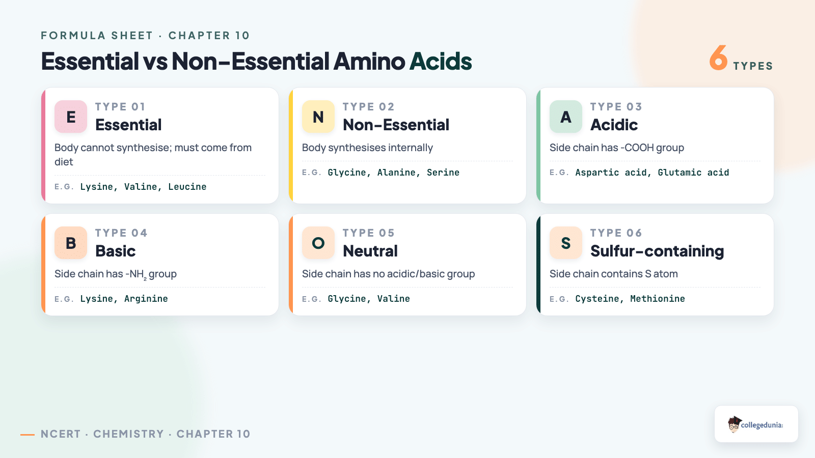
| α-Amino acid general formula | R-CH(NH2)-COOH | R = side chain | 10.2.1 |
| Essential amino acids (10) | Val, Leu, Ile, Thr, Met, Phe, Trp, Lys, Arg, His | Must come from diet | 10.2.2 |
| Zwitter ion | R-CH(NH2)-COOH R-CH(+NH3)-COO- | Net charge 0; amphoteric | 10.2.2 |
| Peptide bond | H2N-CHR1-COOH + H2N-CHR2-COOH -H2O -CHR1-CO-NH-CHR2- | Amide between −COOH and −NH2 | 10.2.3 |
| Four levels of protein structure | 1° sequence → 2° helix/sheet → 3° 3-D fold → 4° sub-unit assembly | 2°: H-bonds between peptide C=O and N−H | 10.2.3 |
| Denaturation | Heat / pH / urea / heavy metal / organic solvent | 2° and 3° lost; 1° preserved | 10.2.3 |
| Enzyme Ea drop (NCERT) | Ea (H+) = 6.22 kJ mol-1; Ea (sucrase) = 2.15 kJ mol-1 | Sucrose hydrolysis | 10.3 |
| Fat-soluble vitamins | A, D, E, K (ADEK) | Stored in liver and adipose tissue | 10.4 |
| Nucleic acid composition | Pentose sugar + H3PO4 + nitrogen base | Sugars: β-D-ribose (RNA), β-D-2-deoxyribose (DNA) | 10.5 |
| Nucleoside vs nucleotide | Base + sugar (N-glycosidic at C1′) vs Base + sugar + phosphate (ester at C5′) | Nucleotide = phosphate ester of nucleoside | 10.5 |
| Phosphodiester linkage | …-Sugar-C3'O-P(=O)(O-)-C5'O-Sugar-… | 3′ of one to 5′ of next | 10.5 |
| Watson-Crick base pairing | A ··· T (2 H-bonds); G ··· C (3 H-bonds) | 1:1 ratio (Chargaff) | 10.5 |
The top CBSE 1-mark slip is the ring swap: glucose forms a pyranose 6-ring, fructose a furanose 5-ring.
Nucleoside vs Nucleotide; DNA vs RNA Composition
| Property | DNA | RNA |
|---|---|---|
| Sugar | beta-D-2-deoxyribose (no -OH at C2') | beta-D-ribose (-OH at C2' present) |
| Bases | A, G, C, T | A, G, C, U (uracil replaces thymine) |
| Strand | Double helix, antiparallel | Single strand |
| Base pairs | A ··· T (2 H-bonds); G ··· C (3 H-bonds) | A-U / G-C in hairpins |
| Function | Stores heredity; replicates | mRNA, tRNA, rRNA in protein synthesis |
Nucleoside = base + sugar; nucleotide = base + sugar + phosphate. Purines (A, G) are double-ring; pyrimidines (C, T, U) are single-ring. Mnemonic: "PURe As Gold".
Vitamins, Sources and Deficiency Reference Table for Biomolecules
These pairs power most 1-mark MCQs on Section 10.4.
| Vitamin | Solubility class | Main source | Deficiency disease |
|---|---|---|---|
| A | Fat-soluble | Fish liver oil, carrots, butter, milk | Xerophthalmia, night blindness |
| B1 (thiamine) | Water-soluble | Yeast, milk, cereals | Beri-beri |
| B2 (riboflavin) | Water-soluble | Milk, egg white, liver, kidney | Cheilosis |
| B6 (pyridoxine) | Water-soluble | Yeast, milk, egg yolk, cereals | Convulsions |
| B12 | Water-soluble (but stored) | Meat, fish, egg, curd | Pernicious anaemia |
| C (ascorbic acid) | Water-soluble | Citrus fruits, amla, leafy vegetables | Scurvy, bleeding gums |
| D | Fat-soluble | Sunlight, fish, egg yolk | Rickets, osteomalacia |
| E | Fat-soluble | Wheat germ oil, sunflower oil | RBC fragility, muscular weakness |
| K | Fat-soluble | Green leafy vegetables | Increased blood-clotting time |
Student Feedback
In a Collegedunia poll of 900 Class 12 students, 78% said the DNA vs RNA and vitamin-deficiency tables on this Biomolecules sheet saved them the most revision time before the Chemistry exam.
Other Resources for Biomolecules Class 12 Chemistry
- Biomolecules Class 12 Chemistry Formula Sheet
- Biomolecules Class 12 Chemistry NCERT Solutions
- Biomolecules Class 12 Chemistry Notes
- Biomolecules Class 12 Chemistry NCERT Book PDF
- Biomolecules Class 12 Chemistry NCERT Exemplar Book PDF
- Biomolecules Class 12 Chemistry NCERT Exemplar Solutions
- Biomolecules Class 12 Chemistry Handwritten Notes
NCERT Formula Sheet for Class 12 Chemistry: All Chapters
Jump to the formula sheet for any other chapter of Class 12 Chemistry below.
| Chapter | Resource |
|---|---|
| Chapter 1 | Solutions Formula Sheet |
| Chapter 2 | Electrochemistry Formula Sheet |
| Chapter 3 | Chemical Kinetics Formula Sheet |
| Chapter 4 | d- and f-Block Elements Formula Sheet |
| Chapter 5 | Coordination Compounds Formula Sheet |
| Chapter 6 | Haloalkanes and Haloarenes Formula Sheet |
| Chapter 7 | Alcohols, Phenols and Ethers Formula Sheet |
| Chapter 8 | Aldehydes, Ketones and Carboxylic Acids Formula Sheet |
| Chapter 9 | Amines Formula Sheet |
Biomolecules Class 12 Chemistry Formula Sheet FAQs
Ques. Where can I download the Biomolecules Class 12 Chemistry Formula Sheet PDF?
Ans. You can download the Biomolecules Class 12 Chemistry Formula Sheet PDF directly from this Collegedunia page. Both the Normal and HD versions are available and free of cost.
Ques. Is this Formula Sheet aligned with the 2026-27 NCERT?
Ans. Yes. This page reflects the current 2026-27 syllabus for Class 12 Chemistry. Biomolecules is fully retained in the new edition with no structural cuts; every concept in Sections 10.1 to 10.6 of the NCERT remains examinable.
Ques. How many pages is the Class 12th Chemistry Biomolecules Formula Sheet PDF?
Ans. The Formula Sheet PDF runs approximately 18 pages and covers the carbohydrate master table, amino-acid and protein structural rules, enzyme activation-energy data, the vitamin-deficiency reference, the DNA-RNA comparison, and the hormone classification.
Ques. What is the difference between glucose and fructose at the structural level?
Ans. Both have the same molecular formula C6H12O6 , but they differ in three structural respects. Glucose is an aldohexose: it carries a −CHO group at C1 and four chiral centres (C2, C3, C4, C5). Fructose is a ketohexose: it carries a >C=O at C2, a primary −OH at C1, and three chiral centres. On cyclisation, the −OH at C5 of glucose attacks the C1 aldehyde to give a six-membered pyranose ring (one O + five C); in fructose the −OH at C5 attacks the C2 keto to give a five-membered furanose ring (one O + four C). Glucose is dextrorotatory ([α]D = +52.5∘) ; fructose is laevorotatory ([α]D = -92.4∘) . The 1 : 1 mixture obtained when sucrose hydrolyses is called invert sugar because the net rotation flips from + to −.
Ques. Why are sucrose, maltose and lactose classified differently on the reducing test?
Ans. A reducing sugar must have at least one free hemiacetal (a C bearing both an −OH and an −OR via the ring oxygen) which can open back to a free −CHO and reduce Fehling or Tollens reagent. Sucrose (α-D-glucose C1 – β-D-fructose C2) uses both anomeric −OH groups (C1 of glucose and C2 of fructose) inside the glycosidic linkage, so neither sugar can open up - sucrose is non-reducing. Maltose (two α-D-glucose units linked C1–C4) and lactose (β-D-galactose C1 – β-D-glucose C4) each leave one anomeric C free, so they retain a hemiacetal and reduce Fehling and Tollens. The same rule explains why all monosaccharides reduce both reagents.
Ques. What exactly happens during the denaturation of a protein, and is it reversible?
Ans. Denaturation is the loss of the secondary and tertiary three-dimensional structure of a protein, triggered by heat, pH change, urea, organic solvents, or heavy-metal salts. The hydrogen bonds that hold the α-helix and the β-pleated sheet, plus the disulphide −S−S− bridges that lock the 3-D fold, are cleaved. The primary structure (the peptide-bond sequence) is preserved because the −CO−NH− bond itself is not broken. The protein loses biological activity (a denatured enzyme cannot catalyse; a denatured hormone cannot signal). Common examples include the coagulation of egg-white on boiling and the curdling of milk by lactic acid from bacteria. In most cases denaturation is irreversible in a laboratory setting; refolding the chain back into its native conformation requires either chaperone proteins or controlled cellular conditions and is rarely complete.
Ques. How does the structure of DNA differ from the structure of RNA?
Ans. DNA and RNA differ at three structural levels. Sugar: DNA contains β-D-2-deoxyribose (no −OH at C2′); RNA contains β-D-ribose (−OH at C2′ retained). Bases: DNA uses adenine, guanine, cytosine, and thymine; RNA uses adenine, guanine, cytosine, and uracil in place of thymine. Strand architecture: DNA is a right-handed double helix with two antiparallel strands held by the Watson-Crick base pairs A···T (two H-bonds) and G···C (three H-bonds), giving Chargaff's 1:1 ratio. RNA is single-stranded and exists in three functional forms (mRNA, rRNA, tRNA) that work together during protein synthesis. DNA stores hereditary information and self-replicates; RNA executes the genetic message, and it does not self-replicate inside cells.
Ques. Which vitamin deficiencies cause scurvy, beri-beri, night blindness, rickets, and increased blood clotting time?
Ans. The five disease-vitamin pairs that NCERT Table 10.3 fixes are: Scurvy → deficiency of Vitamin C (ascorbic acid; sourced from citrus and amla); Beri-beri → deficiency of Vitamin B1 (thiamine); Night blindness / xerophthalmia → deficiency of Vitamin A; Rickets / osteomalacia → deficiency of Vitamin D; Increased blood-clotting time → deficiency of Vitamin K. NCERT exercise 10.19 asks "name the vitamin responsible for the coagulation of blood" and the marking scheme expects only "Vitamin K".
Ques. Why is insulin called a protein when it is only 51 amino acids long?
Ans. The boundary between polypeptide and protein is not a sharp count. NCERT records that compounds with more than 10 amino acid residues are usually called polypeptides, and those with more than 100 residues or a molecular mass above 10,000 u are called proteins. Insulin sits below both of those numerical thresholds (51 residues, M about 5,800 u), but it has a clearly defined three-dimensional fold built from two polypeptide chains held together by disulphide −S−S− bridges, and it performs a well-defined biological function (lowering blood glucose by promoting cellular uptake and storage as glycogen). Because of that well-defined 3-D structure and dedicated function, NCERT and IUPAC convention both classify insulin as a protein hormone, even though by residue count alone it would technically be a polypeptide.
Ques. What is the difference between anomers and epimers and how is mutarotation defined?
Ans. Anomers are stereoisomers of sugars that differ only at the anomeric carbon - C1 in aldoses or C2 in ketoses - which is the new chiral centre created when the open chain cyclises. Alpha- and beta-D-glucopyranose are anomers. Epimers are stereoisomers that differ at one non-anomeric chiral carbon: glucose and galactose are C4 epimers; glucose and mannose are C2 epimers. Mutarotation is the gradual change in specific rotation when a pure anomer of a reducing sugar dissolves in water and equilibrates with the other anomer through the open-chain form. For D-glucose, pure alpha has [α] = +112°, pure beta has [α] = +19°, and the equilibrium mixture has [α] = +52.5°.
Ques. How is haemoglobin an example of quaternary protein structure?
Ans. Haemoglobin is built from four polypeptide subunits - two α chains (141 residues each) and two β chains (146 residues each) - each cradling a heme group with a Fe(II) centre. The four subunits associate through non-covalent interactions and salt bridges; total molecular mass is about 64,500 u. Cooperative O2 binding (positive cooperativity) gives haemoglobin its sigmoidal binding curve, distinguishing it from monomeric myoglobin (a tertiary-only protein). The association of two or more polypeptide chains defines quaternary structure.
Ques. What are purines and pyrimidines and which bases appear in DNA vs RNA?
Ans. Purines are double-ring nitrogen bases - adenine (A) and guanine (G); mnemonic "PURe As Gold". Pyrimidines are single-ring bases - cytosine (C), thymine (T) and uracil (U). DNA contains A, G, C, T; RNA replaces thymine with uracil and so contains A, G, C, U. The Watson-Crick base pairs in DNA are A ··· T (2 H-bonds) and G ··· C (3 H-bonds), each pairing one purine with one pyrimidine.



Comments