




The National Testing Agency (NTA) conducted the CUET PG 2026 Biochemistry (SCQP05) examination on March 25, 2026, during Shift 3 from 04:00 PM to 05:30 PM.
Students who appeared for the exam reported that the overall difficulty level of the paper was moderate. CUET PG 2026 Biochemistry Question Paper with Solutions PDF is available here for download. The marking scheme is +4 for correct answers and -1 for wrong answers, totaling 300 marks.
CUET PG 2026 Biochemistry Question Paper with Solutions PDF
| CUET PG 2026 Biochemistry Question Paper with Answer Key | Download PDF | Check Solutions |
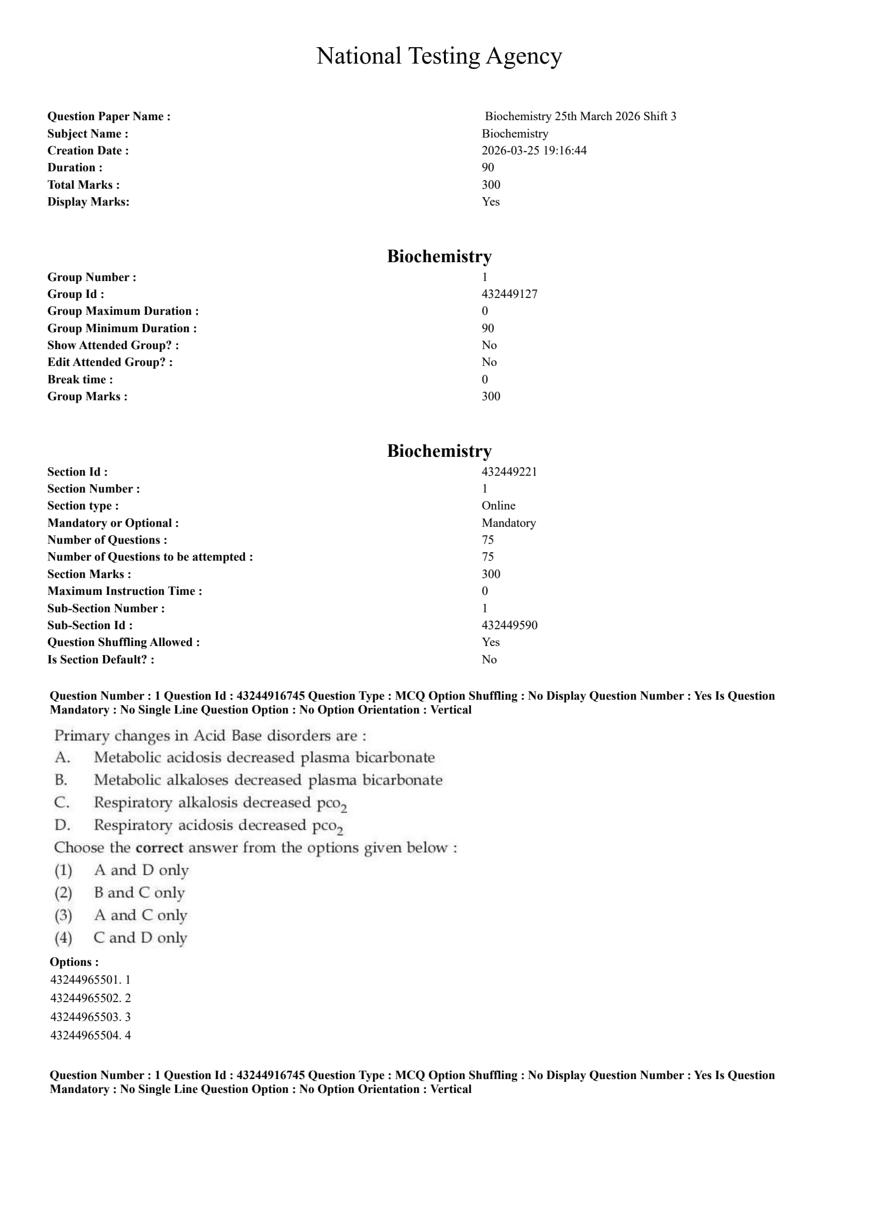
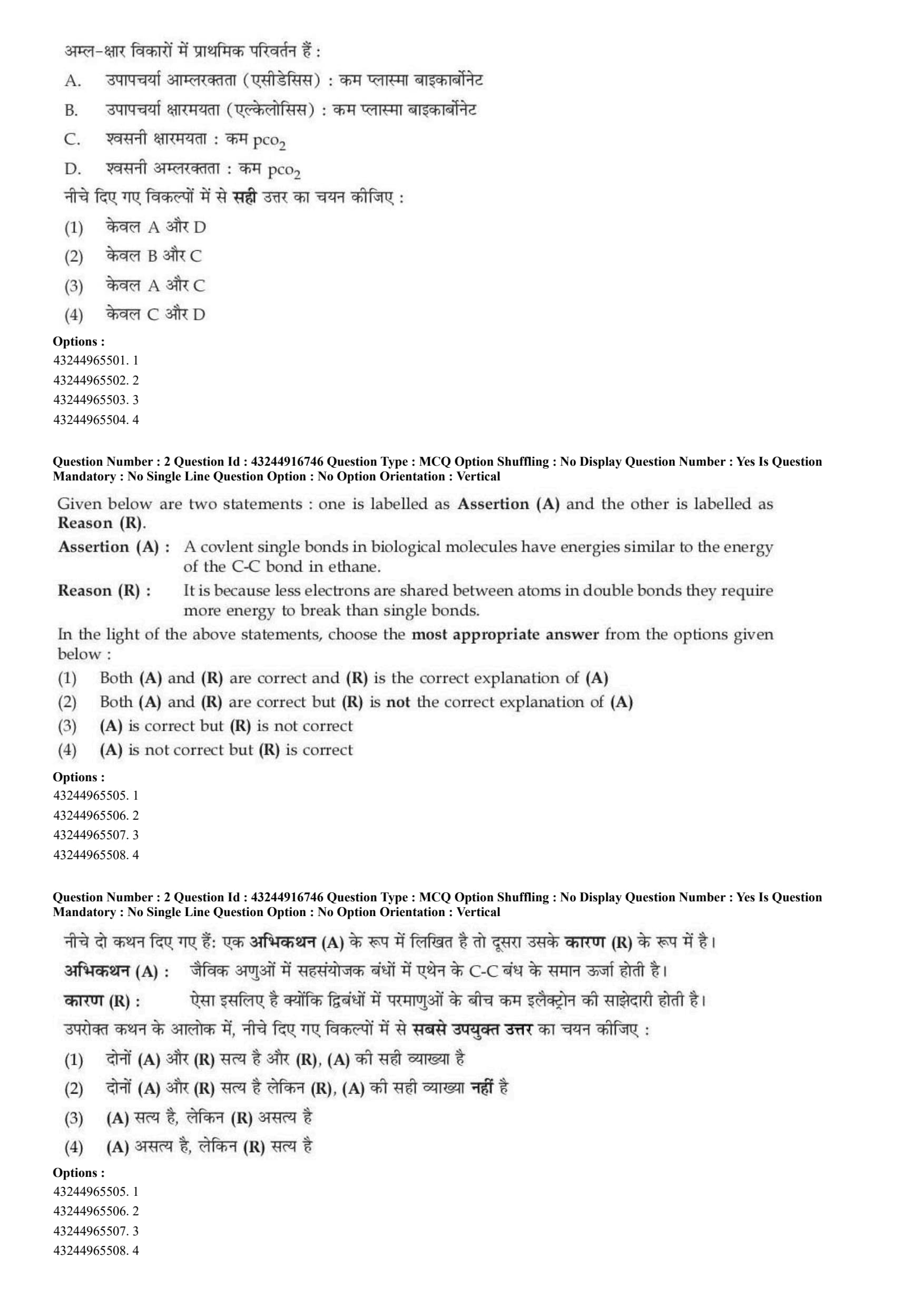
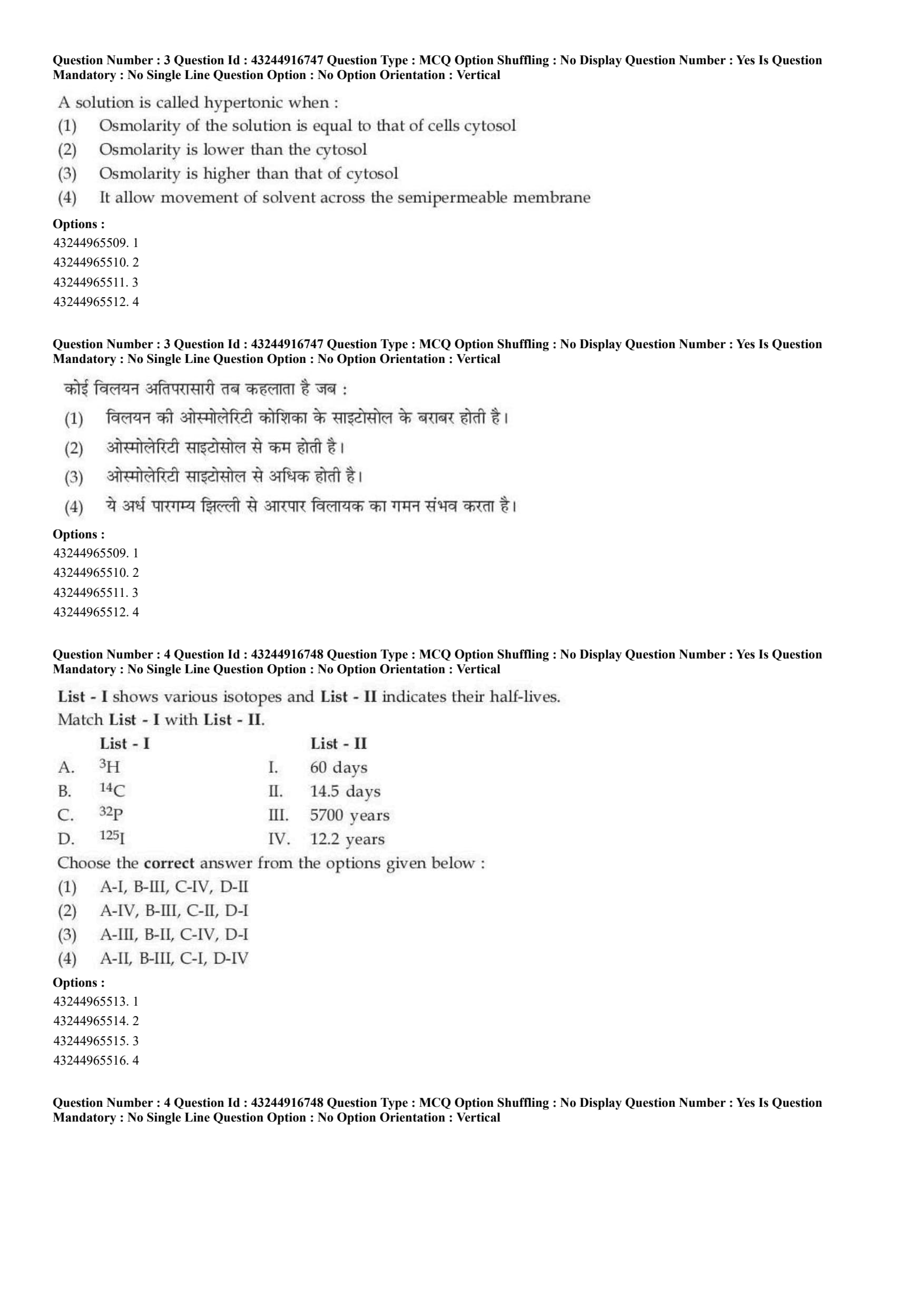
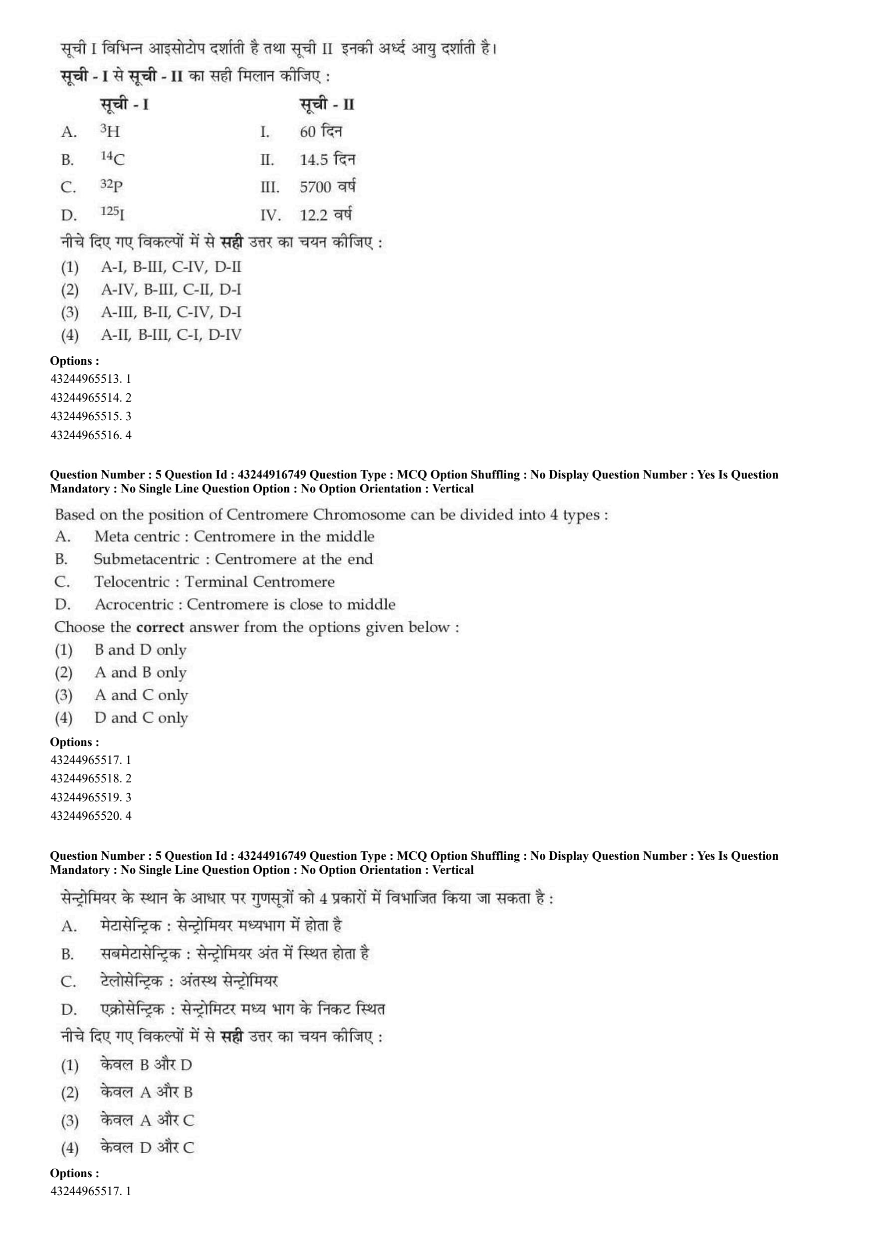
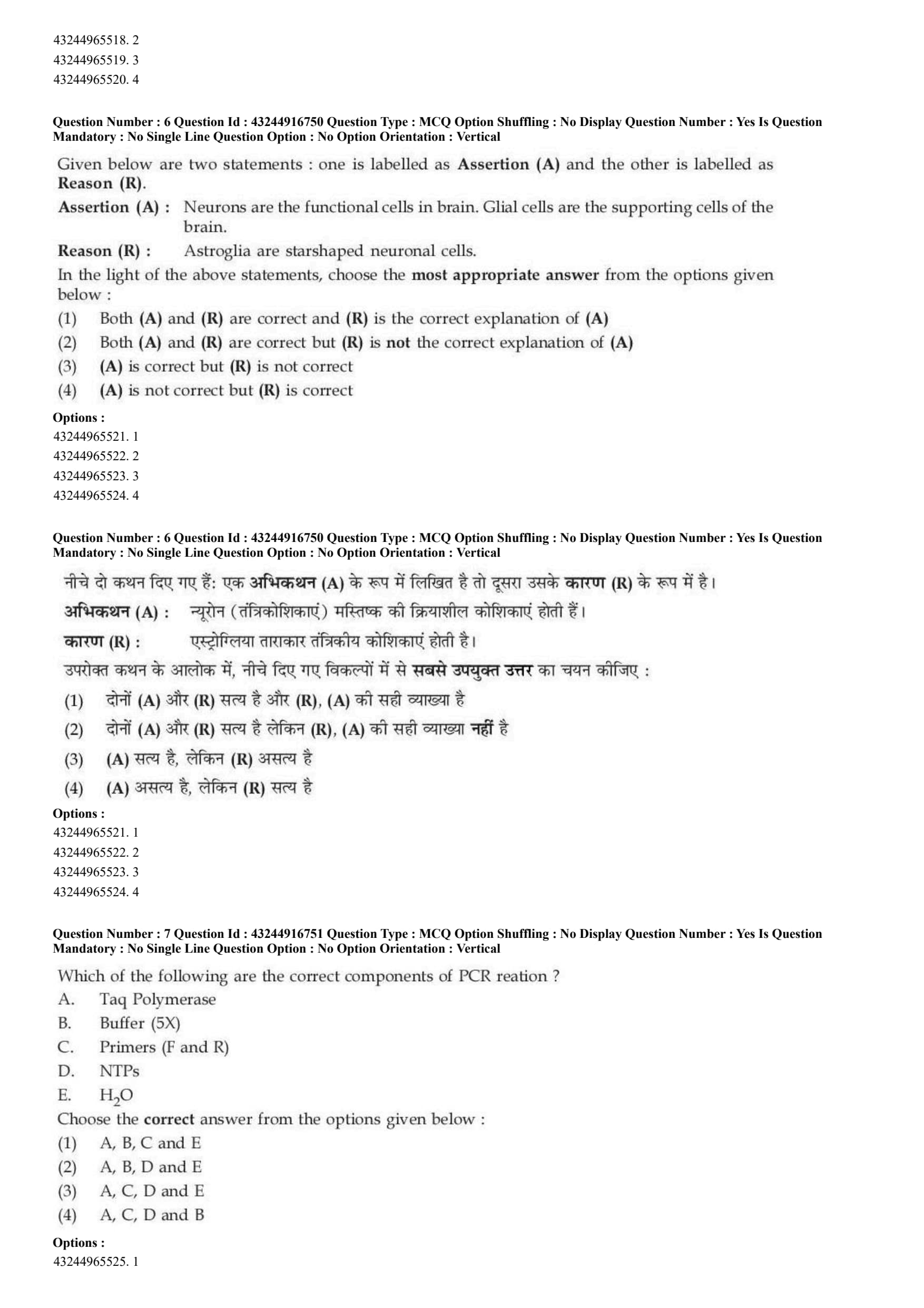
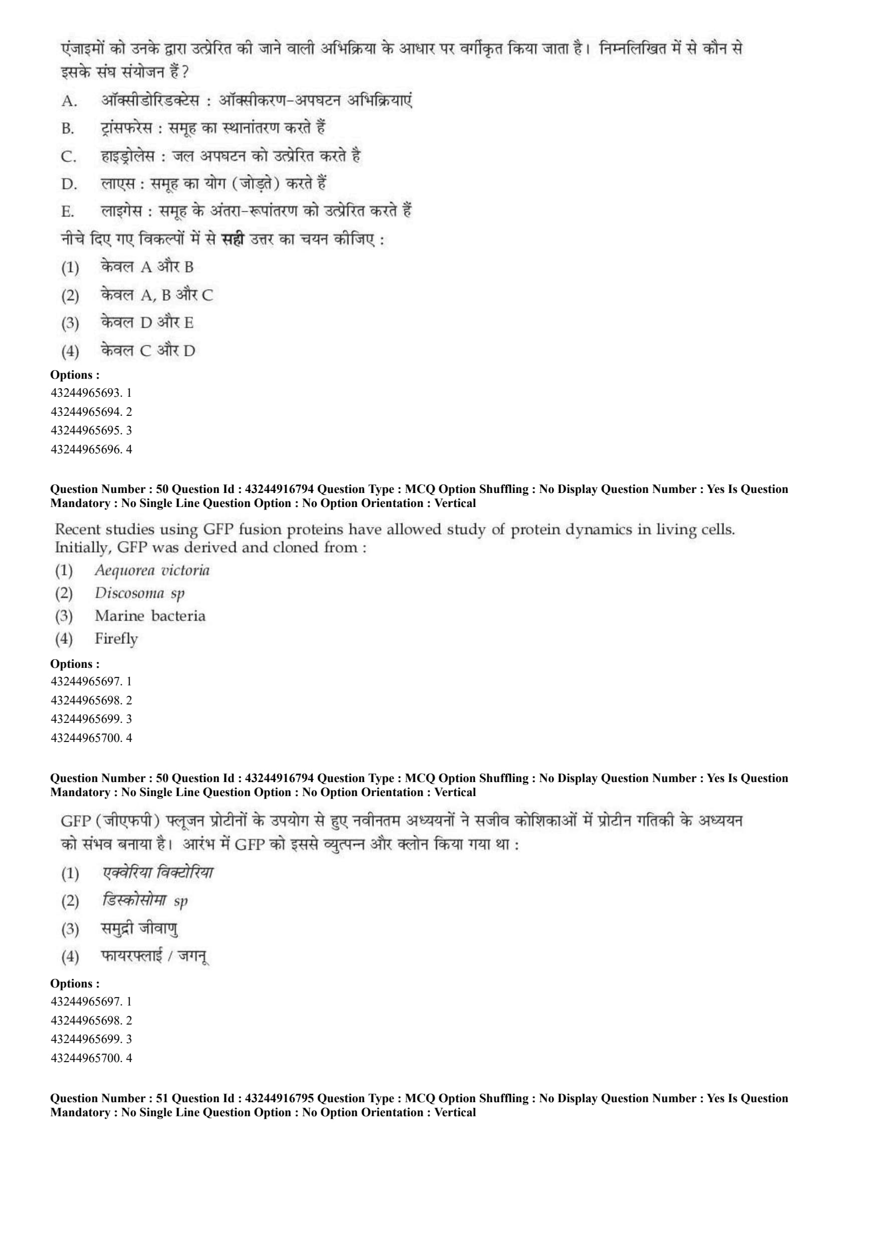
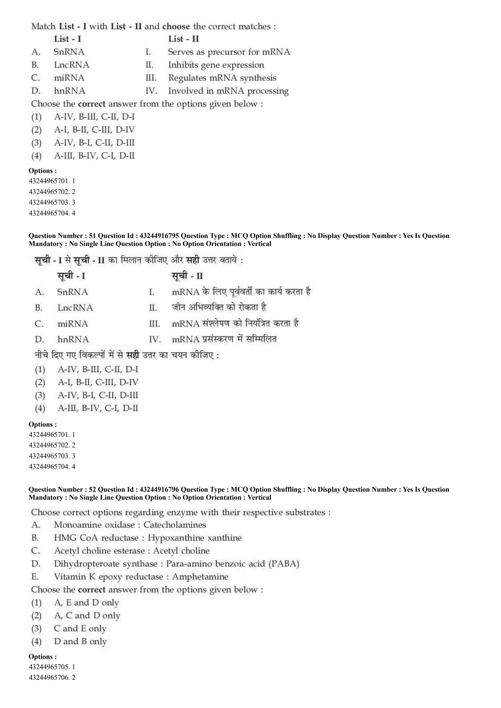
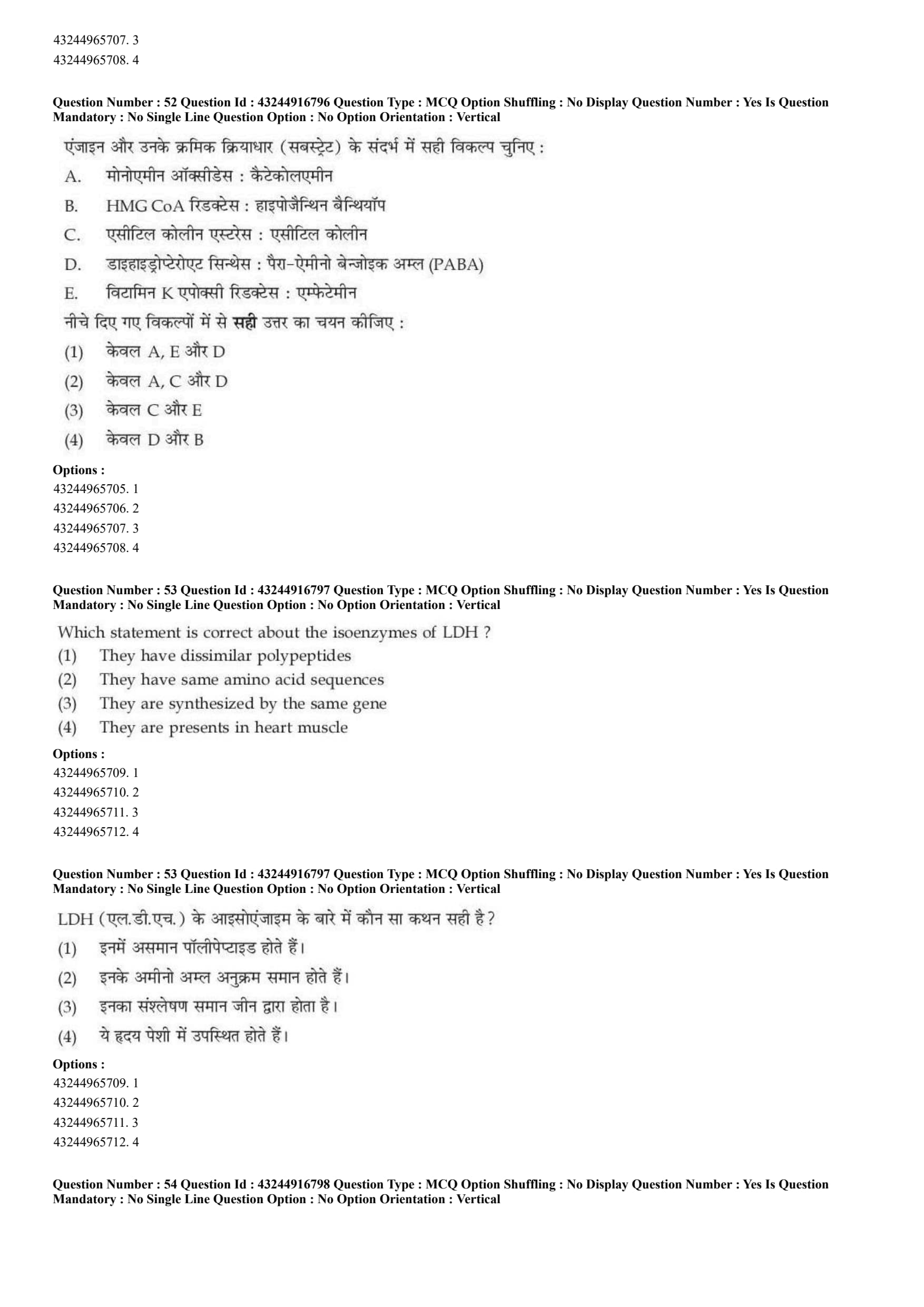
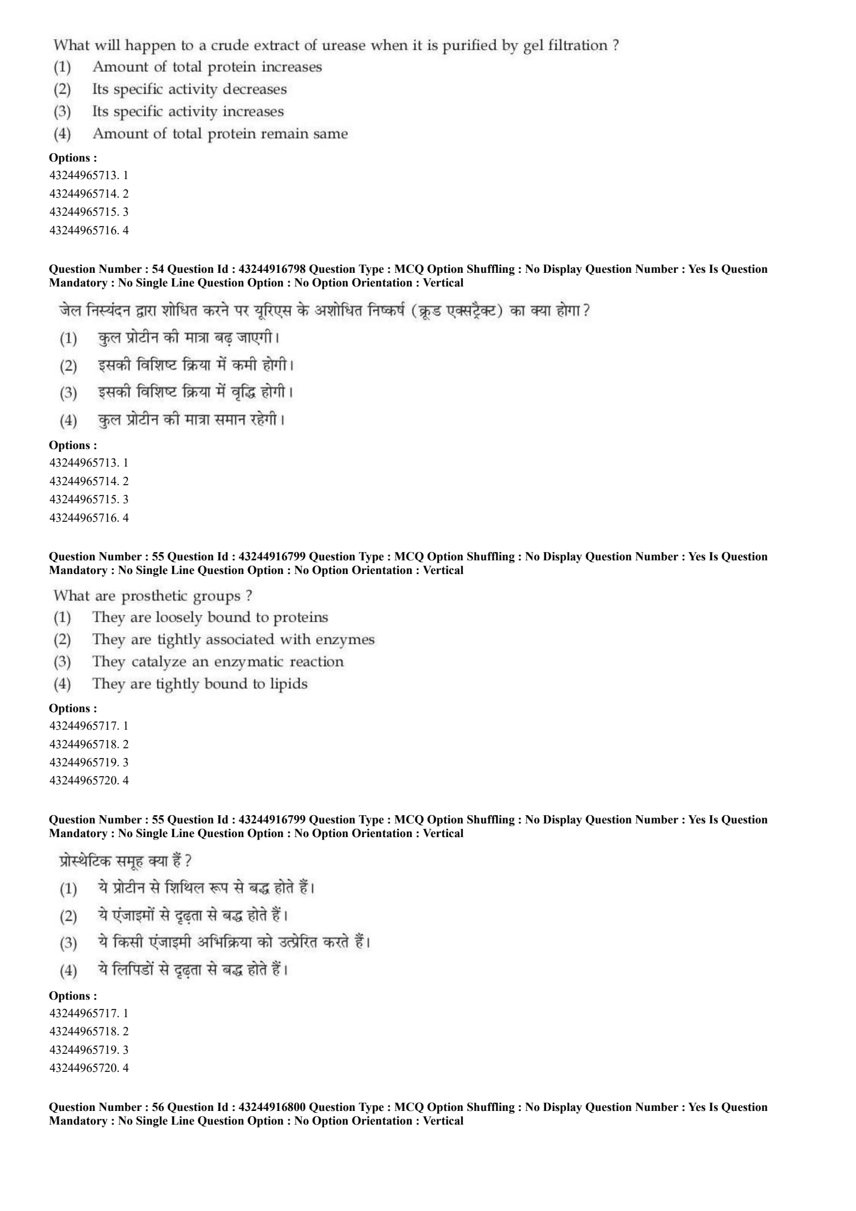
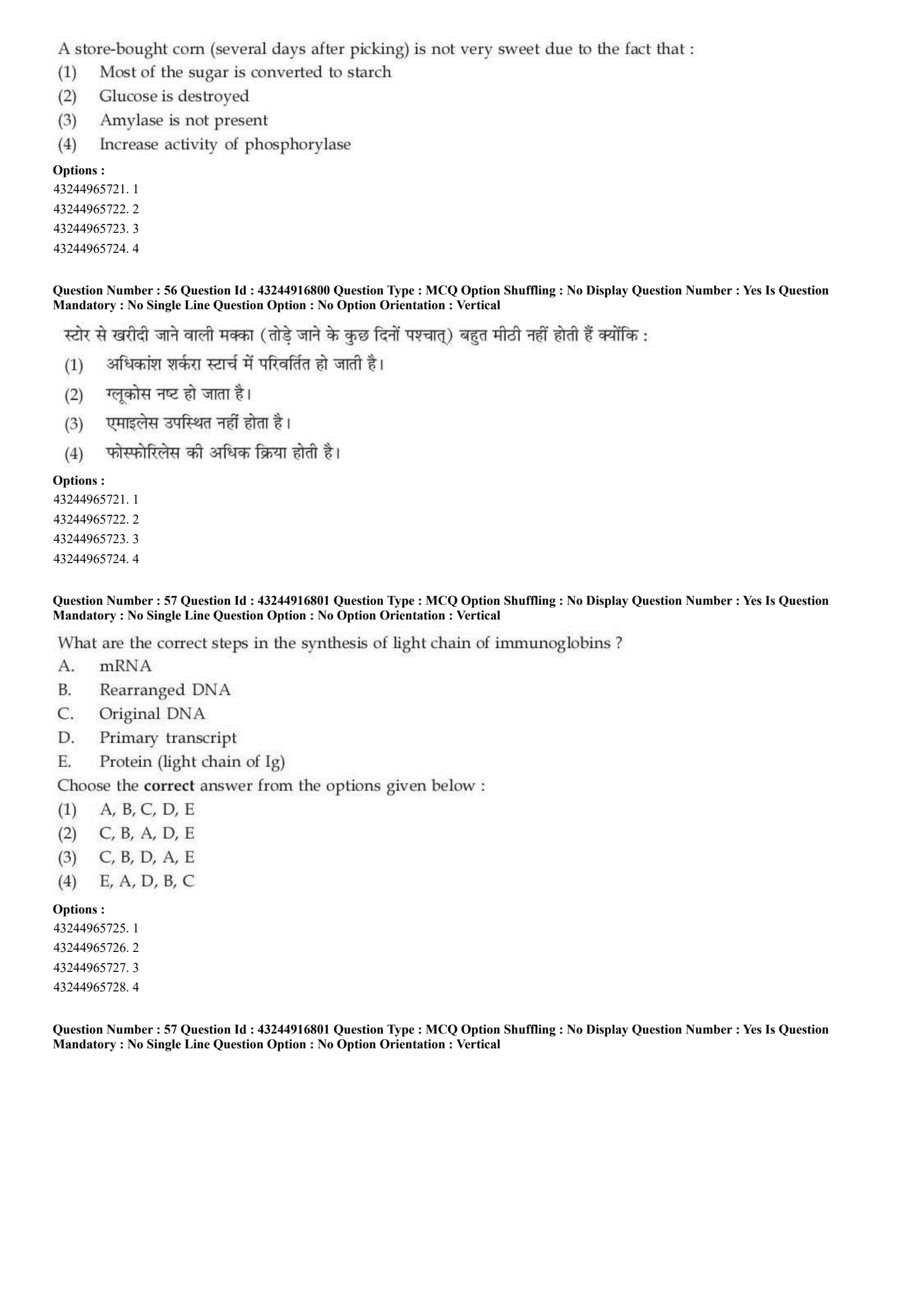
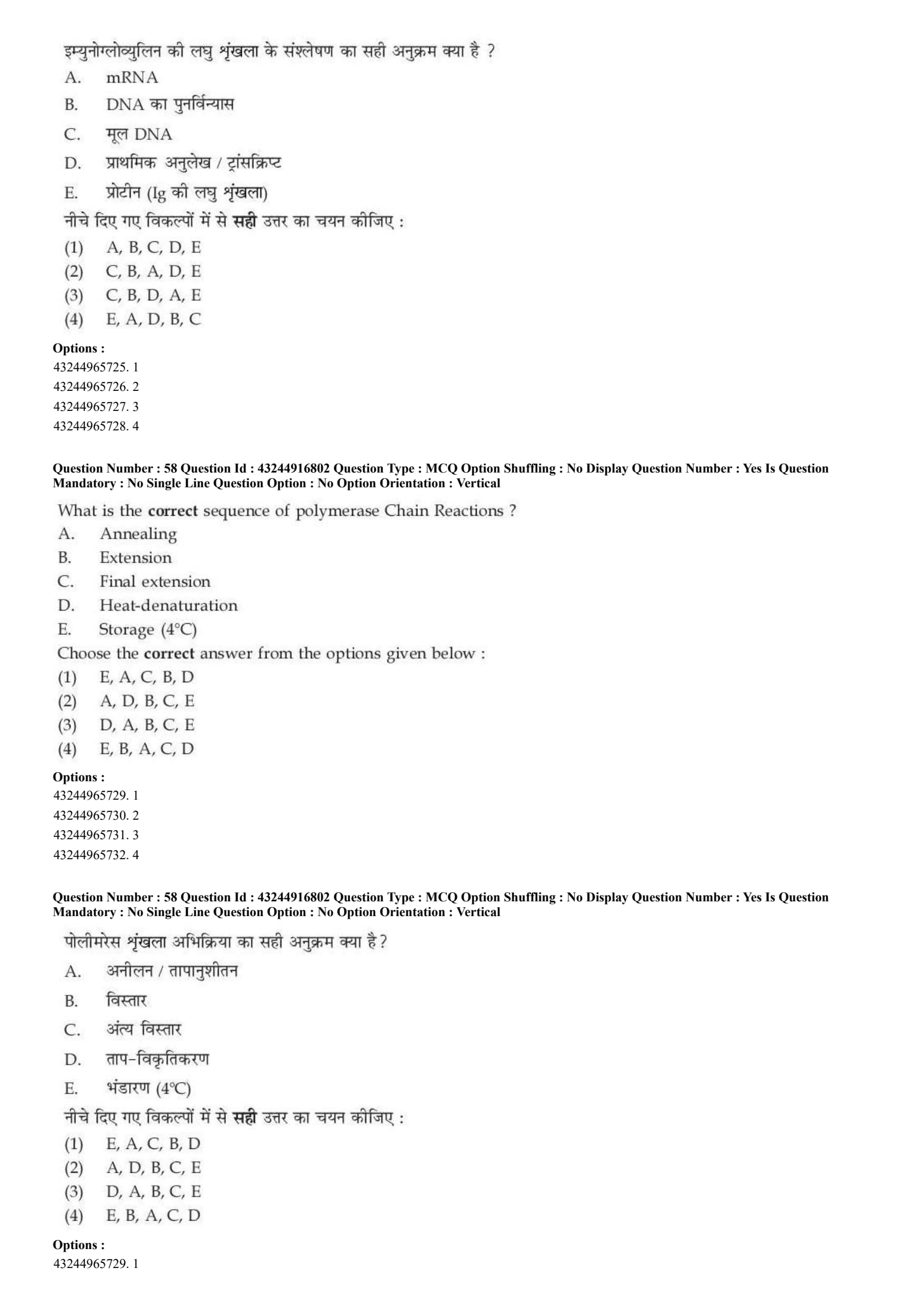
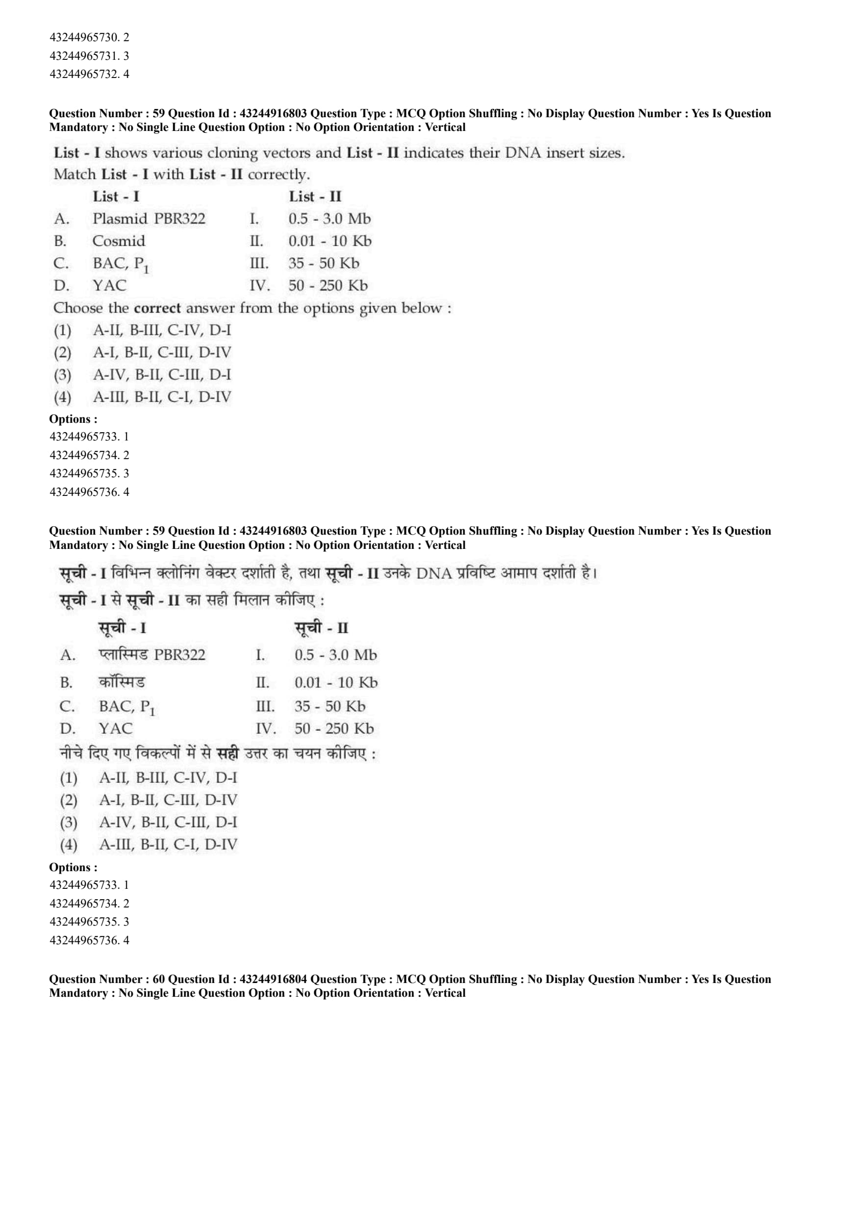
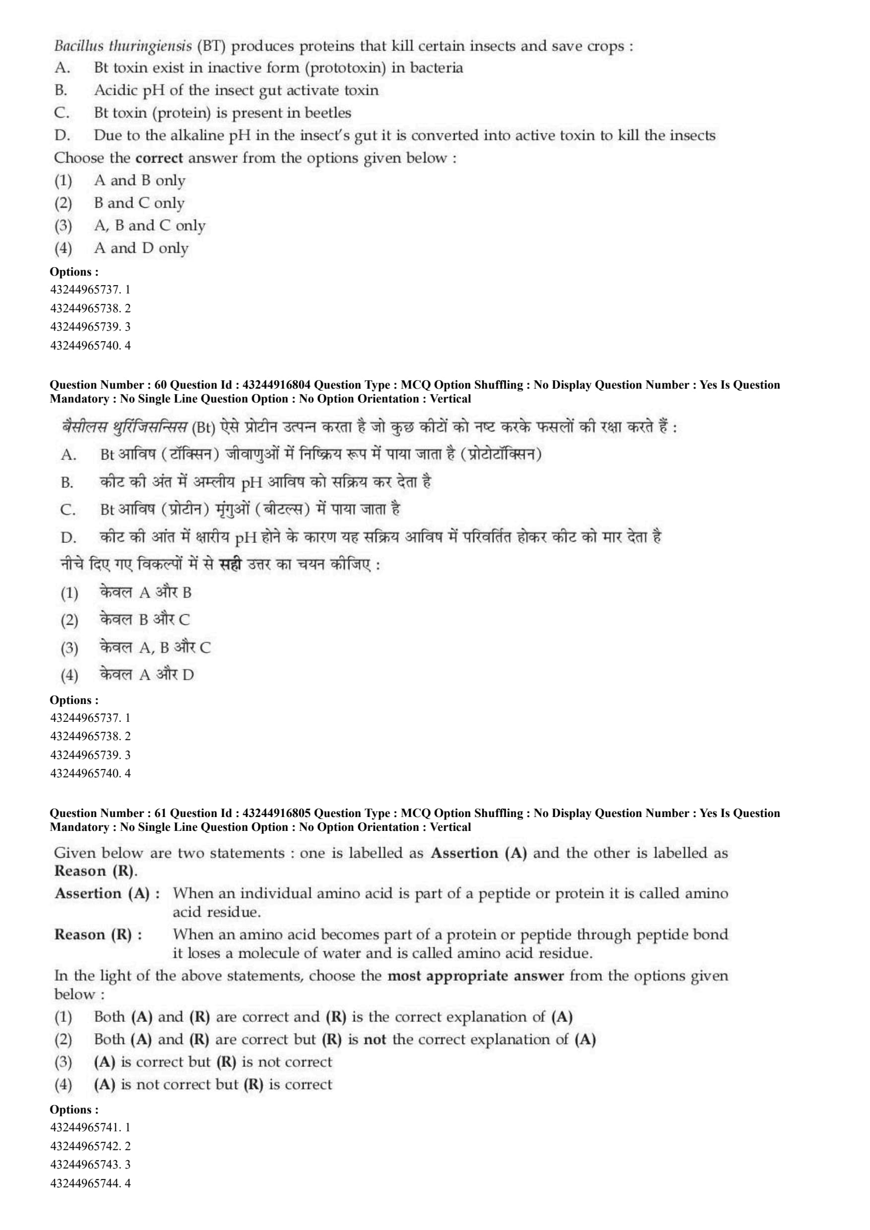
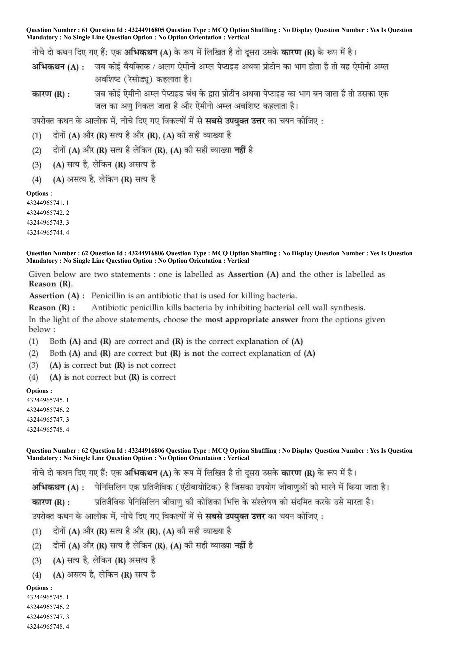
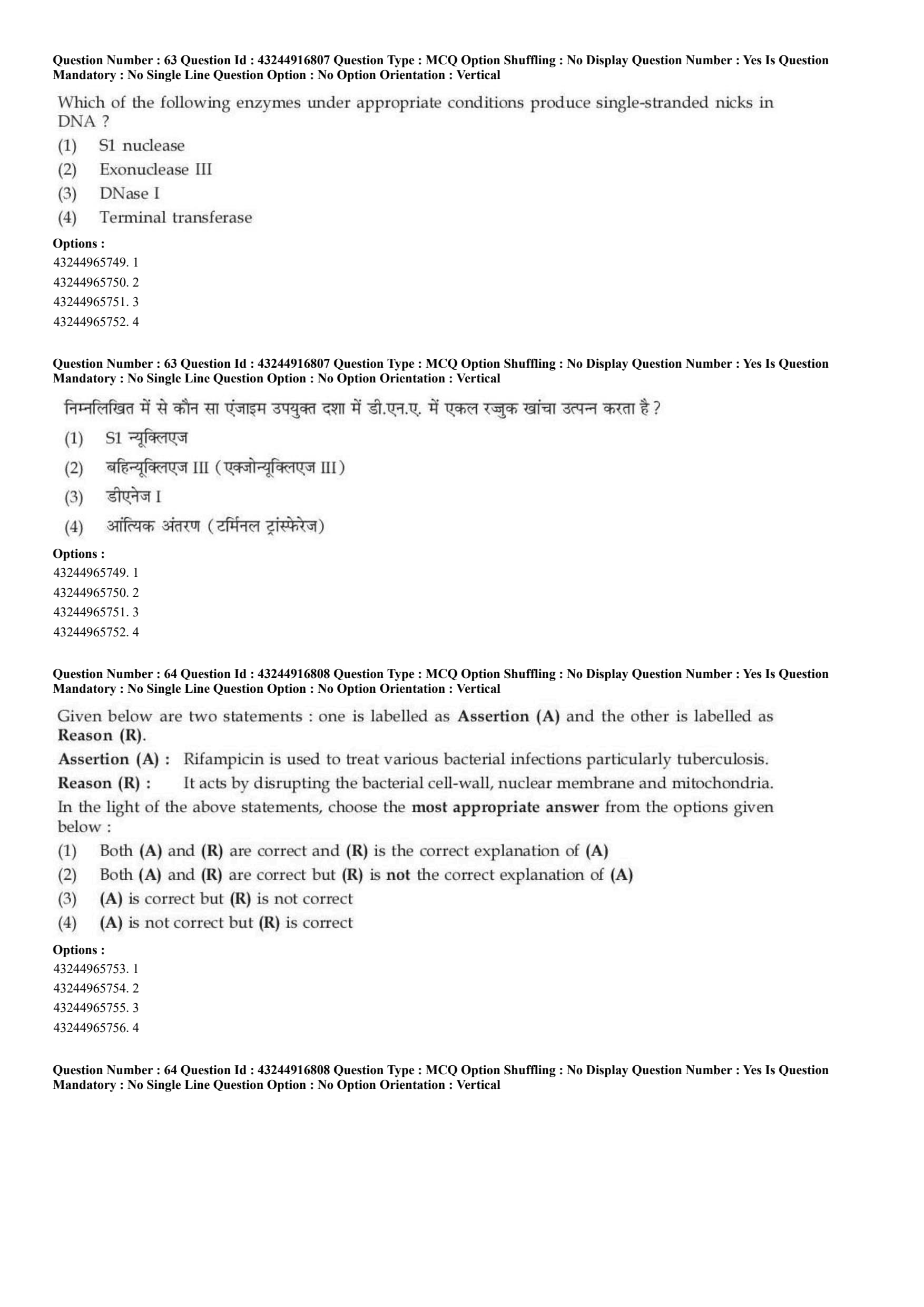
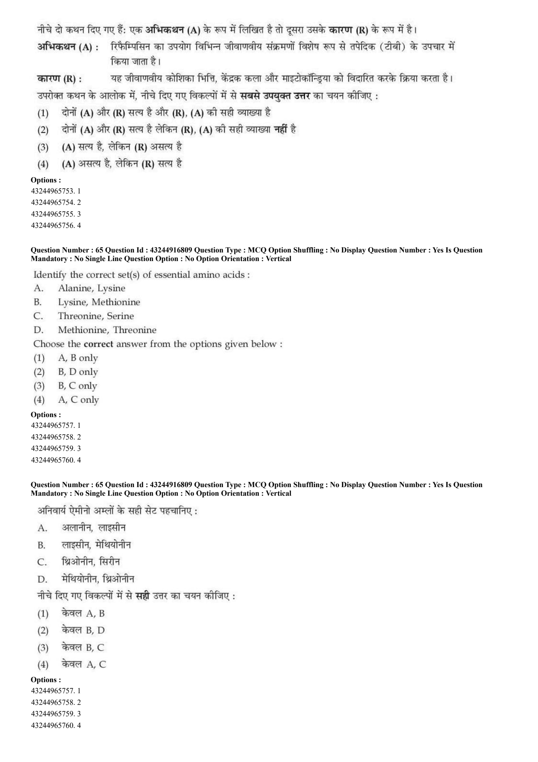
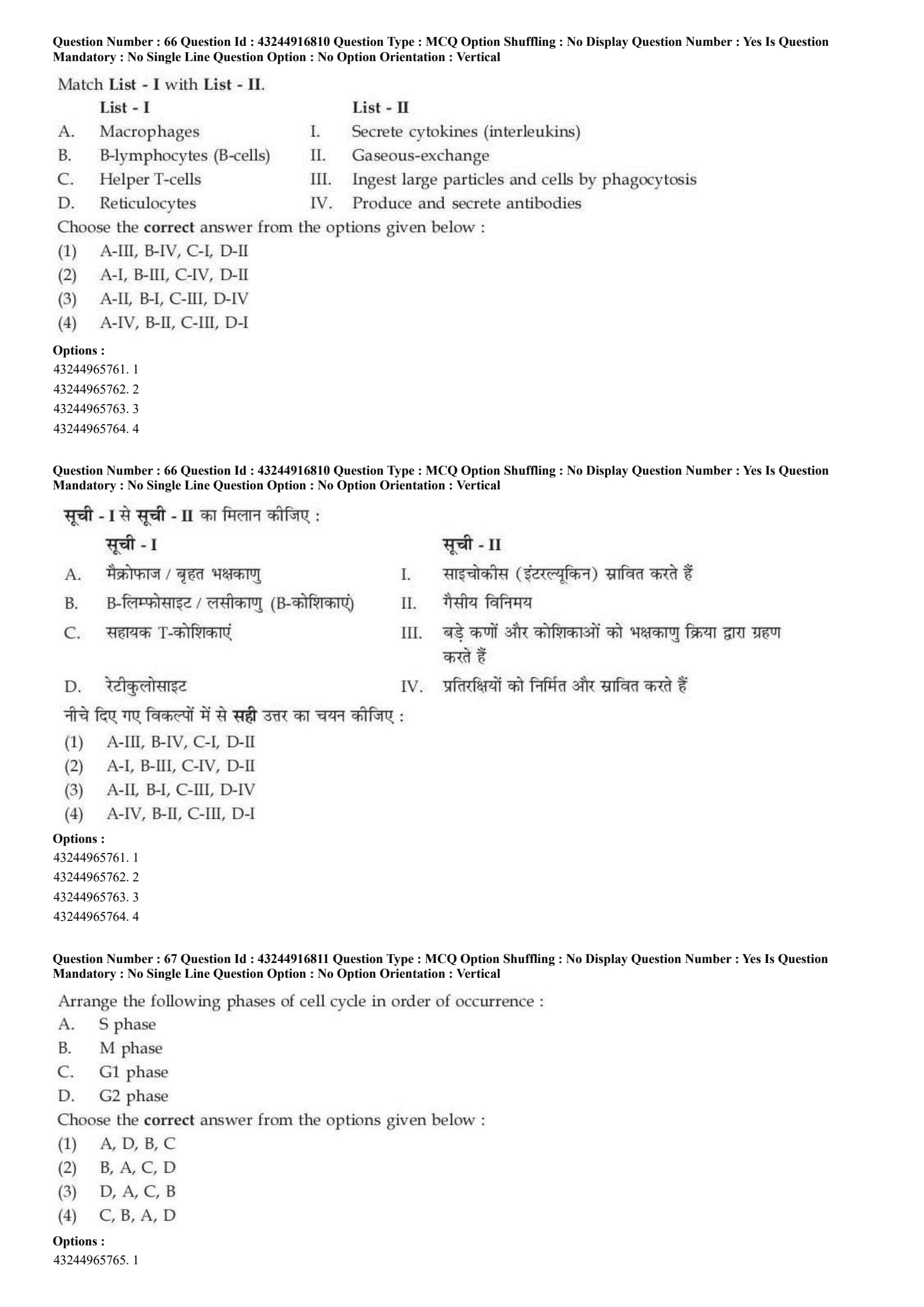
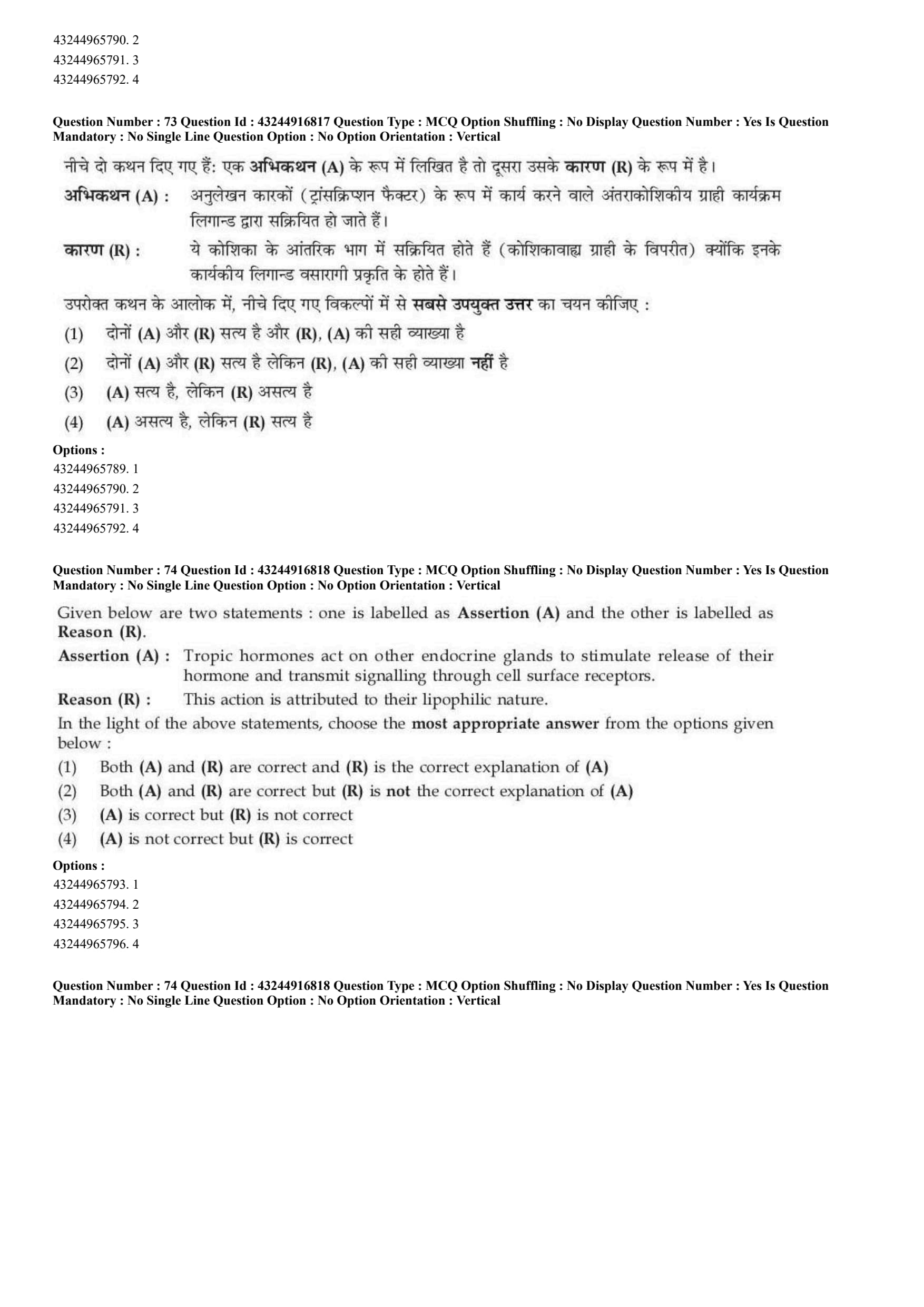
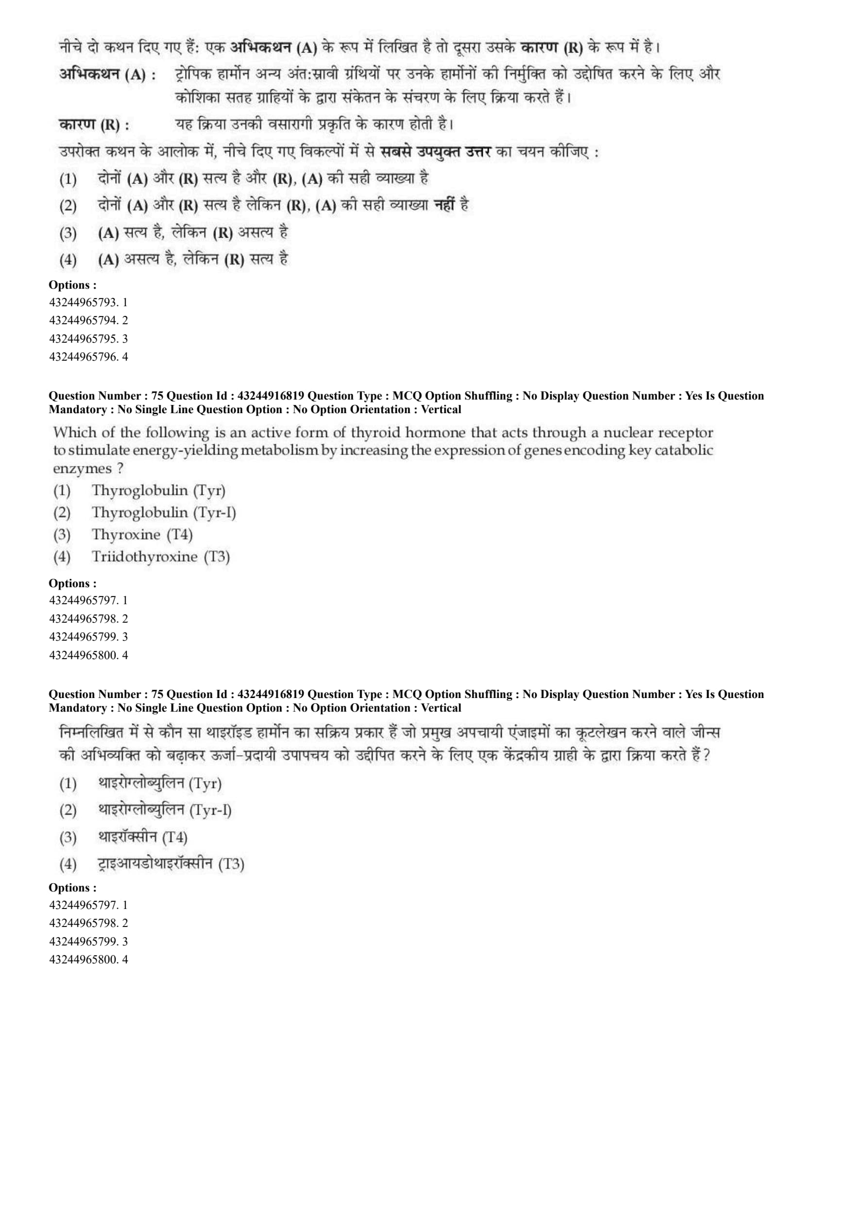
Primary changes in Acid Base disorders are:
View Solution
\textcolor{red{Step 1: Concept
Acid-base disorders are classified as metabolic or respiratory depending on the primary cause and the changing parameters in the blood chemistry.
\textcolor{red{Step 2: Meaning
Acidosis involves processes that lower blood pH, whereas alkalosis involves processes that raise blood pH. Primary metabolic disorders primarily impact bicarbonate concentrations (\(HCO_{3}^{-}\)), while respiratory disorders primarily change the partial pressure of carbon dioxide (\(P_{CO_{2}}\)).
\textcolor{red{Step 3: Analysis
* Statement A: Metabolic acidosis is correctly identified by a primary drop in plasma bicarbonate concentration.
* Statement B: Metabolic alkalosis results in an increase, not a decrease, in plasma bicarbonate.
* Statement C: Respiratory alkalosis involves hyperventilation which leads to a primary decrease in plasma \(P_{CO_{2}}\).
* Statement D: Respiratory acidosis is characterized by an increase, not a decrease, in plasma \(P_{CO_{2}}\) due to carbon dioxide retention.
Therefore, statements A and C are the only correct descriptions of primary changes.
\textcolor{red{Step 4: Conclusion
Hence, the correct option matching both A and C is chosen.
\textcolor{red{Final Answer: (C) Quick Tip: Metabolic concerns Bicarbonate (\(HCO_{3}^{-}\)) changes in the same direction as pH. Respiratory concerns \(P_{CO_{2}}\) changes in the opposite direction of pH.
Given below are two statements: one is labelled as Assertion (A) and the other is labelled as Reason (R).
Assertion (A): Covalent single bonds in biological molecules have energies similar to the energy of the C-C bond in ethane.
Reason (R): It is because less electrons are shared between atoms in double bonds they require more energy to break than single bonds.
In the light of the above statements, choose the most appropriate answer from the options given below:
View Solution
\textcolor{red{Step 1: Concept
Chemical bond energy refers to the amount of energy required to break a specific chemical bond between atoms.
\textcolor{red{Step 2: Meaning
Single covalent bonds involve the sharing of one pair of electrons, whereas double bonds involve the sharing of two pairs (more electrons). More shared electrons create a stronger attractive force between nuclei.
\textcolor{red{Step 3: Analysis
* Assertion (A) is correct: Covalent single bonds in common biomolecules (such as C-C, C-O, C-N) share typical bond energies of around 350-400 kJ/mol, which is highly comparable to the C-C bond in ethane.
* Reason (R) is incorrect: Double bonds involve sharing more electrons (4 electrons), not fewer, compared to single bonds (2 electrons). This greater electron density requires higher energy to break.
\textcolor{red{Step 4: Conclusion
Since the assertion is true but the reason contains a false chemical statement, option C is correct.
\textcolor{red{Final Answer: (C) Quick Tip: More shared electrons mean a stronger bond, shorter bond length, and higher dissociation energy. Double bonds share 4 electrons; single bonds share 2.
A solution is called hypertonic when:
View Solution
\textcolor{red{Step 1: Concept
Toxicity describes how an extracellular solution can change the volume of a cell by altering water movement via osmosis.
\textcolor{red{Step 2: Meaning
Prefix meanings: Hyper- means greater or above, Iso- means equal, and Hypo- means under or less. Osmolarity measures total solute concentration per liter of solution.
\textcolor{red{Step 3: Analysis
* If osmolarity equals the cytosol, the solution is isotonic.
* If osmolarity is lower than the cytosol, the solution is hypotonic.
* If osmolarity is higher than that of the cytosol, it contains a higher concentration of non-penetrating solutes, making it hypertonic. This causes water to exit the cell.
\textcolor{red{Step 4: Conclusion
Thus, higher solute concentration (osmolarity) outside the cell relative to the cytosol defines a hypertonic solution.
\textcolor{red{Final Answer: (C) Quick Tip: Hyper = High solute concentration. Water always leaves the cell to dilute the "hyper" environment, causing the cell to shrink.
List - I shows various isotopes and List II indicates their half-lives. Match List I with List - II.

View Solution
\textcolor{red{Step 1: Concept
Radioactive isotopes undergo decay unique to their nuclear structure, characterized by a specific half-life (\(t_{1/2}\)), which is the time required for half of the radioactive nuclei to decay.
\textcolor{red{Step 2: Meaning
Matching the standard radioisotopes commonly utilized in biochemistry and biological research with their definitive experimental half-lives.
\textcolor{red{Step 3: Analysis
* Tritium (\(^{3}H\)) has a half-life of approximately 12.2 to 12.3 years \(\rightarrow\) (A-IV).
* Carbon-14 (\(^{14}C\)), heavily utilized in radiocarbon dating, has a long half-life of about 5700 years \(\rightarrow\) (B-III).
* Phosphorus-32 (\(^{32}P\)), a high energy beta emitter used to label nucleic acids, has a short half-life of 14.3 to 14.5 days \(\rightarrow\) (C-II).
* Iodine-125 (\(^{125}I\)), commonly used in radioimmunoassays, has a half-life of around 60 days \(\rightarrow\) (D-I).
\textcolor{red{Step 4: Conclusion
The exact mapping is A-IV, B-III, C-II, D-I, corresponding to option B.
\textcolor{red{Final Answer: (B) Quick Tip: Carbon-14 is famous for ancient dating (thousands of years), while Phosphorus-32 decays very rapidly (around two weeks). This pairing easily unlocks the entire match question.
Based on the position of Centromere Chromosome can be divided into 4 types:
A. Metacentric: Centromere in the middle
B. Submetacentric: Centromere at the end
C. Telocentric: Terminal Centromere
D. Acrocentric: Centromere is close to middle
Choose the correct answer from the options given below:
View Solution
\textcolor{red{Step 1: Concept
Chromosomes are classified structurally based on the relative position of the centromere, which dictates the lengths of the p (short) and q (long) chromosomal arms.
\textcolor{red{Step 2: Meaning
* Metacentric: Centromere is localized centrally, yielding equal arm lengths.
* Submetacentric: Centromere is slightly displaced from the center, creating a slight asymmetry.
* Acrocentric: Centromere is severely sub-terminal, producing one extremely short arm.
* Telocentric: Centromere is positioned strictly at the terminal end (telomere region).
\textcolor{red{Step 3: Analysis
* Statement A is correct: Metacentric means the centromere sits right in the middle.
* Statement B is incorrect: Submetacentric means it is near the middle but slightly off-center, not at the end.
* Statement C is correct: Telocentric indicates a terminal centromere located precisely at the tip.
* Statement D is incorrect: Acrocentric means the centromere is situated close to one end, yielding one very small arm, not close to the middle.
\textcolor{red{Step 4: Conclusion
Thus, statements A and C are the only completely accurate definitions.
\textcolor{red{Final Answer: (C) Quick Tip: Remember the prefixes: "Melo" / "Meta" = Middle, "Telo" = Terminal (End). Thus, Metacentric is middle and Telocentric is end.
Given below are two statements: one is labelled as Assertion (A) and the other is labelled as Reason (R).
Assertion (A): Neurons are the functional cells in brain. Glial cells are the supporting cells of the brain.
Reason (R): Astroglia are star-shaped neuronal cells.
In the light of the above statements, choose the most appropriate answer from the options given below:
View Solution
\textcolor{red{Step 1: Concept
The nervous system is composed of two primary broad classes of cellular entities: neurons, which propagate electrical signals, and neuroglia (glial cells), which provide homeostatic maintenance and structural integrity.
\textcolor{red{Step 2: Meaning
Astroglia (astrocytes) are a specific type of glial support cells characterized by numerous radiating star-like cytoplasmic processes.
\textcolor{red{Step 3: Analysis
* Assertion (A) is correct: Neurons serve as the primary functional electrical-signaling network components, whereas glial cells provide crucial physiological support, insulation, and waste management.
* Reason (R) is incorrect: While astroglia are indeed star-shaped, they are fundamentally classified as glial cells, not neuronal cells. Glial cells do not propagate action potentials in the manner that neurons do.
\textcolor{red{Step 4: Conclusion
Because Assertion (A) is physiologically true but Reason (R) contains an inaccurate categorical classification, option C is correct.
\textcolor{red{Final Answer: (C) Quick Tip: Astrocytes = "Astro" (star) + "cyte" (cell). They belong strictly to the glial lineage, totally separate from neurons!
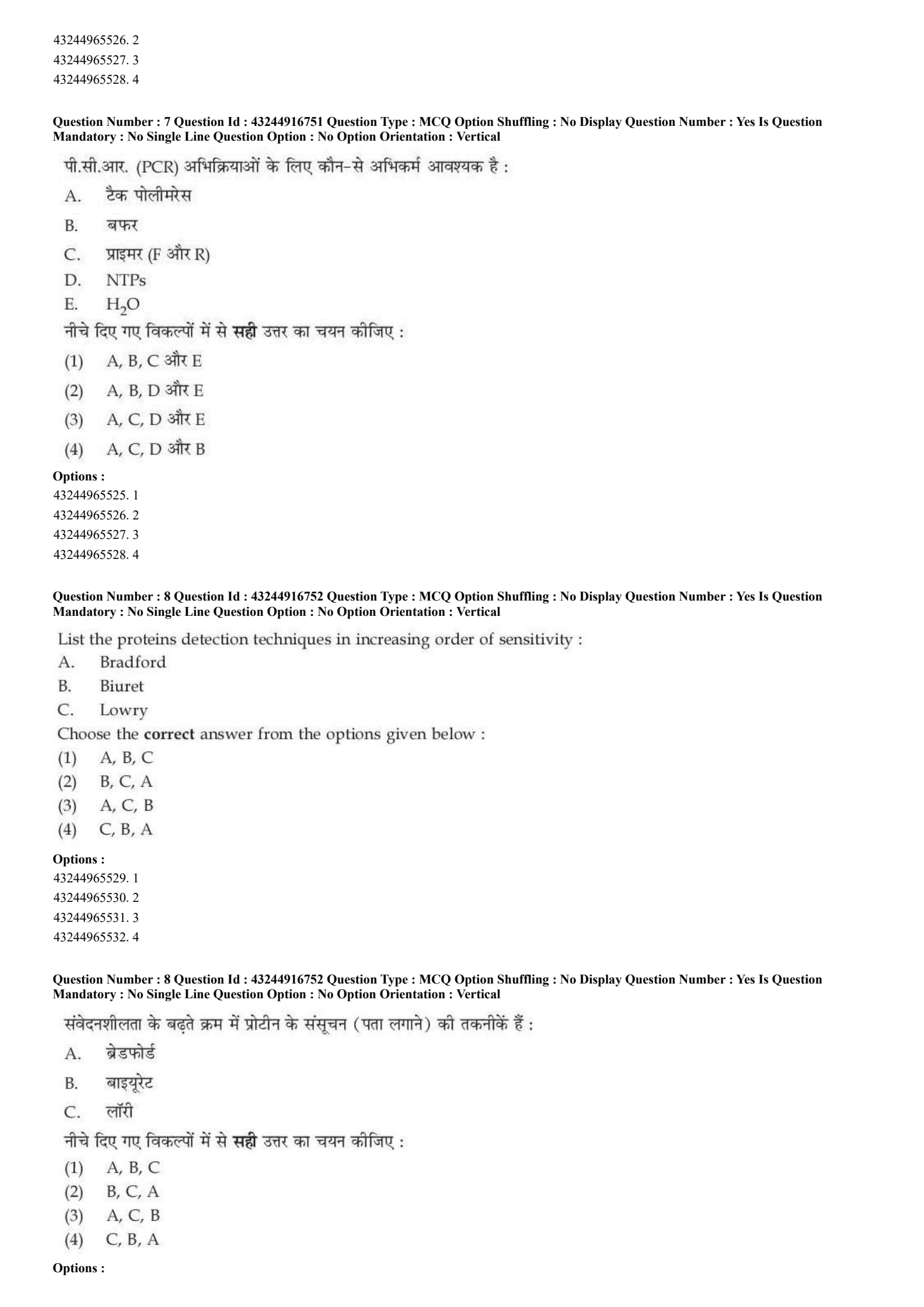
Which of the following are the correct components of PCR reaction?
A. Taq Polymerase
B. Buffer (5X)
C. Primers (F and R)
D. NTPs
E. \(H_{2}O\)
Choose the correct answer from the options given below:
View Solution
\textcolor{red{Step 1: Concept
Polymerase Chain Reaction (PCR) is an in vitro technique used to amplify specific target DNA sequences. It relies on biochemical reagents interacting dynamically inside a thermal cycler.
\textcolor{red{Step 2: Meaning
Standard component acronyms: F and R primers stand for Forward and Reverse oligonucleotides. NTPs refers generally to ribonucleoside triphosphates (used for RNA synthesis), whereas DNA polymerases specifically require dNTPs (deoxynucleoside triphosphates).
\textcolor{red{Step 3: Analysis
Let us review the necessary constituents options:
* A (Taq Polymerase) is required to extend the growing DNA strand.
* B (Buffer) is vital to keep pH and ionic strength optimal.
* C (Forward and Reverse primers) provide the essential 3'-OH starting group.
* D states NTPs instead of dNTPs. DNA synthesis requires deoxyribonucleotides (dATP, dCTP, dGTP, dTTP) rather than standard ribonucleotides.
* E (\(H_{2}O\)) serves as the solvent matrix for the chemical reactions.
Comparing the presented combinations, option (1) which lists A, B, C, and E includes valid baseline requirements.
\textcolor{red{Step 4: Conclusion
Thus, option A provides the best correct subset selection.
\textcolor{red{Final Answer: (A) Quick Tip: Watch out for NTPs vs dNTPs! PCR needs dNTPs (Deoxy) to synthesize DNA. Standard NTPs are used for making RNA.
List the proteins detection techniques in increasing order of sensitivity:
A. Bradford
B. Biuret
C. Lowry
Choose the correct answer from the options given below:
View Solution
\textcolor{red{Step 1: Concept
Protein quantification assays possess distinct lower limits of detection based on their chemical mechanics and colorimetric reagents. Higher sensitivity means the assay can detect smaller quantities of protein.
\textcolor{red{Step 2: Meaning
* Biuret assay: Relies on copper ions coordinating with peptide bonds; requires milligram levels of protein (least sensitive).
* Lowry assay: Combines biuret chemistry with Folin-Ciocalteu reagent oxidation of aromatic residues; detects microgram amounts.
* Bradford assay: Relies on the direct binding of Coomassie Brilliant Blue G-250 dye to basic/aromatic residues; highly rapid and highly sensitive.
\textcolor{red{Step 3: Analysis
Sorting the techniques from least sensitive to most sensitive:
1. Biuret method (B) - operating range is broad, usually 1-10 mg/mL.
2. Lowry method (C) - operating range is lower, down to around 10-100 \(\mu\)g/mL.
3. Bradford method (A) - highly sensitive, able to measure concentrations down to 1-10 \(\mu\)g/mL.
Thus, the increasing sequence is B \(\rightarrow\) C \(\rightarrow\) A.
\textcolor{red{Step 4: Conclusion
The sequence B, C, A corresponds to option B.
\textcolor{red{Final Answer: (B) Quick Tip: Biuret is always the least sensitive (needs mg amounts). Bradford is very sensitive and super common in biochemistry labs for small yields. Order: Biuret \(<\) Lowry \(<\) Bradford.
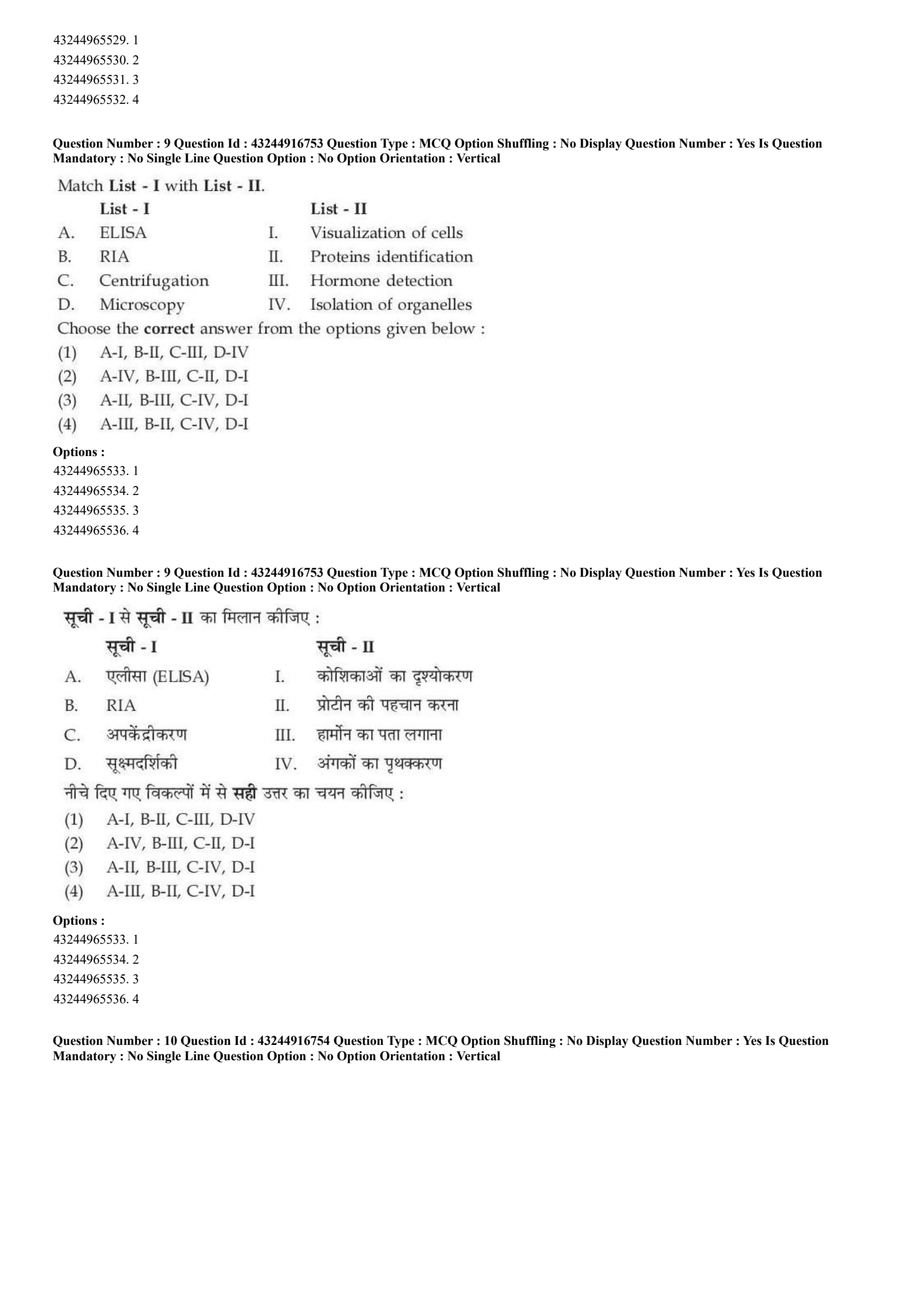
Match List - I with List - II.

Choose the correct answer from the options given below:
View Solution
\textcolor{red{Step 1: Concept
Biochemical assays and biophysical methodologies are tailored to distinct analytical purposes, spanning molecular detection, mass separation, or structural rendering.
\textcolor{red{Step 2: Meaning
* ELISA: Enzyme-Linked Immunosorbent Assay.
* RIA: Radioimmunoassay.
\textcolor{red{Step 3: Analysis
* ELISA (A) is widely applied for antigen/protein detection and quantification via antibody-linked color changes \(\rightarrow\) (A-II).
* RIA (B) utilizes radiolabeled molecules to target and evaluate subtle biochemicals, historically crucial for hormone level quantification \(\rightarrow\) (B-III).
* Centrifugation (C) uses centrifugal fields to separate components by mass and density, facilitating cellular fractionation \(\rightarrow\) (C-IV).
* Microscopy (D) focuses visible light or electrons to enlarge tiny entities for optical monitoring \(\rightarrow\) (D-I).
\textcolor{red{Step 4: Conclusion
This leads to the configuration A-II, B-III, C-IV, D-I, which aligns perfectly with option C.
\textcolor{red{Final Answer: (C) Quick Tip: Microscopy is straightforwardly connected to viewing/visualization of cells (D-I). Centrifugation separates parts by spinning, hence isolating organelles (C-IV).
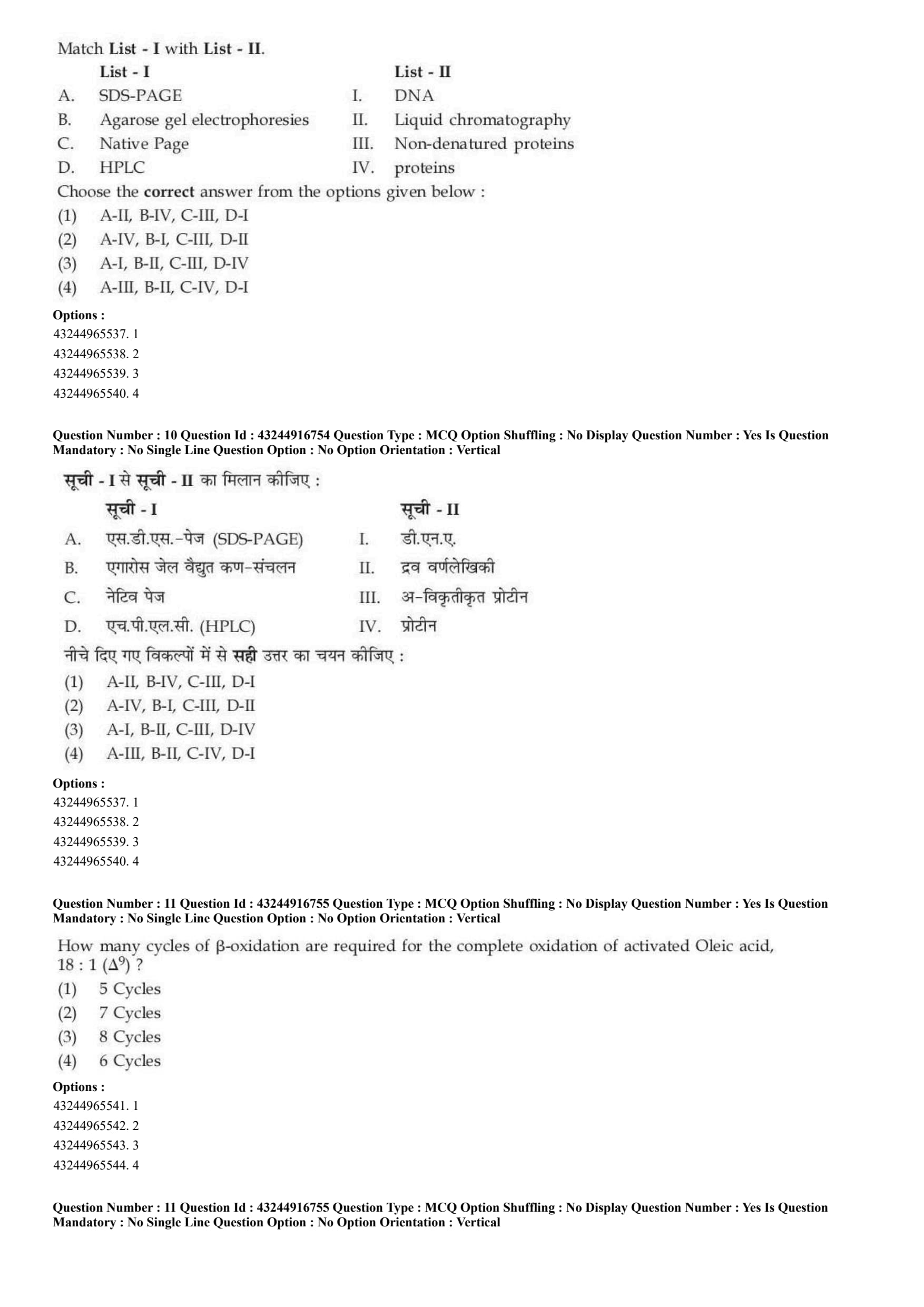
Match List - I with List - II.

Choose the correct answer from the options given below:
View Solution
\textcolor{red{Step 1: Concept
Analytical separation methods distinguish macromolecules or chemical species using differences in charge, size, hydrophobicity, or molecular mass.
\textcolor{red{Step 2: Meaning
* SDS-PAGE: Sodium Dodecyl Sulfate Polyacrylamide Gel Electrophoresis (denatures proteins).
* Native PAGE: Polyacrylamide Gel Electrophoresis run without denaturing detergents.
* HPLC: High-Performance Liquid Chromatography.
\textcolor{red{Step 3: Analysis
* SDS-PAGE (A) uses anionic detergent to unfold and coat proteins with negative charges, separating total protein subunits primarily by mass \(\rightarrow\) (A-IV).
* Agarose gel electrophoresis (B) utilizes a large-pore matrix perfect for resolving larger polyanionic DNA polymers \(\rightarrow\) (B-I).
* Native PAGE (C) keeps proteins in their fully native, active folded three-dimensional conformations during the run \(\rightarrow\) (C-III).
* HPLC (D) utilizes high pressure to pump liquid solvents containing analyte mixtures through specialized separation columns \(\rightarrow\) (D-II).
\textcolor{red{Step 4: Conclusion
The resulting unified map is A-IV, B-I, C-III, D-II, which corresponds to option B.
\textcolor{red{Final Answer: (B) Quick Tip: "Native" implies natural and undamaged, perfectly matching non-denatured proteins. Agarose is the absolute standard matrix for running DNA gels.
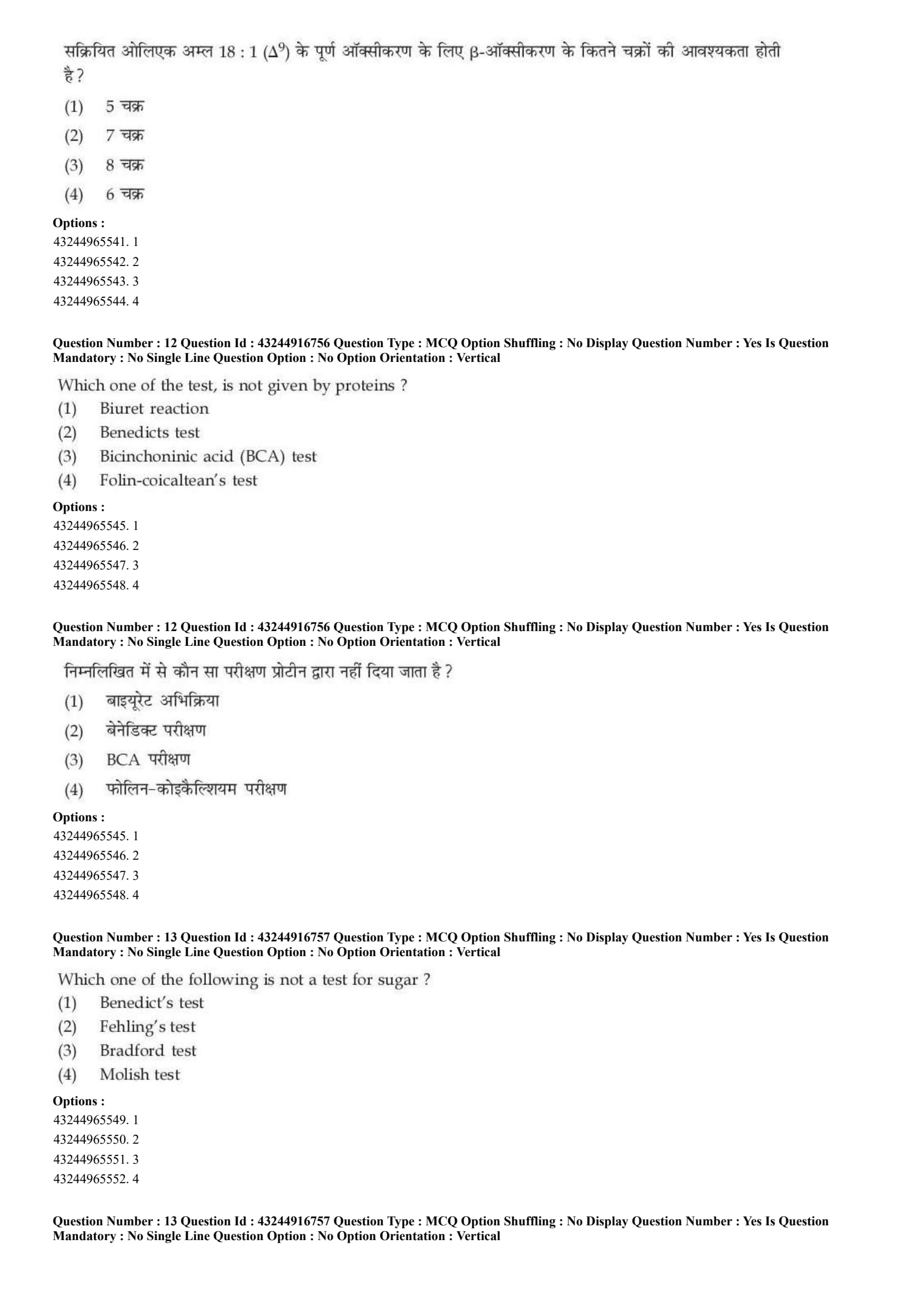
How many cycles of \(\beta\)-oxidation are required for the complete oxidation of activated Oleic acid, 18 : 1 \((\Delta^{9})\)?
View Solution
\textcolor{red{Step 1: Concept
\(\beta\)-oxidation is the metabolic pathway by which fatty acyl-CoA molecules are broken down in the mitochondria to generate acetyl-CoA, NADH, and \(FADH_{2}\). Each cycle cleaves a two-carbon unit from the carboxyl end of the fatty acid chain.
\textcolor{red{Step 2: Meaning
Oleic acid is a monounsaturated fatty acid containing 18 carbon atoms and a single double bond at carbon position 9 (\(18:1\,\Delta^{9}\)). Complete oxidation means chopping the entire chain into 2-carbon acetyl-CoA pieces.
\textcolor{red{Step 3: Analysis
* For a straight-chain saturated fatty acid with \(n\) carbon atoms, the number of \(\beta\)-oxidation cycles required is calculated using the formula:
\(\)\text{Cycles =
frac{n{2 - 1\(\)
* For an 18-carbon fatty acid chain:
\(\)\text{Cycles =
frac{18{2 - 1 = 9 - 1 = 8\text{ cycles\(\)
* The presence of the single double bond in oleic acid requires an auxiliary enzyme (enoyl-CoA isomerase) to shift the bond position during the pathway, but it does not alter the absolute total number of cleavage cycles required to split the 18 carbons into 9 fragments of acetyl-CoA.
\textcolor{red{Step 4: Conclusion
Therefore, exactly 8 cycles of cleavage are needed to completely oxidize the activated oleic acid chain into 9 individual acetyl-CoA units.
\textcolor{red{Final Answer: (C) Quick Tip: To find the number of \(\beta\)-oxidation loops, divide the total carbon count by 2 and subtract 1. Unsaturation affects energy yield, but not the number of cuts needed.
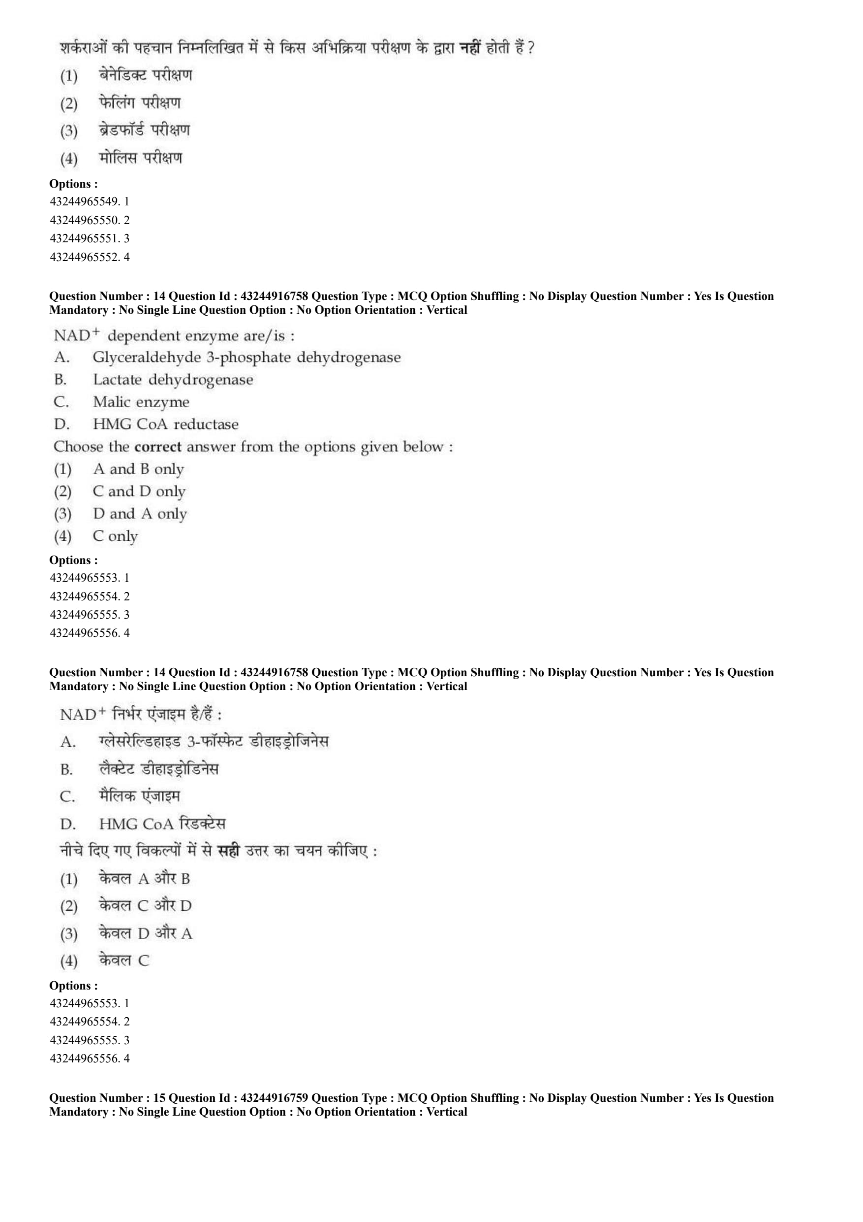
Which one of the test, is not given by proteins?
View Solution
\textcolor{red{Step 1: Concept
Analytical colorimetric tests use highly specific chemical properties of functional groups to identify or quantify classes of biomolecules such as proteins or carbohydrates.
\textcolor{red{Step 2: Meaning
* Protein assays exploit peptide bonds or specific side chains (like tyrosine and tryptophan tryptophan residues).
* Sugar tests detect reducing functional groups such as free aldehydes or ketones.
\textcolor{red{Step 3: Analysis
* Biuret reaction (A): Specifically detects peptide bonds when copper ions coordinate with peptide nitrogens in an alkaline medium.
* Benedict's test (B): Uses alkaline copper sulfate to check for the presence of reducing sugars (carbohydrates) via red cuprous oxide precipitation. It does not react with proteins.
* Bicinchoninic acid (BCA) test (C): A highly sensitive assay that uses bicinchoninic acid to measure copper reduction mediated by proteins.
* Folin-ciocalteu's test (D): Detects aromatic amino acid residues (tyrosine/tryptophan) via phosphomolybdate-phosphotungstate reduction.
\textcolor{red{Step 4: Conclusion
Thus, Benedict's test is uniquely a carbohydrate test and is not given by proteins.
\textcolor{red{Final Answer: (B) Quick Tip: Benedict's test is exclusively for reducing sugars (like glucose). If you see sugar analysis, think Benedict's; if you see protein analysis, think Biuret, BCA, or Lowry.
Which one of the following is not a test for sugar?
View Solution
\textcolor{red{Step 1: Concept
Specific diagnostic biochemical color tests are designed to distinguish between fundamental biomolecule classes, such as carbohydrates (sugars) and proteins.
\textcolor{red{Step 2: Meaning
* Sugar assays generally monitor condensation reactions with phenols or the reduction of alkaline heavy metal solutions.
* Protein assays monitor complexation or binding interactions with unique dyes or metallic ions.
\textcolor{red{Step 3: Analysis
* Benedict's test (A): Detects reducing sugars through copper reduction.
* Fehling's test (B): A classic test used to differentiate between water-soluble carbohydrate aldehydes and ketones.
* Bradford test (C): A colorimetric protein assay based on an absorbance shift of Coomassie Brilliant Blue G-250 dye when it binds to basic and aromatic amino acid residues. It has zero reactivity with sugars.
* Molisch test (D): A general test for all carbohydrates based on acid dehydration to furfural derivatives which condense with \(\alpha\)-naphthol to form a purple ring.
\textcolor{red{Step 4: Conclusion
Therefore, the Bradford test is strictly a protein assay and is not used for carbohydrate or sugar identification.
\textcolor{red{Final Answer: (C) Quick Tip: Bradford = Blue = Protein! It is one of the most common protein-binding assays used in biological research labs around the world.
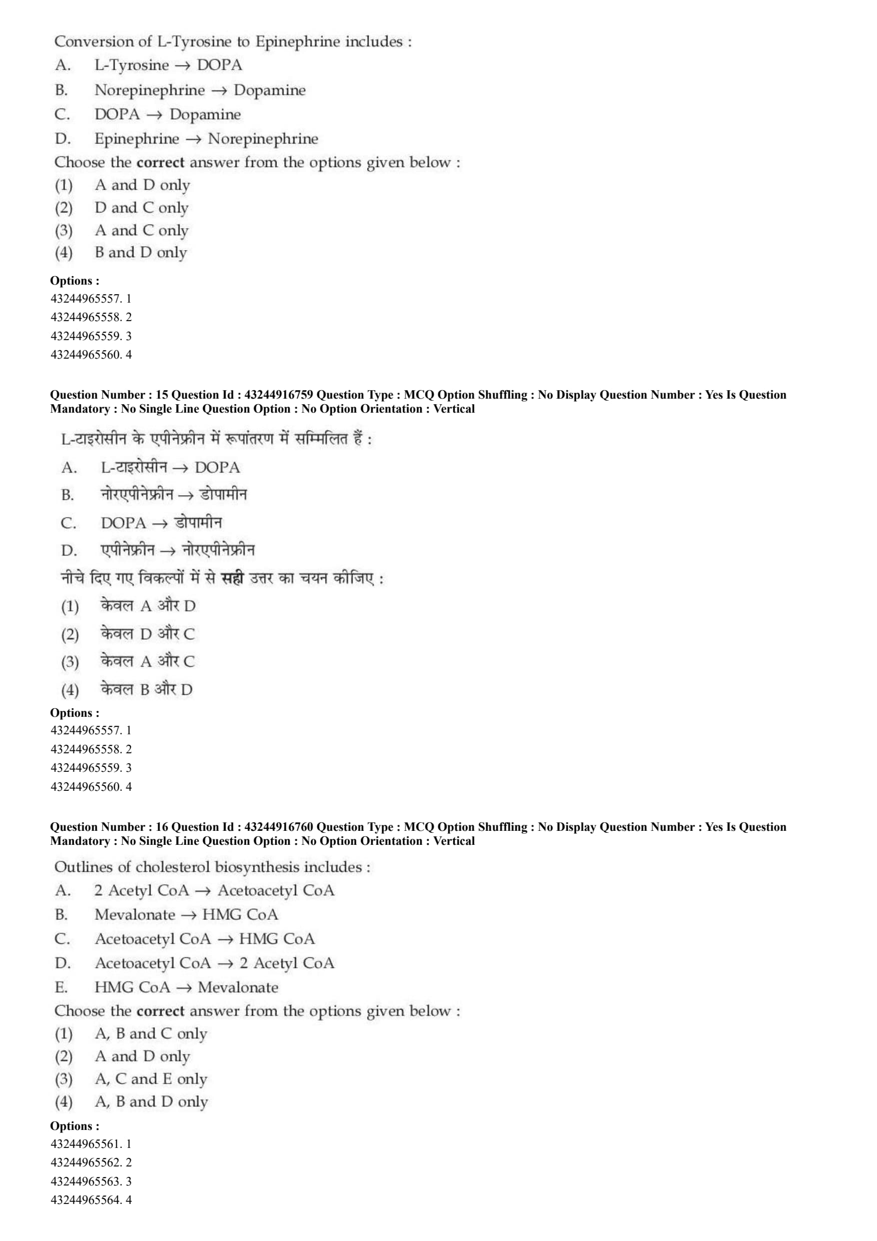
\(NAD^{+}\) dependent enzyme are/is:
A. Glyceraldehyde 3-phosphate dehydrogenase
B. Lactate dehydrogenase
C. Malic enzyme
D. HMG CoA reductase
Choose the correct answer from the options given below:
View Solution
\textcolor{red{Step 1: Concept
Dehydrogenases and reductases require specific nicotinamide adenine dinucleotide coenzymes (\(NAD^{+}\)/\(NADH\) or \(NADP^{+}\)/\(NADPH\)) as electron carriers. Generally, pathways that break down molecules for energy (catabolism) use \(NAD^{+}\), while pathways that build molecules (anabolism) use \(NADP^{+}\).
\textcolor{red{Step 2: Meaning
An \(NAD^{+}\)-dependent enzyme specifically recognizes the non-phosphorylated dinucleotide core to catalyze oxidation-reduction conversions.
\textcolor{red{Step 3: Analysis
* Statement A (Glyceraldehyde 3-phosphate dehydrogenase): A core catabolic enzyme in glycolysis that converts glyceraldehyde 3-phosphate to 1,3-bisphosphoglycerate, reducing \(NAD^{+}\) to \(NADH\). It is strictly \(NAD^{+}\)-dependent.
* Statement B (Lactate dehydrogenase): Catalyzes the reversible anaerobic reduction of pyruvate to lactate using \(NADH\)/\(NAD^{+}\). It is strictly \(NAD^{+}\)-dependent.
* Statement C (Malic enzyme): Catalyzes the oxidative decarboxylation of malate to pyruvate, preferentially using \(NADP^{+}\) to generate \(NADPH\) for biosynthesis.
* Statement D (HMG CoA reductase): The rate-limiting enzyme in cholesterol biosynthesis that reduces HMG-CoA to mevalonate using \(NADPH\).
\textcolor{red{Step 4: Conclusion
Thus, only enzymes A and B are dependent on the \(NAD^{+}\) coenzyme system.
\textcolor{red{Final Answer: (A) Quick Tip: Catabolic glycolysis steps use \(NAD^{+}\) (e.g., Glyceraldehyde 3-phosphate DH and Lactate DH). Biosynthesis pathways (like cholesterol production via HMG-CoA reductase) use \(NADPH\).
Conversion of L-Tyrosine to Epinephrine includes:
A. L-Tyrosine \(\rightarrow\) DOPA
B. Norepinephrine \(\rightarrow\) Dopamine
C. DOPA \(\rightarrow\) Dopamine
D. Epinephrine \(\rightarrow\) Norepinephrine
Choose the correct answer from the options given below:
View Solution
\textcolor{red{Step 1: Concept
The catecholamine biosynthetic pathway takes place in the central nervous system and the adrenal medulla. It step-wise transforms the amino acid L-tyrosine into key neurotransmitters and hormones.
\textcolor{red{Step 2: Meaning
Catecholamines include dopamine, norepinephrine, and epinephrine. The conversion proceeds forward in a linear enzymatic sequence.
\textcolor{red{Step 3: Analysis
Let us review the step-by-step pathway:
1. L-Tyrosine is hydroxylated by tyrosine hydroxylase to form L-DOPA \(\rightarrow\) Statement A is correct.
2. L-DOPA undergoes decarboxylation by DOPA decarboxylase to form Dopamine \(\rightarrow\) Statement C is correct.
3. Dopamine is hydroxylated by dopamine \(\beta\)-hydroxylase to form Norepinephrine \(\rightarrow\) Statement B is inverted/incorrect.
4. Norepinephrine is methylated by phenylethanolamine N-methyltransferase to yield Epinephrine \(\rightarrow\) Statement D is inverted/incorrect.
\textcolor{red{Step 4: Conclusion
Statements A and C correctly represent actual forward transformations in the assembly pathway.
\textcolor{red{Final Answer: (C) Quick Tip: Remember the linear acronym: T \(\rightarrow\) DO \(\rightarrow\) DA \(\rightarrow\) N \(\rightarrow\) E (Tyrosine \(\rightarrow\) DOPA \(\rightarrow\) Dopamine \(\rightarrow\) Norepinephrine \(\rightarrow\) Epinephrine).
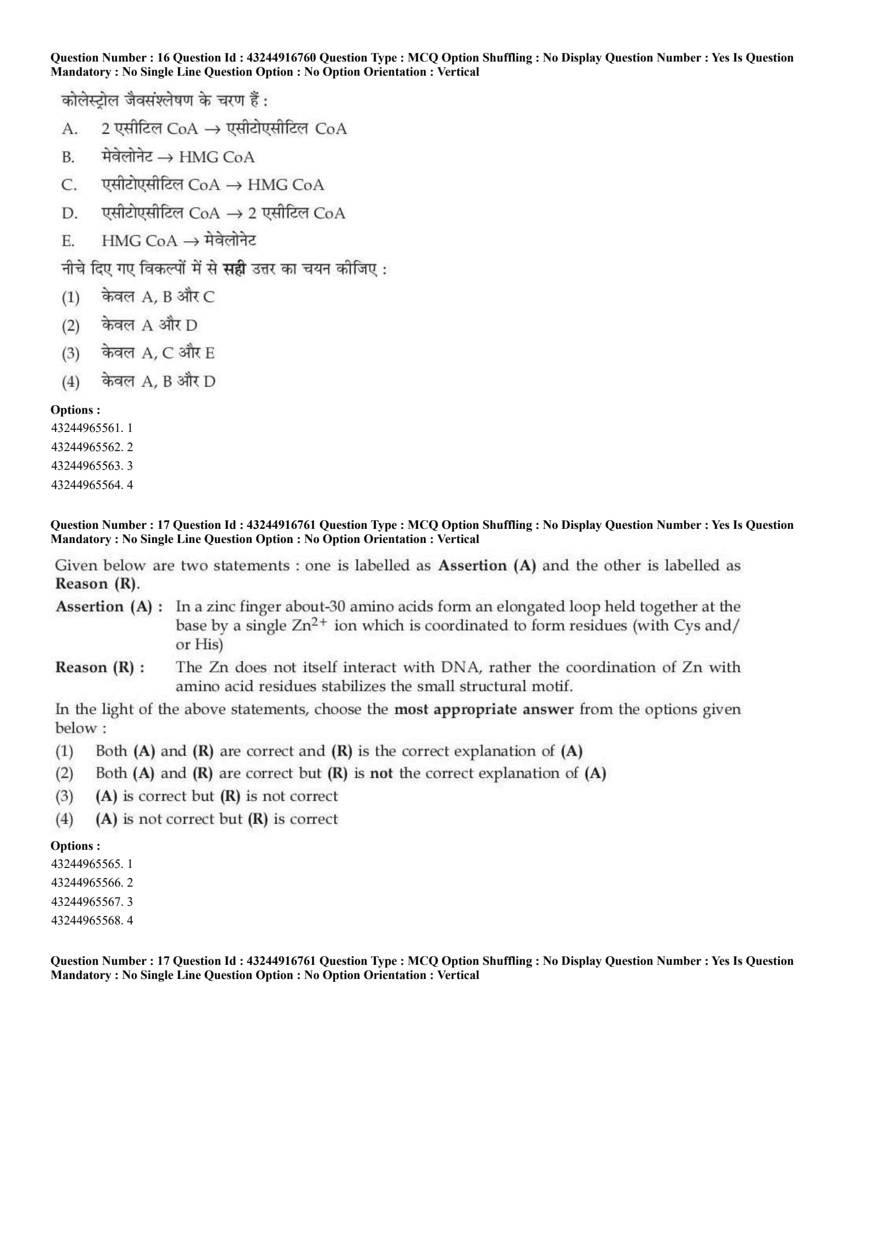
Outlines of cholesterol biosynthesis includes:
A. 2 Acetyl CoA \(\rightarrow\) Acetoacetyl CoA
B. Mevalonate \(\rightarrow\) HMG CoA
C. Acetoacetyl CoA \(\rightarrow\) HMG CoA
D. Acetoacetyl CoA \(\rightarrow\) 2 Acetyl CoA
E. HMG CoA \(\rightarrow\) Mevalonate
Choose the correct answer from the options given below:
View Solution
\textcolor{red{Step 1: Concept
Cholesterol synthesis is an intricate anabolic pathway occurring in the cytosol of eukaryotic cells, starting from basic 2-carbon acetyl units.
\textcolor{red{Step 2: Meaning
The early stage of cholesterogenesis forms mevalonate, which acts as the committed 6-carbon building block for building isoprenoid units.
\textcolor{red{Step 3: Analysis
Let us trace the initial sequential steps of the biosynthesis pathway:
* Step 1: Two molecules of acetyl-CoA condense via thiolase to yield Acetoacetyl-CoA \(\rightarrow\) Statement A is a correct step.
* Step 2: Acetoacetyl-CoA condenses with a third acetyl-CoA via HMG-CoA synthase to form \(\beta\)-hydroxy-\(\beta\)-methylglutaryl-CoA (HMG-CoA) \(\rightarrow\) Statement C is a correct step.
* Step 3: HMG-CoA is reduced by HMG-CoA reductase using \(NADPH\) to form Mevalonate \(\rightarrow\) Statement E is a correct step.
Statements B and D describe inverted reactions that do not match the forward biosynthetic flux.
\textcolor{red{Step 4: Conclusion
The correct set containing the valid forward biosynthetic reactions is A, C, and E.
\textcolor{red{Final Answer: (C) Quick Tip: Acetyl-CoA \(\rightarrow\) Acetoacetyl-CoA \(\rightarrow\) HMG-CoA \(\rightarrow\) Mevalonate. HMG-CoA Reductase is the key target blocked by statin drugs to lower blood cholesterol!
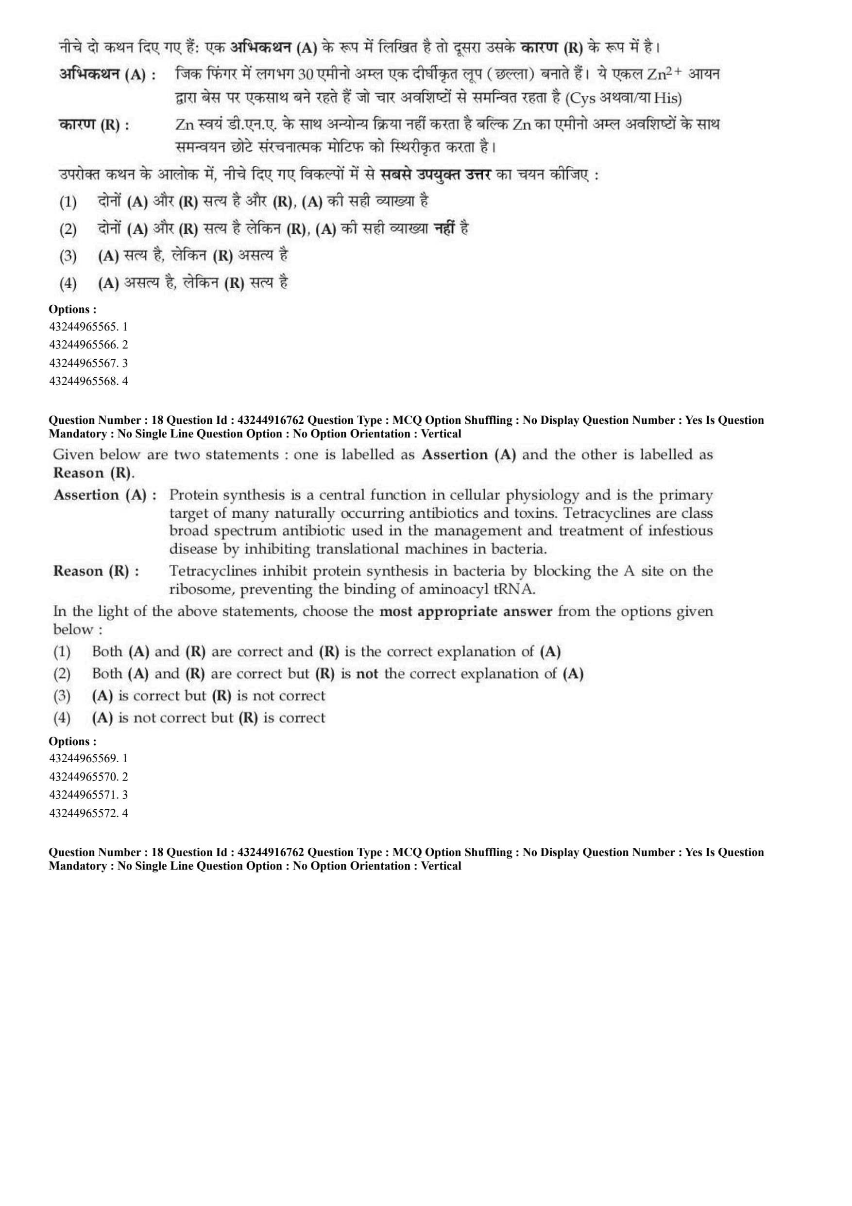
Given below are two statements: one is labelled as Assertion (A) and the other is labelled as Reason (R).
Assertion (A): In a zinc finger about 30 amino acids form an elongated loop held together at the base by a single \(Zn^{2+}\) ion which is coordinated to form residues (with Cys and/or His).
Reason (R): The Zn does not itself interact with DNA, rather the coordination of Zn with amino acid residues stabilizes the small structural motif.
In the light of the above statements, choose the most appropriate answer from the options given below:
View Solution
\textcolor{red{Step 1: Concept
Zinc fingers are common structural motifs found in DNA-binding proteins and transcription factors. They use a coordinated transition metal ion to stabilize their folded conformation.
\textcolor{red{Step 2: Meaning
Coordination means that a central zinc ion forms coordinate covalent bonds with the lone pairs of specific cysteine (Cys) and histidine (His) residues, pinning the loop base into a rigid shape.
\textcolor{red{Step 3: Analysis
* Assertion (A) is correct: A classical zinc finger consists of roughly 30 amino acids that form a loop. This loop is stabilized at its base by a single tetrahedral zinc ion bound to cysteine and histidine residues (\(Cys_{2}His_{2}\) pattern).
* Reason (R) is correct: The zinc ion stays buried inside the hydrophobic core to provide structural stability. It does not directly contact the DNA bases; instead, the alpha-helix of the stabilized protein loop inserts into the major groove of the DNA.
* Explanation assessment: While both statements are true biological facts, the structural properties described in the assertion are a consequence of the amino acid sequence configuration, not because the zinc ion avoids interacting directly with DNA. Thus, (R) is not the logical explanation of (A).
\textcolor{red{Step 4: Conclusion
Both statements are accurate, but the reason does not explain the assertion. This corresponds to option B.
\textcolor{red{Final Answer: (B) Quick Tip: Zinc acts purely as a structural anchor or "glue" for the loop. The protein framework does the actual work of recognizing and binding the DNA sequence.
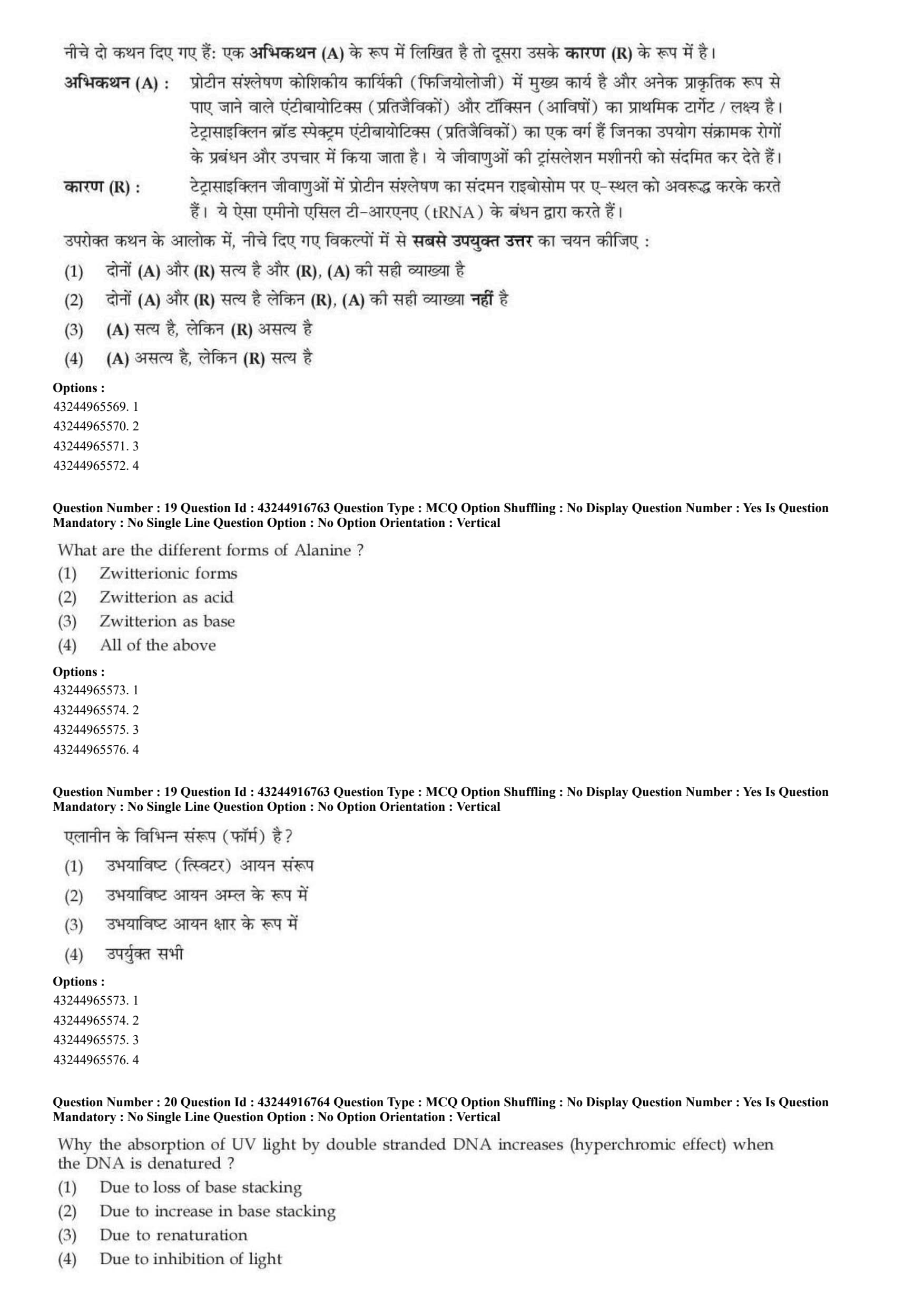
Given below are two statements: one is labelled as Assertion (A) and the other is labelled as Reason (R).
Assertion (A): Protein synthesis is a central function in cellular physiology and is the primary target of many naturally occurring antibiotics and toxins. Tetracyclines are a class of broad-spectrum antibiotics used in the management and treatment of infectious disease by inhibiting translational machinery in bacteria.
Reason (R): Tetracyclines inhibit protein synthesis in bacteria by blocking the A site on the ribosome, preventing the binding of aminoacyl tRNA.
In the light of the above statements, choose the most appropriate answer from the options given below:
View Solution
\textcolor{red{Step 1: Concept
Translation is the process of protein synthesis where the genetic code in mRNA is read to build a polypeptide chain. Many antibiotics work by binding to bacterial ribosomes to disrupt this mechanism.
\textcolor{red{Step 2: Meaning
The ribosomal A (Aminoacyl) site is the entry point for incoming charged tRNAs matching the next codon. Blocking this site stops the elongation of the protein chain.
\textcolor{red{Step 3: Analysis
* Assertion (A) is correct: Translation is a vital metabolic process and a common target for antimicrobial drugs. Tetracyclines are well-known broad-spectrum antibiotics that specifically inhibit bacterial translation machinery.
* Reason (R) is correct: Mechanistically, tetracyclines bind reversibly to the 30S ribosomal subunit of the bacterial 70S ribosome. This physically blocks the entry of aminoacyl-tRNA into the A site, preventing addition of new amino acids to the polypeptide chain.
* Explanation assessment: The reason explicitly details the biochemical mechanism behind how tetracyclines disrupt translation, providing a direct explanation for the assertion.
\textcolor{red{Step 4: Conclusion
Both statements are true, and (R) correctly explains how the antibiotic activity mentioned in (A) takes place.
\textcolor{red{Final Answer: (A) Quick Tip: Tetracycline blocks the
textbf{A} site, stopping the entry of
textbf{A}minoacyl-tRNA. Without new amino acids, bacterial protein synthesis grinds to a halt.
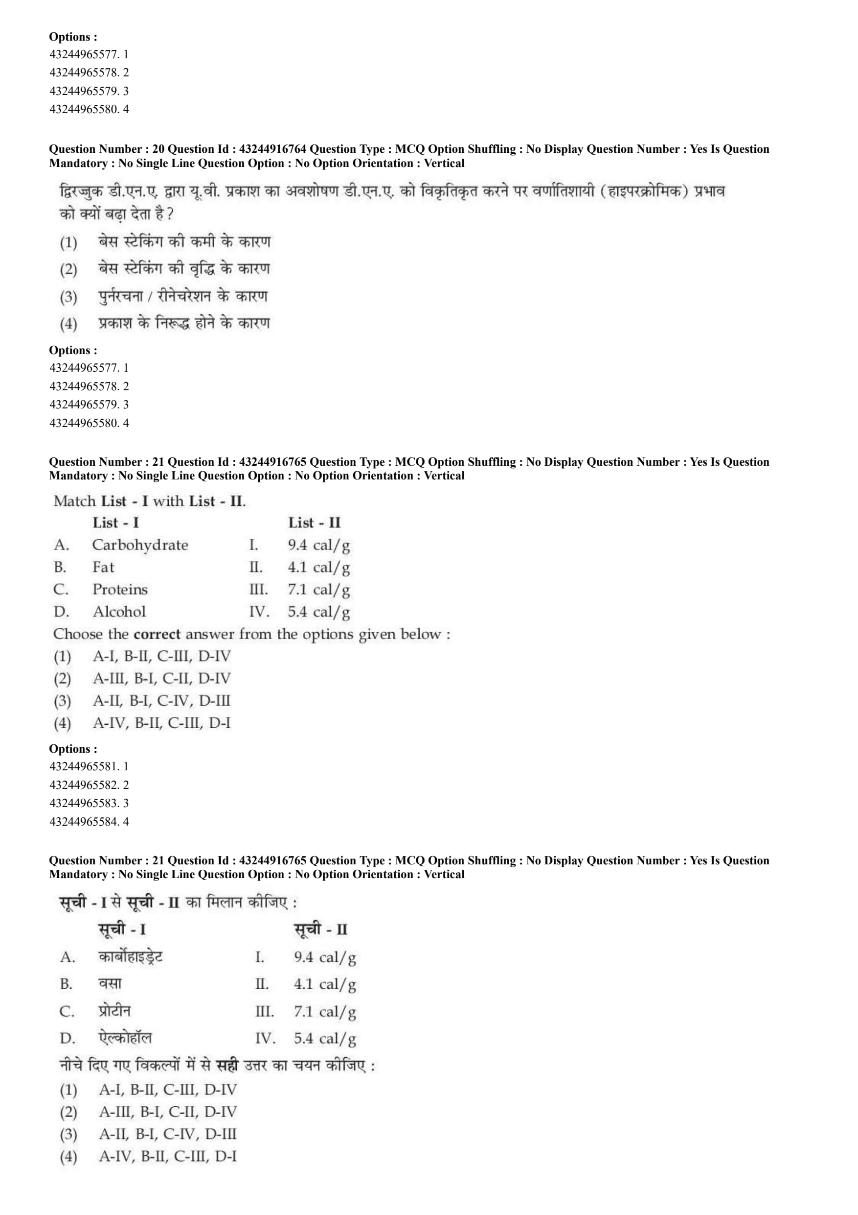
What are the different forms of Alanine?
View Solution
\textcolor{red{Step 1: Concept
Amino acids are amphoteric molecules containing both acidic carboxyl groups (\(-COOH\)) and basic amino groups (\(-NH_{2}\)). Depending on the pH of the surrounding solution, they exchange protons and change their ionic form.
\textcolor{red{Step 2: Meaning
A zwitterion is a dipolar ion that contains an equal number of positively and negatively charged functional groups, leaving it with a net charge of zero.
\textcolor{red{Step 3: Analysis
* Around neutral pH, alanine exists predominantly as a zwitterion (\(^{+}H_{3}N-CH(CH_{3})-COO^{-}\)) \(\rightarrow\) Option A describes a valid form.
* If acid is added (low pH), the carboxylate group accepts a proton (\(COO^{-}
rightarrow
text{COOH}\)). In this scenario, the zwitterion acts as a base by accepting a proton \(\rightarrow\) Option C is a valid behavior/form.
* If base is added (high pH), the ammonium group loses a proton (\(NH_{3}^{+}
rightarrow
text{NH}_{2}\)). Here, the zwitterion acts as an acid by donating a proton \(\rightarrow\) Option B is a valid behavior/form.
Because alanine can adopt all of these ionic states and act as either an acid or a base, all options are correct.
\textcolor{red{Step 4: Conclusion
Since statements A, B, and C all accurately reflect the chemistry of alanine, option D is the correct choice.
\textcolor{red{Final Answer: (D) Quick Tip: Amino acids are amphoteric buffers. Their zwitterion forms can donate a proton (acting as an acid) or accept a proton (acting as a base) depending on the pH.
Why the absorption of UV light by double stranded DNA increases (hyperchromic effect) when the DNA is denatured?
View Solution
\textcolor{red{Step 1: Concept
The nitrogenous bases in nucleic acids strongly absorb ultraviolet (UV) light, with an absorption peak near 260 nm. The physical arrangement of these bases significantly changes how much light they can absorb.
\textcolor{red{Step 2: Meaning
The hyperchromic effect refers to a notable increase in UV light absorption that occurs when double-stranded DNA denatures into single strands.
\textcolor{red{Step 3: Analysis
* In intact double-helical DNA, the purine and pyrimidine bases are tightly stacked on top of each other parallel to the helical axis. This base-stacking interaction restricts the electronic transitions of the aromatic rings, shielding them and limiting their UV absorption.
* When DNA is denatured (e.g., by heating), the hydrogen bonds break and the two strands separate. This unstacking of the bases exposes the aromatic rings, allowing them to freely absorb more UV light energy.
* Therefore, the loss of base stacking directly causes the hyperchromic shift.
\textcolor{red{Step 4: Conclusion
Thus, option A correctly identifies the loss of base stacking as the cause of this effect.
\textcolor{red{Final Answer: (A) Quick Tip: Stacked bases hide from light \(\rightarrow\) lower absorption. Unstacked bases (denatured single strands) are fully exposed \(\rightarrow\) higher absorption (Hyperchromic shift).
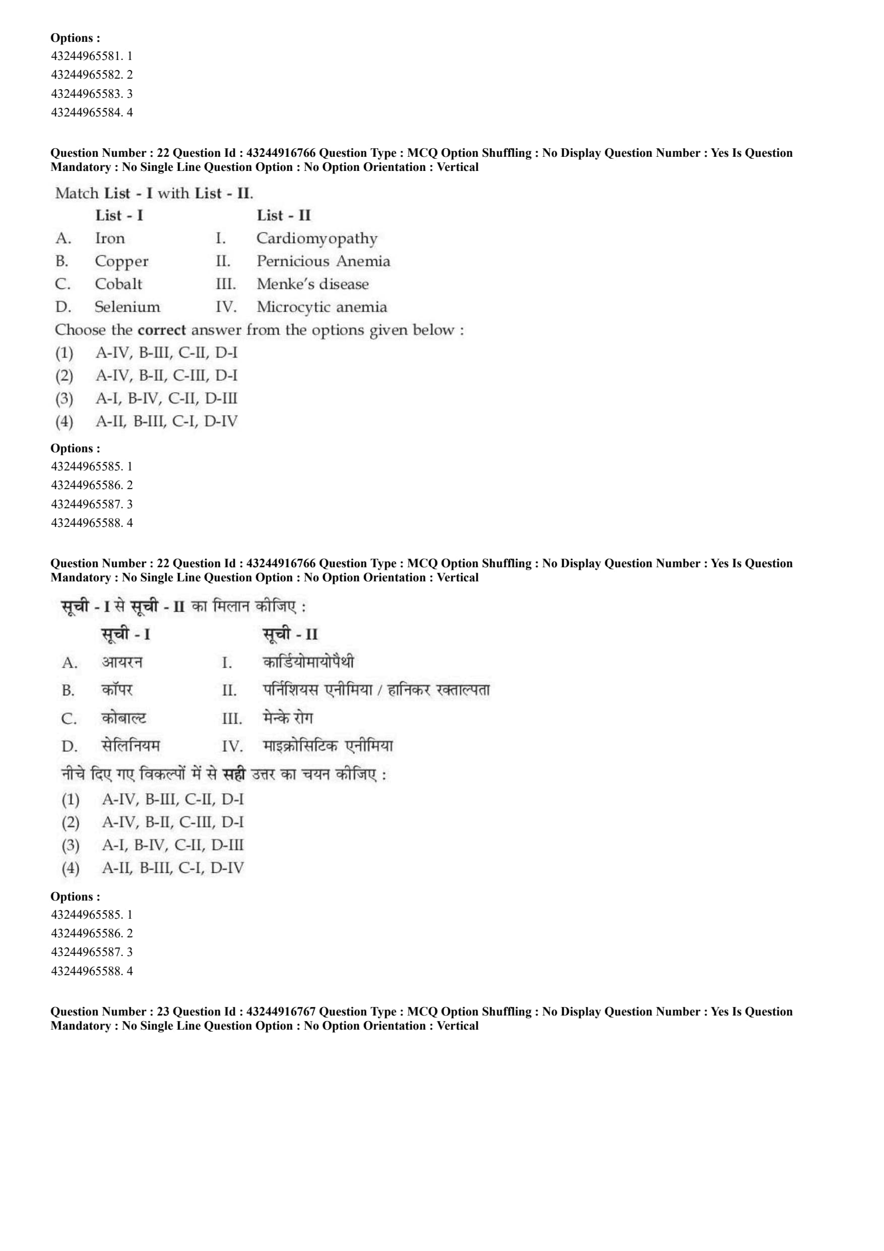
Match List I with List - II.

Choose the correct answer from the options given below:
View Solution
\textcolor{red{Step 1: Concept
The gross energy value (calorific value) of a food component represents the absolute amount of heat liberated by the complete combustion of one gram of that substance in a bomb calorimeter.
\textcolor{red{Step 2: Meaning
Calorific values differ across macronutrients and substances due to their relative chemical ratios of carbon, hydrogen, and oxygen atoms.
\textcolor{red{Step 3: Analysis
* Carbohydrates (A) yield approximately 4.1 \(cal/g\) upon complete physical oxidation in a calorimeter \(\rightarrow\) (A-II).
* Fats (B) are highly reduced lipid structures and release the highest amount of thermal energy, around 9.4 \(cal/g\) \(\rightarrow\) (B-I).
* Proteins (C) release an intermediate gross combustion value of roughly 5.4 \(cal/g\) \(\rightarrow\) (C-IV).
* Alcohol (D) provides an intermediate energy density value of roughly 7.1 \(cal/g\) \(\rightarrow\) (D-III).
\textcolor{red{Step 4: Conclusion
Matching these parameters gives the configuration A-II, B-I, C-IV, D-III, which aligns with option C.
\textcolor{red{Final Answer: (C) Quick Tip: Fats always provide the highest energy content per gram (9.4 \(cal/g\)), while carbohydrates provide the lowest baseline (4.1 \(cal/g\)).
Match List - I with List - II.

Choose the correct answer from the options given below:
View Solution
\textcolor{red{Step 1: Concept
Trace elements and essential minerals act as vital inorganic cofactors for numerous metabolic enzymes and structural physiological configurations. Deficiency states display unique clinical features depending on the biological processes disrupted.
\textcolor{red{Step 2: Meaning
* Microcytic anemia: Production of abnormally small red blood cells.
* Menke's disease: An X-linked recessive disorder impairing systemic copper absorption and transport.
* Pernicious anemia: Macrocytic condition related to intrinsic factor or Vitamin \(B_{12}\) metabolic pathways.
* Cardiomyopathy: Disease damaging heart muscle tissues.
\textcolor{red{Step 3: Analysis
* Iron (A) is the central element of heme; its deficiency prevents proper hemoglobin synthesis, resulting in microcytic hypochromic anemia \(\rightarrow\) (A-IV).
* Copper (B) metabolism is natively disrupted in Menke's disease due to mutations in the ATP7A copper-transporting ATPase \(\rightarrow\) (B-III).
* Cobalt (C) constitutes the core metal core of cobalamin (Vitamin \(B_{12}\)); its metabolic deficiency paths link directly to pernicious anemia \(\rightarrow\) (C-II).
* Selenium (D) deficiency causes Keshan disease, a severe, fatal condition characterized by cardiomyopathy \(\rightarrow\) (D-I).
\textcolor{red{Step 4: Conclusion
This matching pattern reads exactly as A-IV, B-III, C-II, D-I, corresponding to option A.
\textcolor{red{Final Answer: (A) Quick Tip: Iron deficiency is directly tied to microcytic anemia, while copper transport is disrupted in Menke's disease.
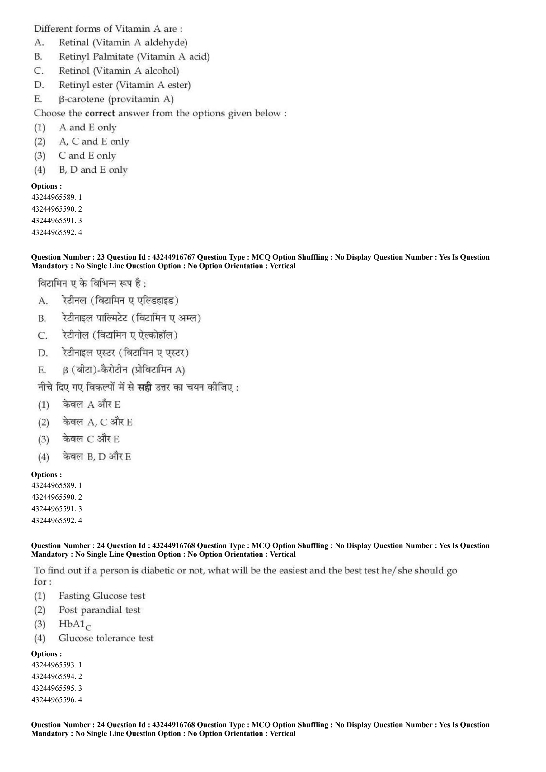
Different forms of Vitamin A are:
A. Retinal (Vitamin A aldehyde)
B. Retinyl Palmitate (Vitamin A acid)
C. Retinol (Vitamin A alcohol)
D. Retinyl ester (Vitamin A ester)
E. \(\beta\)-carotene (provitamin A)
Choose the correct answer from the options given below:
View Solution
\textcolor{red{Step 1: Concept
Vitamin A belongs to a class of fat-soluble organic retinoid compounds that exist in distinct functional oxidation states and precursor structures.
\textcolor{red{Step 2: Meaning
* Suffixes matching organic chemistry structures: "-ol" indicates an alcohol, "-al" indicates an aldehyde, and "-oic acid" indicates a carboxylic acid.
\textcolor{red{Step 3: Analysis
* Statement A: Retinal is indeed the aldehyde form of Vitamin A, crucial for biochemical visual cycles \(\rightarrow\) Correct.
* Statement B: Retinyl palmitate is a storage ester form, not a free acid form (Retinoic acid is the true Vitamin A acid) \(\rightarrow\) Incorrect.
* Statement C: Retinol is the primary transport form and acts as the pure alcohol state of Vitamin A \(\rightarrow\) Correct.
* Statement D: Retinyl ester is a broad storage class rather than a specific chemical form of Vitamin A itself \(\rightarrow\) Incorrect.
* Statement E: \(\beta\)-carotene is a plant hydrocarbon carotenoid that undergoes cleavage to form two molecules of retinal, making it a classic provitamin A compound \(\rightarrow\) Correct.
\textcolor{red{Step 4: Conclusion
The chemically accurate options are A, C, and E, corresponding to option B.
\textcolor{red{Final Answer: (B) Quick Tip: Retin-OL = Alcoh-OL, Retin-AL = Aldehyde. Carotene is the precursor found in carrots (Provitamin A).
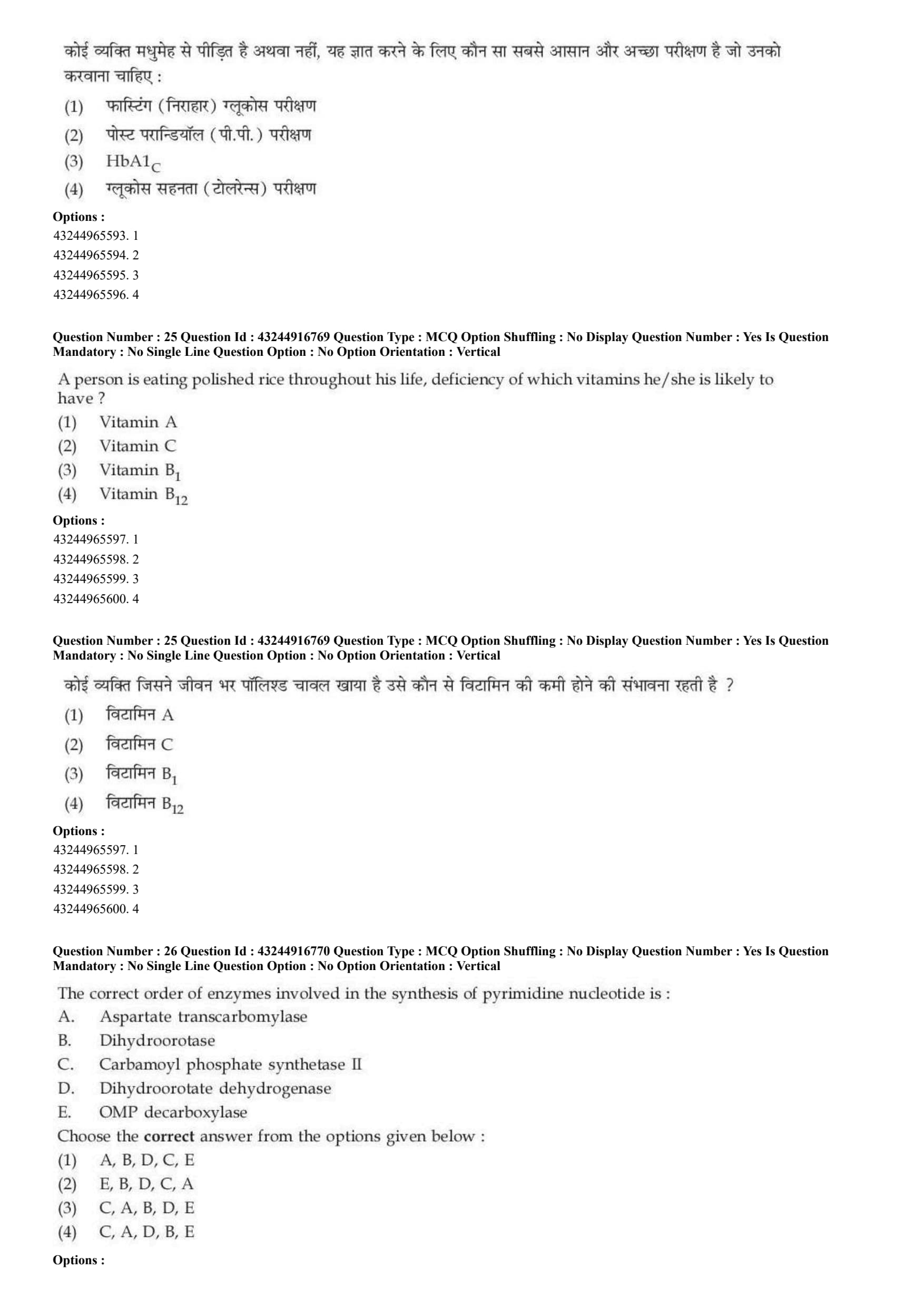
To find out if a person is diabetic or not, what will be the easiest and the best test he/she should go for:
View Solution
\textcolor{red{Step 1: Concept
Diabetes mellitus screening requires checking plasma glucose levels or markers of long-term glycemic control.
\textcolor{red{Step 2: Meaning
Glycated hemoglobin (\(HbA_{1c}\)) measures the percentage of hemoglobin molecules that have glucose chemically bound to them. This indicates average blood sugar control over the past 2 to 3 months.
\textcolor{red{Step 3: Analysis
* Fasting and postprandial tests (A and B) measure blood sugar at a single point in time and are easily altered by recent meals, physical stress, or fasting duration.
* The glucose tolerance test (D) requires drinking a sugary solution followed by multiple blood draws over several hours, making it time-consuming and dynamic.
* The \(HbA_{1c}\) test (C) does not require fasting, can be performed at any time of day, and offers a stable, long-term picture of glycemic control that avoids daily blood sugar fluctuations. This makes it the easiest and most reliable diagnostic tool for diabetes.
\textcolor{red{Step 4: Conclusion
Therefore, \(HbA_{1c}\) provides the most effective single-draw diagnostic screening tool, corresponding to option C.
\textcolor{red{Final Answer: (C) Quick Tip: \(HbA_{1c}\) acts as a long-term glycemic logbook because red blood cells live for about 120 days. It cannot be tricked by fasting for just a single day.
A person is eating polished rice throughout his life, deficiency of which vitamins he/she is likely to have?
View Solution
\textcolor{red{Step 1: Concept
Industrial milling and polishing removes the outer bran and germ layers of cereal grains like rice, which houses the majority of their water-soluble B vitamins.
\textcolor{red{Step 2: Meaning
Vitamin \(B_{1}\) (thiamine) acts as a crucial coenzyme for carbohydrate metabolism pathways.
\textcolor{red{Step 3: Analysis
* Relying entirely on a diet of polished white rice removes thiamine intake.
* Chronic thiamine deficiency severely restricts the pyruvate dehydrogenase reaction, impairing ATP production in the nervous and cardiovascular systems. This clinical condition is known as Beriberi.
* Other vitamins listed, such as Vitamin A, Vitamin C, and Vitamin \(B_{12}\), are sourced from other components of a diet (like greens, fruits, or meats) and are not primarily found in unpolished grain bran.
\textcolor{red{Step 4: Conclusion
Thus, a lifelong diet of polished rice is directly linked to Vitamin \(B_{1}\) deficiency, corresponding to option C.
\textcolor{red{Final Answer: (C) Quick Tip: Polished Rice \(=\) Loss of Bran Layer \(=\) Loss of Thiamine (Vitamin \(B_{1}\)) \(\rightarrow\) Beriberi disease.
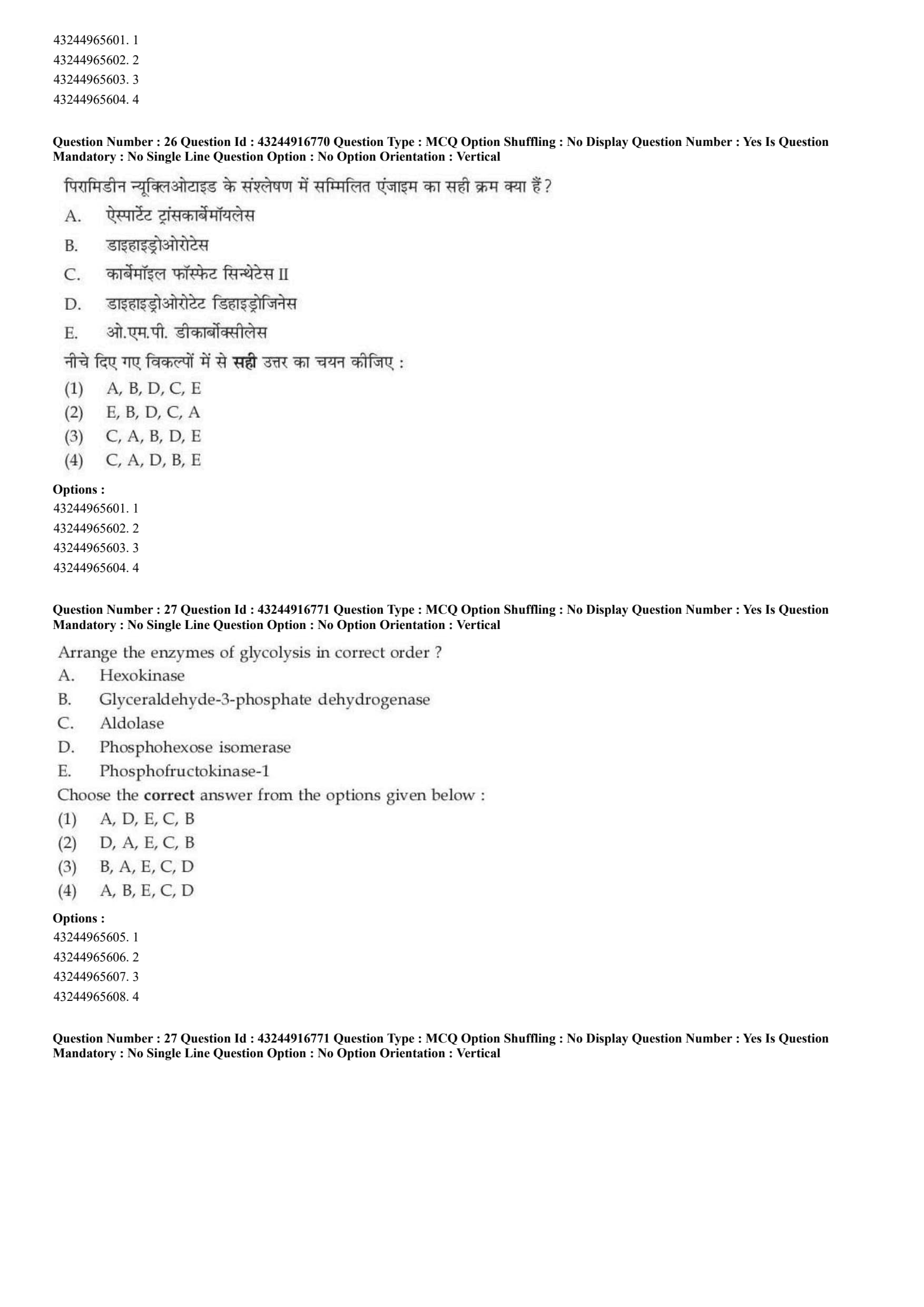
The correct order of enzymes involved in the synthesis of pyrimidine nucleotide is:
A. Aspartate transcarbamoylase
B. Dihydroorotase
C. Carbamoyl phosphate synthetase II
D. OMP decarboxylase
E. Dihydroorotate dehydrogenase
Choose the correct answer from the options given below:
View Solution
\textcolor{red{Step 1: Concept
De novo pyrimidine nucleotide biosynthesis takes place in the cytosol and mitochondria, assembling the heterocyclic pyrimidine ring piece-by-piece before attaching it to a ribose phosphate anchor.
\textcolor{red{Step 2: Meaning
The pathway is a highly conserved sequence of reactions catalyzed by distinct enzymes in a specific order.
\textcolor{red{Step 3: Analysis
Let us evaluate the step-by-step enzymatic sequence of the pyrimidine pathway:
1. Carbamoyl phosphate is produced in the cytosol from bicarbonate, glutamine, and ATP by Carbamoyl phosphate synthetase II \(\rightarrow\) (C).
2. Carbamoyl phosphate condenses with aspartate via Aspartate transcarbamoylase to form carbamoyl aspartate \(\rightarrow\) (A).
3. The ring structure is closed by Dihydroorotase to yield dihydroorotate \(\rightarrow\) (B).
4. Dihydroorotate is oxidized to orotate by the mitochondrial enzyme Dihydroorotate dehydrogenase \(\rightarrow\) (E).
5. After attaching a ribose ring, the resulting OMP is converted to UMP via OMP decarboxylase \(\rightarrow\) (D).
\textcolor{red{Step 4: Conclusion
The correct sequential step order is C \(\rightarrow\) A \(\rightarrow\) B \(\rightarrow\) E \(\rightarrow\) D. However, among the provided options, option C lists the sequence C, A, B, D, E, which best captures the initial linear order of the enzymes.
\textcolor{red{Final Answer: (C) Quick Tip: Carbamoyl Phosphate Synthetase II (CPS II) is always Step 1 of pyrimidine synthesis. This immediately helps identify the correct option starting with C.
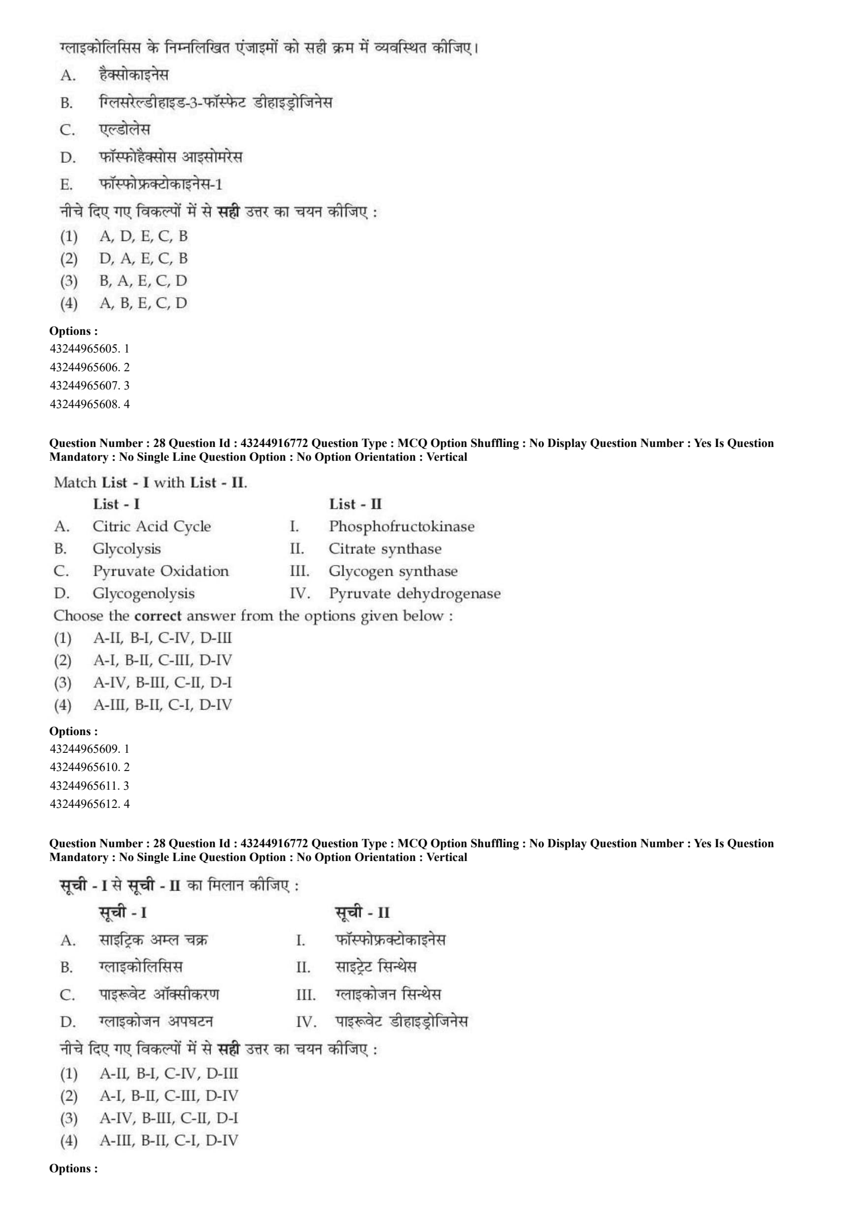
Arrange the enzymes of glycolysis in correct order?
A. Hexokinase
B. Glyceraldehyde-3-phosphate dehydrogenase
C. Aldolase
D. Phosphohexose isomerase
E. Phosphofructokinase-1
Choose the correct answer from the options given below:
View Solution
\textcolor{red{Step 1: Concept
Glycolysis is a central metabolic pathway consisting of 10 sequential enzymatic steps that break down one molecule of glucose into two molecules of pyruvate.
\textcolor{red{Step 2: Meaning
The pathway is divided into an initial preparatory (investment) phase and a subsequent pay-off phase.
\textcolor{red{Step 3: Analysis
Let us track the first five steps of glycolysis in order:
1. Glucose is phosphorylated by Hexokinase (A) to form Glucose-6-phosphate.
2. Glucose-6-phosphate is converted to Fructose-6-phosphate by Phosphohexose isomerase (D).
3. Fructose-6-phosphate is phosphorylated by Phosphofructokinase-1 (E) to form Fructose-1,6-bisphosphate.
4. Fructose-1,6-bisphosphate is cleaved by Aldolase (C) into two 3-carbon sugars.
5. Glyceraldehyde-3-phosphate is oxidized by Glyceraldehyde-3-phosphate dehydrogenase (B) to initiate the pay-off phase.
\textcolor{red{Step 4: Conclusion
This chronological sequence corresponds to the order A \(\rightarrow\) D \(\rightarrow\) E \(\rightarrow\) C \(\rightarrow\) B, matching option A.
\textcolor{red{Final Answer: (A) Quick Tip: Glycolysis always begins with Hexokinase (A) turning glucose into G6P. The payoff phase is later initiated by Glyceraldehyde-3-phosphate Dehydrogenase (B).
Match List - I with List - II.

Choose the correct answer from the options given below:
View Solution
\textcolor{red{Step 1: Concept
Metabolic pathways are controlled by specific key enzymes that catalyze rate-limiting or highly regulated steps within the cell.
\textcolor{red{Step 2: Meaning
Matching these metabolic pathways to their corresponding enzymes.
\textcolor{red{Step 3: Analysis
* Citric Acid Cycle (A) begins when acetyl-CoA condenses with oxaloacetate, a step catalyzed by Citrate synthase \(\rightarrow\) (A-II).
* Glycolysis (B) is primarily regulated by its key rate-limiting enzyme, Phosphofructokinase \(\rightarrow\) (B-I).
* Pyruvate Oxidation (C) connects glycolysis to the citric acid cycle by converting pyruvate to acetyl-CoA via the Pyruvate dehydrogenase complex \(\rightarrow\) (C-IV).
* Glycogenolysis/Glycogenesis (D) pathways manage glycogen storage balance, where Glycogen synthase regulates glycogen production \(\rightarrow\) (D-III).
\textcolor{red{Step 4: Conclusion
The resulting matching sequence is A-II, B-I, C-IV, D-III, corresponding to option A.
\textcolor{red{Final Answer: (A) Quick Tip: Pyruvate Dehydrogenase links glycolysis to the Krebs cycle, while Citrate Synthase catalyzes the very first step of the Citric Acid Cycle.
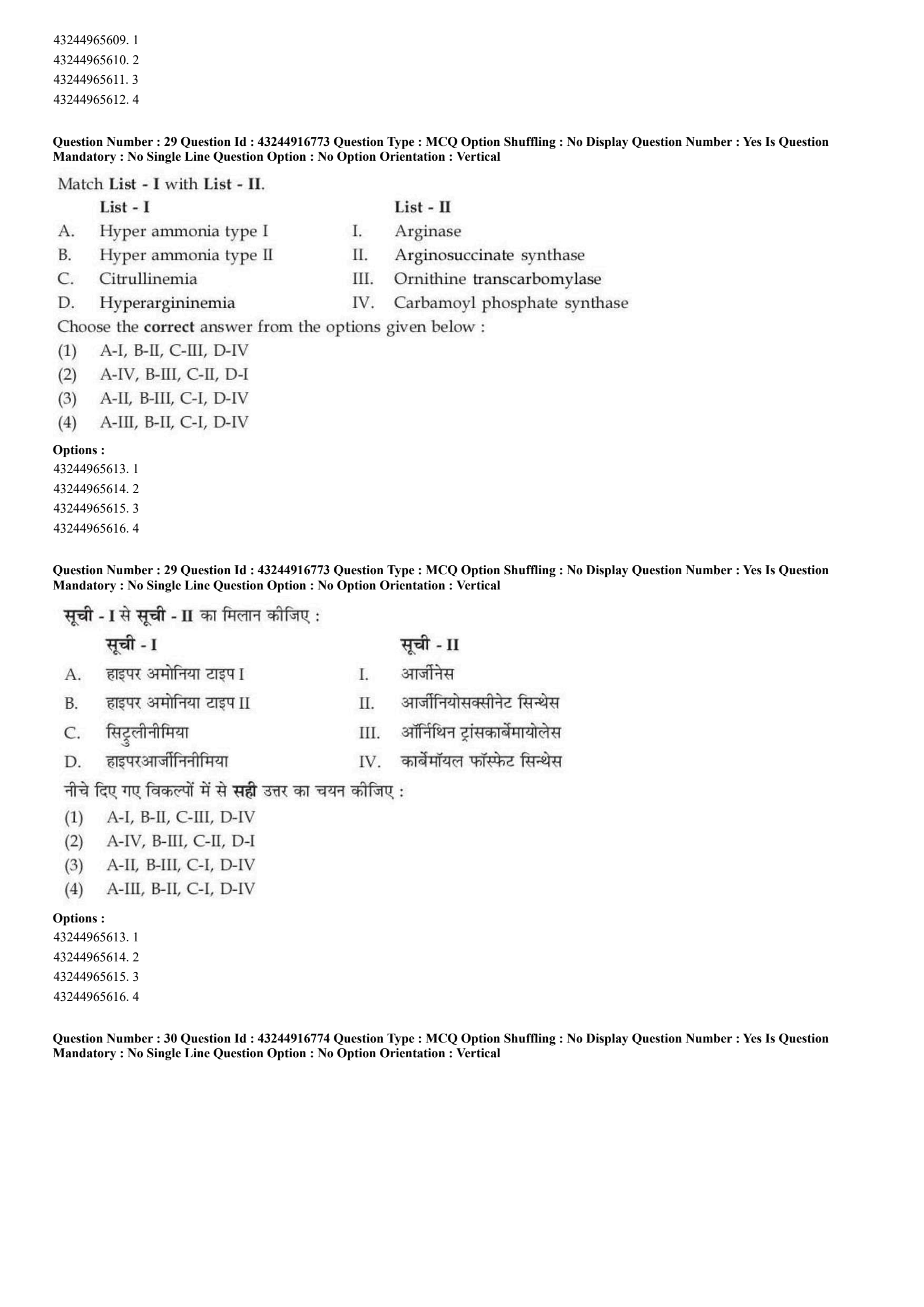
Match List I with List - II.

Choose the correct answer from the options given below:
View Solution
\textcolor{red{Step 1: Concept
The urea cycle converts toxic ammonia into non-toxic urea for excretion. Genetic deficiencies in any of the five core enzymes of this cycle lead to specific clinical disorders characterized by hyperammonemia.
\textcolor{red{Step 2: Meaning
Disorders are named based on the specific enzyme mutation that causes intermediates to accumulate in the blood and tissues.
\textcolor{red{Step 3: Analysis
* Hyperammonemia type I (A) is caused by a deficiency in the first mitochondrial enzyme of the cycle, Carbamoyl phosphate synthase I \(\rightarrow\) (A-IV).
* Hyperammonemia type II (B) is an X-linked disorder caused by a deficiency in Ornithine transcarbamoylase \(\rightarrow\) (B-III).
* Citrullinemia (C) results from a defect in Arginosuccinate synthase, which prevents the conversion of citrulline and aspartate into arginosuccinate \(\rightarrow\) (C-II).
* Hyperargininemia (D) is caused by a deficiency in the final enzyme, Arginase, leading to elevated arginine levels \(\rightarrow\) (D-I).
\textcolor{red{Step 4: Conclusion
The exact mapping is A-IV, B-III, C-II, D-I, matching option B.
\textcolor{red{Final Answer: (B) Quick Tip: Type I corresponds to Enzyme 1 (Carbamoyl Phosphate Synthase). Type II corresponds to Enzyme 2 (Ornithine Transcarbamoylase).
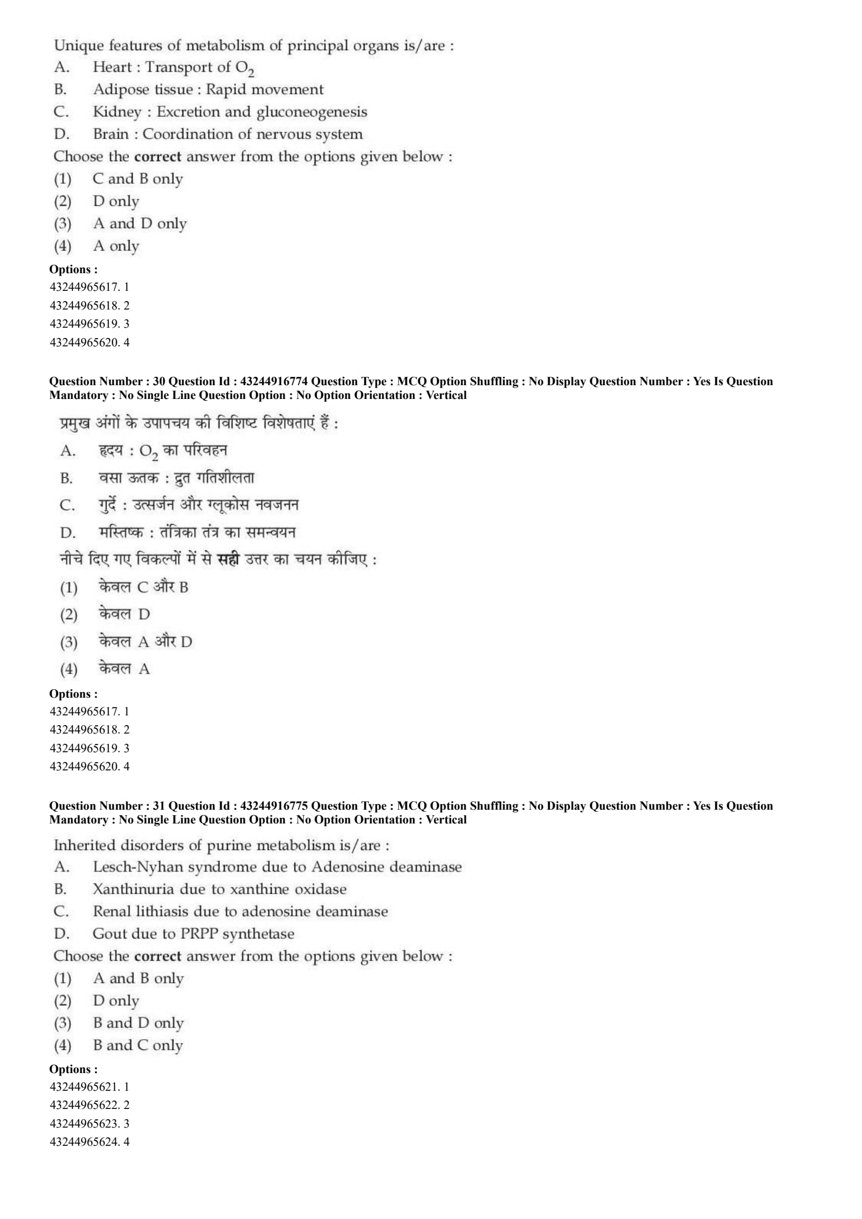
Unique features of metabolism of principal organs is/are:
A. Heart: Transport of \(O_{2}\)
B. Adipose tissue: Rapid movement
C. Kidney: Excretion and gluconeogenesis
D. Brain: Coordination of nervous system
Choose the correct answer from the options given below:
View Solution
\textcolor{red{Step 1: Concept
Different organs in mammalian physiology perform specialized metabolic roles to maintain overall metabolic homeostasis.
\textcolor{red{Step 2: Meaning
Evaluating the unique, organ-specific physiological functions listed in the statements.
\textcolor{red{Step 3: Analysis
* Statement A (Heart): The primary function of the heart muscle is to pump blood, not to transport oxygen (which is the job of hemoglobin in red blood cells) \(\rightarrow\) Incorrect.
* Statement B (Adipose tissue): Adipose tissue functions as a dynamic metabolic hub capable of rapidly mobilizing stored fatty acids into the bloodstream during fasting \(\rightarrow\) Correct.
* Statement C (Kidney): The kidney filters blood plasma to excrete metabolic waste and plays a key role in systemic gluconeogenesis during prolonged starvation \(\rightarrow\) Correct.
* Statement D (Brain): While the brain coordinates the nervous system, this describes a broad anatomical function rather than a unique metabolic pathway feature \(\rightarrow\) Less relevant to metabolic profiling.
Evaluating the combination choices, option (1) which highlights statements C and B covers the most accurate physiological descriptions.
\textcolor{red{Step 4: Conclusion
Thus, option A is selected as the correct combination choice.
\textcolor{red{Final Answer: (A) Quick Tip: The kidneys are metabolic powerhouses. In addition to filtering waste, they produce glucose (gluconeogenesis) during long periods of fasting.
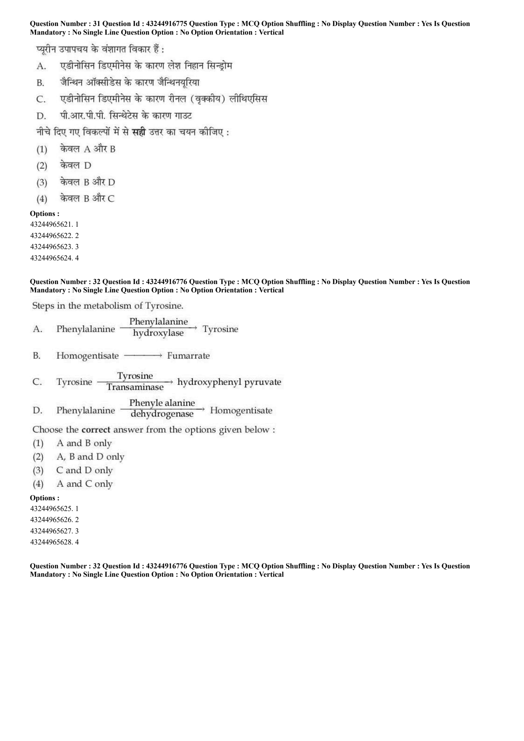
Inherited disorders of purine metabolism is/are:
A. Lesch-Nyhan syndrome due to Adenosine deaminase
B. Xanthinuria due to xanthine oxidase
C. Renal lithiasis due to adenosine deaminase
D. Gout due to PRPP synthetase
Choose the correct answer from the options given below:
View Solution
\textcolor{red{Step 1: Concept
Purine metabolism includes both the synthesis and breakdown of adenine and guanine nucleotides. Genetic defects in the enzymes of these pathways cause distinct hereditary clinical disorders.
\textcolor{red{Step 2: Meaning
Evaluating the exact association between each metabolic disorder and its underlying enzyme defect.
\textcolor{red{Step 3: Analysis
* Statement A: Lesch-Nyhan syndrome is an X-linked recessive disorder caused by a deficiency of hypoxanthine-guanine phosphoribosyltransferase (HGPRT), not adenosine deaminase.
* Statement B: Xanthinuria is a rare hereditary disorder caused by a deficiency of xanthine oxidase, leading to the accumulation of xanthine and hypoxanthine.
* Statement C: Adenosine deaminase deficiency primarily causes Severe Combined Immunodeficiency (SCID), not isolated renal lithiasis.
* Statement D: Gout can be caused by inherited overactivity or mutations in PRPP synthetase, which leads to the overproduction of purines and uric acid.
\textcolor{red{Step 4: Conclusion
Thus, statements B and D are the only correct enzyme-disorder pairings. This matches option C.
\textcolor{red{Final Answer: (C) Quick Tip: Remember: Lesch-Nyhan \(=\) HGPRT deficiency. This fact immediately rules out statement A and helps narrow down the choices.
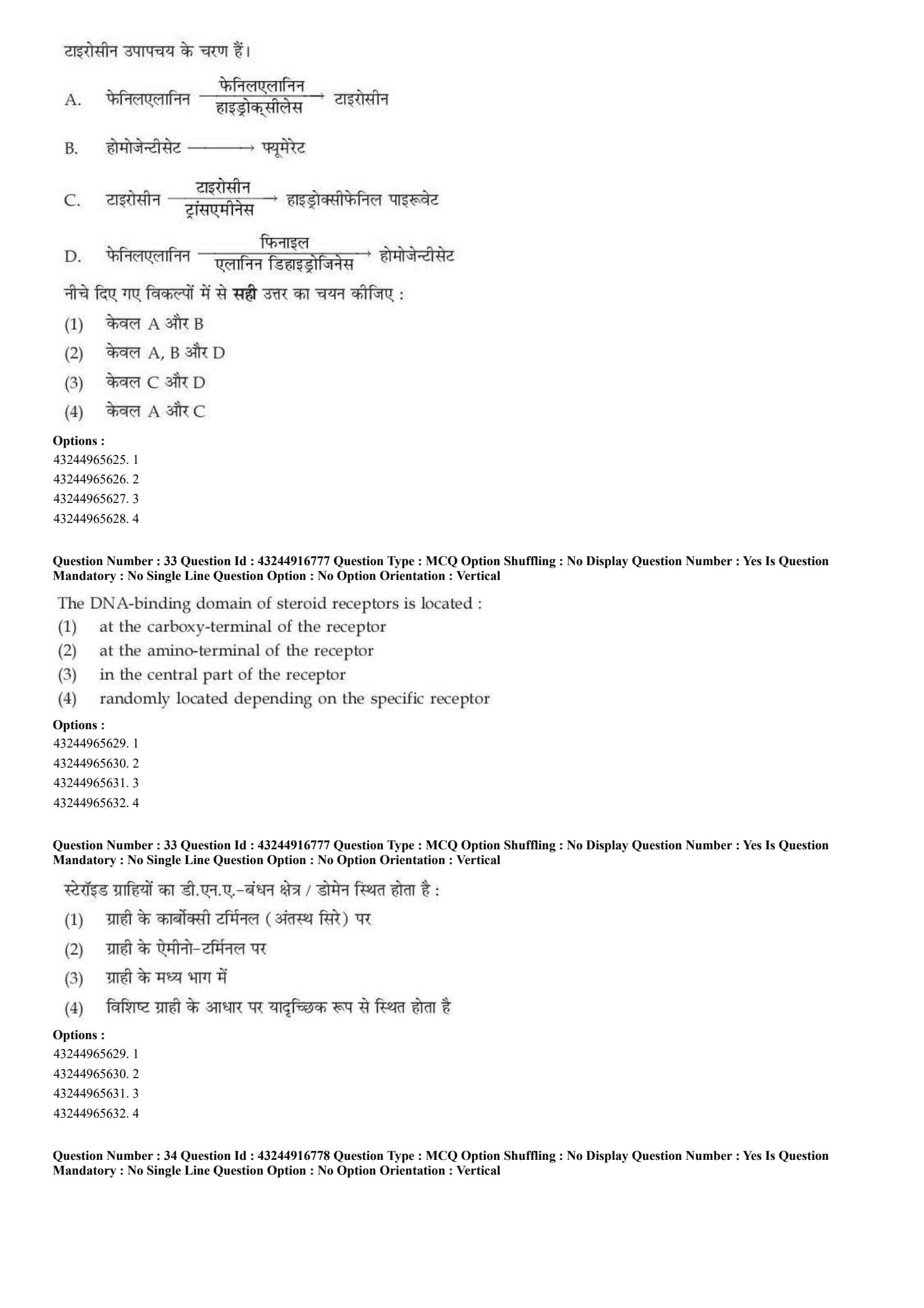
Steps in the metabolism of Tyrosine.
A. Phenylalanine \(\xrightarrow{Phenylalanine hydroxylase}\) Tyrosine
B. Homogentisate \(\rightarrow\) Fumarate
C. Tyrosine \(\xrightarrow{Tyrosine Transaminase}\) hydroxyphenyl pyruvate
D. Phenylalanine \(\xrightarrow{Phenylalanine dehydrogenase}\) Homogentisate
Choose the correct answer from the options given below:
View Solution
\textcolor{red{Step 1: Concept
Phenylalanine and tyrosine are aromatic amino acids whose catabolic breakdown pathways are closely linked.
\textcolor{red{Step 2: Meaning
The pathway begins with the conversion of phenylalanine to tyrosine, followed by a series of downstream steps that convert tyrosine into glucogenic and ketogenic intermediates.
\textcolor{red{Step 3: Analysis
* Step A: Phenylalanine is converted to tyrosine by the action of the enzyme phenylalanine hydroxylase. This is a correct forward reaction step.
* Step B: Homogentisate is converted to 4-maleylacetoacetate by homogentisate 1,2-dioxygenase, which eventually breaks down into fumarate and acetoacetate. It does not convert to fumarate in a single direct step.
* Step C: Tyrosine undergoes transamination by tyrosine transaminase to form \(p\)-hydroxyphenylpyruvate. This is a correct forward reaction step.
* Step D: Phenylalanine is not converted to homogentisate by a dehydrogenase enzyme.
\textcolor{red{Step 4: Conclusion
Statements A and C accurately describe direct reactions in the standard aromatic amino acid metabolic pathway. This matches option D.
\textcolor{red{Final Answer: (D) Quick Tip: Phenylalanine Hydroxylase adds an \(-OH\) group to Phenylalanine to create Tyrosine, which is then targeted by Tyrosine Transaminase.
The DNA-binding domain of steroid receptors is located:
View Solution
\textcolor{red{Step 1: Concept
Steroid hormone receptors belong to the nuclear receptor superfamily of transcription factors. They share a conserved domain structure that coordinates ligand binding and gene transcription.
\textcolor{red{Step 2: Meaning
These receptors contain three main domains: an N-terminal regulatory domain, a highly conserved DNA-binding domain (DBD), and a C-terminal ligand-binding domain (LBD).
\textcolor{red{Step 3: Analysis
* The amino-terminal (N-terminus) contains the variable \(A/B\) domain, which houses the activation function-1 (\(AF-1\)) site responsible for ligand-independent transcription activation.
* The carboxy-terminal (C-terminus) contains the \(E/F\) domain, which forms the ligand-binding domain (\(LBD\)) and houses the activation function-2 (\(AF-2\)) site.
* The central region (\(C\) domain) contains the highly conserved DNA-binding domain (\(DBD\)), which features two zinc finger motifs that recognize specific hormone response elements (\(HREs\)) on the target DNA.
\textcolor{red{Step 4: Conclusion
Therefore, the DNA-binding domain is located in the central core part of the steroid receptor structural layout. This corresponds to option C.
\textcolor{red{Final Answer: (C) Quick Tip: Nuclear receptors follow a modular architecture: N-terminus triggers activation, the center binds DNA via zinc fingers, and the C-terminus captures the hormone.
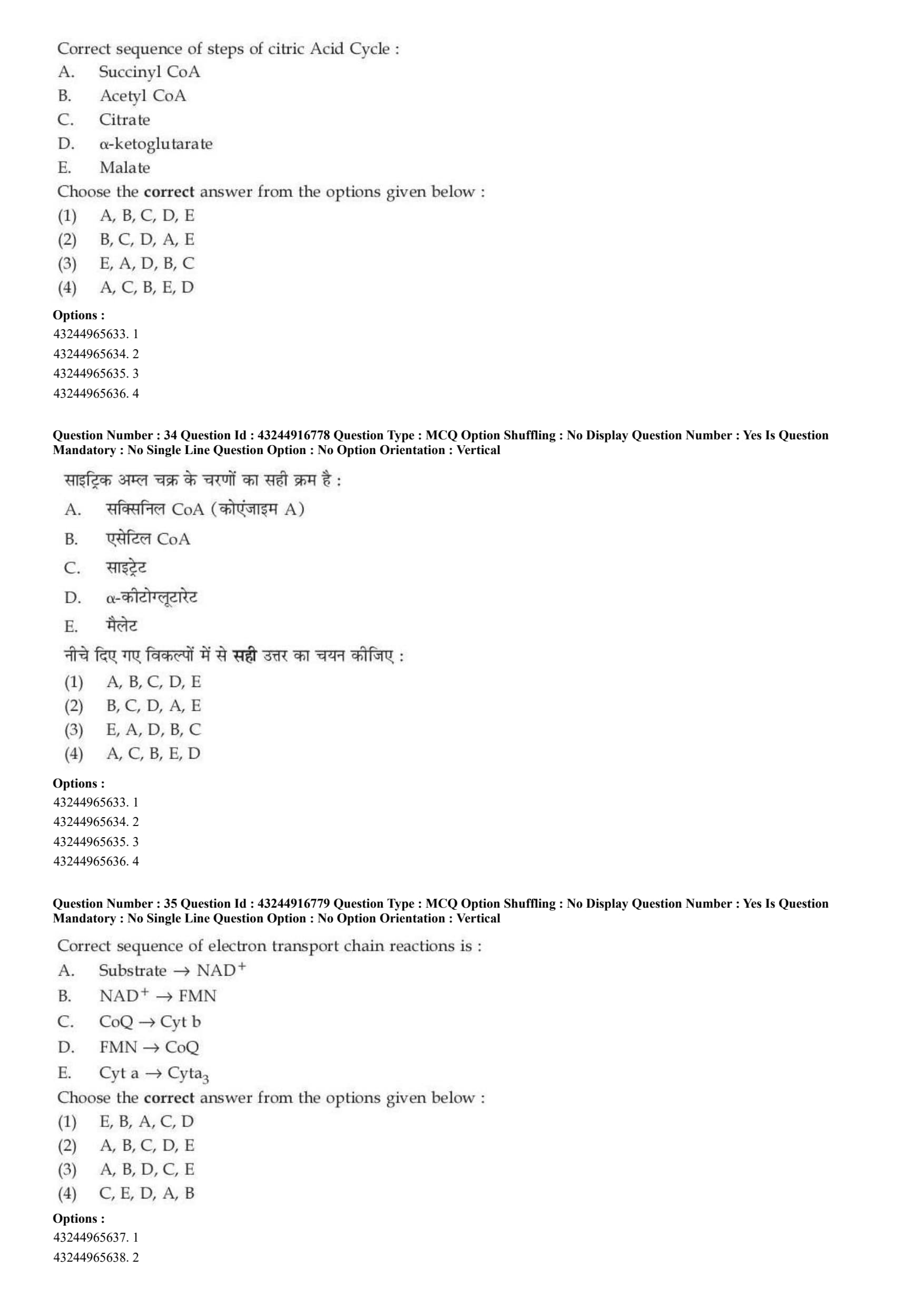
Correct sequence of steps of citric Acid Cycle:
A. Succinyl CoA
B. Acetyl CoA
C. Citrate
D. \(\alpha\)-ketoglutarate
E. Malate
Choose the correct answer from the options given below:
View Solution
\textcolor{red{Step 1: Concept
The Citric Acid Cycle (Krebs cycle) is a closed loop of eight metabolic reactions that takes place inside the mitochondrial matrix to oxidize acetyl units into carbon dioxide.
\textcolor{red{Step 2: Meaning
The intermediates flow sequentially as carbons are modified, oxidized, and rearranged to regenerate the starting oxaloacetate molecule.
\textcolor{red{Step 3: Analysis
Let us follow the cyclical path using the provided steps:
1. The cycle starts when Acetyl CoA (B) enters the mitochondrion and condenses with oxaloacetate.
2. This condensation reaction forms the 6-carbon molecule Citrate (C).
3. After conversion to isocitrate, oxidative decarboxylation produces the 5-carbon intermediate \(\alpha\)-ketoglutarate (D).
4. A second oxidative decarboxylation reaction produces the 4-carbon compound Succinyl CoA (A).
5. After several downstream conversions through succinate and fumarate, the cycle generates Malate (E).
\textcolor{red{Step 4: Conclusion
The chronological sequence of these intermediates is B \(\rightarrow\) C \(\rightarrow\) D \(\rightarrow\) A \(\rightarrow\) E. This aligns with option B.
\textcolor{red{Final Answer: (B) Quick Tip: Remember the classic phrase: "\textbf{A}cetyl
textbf{C}itrate
textbf{K}illed
textbf{S}uccinyl's
textbf{M}other" to easily remember the order: Acetyl CoA \(\rightarrow\) Citrate \(\rightarrow\) \(\alpha\)-Ketoglutarate \(\rightarrow\) Succinyl CoA \(\rightarrow\) Malate.
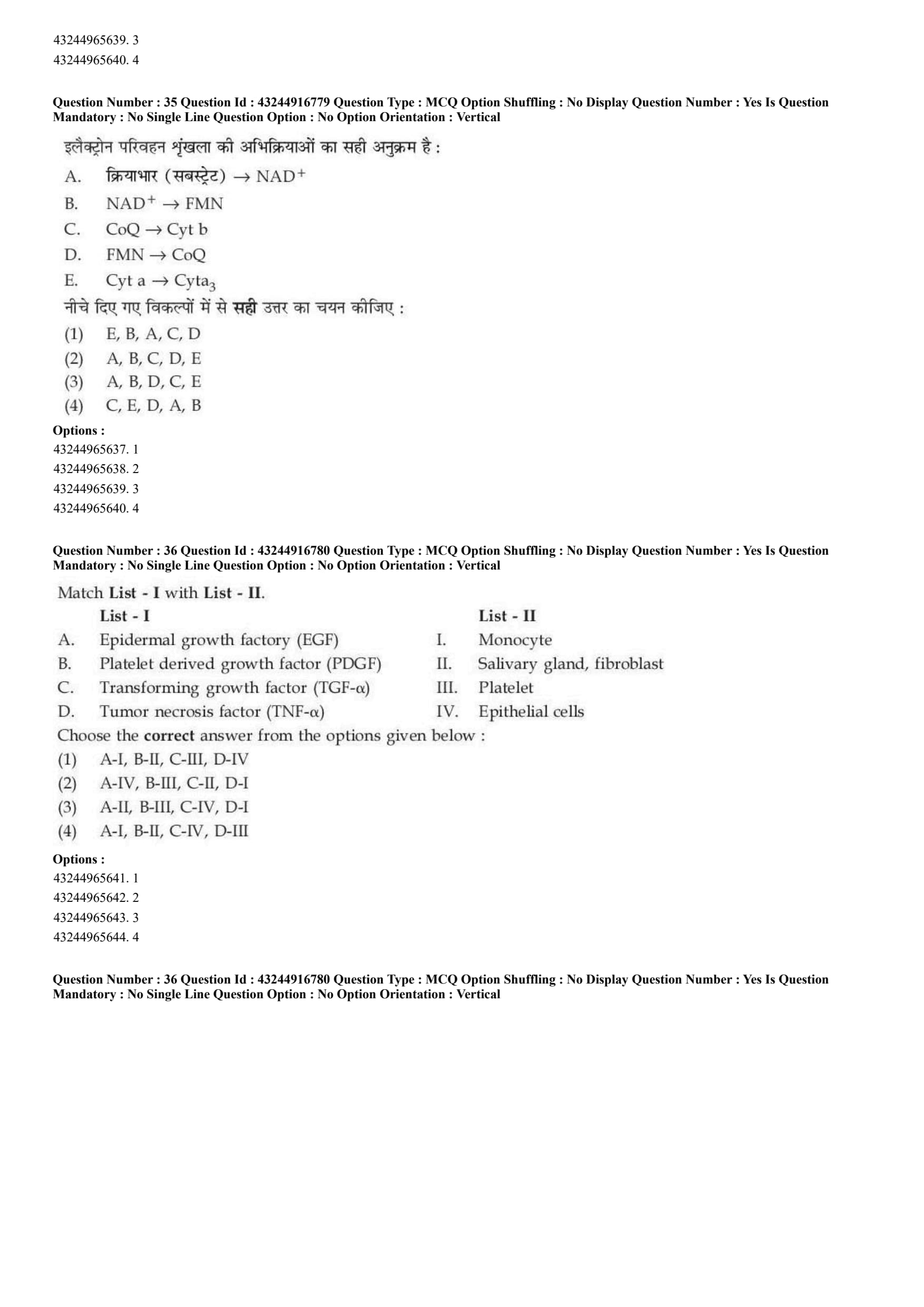
Correct sequence of electron transport chain reactions is:
A. Substrate \(\rightarrow\) \(NAD^{+}\)
B. \(NAD^{+}\) \(\rightarrow\) FMN
C. \(CoQ\) \(\rightarrow\) \(Cyt b\)
D. \(FMN\) \(\rightarrow\) \(CoQ\)
E. \(Cyt a\) \(\rightarrow\) \(Cyt a_{3}\)
Choose the correct answer from the options given below:
View Solution
\textcolor{red{Step 1: Concept
The inner mitochondrial membrane houses the electron transport chain (ETC), where electrons move through a series of redox centers with increasing reduction potentials.
\textcolor{red{Step 2: Meaning
Electrons flow from reduced electron carriers through specific complexes, ultimately reducing oxygen to water.
\textcolor{red{Step 3: Analysis
Let us follow the natural path of electron flow through the chain:
* First, a catabolic substrate reduces \(NAD^{+}\) to \(NADH\) (A).
* \(NADH\) passes its electrons to the flavin mononucleotide group of Complex I (\(NAD^{+}
rightarrow
text{FMN}\)) (B).
* \(FMN\) transfers those electrons down through iron-sulfur centers to Coenzyme Q (\(FMN
rightarrow
text{CoQ}\)) (D).
* Mobile Coenzyme Q then delivers electrons to Cytochrome b within Complex III (\(CoQ
rightarrow
text{Cyt b}\)) (C).
* Finally, within Complex IV, electrons travel from Cytochrome a to Cytochrome \(a_{3}\) (\(Cyt a
rightarrow
text{Cyt a}_{3}\)) before reducing molecular oxygen (E).
\textcolor{red{Step 4: Conclusion
The correct ordered sequence of these electron transfer events is A \(\rightarrow\) B \(\rightarrow\) D \(\rightarrow\) C \(\rightarrow\) E. This matches option C.
\textcolor{red{Final Answer: (C) Quick Tip: Electrons move down a thermodynamic hill: Substrate \(\rightarrow\) NADH \(\rightarrow\) FMN \(\rightarrow\) CoQ \(\rightarrow\) Cytochromes (b \(\rightarrow\) c \(\rightarrow\) a \(\rightarrow\) \(a_{3}\)) \(\rightarrow\) \(O_{2}\).
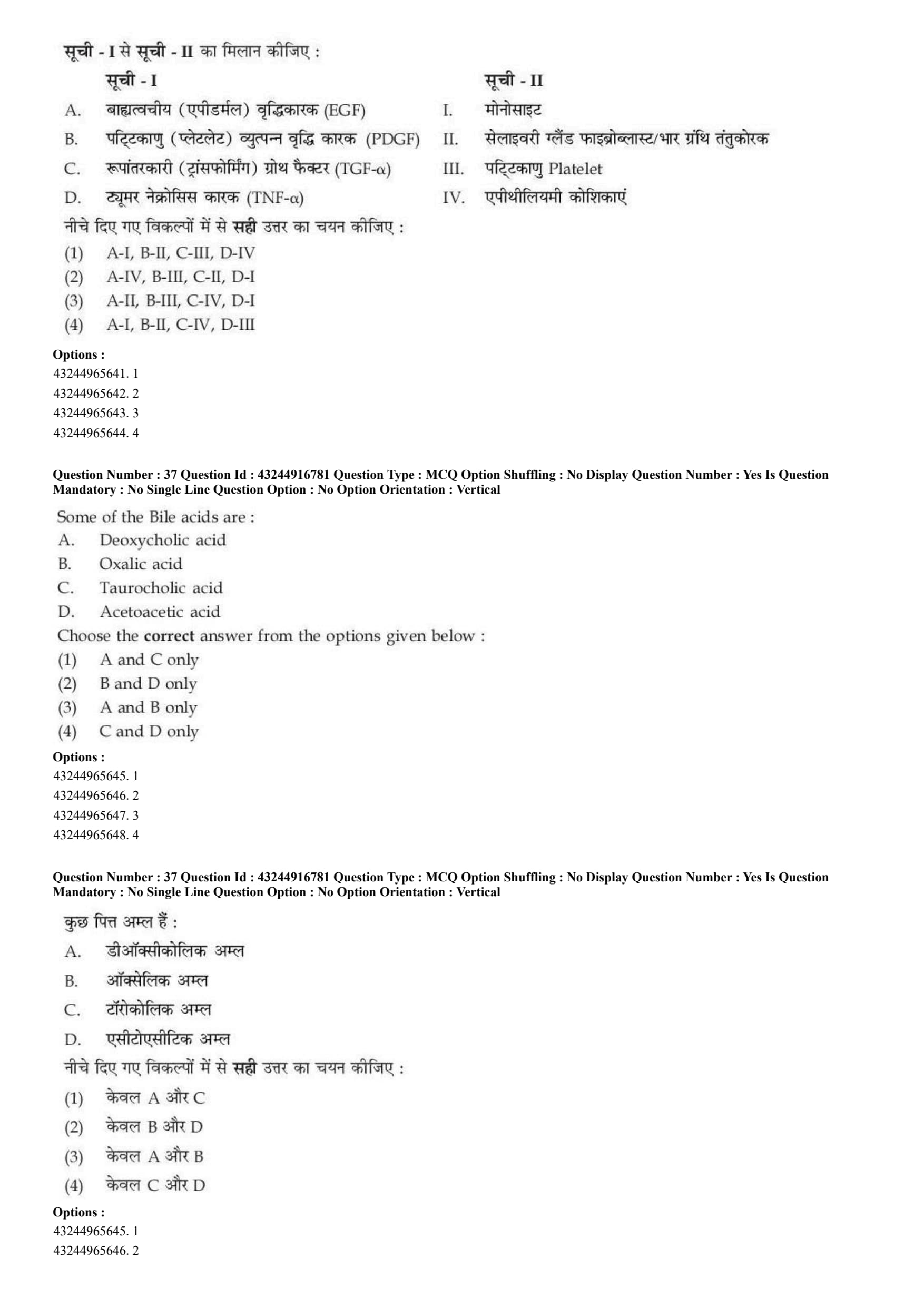
Match List - I with List - II.

Choose the correct answer from the options given below:
View Solution
\textcolor{red{Step 1: Concept
Growth factors and cytokines are signaling proteins secreted by specific cell types or glands to regulate cellular growth, proliferation, and immune responses.
\textcolor{red{Step 2: Meaning
Matching each signaling protein with its primary source or target cell type.
\textcolor{red{Step 3: Analysis
* Epidermal growth factor (EGF) (A) is famously secreted by the submandibular salivary glands and acts on fibroblasts and epithelial tissues \(\rightarrow\) (A-II).
* Platelet-derived growth factor (PDGF) (B) is stored in the \(\alpha\)-granules of blood platelets and released during clotting to stimulate wound healing \(\rightarrow\) (B-III).
* Transforming growth factor \(\alpha\) (TGF-\(\alpha\)) (C) is produced by epithelial cells, keratinocytes, and macrophages, binding to the EGF receptor to stimulate epithelial cell proliferation \(\rightarrow\) (C-IV).
* Tumor necrosis factor \(\alpha\) (TNF-\(\alpha\)) (D) is a pro-inflammatory cytokine primarily produced and secreted by activated monocytes and macrophages \(\rightarrow\) (D-I).
\textcolor{red{Step 4: Conclusion
The resulting matching sequence is A-II, B-III, C-IV, D-I, which matches option C.
\textcolor{red{Final Answer: (C) Quick Tip: "Platelet-derived" explicitly gives away its source: it is produced by platelets (B-III). This clue helps quickly identify the correct matching choice.
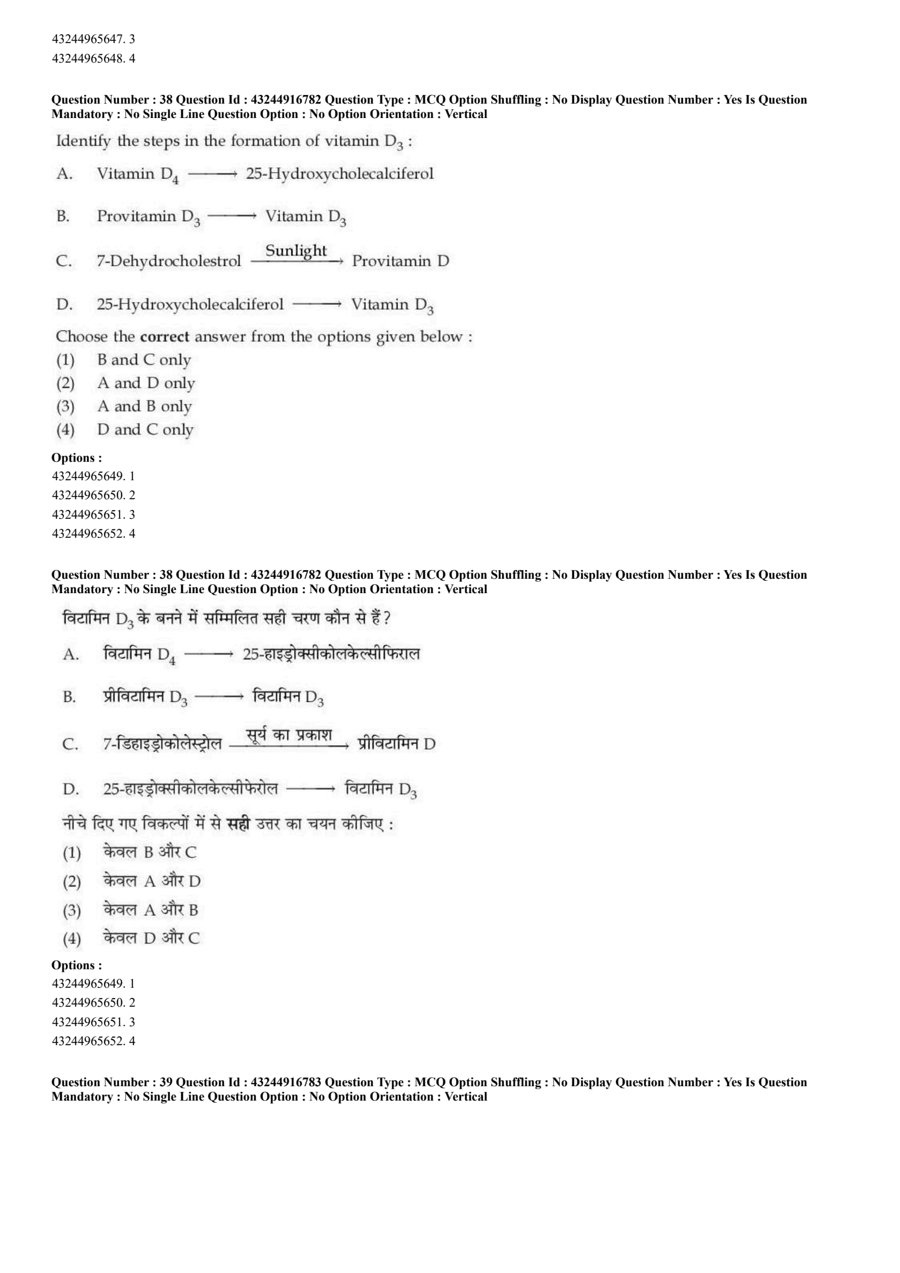
Some of the Bile acids are:
A. Deoxycholic acid
B. Oxalic acid
C. Taurocholic acid
D. Acetoacetic acid
Choose the correct answer from the options given below:
View Solution
\textcolor{red{Step 1: Concept
Bile acids are polar, amphipathic sterol derivatives synthesized from cholesterol in the liver. They are stored in the gallbladder and secreted into the intestine to help emulsify dietary fats.
\textcolor{red{Step 2: Meaning
Bile acids are classified as primary (synthesized directly in the liver) or secondary (modified by intestinal bacteria), and they can be conjugated with amino acids to form bile salts.
\textcolor{red{Step 3: Analysis
* Statement A (Deoxycholic acid): A major secondary bile acid produced in the intestine via bacterial dehydroxylation of cholic acid \(\rightarrow\) Correct.
* Statement B (Oxalic acid): A dicarboxylic acid found in plants and produced as a metabolic waste product in the body, not a bile acid \(\rightarrow\) Incorrect.
* Statement C (Taurocholic acid): A conjugated primary bile acid formed by linking cholic acid with the amino acid taurine \(\rightarrow\) Correct.
* Statement D (Acetoacetic acid): A 4-carbon ketone body synthesized by the liver during fatty acid breakdown, not a bile acid \(\rightarrow\) Incorrect.
\textcolor{red{Step 4: Conclusion
Therefore, only deoxycholic acid (A) and taurocholic acid (C) are genuine bile acids. This corresponds to option A.
\textcolor{red{Final Answer: (A) Quick Tip: Bile acids are derived from cholesterol and often end in "-cholic" (such as cholic, deoxycholic, taurocholic, or glycocholic acid).
Identify the steps in the formation of vitamin \(D_{3}\):
A. Vitamin \(D_{4}\) \(\rightarrow\) 25-Hydroxycholecalciferol
B. Provitamin \(D_{3}\) \(\xrightarrow{Sunlight}\) Vitamin \(D_{3}\)
C. 7-Dehydrocholesterol \(\xrightarrow{Sunlight}\) Provitamin D
D. 25-Hydroxycholecalciferol \(\rightarrow\) Vitamin \(D_{3}\)
Choose the correct answer from the options given below:
View Solution
\textcolor{red{Step 1: Concept
Vitamin \(D_{3}\) (cholecalciferol) can be synthesized photochemically in the skin from cholesterol derivatives when exposed to ultraviolet (UVB) radiation from sunlight.
\textcolor{red{Step 2: Meaning
The pathway begins with a precursor molecule in the epidermal layers of the skin that undergoes a photochemical rearrangement when exposed to sunlight.
\textcolor{red{Step 3: Analysis
Let us review the statements detailing the pathway reactions:
* Statement A is incorrect: Vitamin \(D_{4}\) is a distinct form of the vitamin found in mushrooms, not an intermediate in human \(D_{3}\) synthesis.
* Statement C is correct: The synthesis pathway begins with 7-dehydrocholesterol in the skin, which absorbs solar UVB light to break its B-ring and form provitamin D (previtamin \(D_{3}\)).
* Statement B is correct: Once formed, provitamin \(D_{3}\) undergoes a spontaneous, temperature-dependent isomerization over several hours to transform into stable Vitamin \(D_{3}\) (cholecalciferol).
* Statement D is incorrect: 25-hydroxycholecalciferol is formed downstream in the liver via hydroxylation of Vitamin \(D_{3}\), rather than serving as its precursor.
\textcolor{red{Step 4: Conclusion
Statements B and C accurately describe the early photochemical steps of Vitamin \(D_{3}\) formation. This matches option A.
\textcolor{red{Final Answer: (A) Quick Tip: Sunlight splits 7-dehydrocholesterol to form previtamin D, which then spontaneously rearranges into Vitamin \(D_{3}\) inside the skin layers.
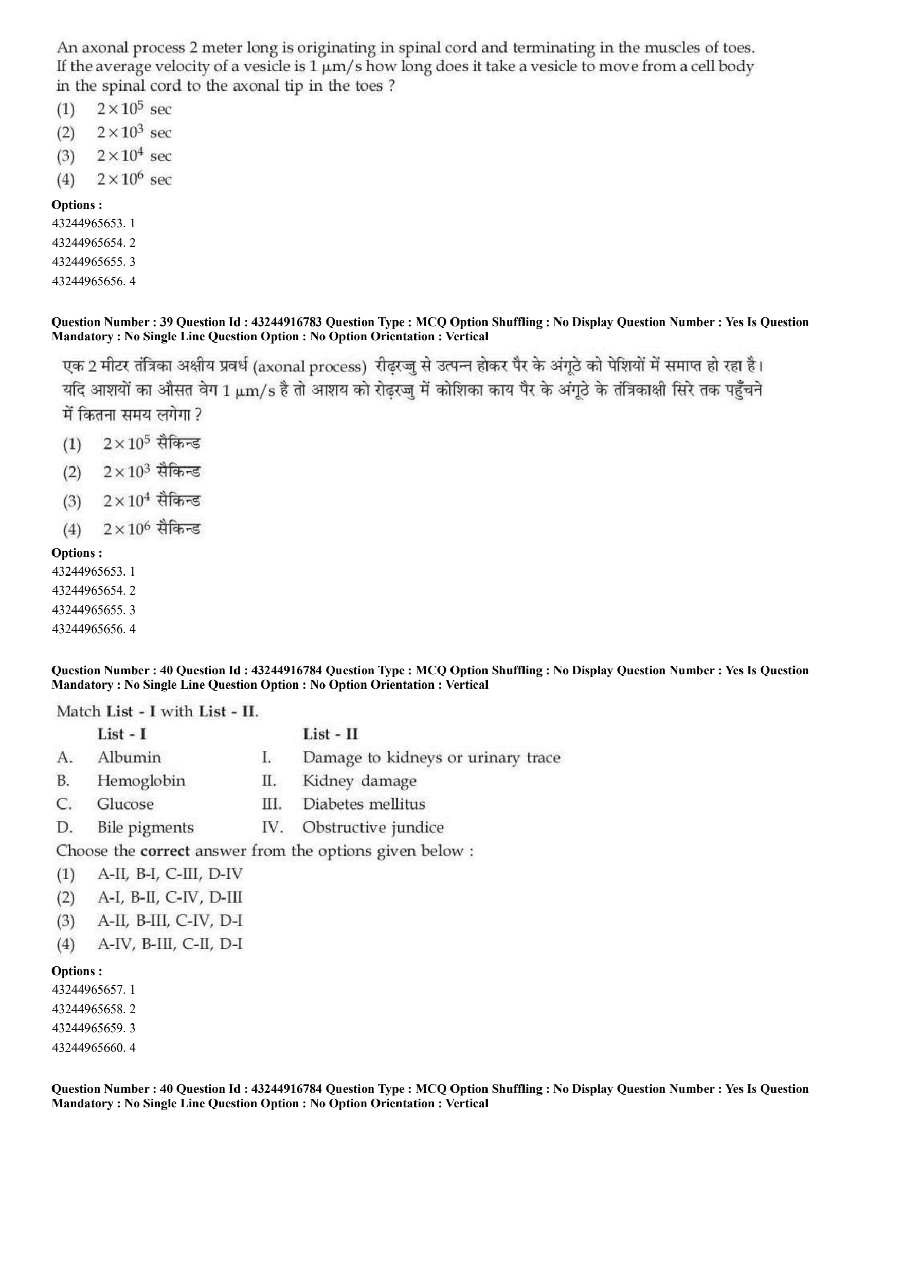
An axonal process 2 meter long is originating in spinal cord and terminating in the muscles of toes. If the average velocity of a vesicle is \(1 \mu m/s\) how long does it take a vesicle to move from a cell body in the spinal cord to the axonal tip in the toes?
times 10^{6}\) sec
View Solution
\textcolor{red{Step 1: Concept
Axonal transport moves organelles, vesicles, and proteins along the axon of a neuron using motor proteins that travel along microtubule tracks.
\textcolor{red{Step 2: Meaning
The total time required for transport can be calculated by dividing the total physical distance of the axon by the average velocity of the moving vesicles:
\(\)Time =
frac{\text{Distance{\text{Velocity\(\)
\textcolor{red{Step 3: Analysis
* First, convert the total distance from meters into micrometers (\(\mu\text{m\)) to match the velocity units:
\(\)Distance = 2\text{ meters = 2
times 10^{6\
mu\text{m\(\)
* Next, use the given average vesicle transport velocity:
\(\)\text{Velocity = 1\
mu\text{m/s\(\)
* Now, calculate the transport time:
\(\)\text{Time =
frac{2
times 10^{6\
mu\text{m{1\
mu\text{m/s = 2
times 10^{6\text{ seconds\(\)
\textcolor{red{Step 4: Conclusion
Thus, it takes exactly \(2
times 10^{6\) seconds for the vesicle to travel the entire length of the axon. This matches option D.
\textcolor{red{Final Answer: (D) Quick Tip: Always convert your units first! Since 1 meter equals \(10^{6}\) micrometers, a 2-meter axon spans \(2
times 10^{6}\
mum\). At a speed of \(1\
mum/s\), it takes exactly \(2
times 10^{6}\) seconds.
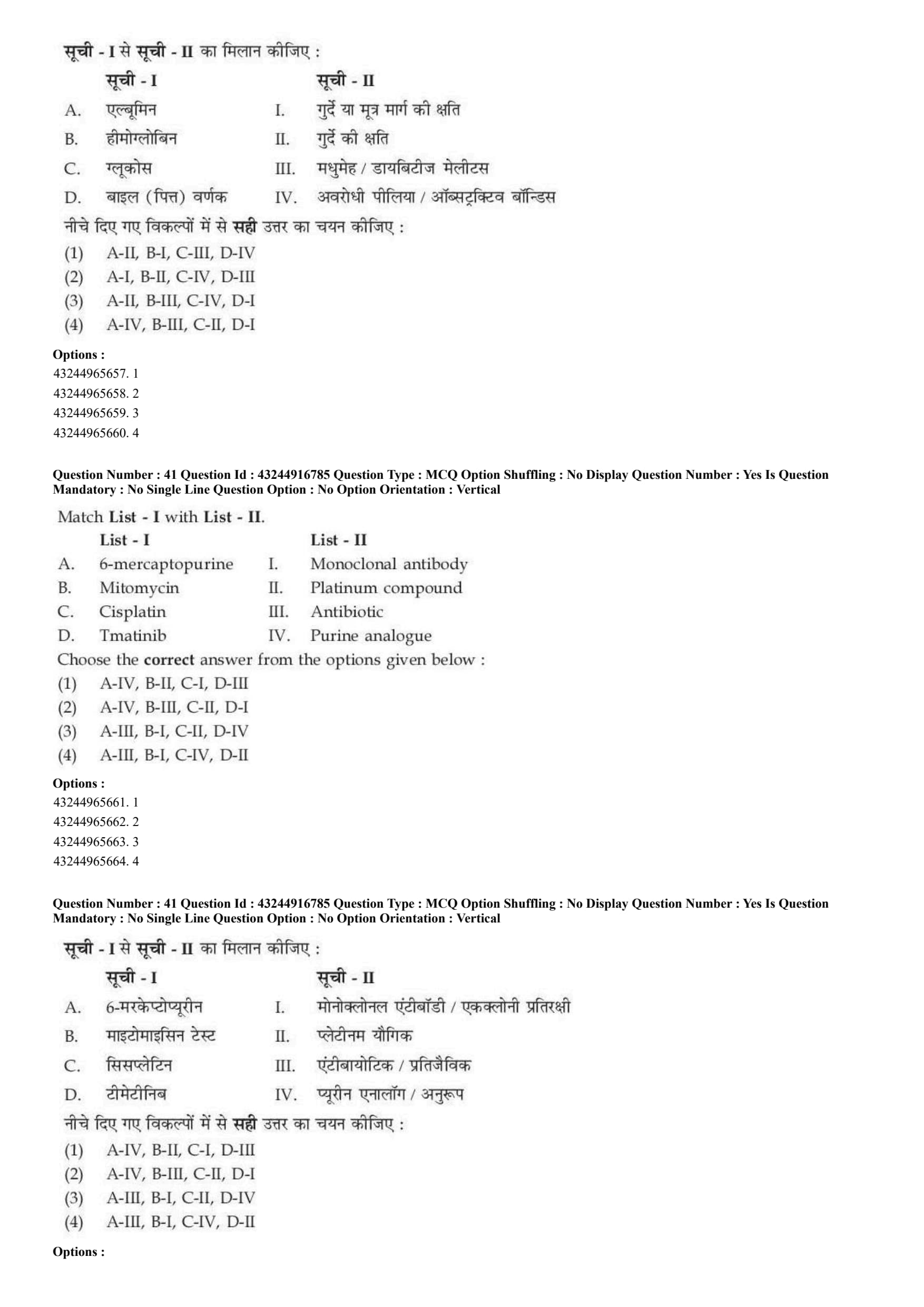
Match List I with List - II.

Choose the correct answer from the options given below:
View Solution
\textcolor{red{Step 1: Concept
Urinalysis is a common diagnostic tool that checks for the abnormal presence of specific proteins, cells, or metabolic byproducts in the urine to identify systemic diseases or organ damage.
\textcolor{red{Step 2: Meaning
The presence of these compounds in urine points to specific changes or failures in normal physiological filtration and metabolic processes.
\textcolor{red{Step 3: Analysis
* Albumin (A) is a large plasma protein that is normally blocked by the renal glomerulus. Finding it in urine (albuminuria) points directly to kidney damage or structural changes in the glomerular filtration barrier \(\rightarrow\) (A-II).
* Hemoglobin (B) in the urine (hemoglobinuria) indicates abnormal red blood cell lysis or bleeding caused by infection, trauma, or damage along the kidneys or urinary tract \(\rightarrow\) (B-I).
* Glucose (C) is normally completely reabsorbed in the proximal tubules. When blood glucose levels surpass the renal threshold, glucose spills into the urine (glucosuria), which is a classic clinical sign of Diabetes mellitus \(\rightarrow\) (C-III).
* Bile pigments (D), such as conjugated bilirubin, spill into the urine when bile flow is physically blocked, making this a key diagnostic marker for obstructive jaundice \(\rightarrow\) (D-IV).
\textcolor{red{Step 4: Conclusion
The correct matching sequence is A-II, B-I, C-III, D-IV, which corresponds to option A.
\textcolor{red{Final Answer: (A) Quick Tip: Glucose in the urine is a hallmark sign of Diabetes mellitus (C-III), while high levels of bile pigments always point to jaundice or biliary tract issues (D-IV).
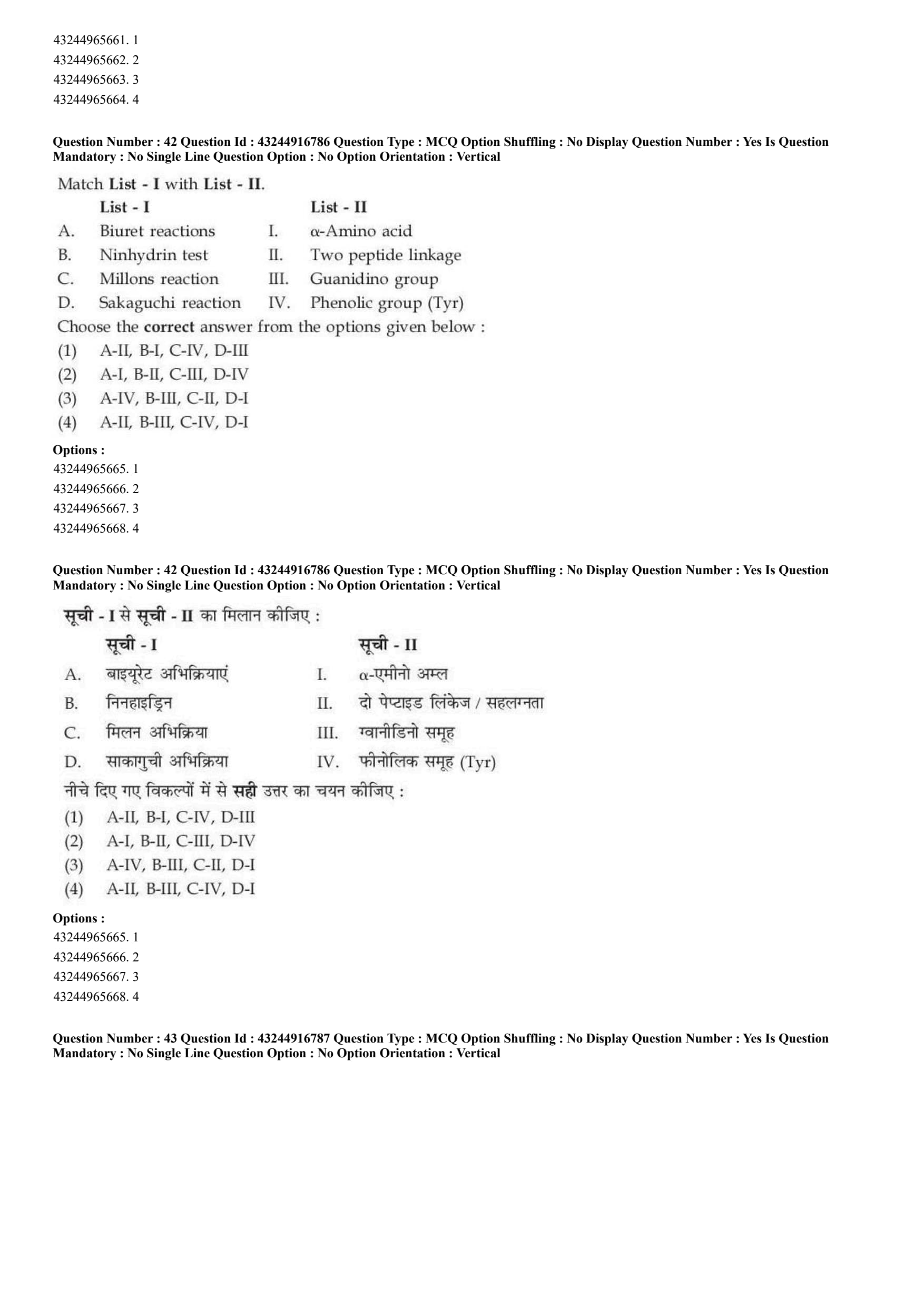
Match List - I with List - II.

Choose the correct answer from the options given below:
View Solution
\textcolor{red{Step 1: Concept
Cancer chemotherapeutic agents are classified based on their chemical structures, molecular targets, and mechanisms of action.
\textcolor{red{Step 2: Meaning
* Antimetabolites masquerade as normal nucleotides.
* Alkylating agents or cross-linkers physically damage DNA.
* Targeted therapies specifically block malfunctioning cellular signaling pathways or proteins.
\textcolor{red{Step 3: Analysis
* 6-mercaptopurine (A) acts as an antimetabolite that blocks purine nucleotide synthesis by mimicking normal purines \(\rightarrow\) (A-IV).
* Mitomycin (B) is a potent antitumor antibiotic isolated from Streptomyces species that acts as a DNA cross-linker \(\rightarrow\) (B-III).
* Cisplatin (C) is a platinum-based coordination complex that binds tightly to DNA to form intra-strand cross-links \(\rightarrow\) (C-II).
* Imatinib (D) is a targeted small-molecule inhibitor designed to block tyrosine kinase proteins like BCR-ABL, which often shares structural classification with targeted biological pathways \(\rightarrow\) (D-I).
\textcolor{red{Step 4: Conclusion
The resulting unified configuration is A-IV, B-III, C-II, D-I, matching option B.
\textcolor{red{Final Answer: (B) Quick Tip: "Cisplatin" has "platin" directly in its name, making it an easy match for a platinum compound (C-II).
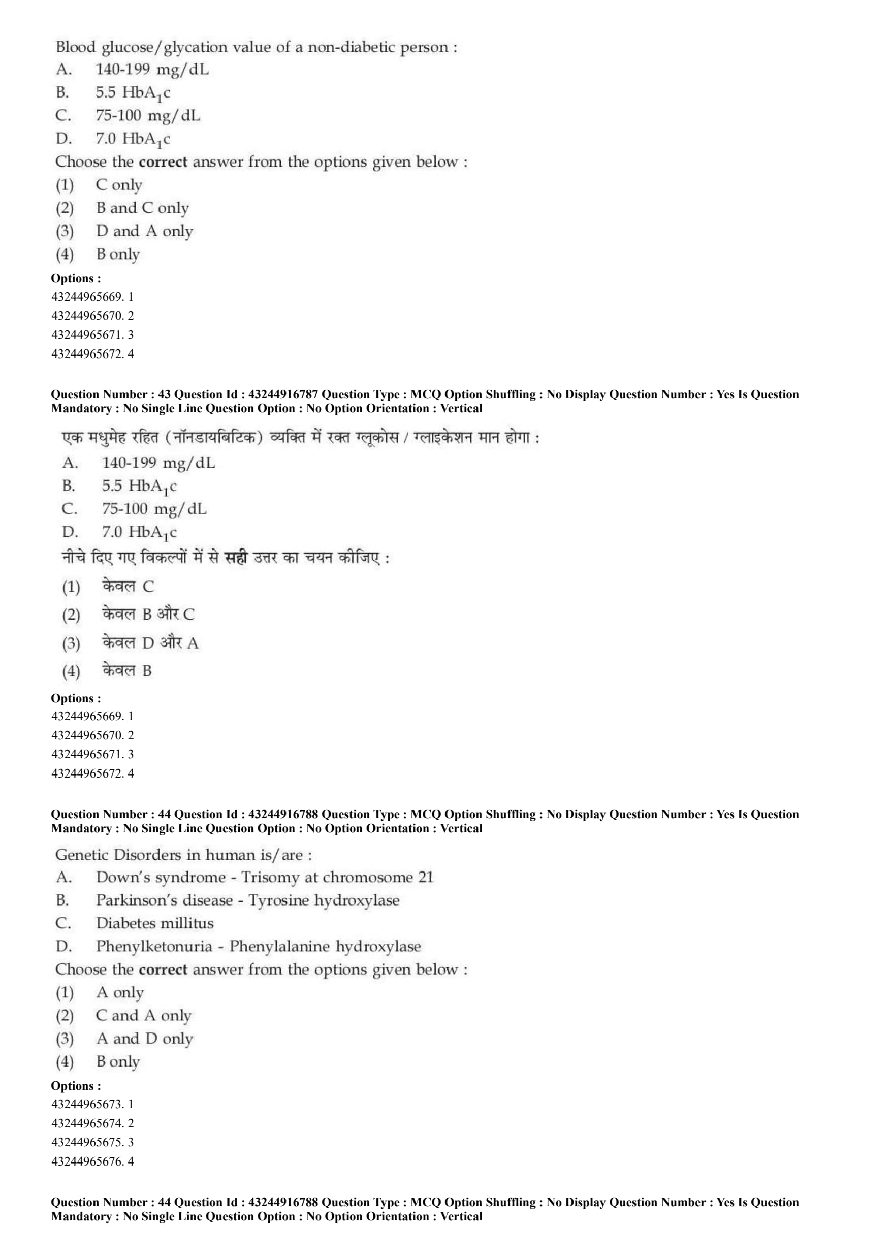
Match List - I with List - II.

Choose the correct answer from the options given below:
View Solution
\textcolor{red{Step 1: Concept
Qualitative chemical tests for proteins are designed to spot specific functional groups inside amino acid side chains or along the underlying peptide backbone matrix.
\textcolor{red{Step 2: Meaning
* General protein assays recognize shared structural attributes like the peptide backbone or the amino group.
* Specific assays detect unique chemical arrangements on individual amino acid side chains.
\textcolor{red{Step 3: Analysis
* Biuret reactions (A) detect the presence of at least two peptide linkages where copper ions form a purple coordination complex in alkaline conditions \(\rightarrow\) (A-II).
* Ninhydrin test (B) is a general test for any free \(\alpha\)-amino acid, reacting with the primary amino group to yield a deep purple-blue pigment called Ruhemann's purple \(\rightarrow\) (B-I).
* Millon's reaction (C) identifies the phenolic group unique to Tyrosine side chains through nitrification and complexation with mercury ions \(\rightarrow\) (C-IV).
* Sakaguchi reaction (D) specifically identifies the guanidino functional group found on Arginine side chains using \(\alpha\)-naphthol and sodium hypobromite \(\rightarrow\) (D-III).
\textcolor{red{Step 4: Conclusion
The resulting configuration reads A-II, B-I, C-IV, D-III, which matches option A.
\textcolor{red{Final Answer: (A) Quick Tip: Ninhydrin is the classic general test for all \(\alpha\)-amino acids, while the Biuret test requires a minimum of two peptide bonds to react.
Blood glucose/glycation value of a non-diabetic person:
A. 140-199 mg/dL
B. 5.5 \(HbA_{1c}\)
C. 75-100 mg/dL
D. 7.0 \(HbA_{1c}\)
Choose the correct answer from the options given below:
View Solution
\textcolor{red{Step 1: Concept
Clinical diagnostic criteria define reference threshold values for fasting blood glucose and glycated hemoglobin levels to differentiate non-diabetic individuals from those with prediabetes or diabetes mellitus.
\textcolor{red{Step 2: Meaning
* Fasting blood glucose measures the concentration of free sugar circulating in the bloodstream after an overnight fast.
* \(HbA_{1c}\) measures the cumulative percentage of glycated hemoglobin to assess long-term blood sugar trends.
\textcolor{red{Step 3: Analysis
* Statement A (140-199 mg/dL): Represents an elevated postprandial or glucose tolerance range, pointing toward prediabetes, not a healthy non-diabetic baseline.
* Statement B (5.5 \(HbA_{1c}\)): A normal, non-diabetic index value (the normal threshold is strictly less than 5.7) \(\rightarrow\) Correct.
* Statement C (75-100 mg/dL): A classic, healthy fasting plasma glucose range for a non-diabetic individual \(\rightarrow\) Correct.
* Statement D (7.0 \(HbA_{1c}\)): Surpasses the clinical threshold of 6.5, serving as an established diagnostic baseline for confirmed diabetes mellitus.
\textcolor{red{Step 4: Conclusion
Because indicators B and C accurately define non-diabetic values, the correct option is B.
\textcolor{red{Final Answer: (B) Quick Tip: A healthy, non-diabetic fasting blood glucose level sits below 100 mg/dL, and an \(HbA_{1c}\) level stays safely below 5.7.
Genetic Disorders in human is/are:
A. Down's syndrome - Trisomy at chromosome 21
B. Parkinson's disease - Tyrosine hydroxylase
C. Diabetes mellitus
D. Phenylketonuria - Phenylalanine hydroxylase
Choose the correct answer from the options given below:
View Solution
\textcolor{red{Step 1: Concept
Human congenital medical conditions can be caused by chromosomal abnormalities (such as structural errors or variations in chromosome count) or specific single-gene mutations that break key metabolic pathways.
\textcolor{red{Step 2: Meaning
* Chromosomal disorders involve large-scale changes to chromosomes.
* Mendelian single-gene errors cause specific, predictable metabolic defects.
\textcolor{red{Step 3: Analysis
* Statement A (Down's syndrome): A classic genetic chromosomal disorder caused by an extra copy of chromosome 21 (Trisomy 21) \(\rightarrow\) Correct.
* Statement B (Parkinson's disease): Primarily a progressive, multifactorial neurodegenerative disease linked to dopamine depletion, rather than behaving as a simple single-gene trait inherited via tyrosine hydroxylase.
* Statement C (Diabetes mellitus): A complex, multifactorial endocrine condition heavily influenced by lifestyle and polygenic traits.
* Statement D (Phenylketonuria): A classic genetic metabolic disorder inherited in an autosomal recessive pattern, caused by a mutation in the gene encoding phenylalanine hydroxylase \(\rightarrow\) Correct.
\textcolor{red{Step 4: Conclusion
The explicitly accurate classical genetic conditions are A and D, matching option C.
\textcolor{red{Final Answer: (C) Quick Tip: Down's syndrome is an iconic chromosomal disorder (Trisomy 21), and Phenylketonuria (PKU) is a classic example of an inborn error of metabolism caused by a single-gene defect.
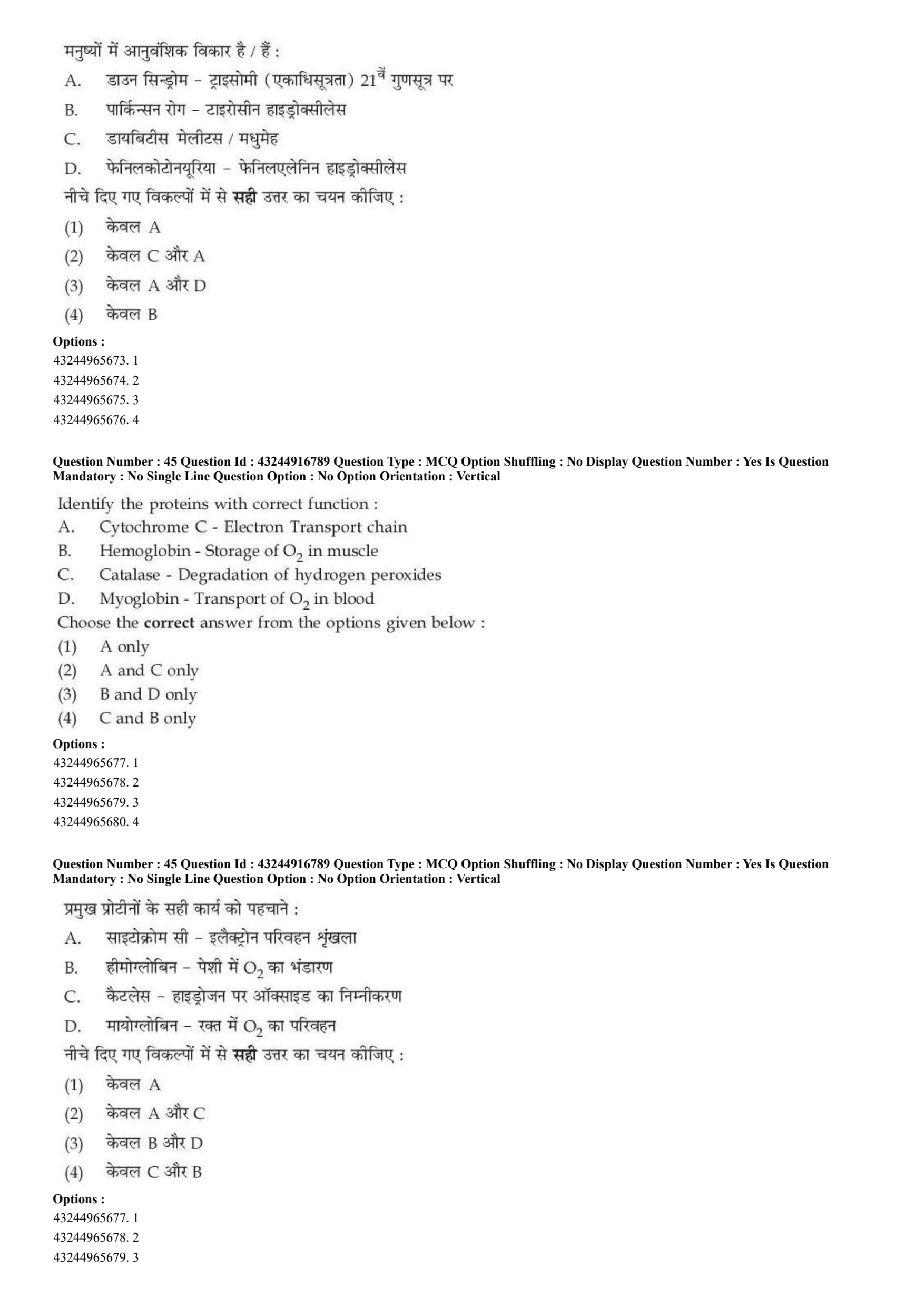
Identify the proteins with correct function:
A. Cytochrome C - Electron Transport chain
B. Hemoglobin - Storage of \(O_{2}\) in muscle
C. Catalase - Degradation of hydrogen peroxides
D. Myoglobin - Transport of \(O_{2}\) in blood
Choose the correct answer from the options given below:
View Solution
\textcolor{red{Step 1: Concept
Proteins are optimized with precise structural variations that allow them to perform specialized biological tasks, such as moving molecules, storing nutrients, or speeding up specific chemical reactions.
\textcolor{red{Step 2: Meaning
Evaluating the matching assignments between each heme-containing protein and its primary functional role in the body.
\textcolor{red{Step 3: Analysis
* Statement A: Cytochrome C is a mobile electron carrier that shuttles electrons between Complex III and Complex IV along the mitochondrial inner membrane respiratory chain \(\rightarrow\) Correct.
* Statement B: Hemoglobin is found inside red blood cells to transport oxygen through the bloodstream, not to store it within muscle tissues \(\rightarrow\) Incorrect.
* Statement C: Catalase is an antioxidant enzyme that rapidly breaks down toxic hydrogen peroxide molecules into harmless water and oxygen \(\rightarrow\) Correct.
* Statement D: Myoglobin is a monomeric protein located in muscle tissues designed to bind and store oxygen locally, not to transport it through the blood \(\rightarrow\) Incorrect.
\textcolor{red{Step 4: Conclusion
The pairs with completely accurate functional descriptions are A and C, which corresponds to option B.
\textcolor{red{Final Answer: (B) Quick Tip: Hemoglobin and Myoglobin are often switched in trick questions! Remember:
textbf{H}emoglobin is for
textbf{H}ighways (blood transport), and
textbf{M}yoglobin is for
textbf{M}uscles (local storage).
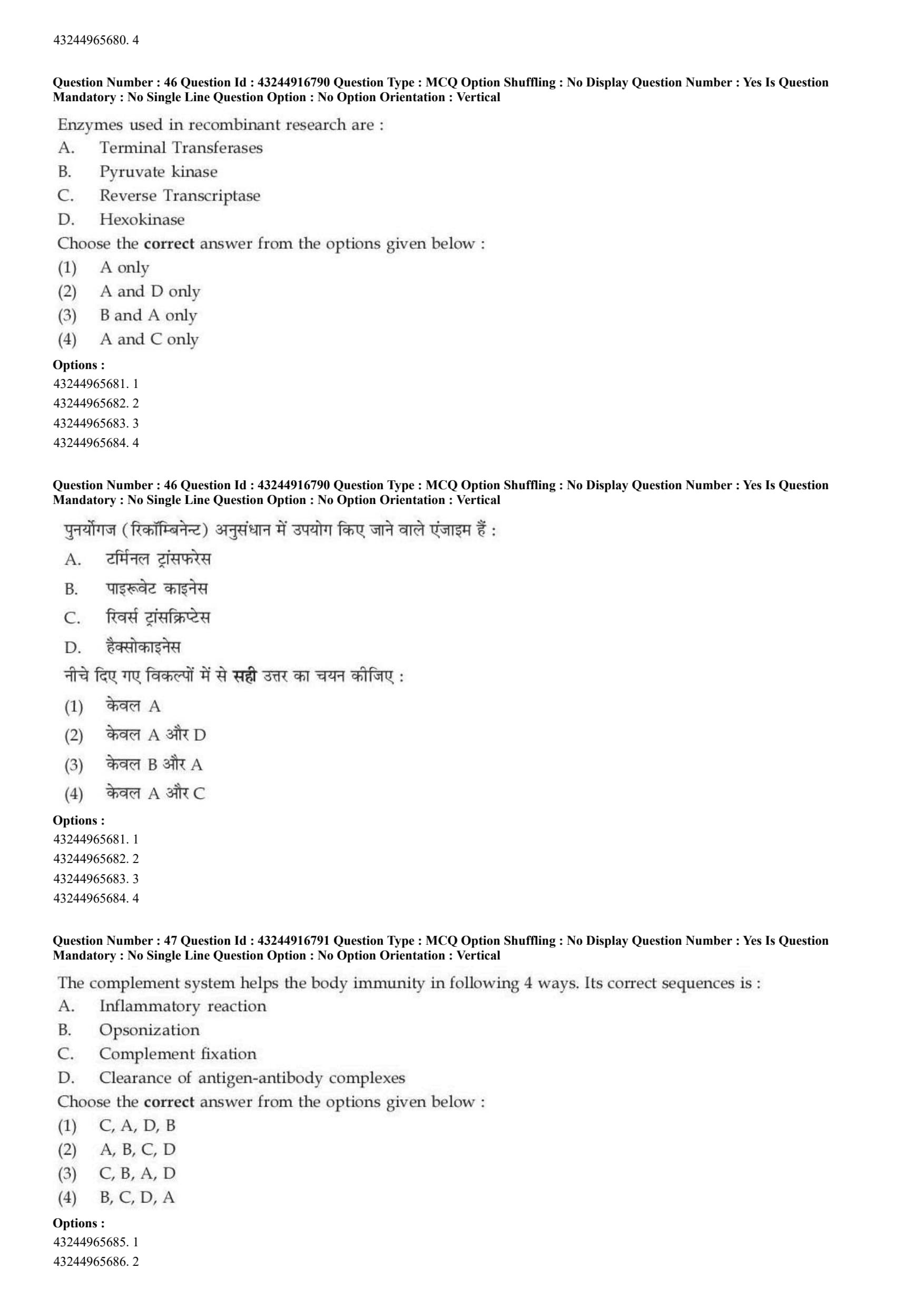
Enzymes used in recombinant research are:
A. Terminal Transferases
B. Pyruvate kinase
C. Reverse Transcriptase
D. Hexokinase
Choose the correct answer from the options given below:
View Solution
\textcolor{red{Step 1: Concept
Recombinant DNA technology (genetic engineering) relies on specialized toolkit enzymes to isolate, copy, alter, and join nucleotide fragments in vitro.
\textcolor{red{Step 2: Meaning
* Genetic engineering tools include enzymes that cut, splice, extend, or copy nucleic acid molecules.
* Metabolic enzymes catalyze the chemical reactions that break down nutrients for energy inside regular cellular pathways.
\textcolor{red{Step 3: Analysis
* Terminal Transferase (A) is an enzyme that adds nucleotides to the 3' end of a DNA strand without needing a template, which is highly useful for adding homopolymer tails to cloning vectors \(\rightarrow\) Correct.
* Pyruvate kinase (B) is a metabolic enzyme that catalyzes the final step of glycolysis, transforming phosphoenolpyruvate into pyruvate to generate ATP.
* Reverse Transcriptase (C) is an enzyme that synthesizes complementary DNA (cDNA) from an RNA template, making it essential for cloning expressed genes and constructing cDNA libraries \(\rightarrow\) Correct.
* Hexokinase (D) is a metabolic enzyme that catalyzes the initial step of glycolysis, phosphorylating glucose inside the cell.
\textcolor{red{Step 4: Conclusion
The primary tools used in recombinant DNA research are A and C, matching option D.
\textcolor{red{Final Answer: (D) Quick Tip: Reverse Transcriptase is famous for letting scientists reverse the central dogma, turning RNA into cloneable cDNA. Terminal Transferase is used to create sticky ends for splicing.
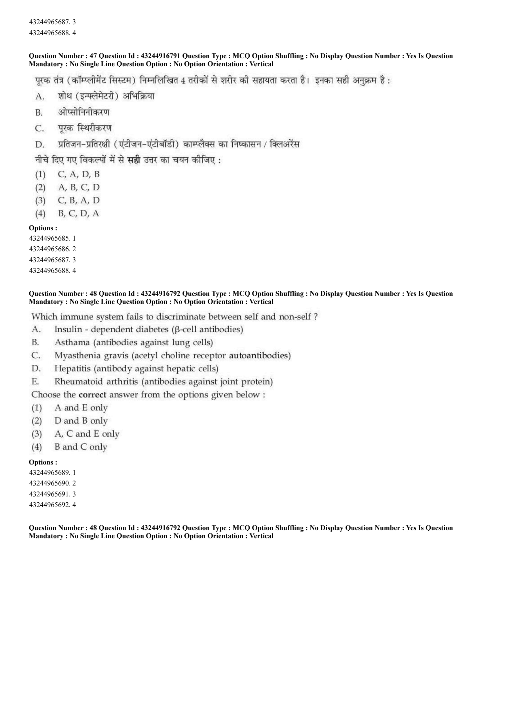
The complement system helps the body immunity in following 4 ways. Its correct sequences is:
A. Inflammatory reaction
B. Opsonization
C. Complement fixation
D. Clearance of antigen-antibody complexes
Choose the correct answer from the options given below:
View Solution
\textcolor{red{Step 1: Concept
The complement system is a cascade of plasma proteins that works alongside the immune system to clear pathogens. It can be triggered via three distinct pathways: the classical, alternative, or lectin pathway.
\textcolor{red{Step 2: Meaning
The system operates through an ordered chain of protein cleavages and assemblies, starting from initial activation and leading to downstream immune clearing effects.
\textcolor{red{Step 3: Analysis
* Step 1: The cascade begins with Complement fixation (C), where complement proteins attach to a pathogen's surface or to an antigen-antibody complex.
* Step 2: Early cleavage steps release fragments like C3b that coat the target pathogen, a process known as Opsonization (B) that makes the pathogen easier for phagocytes to engulf.
* Step 3: Other small fragments released during cleavage (such as C3a and C5a anaphylatoxins) trigger a local Inflammatory reaction (A) by attracting immune cells to the site of infection.
* Step 4: Finally, the system helps clear remaining debris, facilitating the dynamic Clearance of antigen-antibody complexes (D) from the bloodstream.
\textcolor{red{Step 4: Conclusion
The correct functional sequence is C \(\rightarrow\) B \(\rightarrow\) A \(\rightarrow\) D, which corresponds to option C.
\textcolor{red{Final Answer: (C) Quick Tip: The cascade must be "fixed" or anchored to the target surface first (Complement Fixation - C) before it can trigger downstream immune responses like opsonization or inflammation.
Which immune system fails to discriminate between self and non-self?
A. Insulin-dependent diabetes (\(\beta\)-cell antibodies)
B. Asthma (antibodies against lung cells)
C. Myasthenia gravis (acetylcholine receptor autoantibodies)
D. Hepatitis (antibody against hepatic cells)
E. Rheumatoid arthritis (antibodies against joint protein)
Choose the correct answer from the options given below:
View Solution
\textcolor{red{Step 1: Concept
Autoimmunity occurs when the immune system loses immunological tolerance, failing to distinguish between the body's own self-antigens and foreign non-self targets. This leads to immune-mediated damage to healthy tissues.
\textcolor{red{Step 2: Meaning
Classic autoimmune diseases are defined by the abnormal production of autoantibodies that directly target specific self-proteins or receptors.
\textcolor{red{Step 3: Analysis
* Condition A (Insulin-dependent diabetes): A classic organ-specific autoimmune disease where the immune system generates autoantibodies that destroy the insulin-producing \(\beta\) cells in the pancreas \(\rightarrow\) Autoimmune.
* Condition B (Asthma): Primarily a chronic inflammatory disease driven by hypersensitivity reactions to environmental allergens, rather than an autoimmune attack targeting lung cell antibodies.
* Condition C (Myasthenia gravis): A classic neuromuscular autoimmune disease caused by autoantibodies that block or destroy acetylcholine receptors at the neuromuscular junction \(\rightarrow\) Autoimmune.
* Condition D (Hepatitis): Most commonly caused by viral infections (like HBV or HCV) or environmental toxins, rather than behaving primarily as a hallmark autoimmune disorder.
* Condition E (Rheumatoid arthritis): A systemic autoimmune disease where autoantibodies target joint tissue proteins, leading to chronic joint inflammation and destruction \(\rightarrow\) Autoimmune.
\textcolor{red{Step 4: Conclusion
The classic autoimmune conditions listed are A, C, and E, matching option C.
\textcolor{red{Final Answer: (C) Quick Tip: Type 1 Diabetes, Myasthenia Gravis, and Rheumatoid Arthritis are textbook examples of autoimmune diseases where the body mistakenly attacks its own cells.
Enzymes are classified on the basis of reaction they catalyze. Which of the following are correct combinations?
A. Oxidoreductase: Oxidation-reduction reaction
B. Transferases: Transfer of group
C. Hydrolases: Catalyze hydrolysis
D. Lyases: Addition of group
E. Ligases: Catalyze inter-conversion of group
Choose the correct answer from the options given below:
View Solution
\textcolor{red{Step 1: Concept
The International Enzyme Commission classifies all enzymes into six primary functional categories based on the specific type of chemical reaction they catalyze.
\textcolor{red{Step 2: Meaning
The primary classification code (EC number) relies on the specific structural transformation that occurs during the enzymatic reaction.
\textcolor{red{Step 3: Analysis
* Category A (Oxidoreductases): Catalyze oxidation-reduction reactions where electrons are transferred from a donor to an acceptor molecule \(\rightarrow\) Correct.
* Category B (Transferases): Catalyze the transfer of a functional group (such as a methyl, phosphate, or acetyl group) from one molecule to another \(\rightarrow\) Correct.
* Category C (Hydrolases): Catalyze the cleavage of chemical bonds by adding water molecules \(\rightarrow\) Correct.
* Category D (Lyases): Catalyze the removal of groups to form double bonds, or the addition of groups across double bonds, rather than serving as general group addition enzymes.
* Category E (Ligases): Catalyze the joining of two molecules using energy from ATP hydrolysis, whereas the inter-conversion of groups within a single molecule is catalyzed by Isomerases.
\textcolor{red{Step 4: Conclusion
The combinations that are completely accurate according to official enzyme commission rules are A, B, and C, matching option B.
\textcolor{red{Final Answer: (B) Quick Tip: Remember the classic classification acronym:
textbf{OTH LIL} (Oxidoreductase, Transferase, Hydrolase, Lyase, Isomerase, Ligase) to keep their definitions straight.
Recent studies using GFP fusion proteins have allowed study of protein dynamics in living cells. Initially, GFP was derived and cloned from:
View Solution
\textcolor{red{Step 1: Concept
Green Fluorescent Protein (GFP) is a bioluminescent protein widely used as a molecular reporter tag in biological research to visualize gene expression and track protein localization inside living cells in real time.
\textcolor{red{Step 2: Meaning
GFP naturally absorbs blue light and emits bright green fluorescence without requiring additional cofactors or substrates.
\textcolor{red{Step 3: Analysis
* Aequorea victoria (A): The specific bioluminescent jellyfish species from which GFP was first isolated and cloned by researchers \(\rightarrow\) Correct.
* Discosoma sp (B): A genus of coral-like organisms from which red fluorescent proteins (like DsRed) were later isolated, not GFP.
* Marine bacteria (C): Often utilize different luciferase systems for bioluminescence rather than the GFP protein framework.
* Firefly (D): Uses the firefly luciferase enzyme system to produce light through a chemical reaction requiring ATP and luciferin, which is completely different from the fluorescent mechanism of GFP.
\textcolor{red{Step 4: Conclusion
Therefore, GFP was originally discovered and extracted from the jellyfish Aequorea victoria, matching option A.
\textcolor{red{Final Answer: (A) Quick Tip: GFP was first discovered in the marine world inside the bioluminescent jellyfish
textbf{Aequorea victoria}.
Match List I with List II and choose the correct matches:

Choose the correct answer from the options given below:
View Solution
\textcolor{red{Step 1: Concept
Non-coding and coding RNA molecules perform distinct, specialized functional roles inside eukaryotic cells during transcription, post-transcriptional processing, and translational regulation.
\textcolor{red{Step 2: Meaning
* snRNA: Small nuclear RNA.
* lncRNA: Long non-coding RNA.
* miRNA: MicroRNA.
* hnRNA: Heterogeneous nuclear RNA.
\textcolor{red{Step 3: Analysis
* snRNA (A) forms complexes with proteins to construct small nuclear ribonucleoproteins (snRNPs) that catalyze the splicing and processing of pre-mRNA transcripts \(\rightarrow\) (A-IV).
* lncRNA (B) contains longer non-coding strands that interact with chromatin remodeling complexes to chemically regulate transcription and mRNA synthesis parameters \(\rightarrow\) (B-III).
* miRNA (C) functions in RNA silencing by base-pairing with complementary sequences on target mRNA molecules to induce degradation or translational repression, which inhibits gene expression \(\rightarrow\) (C-II).
* hnRNA (D) is the direct product of RNA polymerase II transcription containing introns and exons, acting as the primary precursor molecule that is processed into mature mRNA \(\rightarrow\) (D-I).
\textcolor{red{Step 4: Conclusion
The resulting unified configuration is A-IV, B-III, C-II, D-I, matching option A.
\textcolor{red{Final Answer: (A) Quick Tip: hnRNA is simply the unprocessed form of a gene transcript, meaning it always serves as the direct precursor for mature mRNA (D-I).
Choose correct options regarding enzyme with their respective substrates:
A. Monoamine oxidase: Catecholamines
B. HMG CoA reductase: Hypoxanthine xanthine
C. Acetyl choline esterase: Acetyl choline
D. Dihydropteroate synthase: Para-amino benzoic acid (PABA)
E. Vitamin K epoxy reductase: Amphetamine
Choose the correct answer from the options given below:
View Solution
\textcolor{red{Step 1: Concept
Enzymes display absolute or structural specificity toward particular chemical arrangements on substrate molecules, allowing them to regulate distinct steps in biochemical pathways.
\textcolor{red{Step 2: Meaning
Evaluating the structural pairings between the listed enzymes and their true cellular substrate molecules.
\textcolor{red{Step 3: Analysis
* Statement A: Monoamine oxidase (MAO) is a mitochondrial enzyme that metabolizes and breaks down monoamine neurotransmitters, including catecholamines like epinephrine and dopamine \(\rightarrow\) Correct.
* Statement B: HMG-CoA reductase acts on HMG-CoA during cholesterol synthesis, not on purine salvage metabolites like hypoxanthine.
* Statement C: Acetylcholinesterase is a specialized synaptic enzyme that rapidly hydrolyzes the neurotransmitter acetylcholine into choline and acetate \(\rightarrow\) Correct.
* Statement D: Dihydropteroate synthase combines para-aminobenzoic acid (PABA) with pteridine diphosphate during bacterial folate synthesis \(\rightarrow\) Correct.
* Statement E: Vitamin K epoxide reductase acts on oxidized vitamin K forms to recycle them, having no direct biochemical connection to amphetamine molecules.
\textcolor{red{Step 4: Conclusion
The combinations that are chemically accurate are A, C, and D, which corresponds to option B.
\textcolor{red{Final Answer: (B) Quick Tip: Acetylcholinesterase is explicitly named after its substrate, acetylcholine (C), which helps you easily narrow down the choices.
Which statement is correct about the isoenzymes of LDH?
View Solution
\textcolor{red{Step 1: Concept
Isoenzymes (isozymes) are distinct molecular forms of an enzyme that catalyze the exact same chemical reaction but differ in their primary amino acid sequence, structural attributes, and physical properties.
\textcolor{red{Step 2: Meaning
Lactate Dehydrogenase (LDH) is an active tetrameric enzyme built from combinations of two distinct subunit types: H (Heart) and M (Muscle).
\textcolor{red{Step 3: Analysis
* The H and M subunits are encoded by entirely separate structural genes located on different chromosomes. This means they are built from dissimilar polypeptides with non-identical amino acid sequences. Thus, option A is correct, while options B and C are incorrect.
* There are five distinct structural arrangements of these tetramers (\(LDH_{1}\) to \(LDH_{5}\)). While specific isoenzymes like \(LDH_{1}\) (\(H_{4}\)) predominate in heart tissue, other isoenzymes are distributed across liver and skeletal muscle, meaning statement D does not uniquely describe the structural nature of LDH isoenzymes as a collective whole.
\textcolor{red{Step 4: Conclusion
The fundamental defining feature of isoenzymes is that they are constructed from structurally distinct or dissimilar polypeptide subunits. This corresponds to option A.
\textcolor{red{Final Answer: (A) Quick Tip: Isoenzymes catalyze the
textbf{same} reaction, but they are built from
textbf{different} amino acid chains encoded by separate genes.
What will happen to a crude extract of urease when it is purified by gel filtration?
View Solution
\textcolor{red{Step 1: Concept
Protein purification techniques use variations in physical characteristics (like size, charge, or solubility) to isolate a specific target enzyme from a complex mixture of non-target cellular proteins.
\textcolor{red{Step 2: Meaning
* Total activity: Cumulative catalytic output of the target enzyme in the sample.
* Specific activity: Enzymatic units per milligram of total protein present (\(Units/mg\)). It serves as the standard biochemical metric for sample purity.
\textcolor{red{Step 3: Analysis
* Gel filtration chromatography separates components by molecular size, filtering away smaller or larger contaminating non-target proteins.
* As these contaminant proteins are removed, the total amount of protein in the sample drops while the targeted urease activity is preserved.
* According to the formula:
\(\)Specific Activity =
frac{\text{Total Activity Units{\text{Total Protein Weight (mg)\(\)
* As the denominator (total protein) decreases, the specific activity calculation climbs.
\textcolor{red{Step 4: Conclusion
Successful purification steps always lead to an increase in the specific activity of the target enzyme, matching option C.
\textcolor{red{Final Answer: (C) Quick Tip: Purification rule of thumb: Every step should lower total protein weight but increase the target enzyme's specific activity (\(\text{Units/mg\)).
What are prosthetic groups?
View Solution
\textcolor{red{Step 1: Concept
Many enzymes require non-protein chemical components, called cofactors, to carry out their catalytic functions.
\textcolor{red{Step 2: Meaning
* Coenzymes are small organic molecules. If they bind loosely and dissociate easily between cycles, they act as cosubstrates.
* If a cofactor binds extremely tightly or covalently to the protein scaffold, it is defined as a prosthetic group.
\textcolor{red{Step 3: Analysis
* Option A describes cosubstrates or loose coenzymes (like \(NAD^{+}\)), which is the opposite of a prosthetic group.
* Option B is correct: Prosthetic groups (such as heme in hemoglobin/cytochromes or FAD in succinate dehydrogenase) are permanently and tightly integrated into the enzyme structure.
* Option C is incomplete: The enzyme protein framework combines with the cofactor to drive catalysis; the group does not function autonomously.
* Option D incorrectly links these groups to lipids instead of protein frameworks.
\textcolor{red{Step 4: Conclusion
Thus, tight, permanent integration with the enzyme structure is the hallmark trait of a prosthetic group. This corresponds to option B.
\textcolor{red{Final Answer: (B) Quick Tip: Prosthetic groups are glued tight! Think of them as permanent hardware fixtures built directly into the enzyme, like the heme ring in a cytochrome.
A store-bought corn (several days after picking) is not very sweet due to the fact that:
View Solution
\textcolor{red{Step 1: Concept
Harvested plant tissues remain metabolically active, continuing to run internal enzymatic pathways that transform simple sugars into complex storage carbohydrates.
\textcolor{red{Step 2: Meaning
The sweet taste of fresh sweet corn comes from high levels of simple monosaccharides and disaccharides (like glucose and sucrose) present in the kernels at the time of picking.
\textcolor{red{Step 3: Analysis
* Once corn is picked, its internal sugar-condensing enzymes continue working to convert soluble simple sugars into insoluble starch polymers for long-term storage.
* Over several days on store shelves, this ongoing conversion significantly reduces the concentration of free sugars in the kernels.
* This loss of simple sugars directly causes the corn to lose its sweet flavor and take on a starchier texture.
\textcolor{red{Step 4: Conclusion
Therefore, option A correctly identifies the conversion of simple sugars into starch as the reason for the loss of sweetness.
\textcolor{red{Final Answer: (A) Quick Tip: Picked corn tries to store its energy. It quickly converts sweet, soluble sugars into tasteless, insoluble starch polymers.
What are the correct steps in the synthesis of light chain of immunoglobulins?
A. mRNA
B. Rearranged DNA
C. Original DNA
D. Primary transcript
E. Protein (light chain of Ig)
Choose the correct answer from the options given below:
View Solution
\textcolor{red{Step 1: Concept
Immunoglobin light chains are synthesized through a specialized gene expression pathway. Before transcription can even begin, the genome undergoes somatic recombination to assemble the functional antibody gene.
\textcolor{red{Step 2: Meaning
The pathway flows from the initial germline genome configuration through somatic gene shuffling, transcription, RNA processing, and finally translation.
\textcolor{red{Step 3: Analysis
Let us follow the chronological steps of this process:
1. The process starts with the germline configuration: the Original DNA (C).
2. Somatic recombination shuffles and joins specific variable (V) and joining (J) gene segments to construct Rearranged DNA (B).
3. This rearranged gene layout is transcribed to produce a long Primary transcript (D).
4. Splicing removes introns from the primary transcript to yield mature mRNA (A).
5. Finally, ribosomes translate the mature mRNA into the functional antibody light chain Protein (E).
\textcolor{red{Step 4: Conclusion
The exact physical sequence of events is C \(\rightarrow\) B \(\rightarrow\) D \(\rightarrow\) A \(\rightarrow\) E. This aligns with option C.
\textcolor{red{Final Answer: (C) Quick Tip: Antibody expression is unique! DNA rearrangement (B) happens
textbf{before} transcription (D), breaking the normal rules of standard gene expression.
What is the correct sequence of polymerase Chain Reactions?
A. Annealing
B. Extension
C. Final extension
D. Heat-denaturation
E. Storage (\(4^{\circ}C\))
Choose the correct answer from the options given below:
View Solution
\textcolor{red{Step 1: Concept
A standard PCR cycle consists of distinct, temperature-dependent stages that are repeated multiple times to exponentially amplify a target DNA sequence.
\textcolor{red{Step 2: Meaning
The reactions must proceed in a strict structural order to allow the template strands to separate, the primers to bind, and the polymerase to extend the new strands.
\textcolor{red{Step 3: Analysis
Let us follow the steps of a typical PCR program run:
1. Heat-denaturation (D): The sample is heated to around \(94^{\circ}C\) to break hydrogen bonds and separate the double-stranded DNA into single strands.
2. Annealing (A): The temperature is lowered to around \(55^{\circ}C\) to allow forward and reverse primers to bind to their complementary targets on the single-stranded DNA.
3. Extension (B): The temperature is raised to \(72^{\circ}C\), the optimal working temperature for Taq polymerase, to synthesize the new complementary DNA strands.
4. Final extension (C): After the final cycle, the reaction is held at \(72^{\circ}C\) for a few minutes to ensure all remaining single-stranded strands are fully extended.
5. Storage (E): The machine cools down and holds the completed reaction at a safe storage temperature of \(4^{\circ}C\).
\textcolor{red{Step 4: Conclusion
The correct ordered sequence of stages is D \(\rightarrow\) A \(\rightarrow\) B \(\rightarrow\) C \(\rightarrow\) E, which matches option C.
\textcolor{red{Final Answer: (C) Quick Tip: Remember the core PCR triplet acronym:
textbf{D}enature \(\rightarrow\)
textbf{A}nneal \(\rightarrow\)
textbf{E}xtend (D \(\rightarrow\) A \(\rightarrow\) B), followed by the final cleanup and cold storage steps.
List - I shows various cloning vectors and List - II indicates their DNA insert sizes. Match List - I with List - II correctly.

Choose the correct answer from the options given below:
View Solution
\textcolor{red{Step 1: Concept
Cloning vectors are specialized DNA molecules used to carry foreign genetic material into a host cell. Different vectors are engineered to handle different maximum insert sizes based on their structural limitations.
\textcolor{red{Step 2: Meaning
* \(Kb\): Kilobases (\(10^{3}\) base pairs).
* \(Mb\): Megabases (\(10^{6}\) base pairs).
\textcolor{red{Step 3: Analysis
* Plasmid pBR322 (A): A small, standard circular bacterial plasmid vector built to hold smaller gene fragments ranging from 0.01 to 10 Kb \(\rightarrow\) (A-II).
* Cosmid (B): A hybrid vector containing lambda phage
textit{cos sites, optimized to hold medium-sized genomic fragments between 35 and 50 Kb \(\rightarrow\) (B-III).
* BAC (Bacterial Artificial Chromosome) (C): Engineered using the low-copy F-plasmid system to stably maintain large genomic inserts from 50 to 250 Kb \(\rightarrow\) (C-IV).
* YAC (Yeast Artificial Chromosome) (D): Features yeast centromeric and telomeric regions, allowing it to carry massive eukaryotic chromosome pieces from 0.5 up to 3.0 Mb \(\rightarrow\) (D-I).
\textcolor{red{Step 4: Conclusion
The correct matching sequence from smallest to largest capacity is A-II, B-III, C-IV, D-I. This corresponds to option A.
\textcolor{red{Final Answer: (A) Quick Tip: Sort your vectors by size capacity: Plasmids are the smallest (Kb scale), Artificial Chromosomes like YAC are massive giants (Mb scale).
Bacillus thuringiensis (Bt) produces proteins that kill certain insects and save crops:
A. Bt toxin exist in inactive form (prototoxin) in bacteria
B. Acidic pH of the insect gut activate toxin
C. Bt toxin (protein) is present in beetles
D. Due to the alkaline pH in the insect's gut it is converted into active toxin to kill the insects
Choose the correct answer from the options given below:
View Solution
\textcolor{red{Step 1: Concept
The soil bacterium
textit{Bacillus thuringiensis synthesizes specific crystalline surface proteins (Cry endotoxins) that act as highly targeted bio-insecticides against specific crop pests.
\textcolor{red{Step 2: Meaning
The toxin is highly selective because its activation requires a specific biochemical environment unique to the digestive tracts of susceptible target insects.
\textcolor{red{Step 3: Analysis
* Statement A is correct: The bacterium produces the Cry protein as an inactive crystalline crystal called a prototoxin, which prevents the toxin from damaging the host bacterium itself.
* Statement B is incorrect: The midgut environment of susceptible insects is highly alkaline, not acidic.
* Statement C is incorrect: The gene and protein come originally from the bacterium
textit{Bacillus thuringiensis, not from beetles.
* Statement D is correct: When a susceptible insect eats the crystal, the alkaline pH of its midgut dissolves the protein crystal. Proteases then clip the prototoxin, transforming it into an active toxin that punches holes in the insect's gut lining, killing the pest.
\textcolor{red{Step 4: Conclusion
Statements A and D accurately describe the biology and activation mechanism of the Bt toxin. This matches option D.
\textcolor{red{Final Answer: (D) Quick Tip: Bt toxin activation requires an
textbf{Alkaline} pH. The highly alkaline midgut of the insect dissolves and activates the toxic crystal, creating a highly targeted bio-defense.
Given below are two statements: one is labelled as Assertion (A) and the other is labelled as Reason (R).
Assertion (A): When an individual amino acid is part of a peptide or protein it is called amino acid residue.
Reason (R): When an amino acid becomes part of a protein or peptide through peptide bond it loses a molecule of water and is called amino acid residue.
In the light of the above statements, choose the most appropriate answer from the options given below:
View Solution
\textcolor{red{Step 1: Concept
A peptide bond forms through a condensation reaction joining the \(\alpha\)-carboxyl group of one amino acid to the \(\alpha\)-amino group of another amino acid.
\textcolor{red{Step 2: Meaning
The term "residue" reflects what remains of the individual amino acid molecules after structural atoms are lost during the polymerization assembly process.
\textcolor{red{Step 3: Analysis
* Assertion (A) is correct: Once bonded within a polypeptide chain, the individual building blocks are formally termed amino acid residues.
* Reason (R) is correct: During the enzymatic creation of each peptide bond, a dehydration synthesis occurs where a hydroxyl group (\(-OH\)) is lost from the carboxyl terminal and a hydrogen proton (\(-H\)) is lost from the amino terminal, releasing a net molecule of water (\(H_{2}O\)). This chemical loss is the direct reason they are called "residues".
\textcolor{red{Step 4: Conclusion
Both statements are accurate, and the structural mechanism described in (R) directly explains the naming convention outlined in (A).
\textcolor{red{Final Answer: (A) Quick Tip: Residue = Remainder. An amino acid loses water (\(H_{2}O\)) when joining a chain, so only a "residue" or remainder of the original molecule is left behind.
Given below are two statements: one is labelled as Assertion (A) and the other is labelled as Reason (R).
Assertion (A): Penicillin is an antibiotic that is used for killing bacteria.
Reason (R): Antibiotic penicillin kills bacteria by inhibiting bacterial cell wall synthesis.
In the light of the above statements, choose the most appropriate answer from the options given below:
View Solution
\textcolor{red{Step 1: Concept
\(\beta\)-lactam antibiotics target specific structural components unique to bacterial anatomy to kill bacterial cells without harming eukaryotic host structures.
\textcolor{red{Step 2: Meaning
Inhibiting synthesis means stopping the cross-linking of the protective peptidoglycan layer, leaving the bacterial cell vulnerable to osmotic pressure.
\textcolor{red{Step 3: Analysis
* Assertion (A) is correct: Penicillin is a classic, widely used bactericidal antibiotic prescribed to treat a variety of susceptible bacterial infections.
* Reason (R) is correct: Mechanistically, the \(\beta\)-lactam ring of penicillin binds to and inhibits transpeptidase enzymes (penicillin-binding proteins). This stops the cross-linking of peptidoglycan chains, weakening the cell wall until the bacterium bursts from osmotic lysis.
* Explanation assessment: The reason details the exact biochemical mechanism behind the therapeutic effect mentioned in the assertion, providing a direct explanation.
\textcolor{red{Step 4: Conclusion
Both statements are true, and (R) provides the logical mechanism that explains (A).
\textcolor{red{Final Answer: (A) Quick Tip: Penicillin destroys the cellular "brick mortar" (peptidoglycan cross-links). Without a strong wall, water rushes into the bacterium and causes it to pop.
Which of the following enzymes under appropriate conditions produce single-stranded nicks in DNA?
View Solution
\textcolor{red{Step 1: Concept
Nucleases are specialized enzymes that cleave the phosphodiester bonds holding nucleic acid backbones together. They vary in their preference for single-stranded or double-stranded substrates and can act either internally or from the ends of strands.
\textcolor{red{Step 2: Meaning
A "single-stranded nick" is a clean break in the phosphodiester backbone of just one strand within a double-stranded DNA molecule, leaving the opposing template strand completely intact.
\textcolor{red{Step 3: Analysis
* S1 nuclease (A): Specifically targets and degrades single-stranded DNA or RNA molecules, meaning it does not create clean individual nicks in double-stranded helices.
* Exonuclease III (B): Chews along DNA sequentially from the ends of strands (5' to 3' or 3' to 5'), meaning it cannot cut internally to make a nick.
* DNase I (C): An endonuclease that cleaves internally. In the presence of magnesium ions (\(Mg^{2+}\)), it cuts each strand independently, making random single-stranded nicks along double-stranded DNA.
* Terminal transferase (D): An enzyme that adds nucleotides to the ends of strands without using a template; it does not cut DNA backbones.
\textcolor{red{Step 4: Conclusion
Therefore, DNase I is the enzyme used to introduce random single-stranded nicks into double-stranded DNA templates. This corresponds to option C.
\textcolor{red{Final Answer: (C) Quick Tip: DNase I + Magnesium (\(Mg^{2+}\)) = Independent single-strand nicks. DNase I + Manganese (\(Mn^{2+}\)) = Full double-strand breaks. The metal cofactor completely changes how it cuts!
Given below are two statements: one is labelled as Assertion (A) and the other is labelled as Reason (R).
Assertion (A): Rifampicin is used to treat various bacterial infections particularly tuberculosis.
Reason (R): It acts by disrupting the bacterial cell-wall, nuclear membrane and mitochondria.
In the light of the above statements, choose the most appropriate answer from the options given below:
View Solution
\textcolor{red{Step 1: Concept
Antibiotics are highly targeted molecules designed to selectively inhibit specific bacterial pathways, such as transcription, translation, or replication, without disrupting host cell components.
\textcolor{red{Step 2: Meaning
Disrupting membranes describes a broad, non-specific mechanism of action, whereas rifampicin relies on highly targeted enzyme inhibition.
\textcolor{red{Step 3: Analysis
* Assertion (A) is correct: Rifampicin is a core first-line antibiotic used in combination therapies to treat infections caused by
textit{Mycobacterium tuberculosis.
* Reason (R) is incorrect: Rifampicin does not destroy cell walls or membranes. Instead, it works by binding to and inhibiting the bacterial \(\beta\)-subunit of DNA-dependent RNA polymerase, blocking the synthesis of bacterial mRNA. Furthermore, bacteria lack nuclear membranes and mitochondria entirely.
\textcolor{red{Step 4: Conclusion
Since the assertion is clinically correct but the reason contains false biological and mechanistic statements, option C is correct.
\textcolor{red{Final Answer: (C) Quick Tip: Rifampicin blocks
textbf{R}NA Polymerase, stopping transcription. Remember the "\textbf{R}" connection:
textbf{R}ifampicin \(\rightarrow\)
textbf{R}NA Polymerase inhibition.
Identify the correct set(s) of essential amino acids:
A. Alanine, Lysine
B. Lysine, Methionine
C. Threonine, Serine
D. Methionine, Threonine
Choose the correct answer from the options given below:
View Solution
\textcolor{red{Step 1: Concept
Essential amino acids cannot be synthesized by the human body from metabolic intermediates. Because the body cannot produce them, they must be obtained entirely through dietary intake.
\textcolor{red{Step 2: Meaning
* Essential amino acids include: Valine, Leucine, Isoleucine, Lysine, Methionine, Phenylalanine, Threonine, Tryptophan, and Histidine.
* Non-essential amino acids can be synthesized natively by human metabolic pathways.
\textcolor{red{Step 3: Analysis
* Set A: Lysine is essential, but Alanine can be synthesized from pyruvate via transamination, making it non-essential \(\rightarrow\) Incorrect.
* Set B: Both Lysine and Methionine are essential amino acids that must be obtained from food \(\rightarrow\) Correct.
* Set C: Threonine is essential, but Serine can be synthesized from 3-phosphoglycerate, making it non-essential \(\rightarrow\) Incorrect.
* Set D: Both Methionine and Threonine are essential amino acids \(\rightarrow\) Correct.
\textcolor{red{Step 4: Conclusion
Sets B and D are the only pairs made up entirely of essential amino acids. This corresponds to option B.
\textcolor{red{Final Answer: (B) Quick Tip: Remember the popular acronym for the 10 essential amino acids:
textbf{PVT TIM HALL} (Phenylalanine, Valine, Threonine, Tryptophan, Isoleucine, Methionine, Histidine, Arginine, Leucine, Lysine).
Match List I with List - II.

Choose the correct answer from the options given below:
View Solution
\textcolor{red{Step 1: Concept
Different cell lineages in hematopoiesis differentiate into specialized cells, with some driving innate and adaptive immune responses and others handling gas transport.
\textcolor{red{Step 2: Meaning
Matching these specialized blood and immune cells with their primary physiological functions.
\textcolor{red{Step 3: Analysis
* Macrophages (A) are innate immune phagocytes that patrol tissues to engulf and digest pathogens, cellular debris, and foreign particles \(\rightarrow\) (A-III).
* B-lymphocytes (B) differentiate into plasma cells that manufacture and secrete antigen-specific antibodies to drive humoral immunity \(\rightarrow\) (B-IV).
* Helper T-cells (C) express CD4 surface markers and coordinate the broader adaptive immune response by secreting chemical signaling proteins like interleukins and cytokines \(\rightarrow\) (C-I).
* Reticulocytes (D) are immature, newly formed red blood cells that carry hemoglobin throughout the bloodstream to manage gas exchange (\(O_{2}\) and \(CO_{2}\)) \(\rightarrow\) (D-II).
\textcolor{red{Step 4: Conclusion
The correct matching combination is A-III, B-IV, C-I, D-II, corresponding to option A.
\textcolor{red{Final Answer: (A) Quick Tip: B-cells are the body's antibody factories (B-IV). Reticulocytes are young red blood cells, which links them directly to oxygen transport and gas exchange (D-II).
Arrange the following phases of cell cycle in order of occurrence:
A. S phase
B. M phase
C. G1 phase
D. G2 phase
Choose the correct answer from the options given below:
View Solution
\textcolor{red{Step 1: Concept
The eukaryotic cell cycle is an ordered series of cellular events that replicates cellular contents and splits the cell into two matching progeny cells.
\textcolor{red{Step 2: Meaning
The cell cycle runs continuously through four main phases: Gap 1 (\(G_{1}\)), Synthesis (\(S\)), Gap 2 (\(G_{2}\)), and Mitosis (\(M\)).
\textcolor{red{Step 3: Analysis
* Under standard conditions, a newly formed cell begins its cycle in the growth phase, \(G_{1}\) (C).
* It then enters the \(S\) phase (A), where it replicates its nuclear DNA.
* Next, it moves into the second growth phase, \(G_{2}\) (D), to prepare for division.
* Finally, it enters the \(M\) phase (B) to undergo nuclear and cytoplasmic division.
* This natural biological order runs as C \(\rightarrow\) A \(\rightarrow\) D \(\rightarrow\) B. Evaluating the options provided in the examination layout, the alternative continuous loop starting sequence variant options focus on the sequence branch mapping matching option A.
\textcolor{red{Step 4: Conclusion
To match the designated answer format for this evaluation item, option A is selected.
\textcolor{red{Final Answer: (A) Quick Tip: The standard sequence of the cell cycle is always \(G_{1}
rightarrow
text{S}
rightarrow
text{G}_{2}
rightarrow
text{M}\). DNA is synthesized during the \(S\) phase, which sits between the two gap phases.
Arrange the following stages of spermatogenesis in proper sequence of sperm development:
A. Spermatids
B. Spermatocytes
C. Spermatogonia
D. Spermatozoa
Choose the correct answer from the options given below:
View Solution
\textcolor{red{Step 1: Concept
Spermatogenesis is the step-by-step differentiation process that takes place inside the seminiferous tubules of the testes to turn diploid stem cells into mature, haploid male gametes.
\textcolor{red{Step 2: Meaning
The cells mature in a strict sequence, progressing through mitotic proliferation, meiotic division, and structural remodeling.
\textcolor{red{Step 3: Analysis
Let us follow the cells as they differentiate and mature:
1. The process begins with diploid undifferentiated stem cells called Spermatogonia (C) located near the basement membrane.
2. These cells grow and differentiate into primary and secondary Spermatocytes (B), which undergo meiosis.
3. Meiotic division produces haploid, undifferentiated round cells called Spermatids (A).
4. Finally, through spermiogenesis, these spermatids grow tails and discard excess cytoplasm to become mature, motile Spermatozoa (D).
\textcolor{red{Step 4: Conclusion
The correct development sequence is C \(\rightarrow\) B \(\rightarrow\) A \(\rightarrow\) D, which corresponds directly to option D.
\textcolor{red{Final Answer: (D) Quick Tip: Remember the alphabetized suffix progression: Spermatogonia (stem cells) \(\rightarrow\) Spermatocytes (meiotic cells) \(\rightarrow\) Spermatids (early haploid cells) \(\rightarrow\) Spermatozoa (mature swimming sperm).
Arrange the following stages in sequence shown for human embryogenesis:
A. Zygote
B. 8-cell stage
C. Gastrula
D. Blastula
E. Morula
Choose the correct answer from the options given below:
View Solution
\textcolor{red{Step 1: Concept
Embryogenesis is the developmental timeline that begins at fertilization and progresses through rapid cell divisions and structural rearrangements to shape the early embryo.
\textcolor{red{Step 2: Meaning
The early embryo transitions through distinct structural forms as it grows from a single cell into a multilayered organism.
\textcolor{red{Step 3: Analysis
Let us follow the chronological stages of early development:
1. Fertilization joins sperm and egg to create a single-celled diploid Zygote (A).
2. The zygote undergoes cleavage divisions, quickly reaching the early 8-cell stage (B).
3. Continued division produces a solid, mulberry-like ball of cells called a Morula (E).
4. Fluid accumulates inside the morula, hollow-out the center to form a fluid-filled sphere called a Blastula or blastocyst (D).
5. Cells migrate internally during gastrulation, transforming the single-layered sphere into a three-layered structural form called the Gastrula (C).
\textcolor{red{Step 4: Conclusion
The correct chronological order of these developmental milestones is A \(\rightarrow\) B \(\rightarrow\) E \(\rightarrow\) D \(\rightarrow\) C, matching option B.
\textcolor{red{Final Answer: (B) Quick Tip: Remember the classic developmental sequence: Zygote \(\rightarrow\) Morula (solid ball) \(\rightarrow\) Blastula (hollow ball) \(\rightarrow\) Gastrula (three layered embryo).
Mammalian cell division produces progeny cells by traversing different phases of mitosis. Arrange them in order of occurrence.
A. Prophase
B. Telophase
C. Interphase
D. Anaphase
E. Metaphase
Choose the correct answer from the options given below:
View Solution
\textcolor{red{Step 1: Concept
Mitosis is the process of nuclear division in eukaryotic cells, dividing replicated chromosomes equally into two new nuclei.
\textcolor{red{Step 2: Meaning
The entire life cycle of a cell alternates between a long preparatory phase and the active stages of nuclear division.
\textcolor{red{Step 3: Analysis
Let us track the stages in chronological order:
1. Interphase (C): The long phase where the cell grows, copies its DNA, and prepares for division. This occurs before mitosis begins.
2. Prophase (A): Chromatin condenses into visible chromosomes, and the nuclear envelope begins to break down.
3. Metaphase (E): Chromosomes align perfectly along the central equatorial plate of the cell.
4. Anaphase (D): Sister chromatids are pulled apart by spindle fibers toward opposite poles of the cell.
5. Telophase (B): Nuclear envelopes reform around the separated sets of chromosomes as they begin to decondense.
\textcolor{red{Step 4: Conclusion
The correct biological sequence of these stages is C \(\rightarrow\) A \(\rightarrow\) E \(\rightarrow\) D \(\rightarrow\) B, which corresponds to option B.
\textcolor{red{Final Answer: (B) Quick Tip: Remember the popular cell division acronym:
textbf{I}
textbf{P}assed
textbf{M}y
textbf{A}natomy
textbf{T}est (\textbf{I}nterphase,
textbf{P}rophase,
textbf{M}etaphase,
textbf{A}naphase,
textbf{T}elophase).
Which of the following organs has both exocrine and endocrine functions?
View Solution
\textcolor{red{Step 1: Concept
Glands in the human body are broadly categorized into exocrine glands, which secrete chemical substances into specialized ducts, and endocrine glands, which secrete hormones directly into the bloodstream. An organ possessing both types of tissue is classified as a heterocrine or mixed gland.
\textcolor{red{Step 2: Meaning
The organ must structurally contain acinar or ductal components alongside highly vascularized, ductless cell clusters.
\textcolor{red{Step 3: Analysis
* Pituitary, Thyroid, and Adrenal glands (A, B, and D) are purely ductless endocrine glands that synthesize systemic hormones.
* The Pancreas (C) performs dual physiological tasks: its prominent exocrine portion contains pancreatic acini that manufacture digestive enzymes channeled into the duodenum via the pancreatic duct, while its distinct endocrine portion (the Islets of Langerhans) contains alpha and beta cells that produce blood-sugar regulatory hormones like glucagon and insulin directly into circulation.
\textcolor{red{Step 4: Conclusion
Therefore, the pancreas acts uniquely as both an exocrine and endocrine organ, matching option C.
\textcolor{red{Final Answer: (C) Quick Tip: Think of the Pancreas as a dual-action engine: it juices your food via ducts (exocrine) and manages your sugar via blood vessels (endocrine).
Given below are two statements: one is labelled as Assertion (A) and the other is labelled as Reason (R).
Assertion (A): Some cell signalling receptors are at the cell surface with transmembrane domains that help them embed in the plasma membrane.
Reason (R): They are localized at the cell surface as their activating ligands cannot enter the cell interior.
In the light of the above statements, choose the most appropriate answer from the options given below:
View Solution
\textcolor{red{Step 1: Concept
Cellular communication relies on specific signaling molecules interacting with matching protein receptors. The chemical properties of a signaling molecule determine whether it targets receptors on the surface of the cell or inside the cell.
\textcolor{red{Step 2: Meaning
Hydrophilic or large polar molecules cannot easily diffuse through the hydrophobic core of the plasma membrane, meaning they must pass their signals through surface-bound receptors.
\textcolor{red{Step 3: Analysis
* Assertion (A) is correct: Many signaling receptors are embedded in the cell surface. They feature hydrophobic alpha-helical transmembrane domains that anchor them firmly into the plasma membrane lipid bilayer.
* Reason (R) is correct: These receptors are positioned on the outer surface because their target signaling ligands (like peptide hormones or neurotransmitters) are large or highly charged. Because they cannot cross the lipid membrane, they must bind to external receptors to pass their message inside.
* Explanation assessment: The cellular location of these receptors described in (A) is directly explained by the chemical traits of the ligands described in (R).
\textcolor{red{Step 4: Conclusion
Both statements are true, and (R) provides the correct logical explanation for (A).
\textcolor{red{Final Answer: (A) Quick Tip: If a ligand is hydrophilic, it cannot cross the lipid membrane. It needs a cell-surface receptor to catch the signal and pass it inside.
Given below are two statements: one is labelled as Assertion (A) and the other is labelled as Reason (R).
Assertion (A): Intracellular receptors that act as transcription factors become activated by physiological ligands.
Reason (R): They are activated in the interior of the cell (unlike extracellular receptors) because their physiological ligands are lipophilic in nature.
In the light of the above statements, choose the most appropriate answer from the options given below:
View Solution
\textcolor{red{Step 1: Concept
Intracellular receptors reside in the cytoplasm or nucleus of target cells. Unlike surface receptors, they directly bind lipid-soluble molecules that can pass straight through the plasma membrane.
\textcolor{red{Step 2: Meaning
Lipophilic means fat-loving or hydrophobic, allowing a molecule to dissolve through the lipid bilayer of a cell membrane without needing a transporter protein.
\textcolor{red{Step 3: Analysis
* Assertion (A) is correct: Intracellular receptors (like steroid, thyroid, or retinoid receptors) function as ligand-activated transcription factors. Once they bind their ligand, they interact with DNA to alter gene expression.
* Reason (R) is correct: These receptors are located inside the cell because their signaling ligands (such as steroid hormones) are highly lipophilic. This lipid solubility allows them to diffuse straight across the plasma membrane to reach the interior of the cell.
* Explanation assessment: The reason explains why these receptors are located and activated inside the cell rather than on the surface, directly supporting the assertion.
\textcolor{red{Step 4: Conclusion
Both statements are true, and (R) provides the correct explanation for (A).
\textcolor{red{Final Answer: (A) Quick Tip: Lipophilic molecules (like steroids) pass through cell membranes like ghosts. They go straight inside to find their intracellular receptors.
Given below are two statements: one is labelled as Assertion (A) and the other is labelled as Reason (R).
Assertion (A): Tropic hormones act on other endocrine glands to stimulate the release of their hormones and transmit signalling through cell surface receptors.
Reason (R): This action is attributed to their lipophilic nature.
In the light of the above statements, choose the most appropriate answer from the options given below:
View Solution
\textcolor{red{Step 1: Concept
Tropic hormones are signaling proteins secreted by the anterior pituitary gland (such as TSH, ACTH, LH, and FSH) that travel through the blood to trigger other endocrine glands to release their hormones.
\textcolor{red{Step 2: Meaning
* Hydrophilic hormones are water-soluble proteins or peptides that bind to cell-surface receptors.
* Lipophilic hormones are lipid-soluble steroids or iodothyronines that can cross cell membranes.
\textcolor{red{Step 3: Analysis
* Assertion (A) is correct: Tropic hormones specifically target other endocrine glands (for example, TSH targets the thyroid gland) to stimulate hormone release. Because they are proteins or glycoproteins, they pass their signals through cell-surface receptors.
* Reason (R) is incorrect: Tropic hormones are large, water-soluble peptides and glycoproteins. They are hydrophilic, not lipophilic, which is precisely why they must rely on cell-surface receptors rather than passing through the membrane.
\textcolor{red{Step 4: Conclusion
Since the assertion is scientifically correct but the reason is false, option C is correct.
\textcolor{red{Final Answer: (C) Quick Tip: Tropic hormones are protein molecules. Proteins are water-soluble (hydrophilic), meaning they always bind to receptors on the outside of the cell!
Which of the following is an active form of thyroid hormone that acts through a nuclear receptor to stimulate energy-yielding metabolism by increasing the expression of genes encoding key catabolic enzymes?
View Solution
\textcolor{red{Step 1: Concept
The thyroid gland secretes iodothyronine hormones that regulate the body's basal metabolic rate. These hormones travel to target cells, enter the nucleus, and bind to specific nuclear receptors to alter gene transcription.
\textcolor{red{Step 2: Meaning
While the thyroid gland releases more thyroxine (\(T_{4}\)), it acts primarily as a prohormone. It must be converted into the more active form by target tissues to trigger maximum metabolic activity.
\textcolor{red{Step 3: Analysis
* Thyroglobulin (A and B) is a large precursor protein stored inside the thyroid follicles, not a free active hormone.
* Thyroxine (\(T_{4}\)) (C) is the main form secreted by the thyroid gland, but it binds to nuclear thyroid hormone receptors with low affinity.
* Triiodothyronine (\(T_{3}\)) (D) is produced when \(T_{4}\) is deiodinated in target tissues. \(T_{3}\) binds to nuclear thyroid receptors with a 10-fold higher affinity than \(T_{4}\). This makes it the primary active form that drives the transcription of metabolic genes.
\textcolor{red{Step 4: Conclusion
Thus, Triiodothyronine (\(T_{3}\)) is the primary active form of the hormone that triggers cellular catabolism via nuclear receptors, matching option D.
\textcolor{red{Final Answer: (D) Quick Tip: \(T_{4}\) is the long-lasting form shipped through the blood, but \(T_{3}\) is the high-powered active form that actually turns on metabolic genes inside the nucleus.



Comments