




The National Testing Agency (NTA) conducted the CUET PG 2026 Life Science (SCQP17) examination on March 13, 2026, during Shift 3 from 04:00 PM to 05:30 PM.
Students who appeared for the exam reported that the overall difficulty level of the paper was moderate. CUET PG 2026 Life Science Question Paper with Solutions PDF is available here for download. The marking scheme is +4 for correct answers and -1 for wrong answers, totaling 300 marks.
CUET PG 2026 Life Science Question Paper with Solutions PDF
| CUET PG 2026 Life Science Question Paper | Download PDF | Check Solutions |
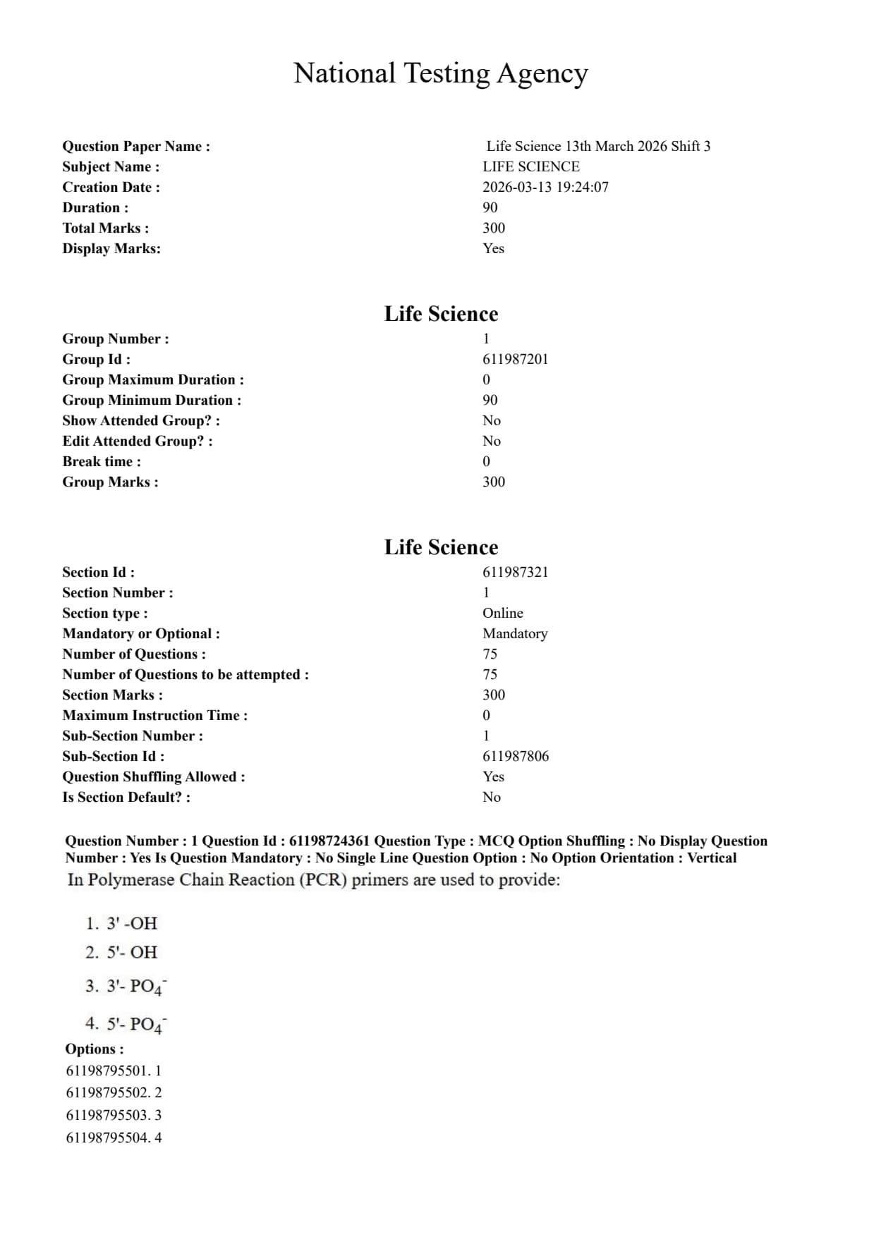
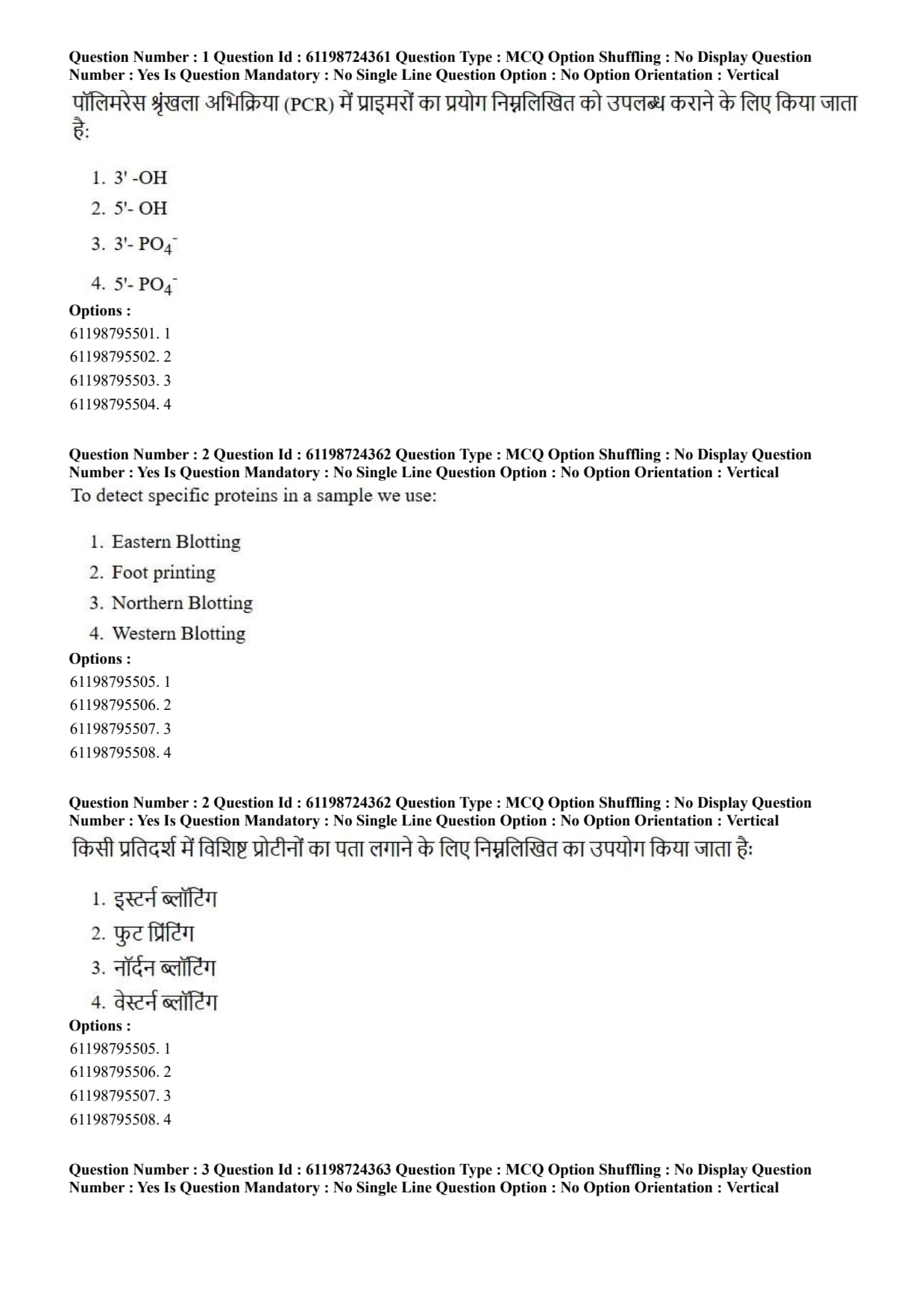
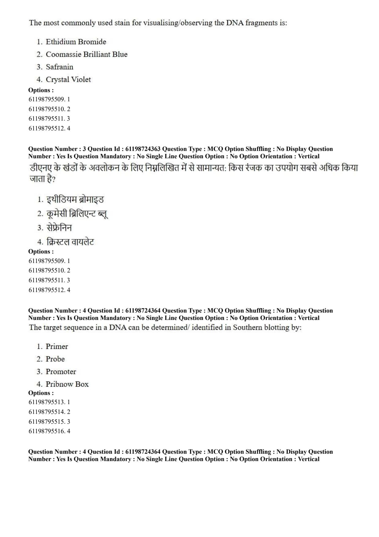
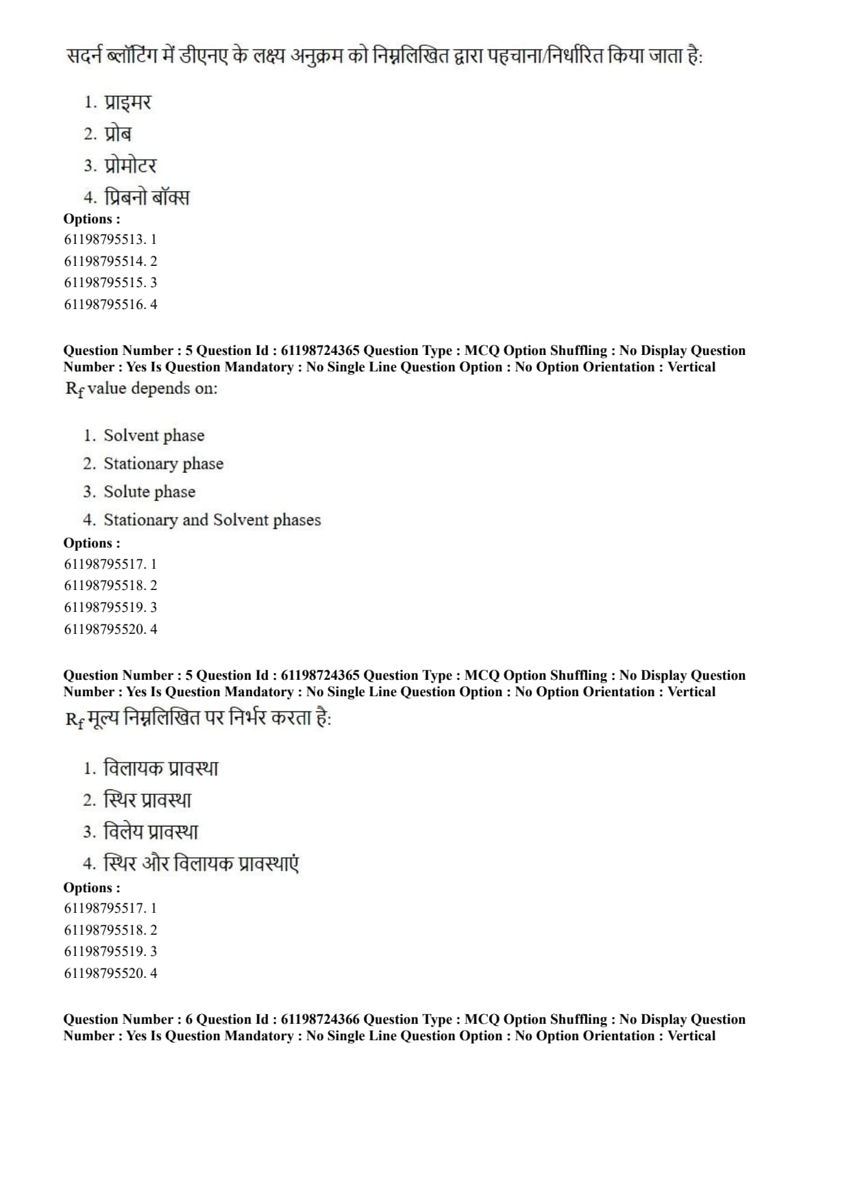
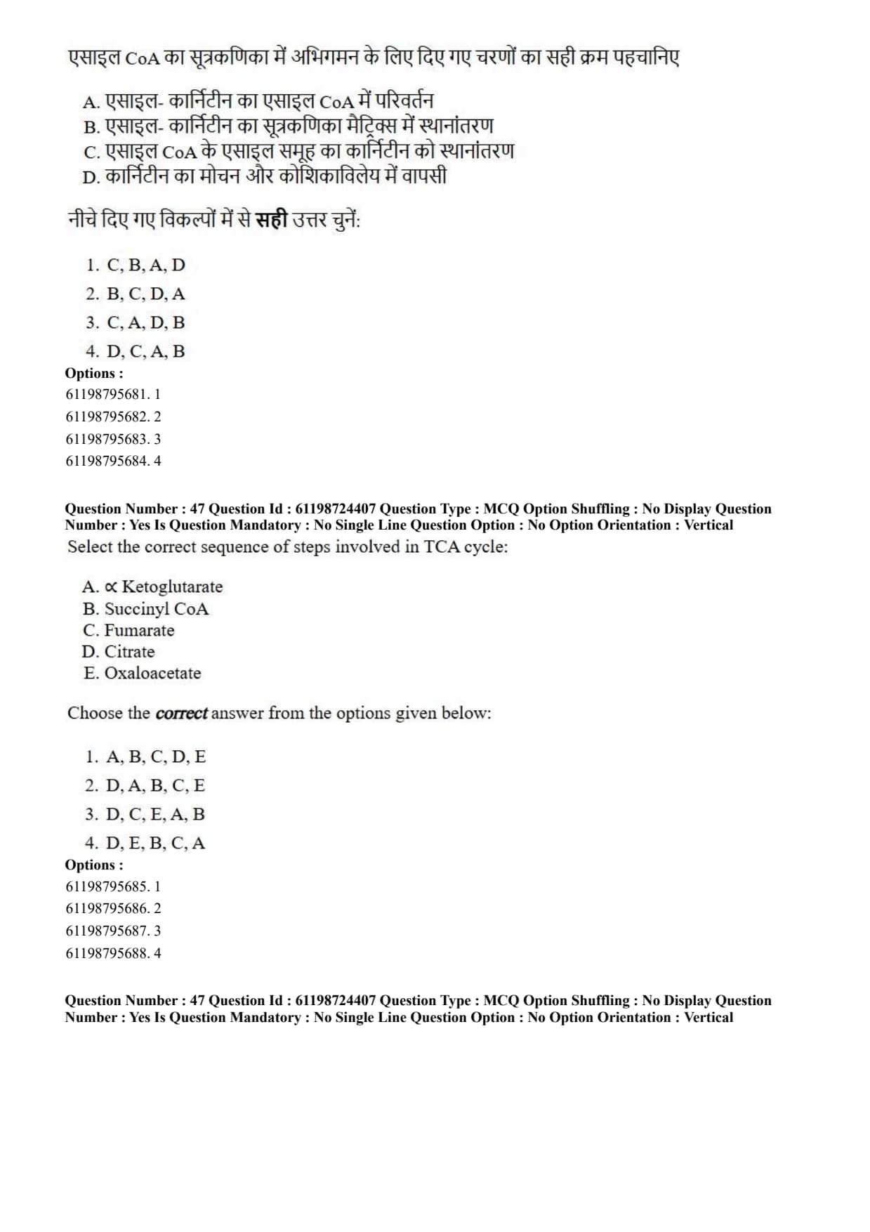
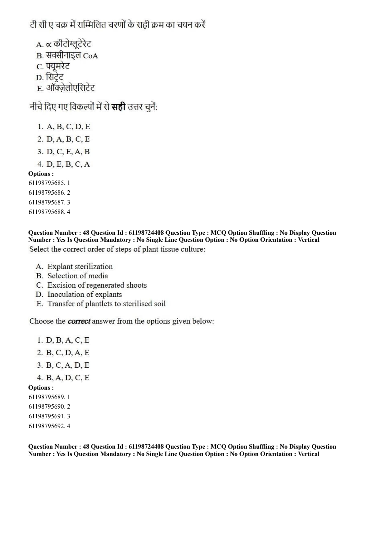
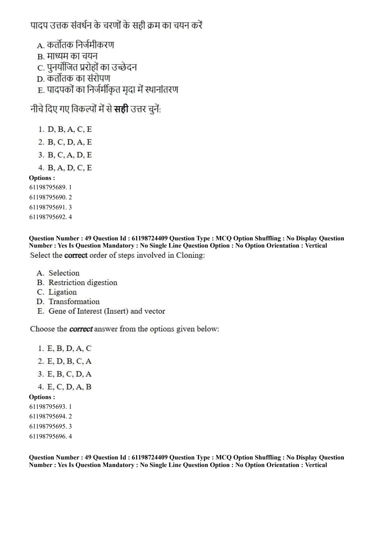
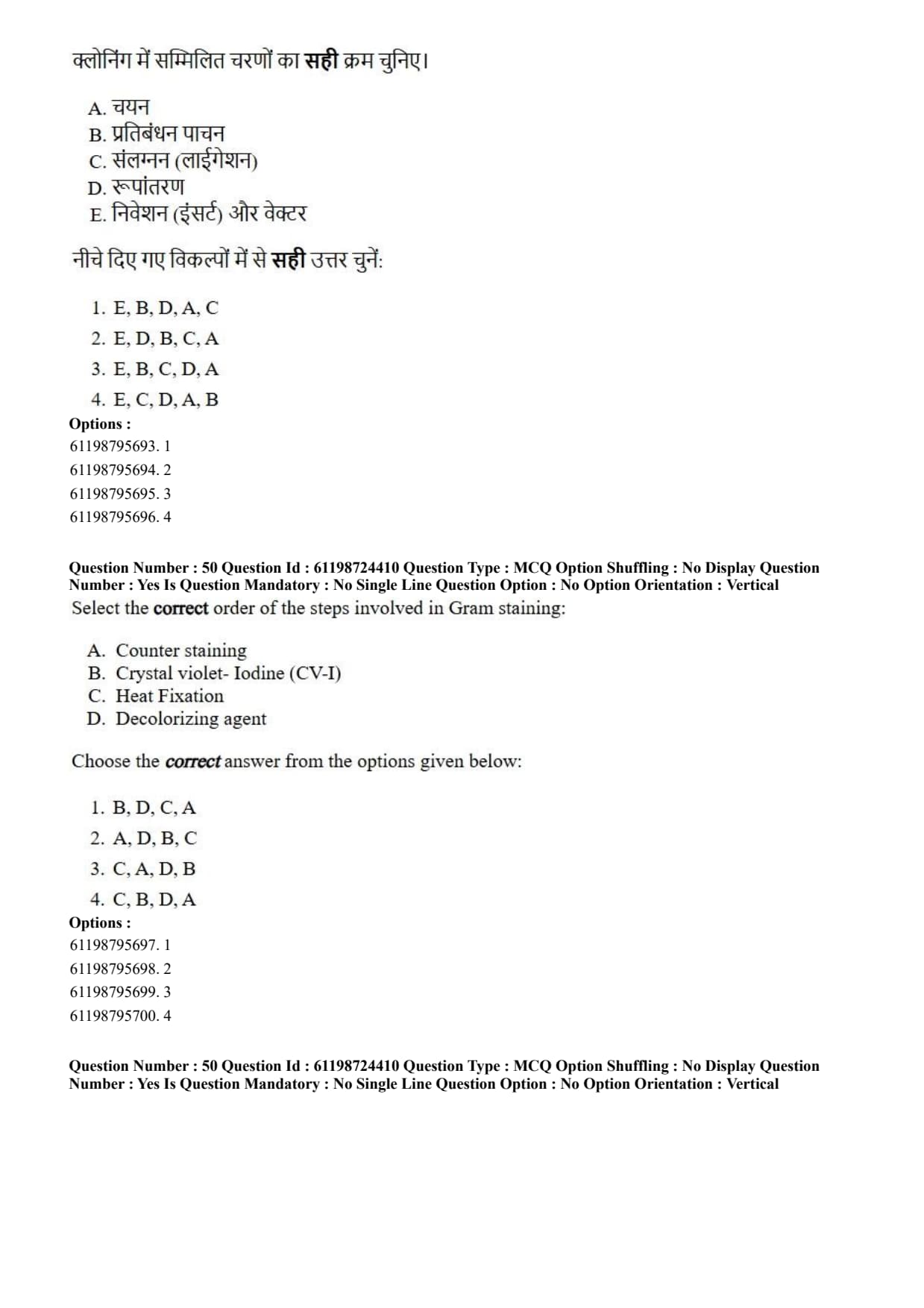
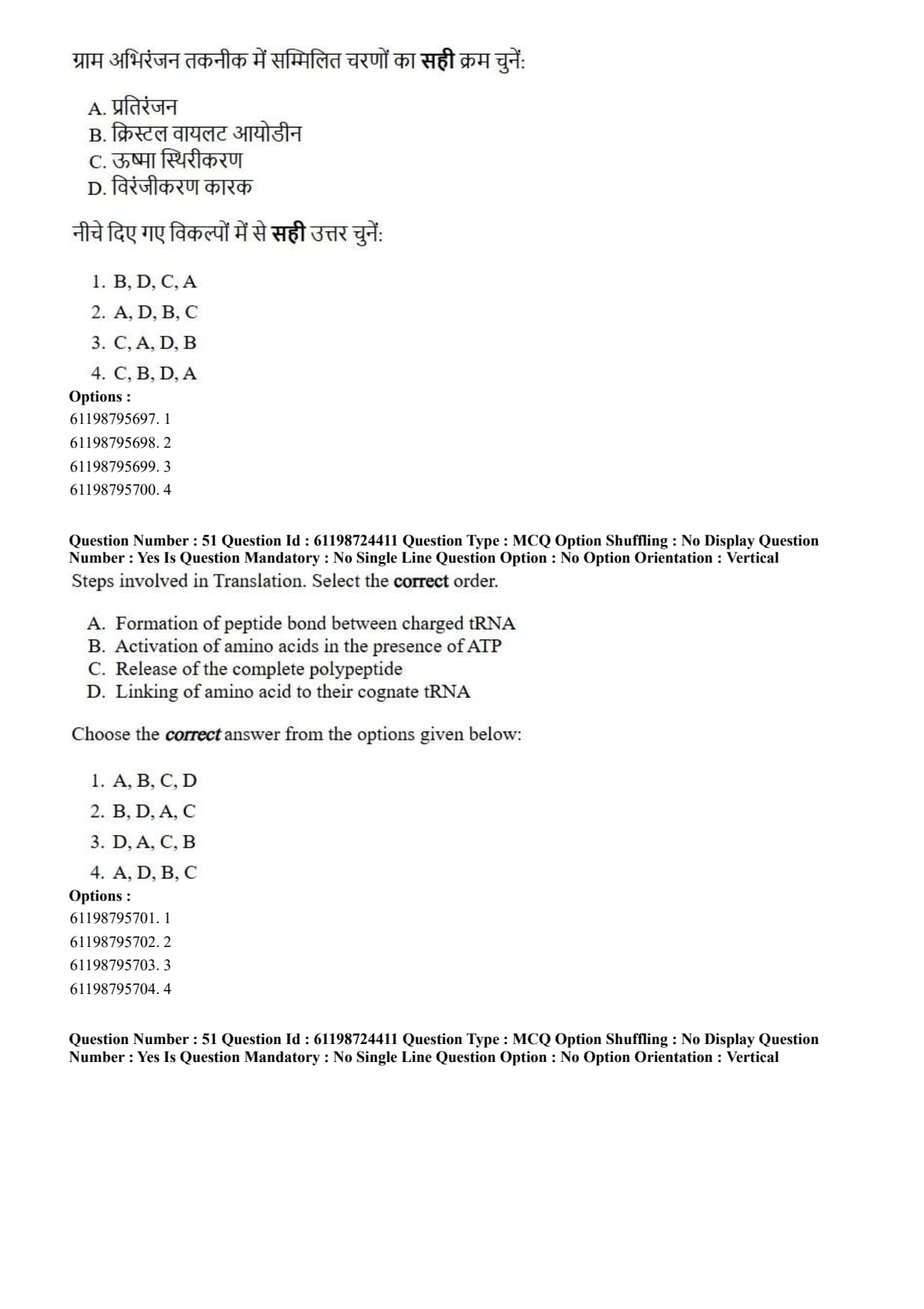
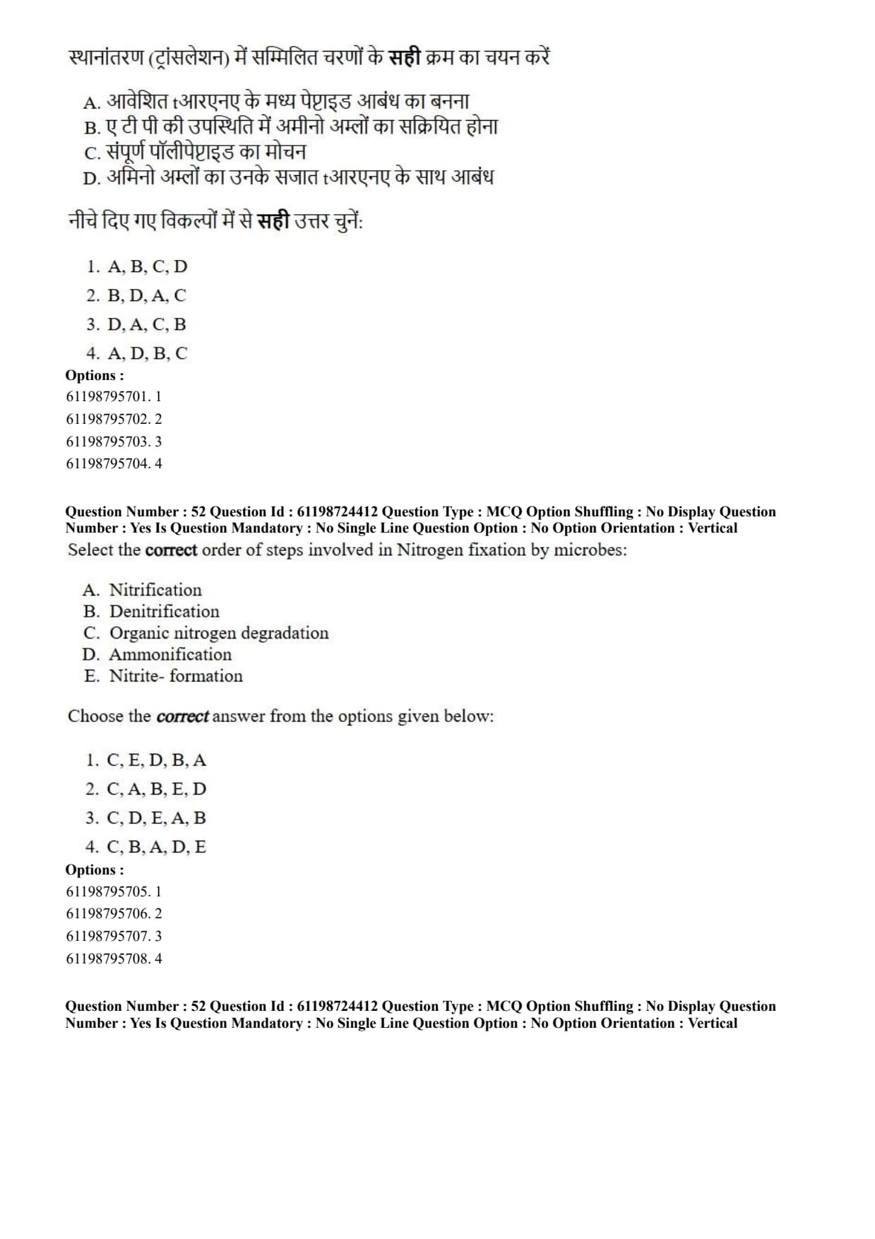
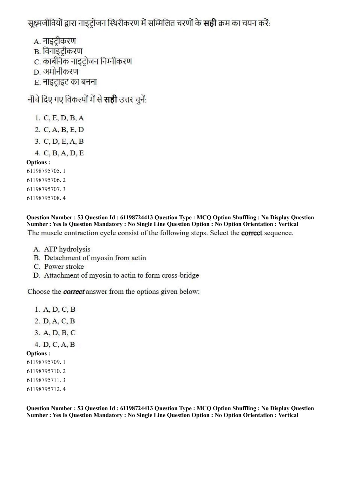
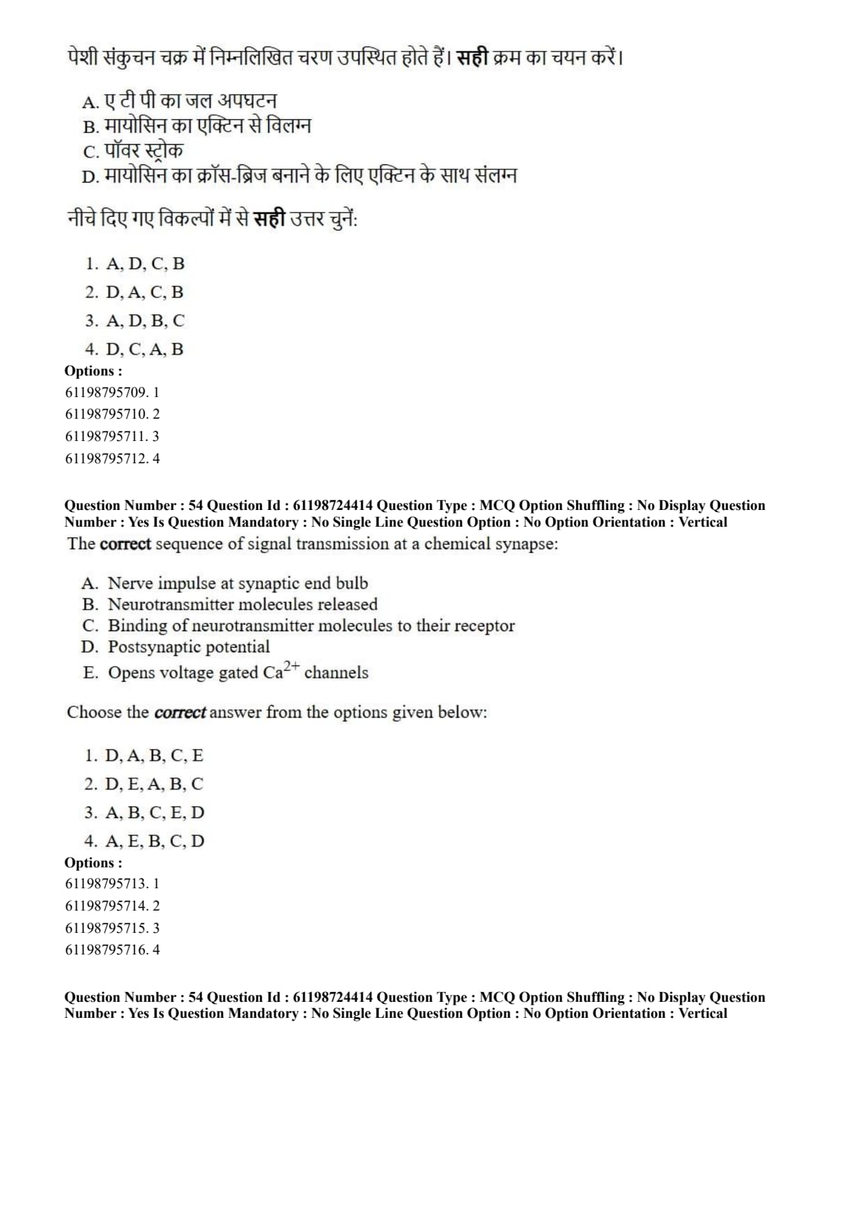
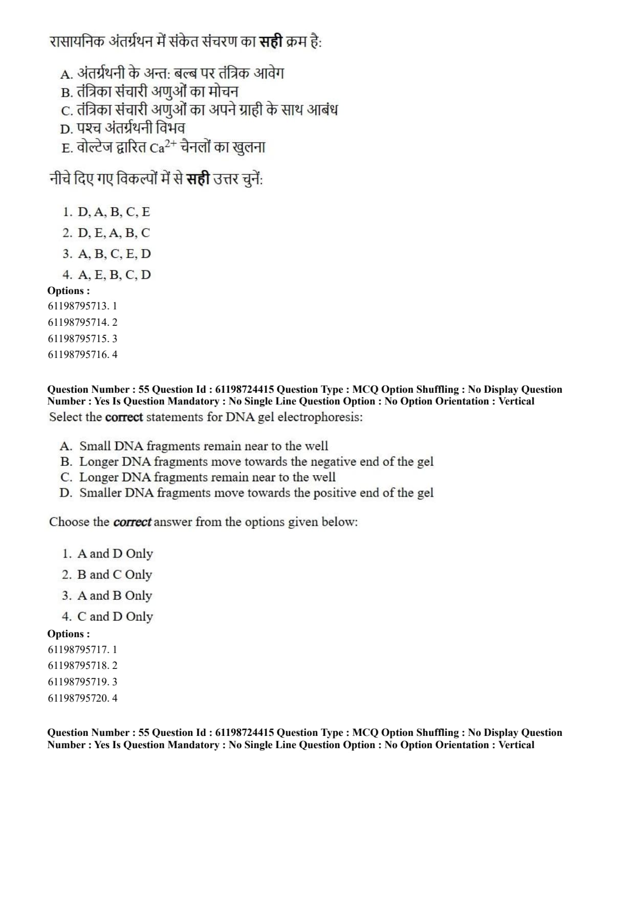
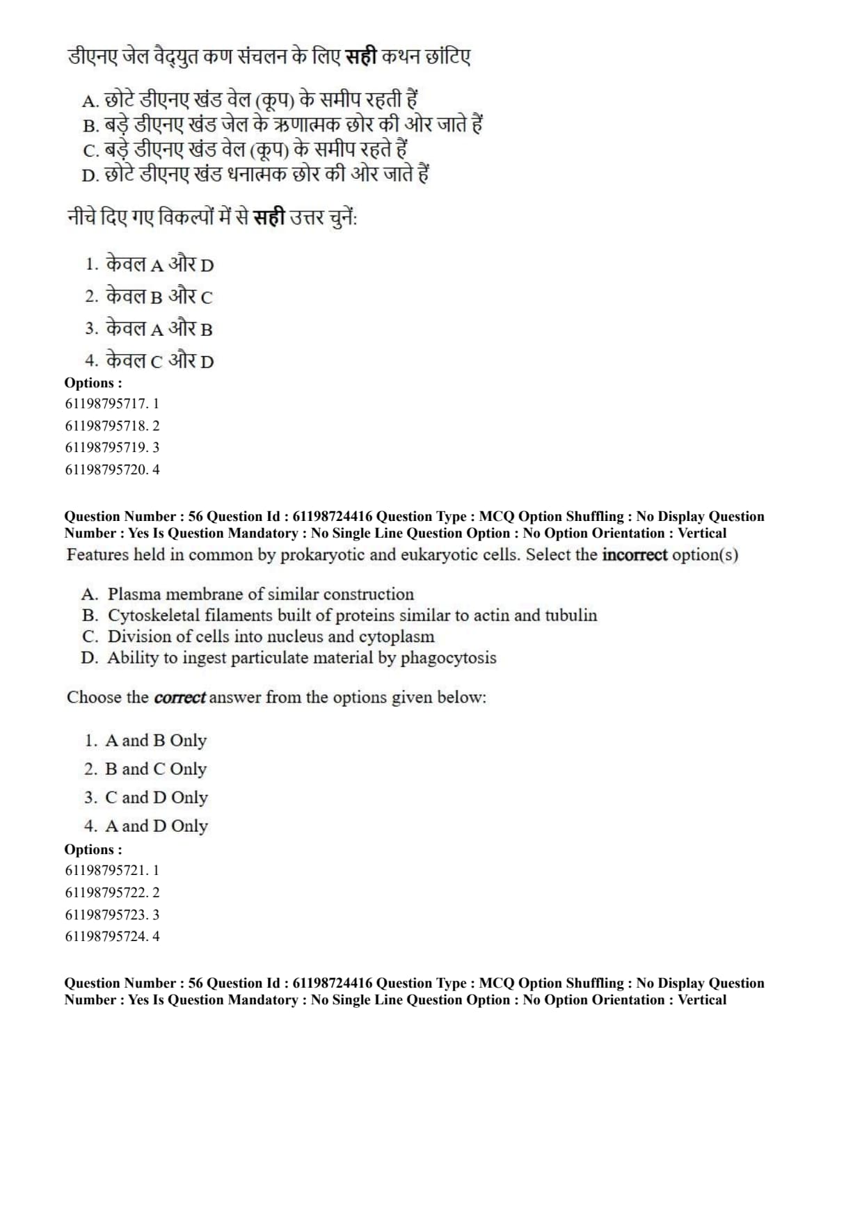
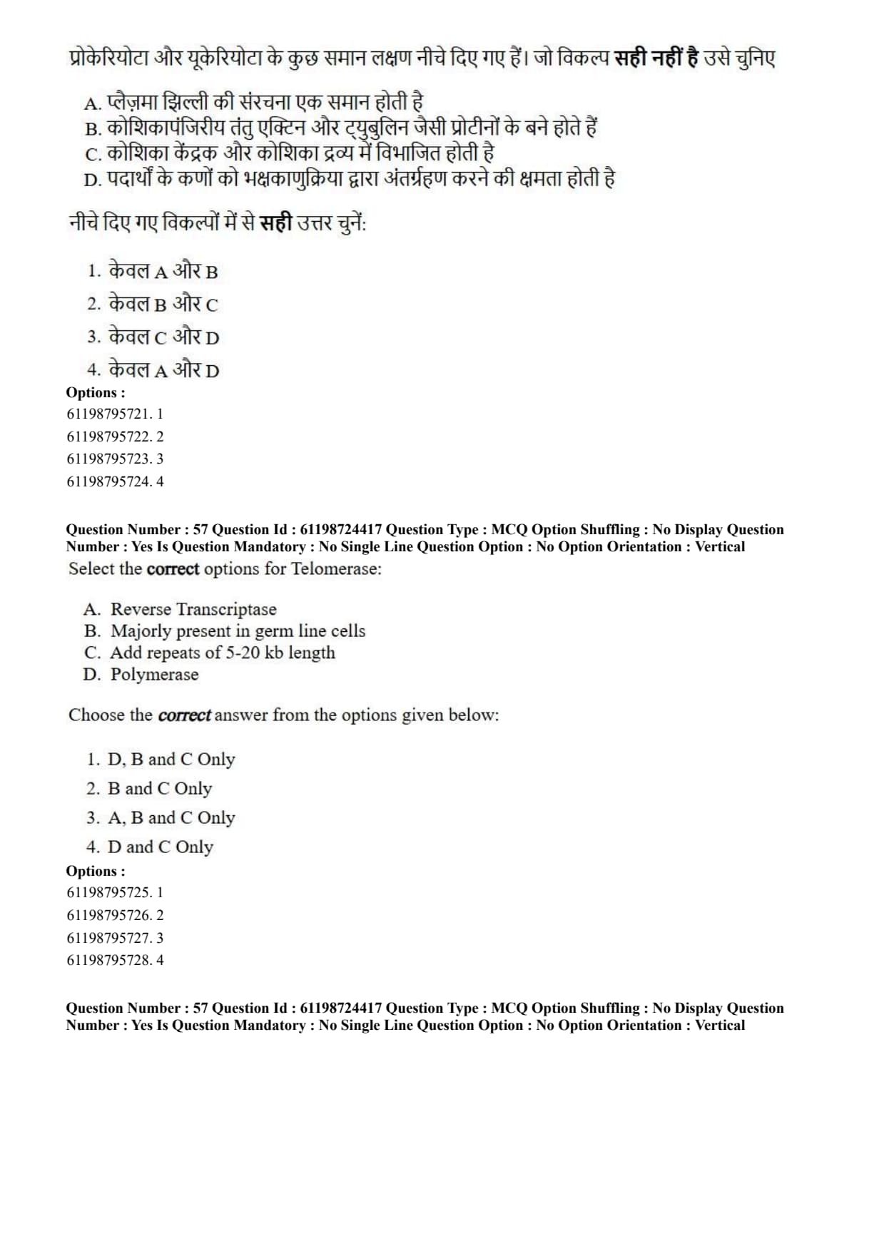
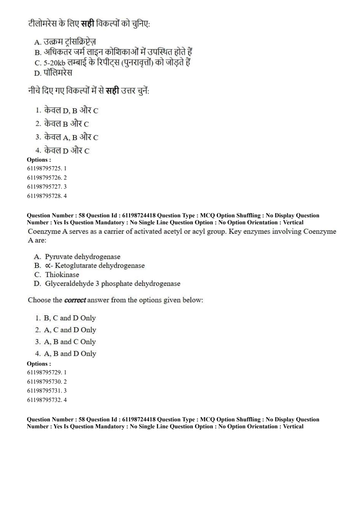
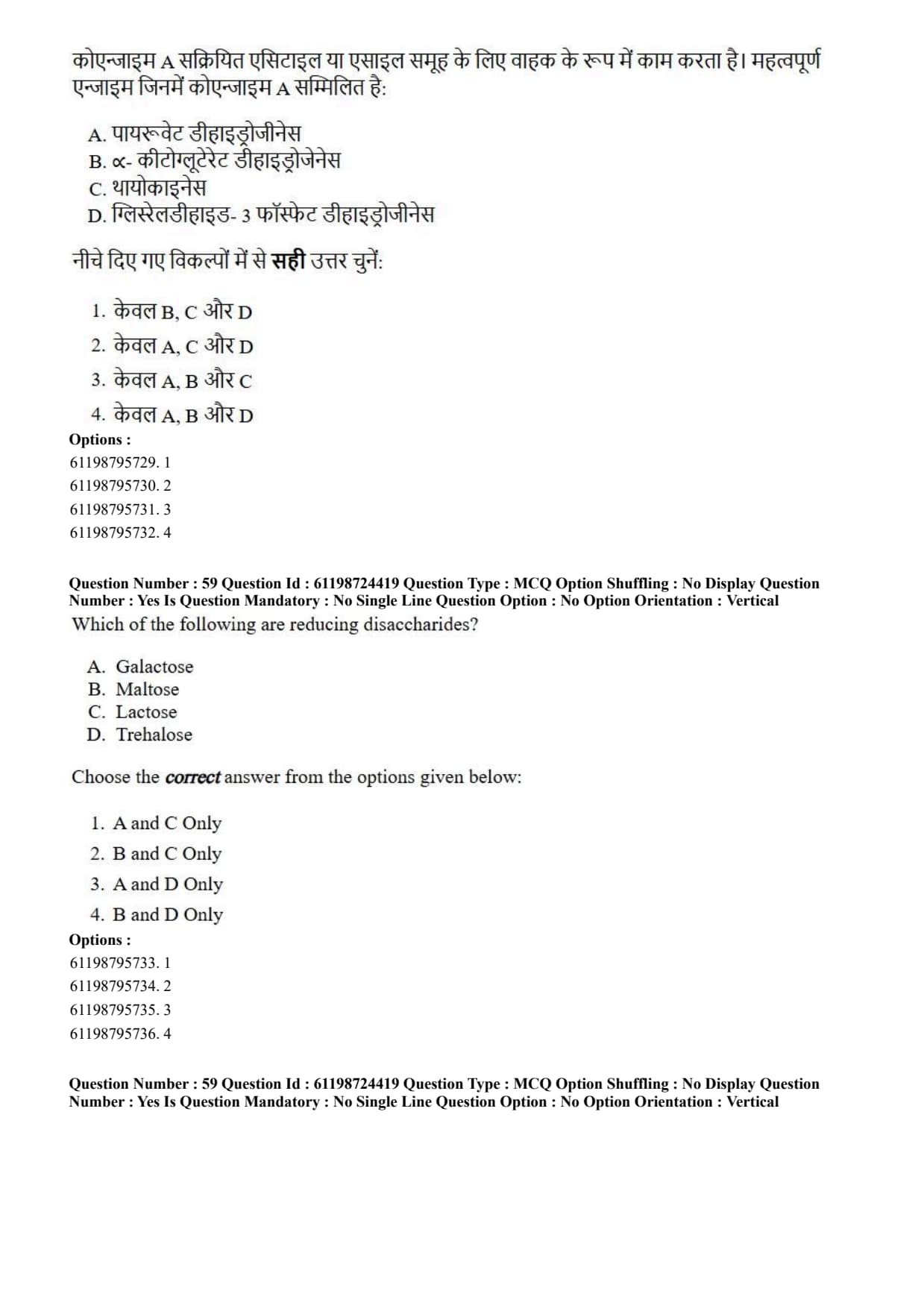
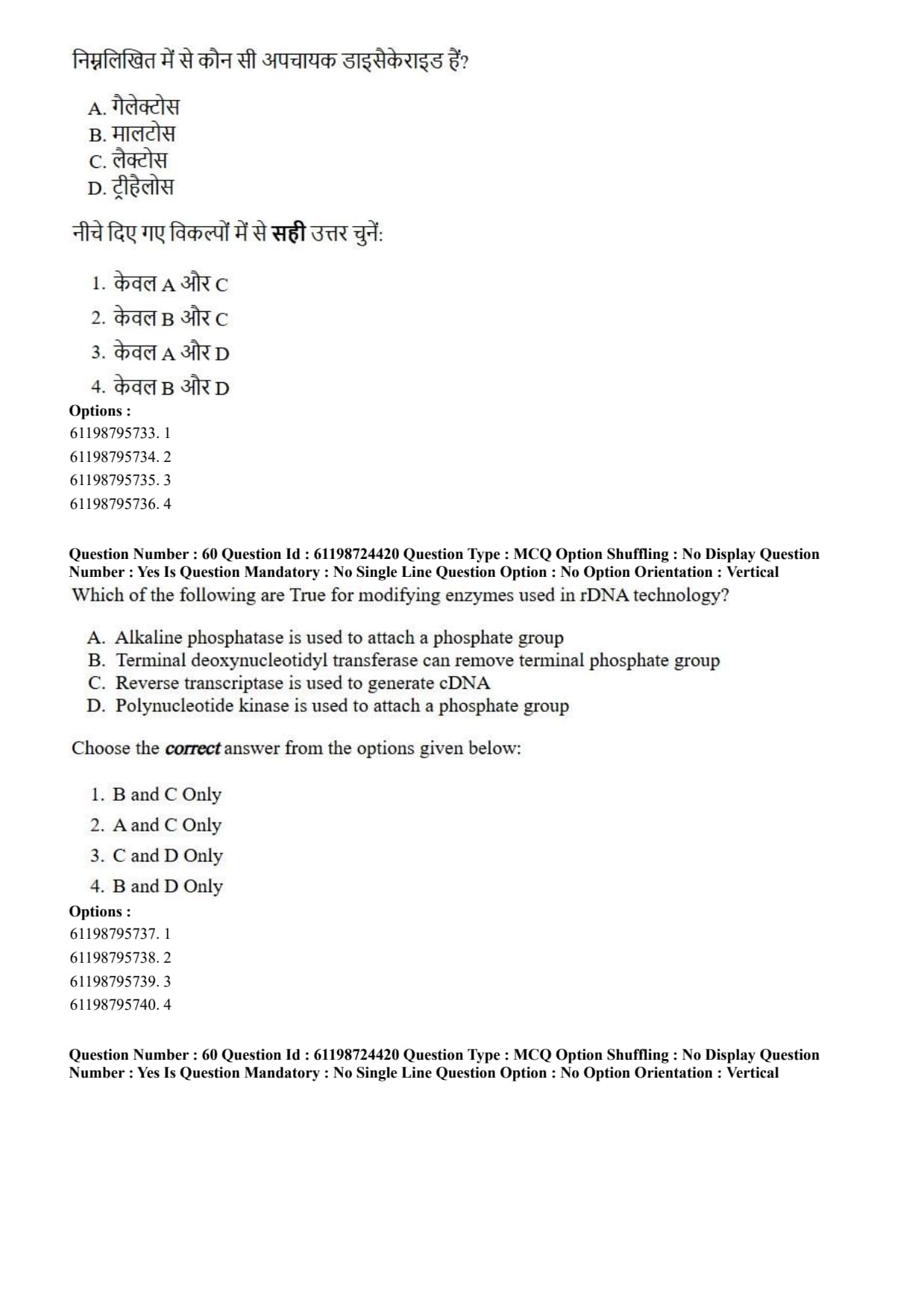
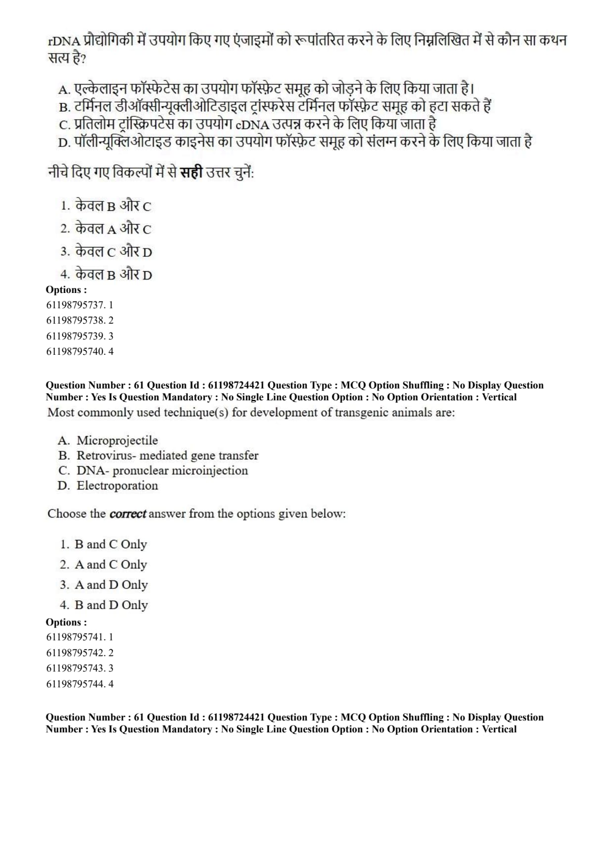
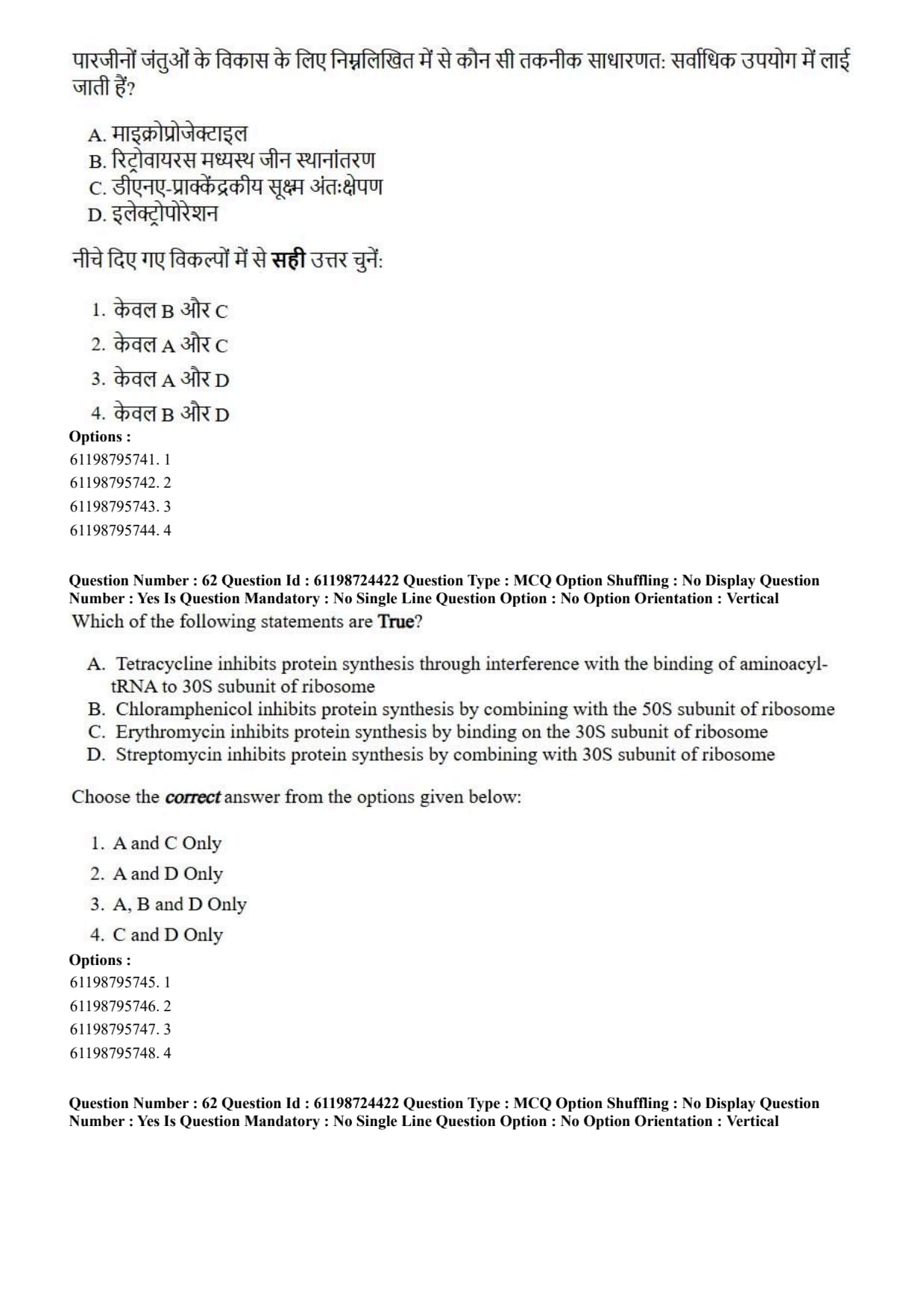
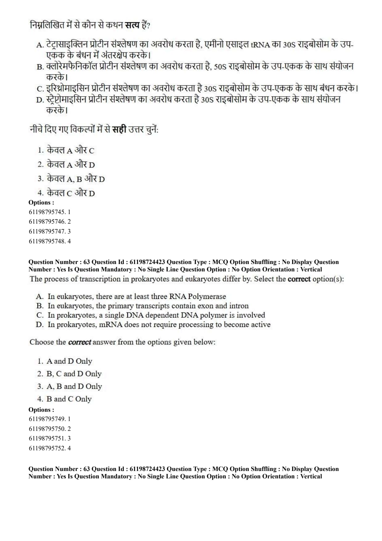
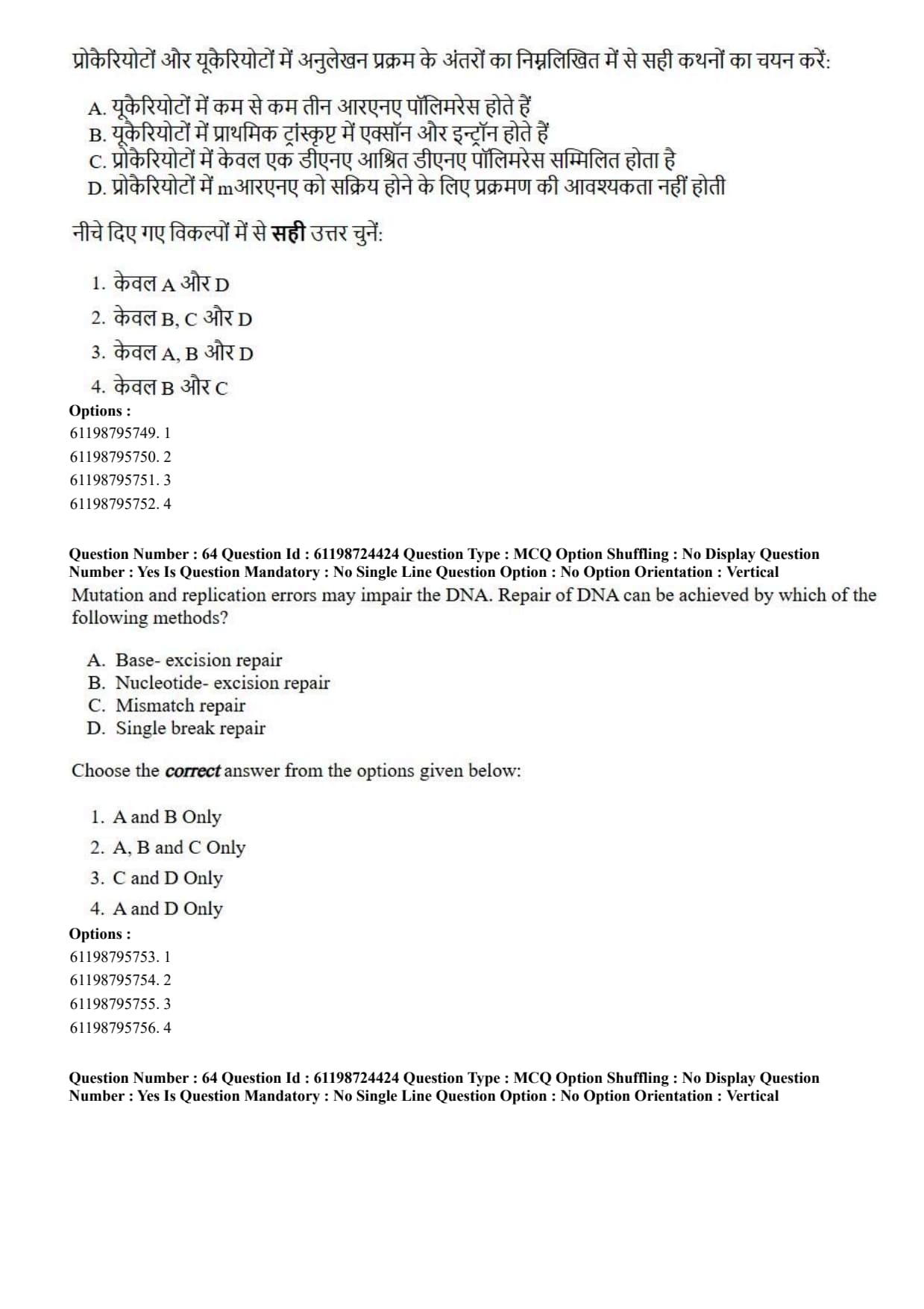
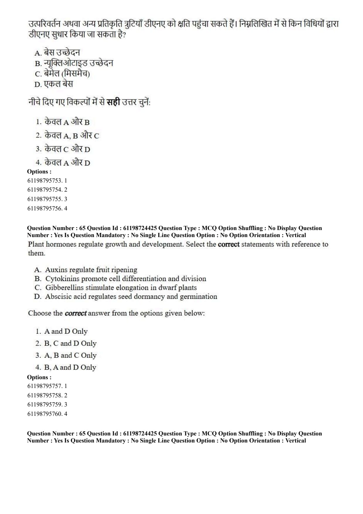
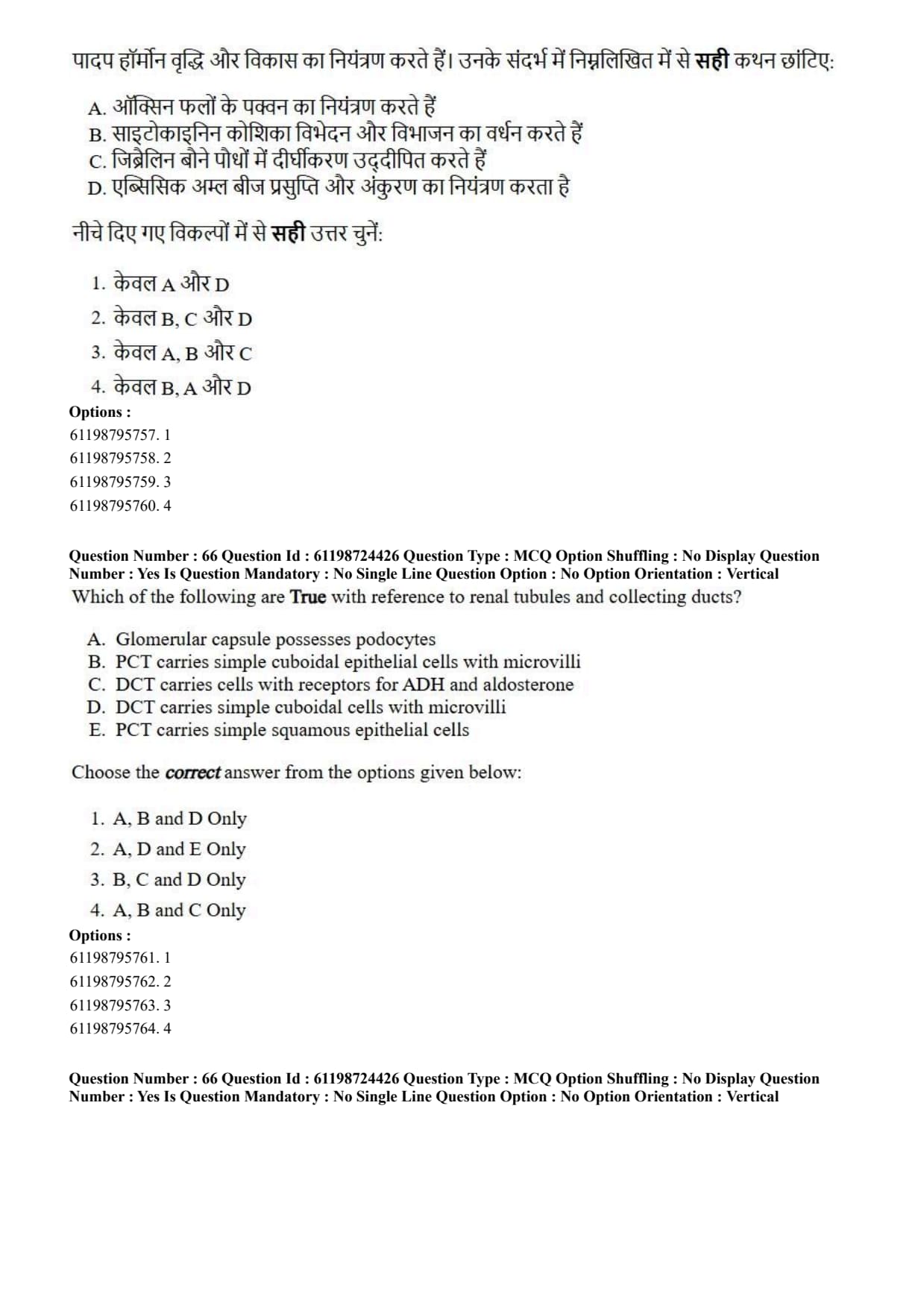
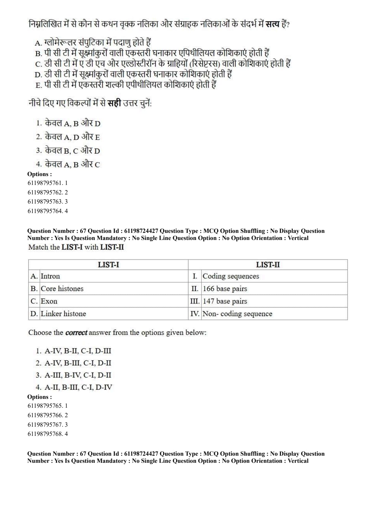
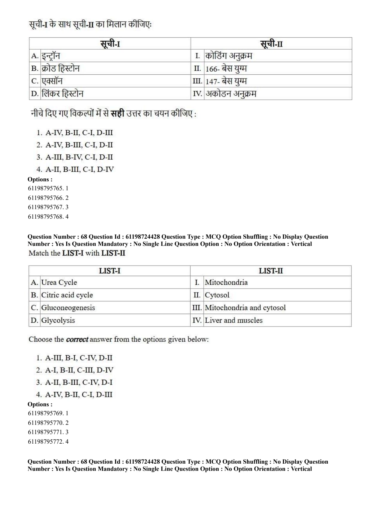
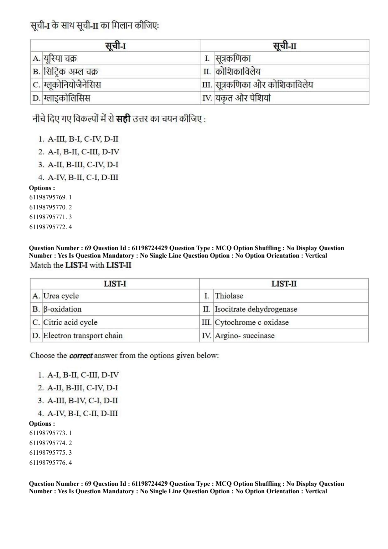
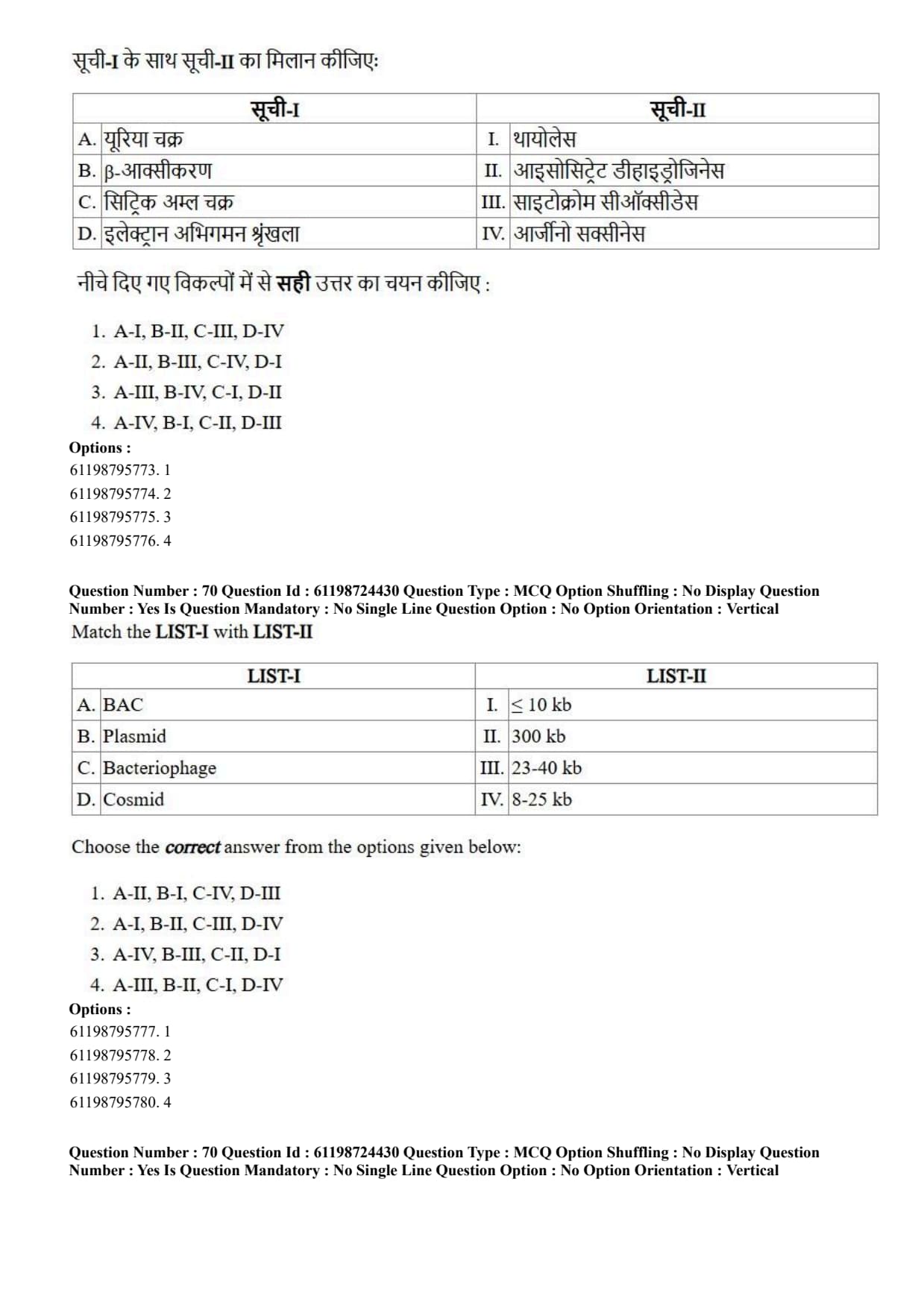
In Polymerase Chain Reaction (PCR) primers are used to provide:
View Solution
Step 1: Understanding the Question:
The question asks about the fundamental role of primers in the Polymerase Chain Reaction (PCR), specifically identifying which chemical group at the end of the primer allows DNA polymerase to attach new nucleotides.
Step 2: Key Formula or Approach:
All DNA polymerases synthesize new DNA strands exclusively in the \(5' \to 3'\) direction.
The catalytic core of DNA polymerase requires a free hydroxyl group (\(-OH\)) at the \(3'\) position of the deoxyribose sugar to perform a nucleophilic attack on the \(\alpha\)-phosphate of an incoming deoxyribonucleoside triphosphate (dNTP).
Step 3: Detailed Explanation:
Role of Primers in PCR: Primers are short, synthetic single-stranded oligonucleotides (usually 18 to 30 nucleotides long) designed to complement specific target sequences on template DNA.
Requirement for Polymerization: DNA polymerases cannot initiate DNA synthesis \(de\ novo\); they can only add nucleotides to an existing pre-formed polynucleotide strand.
Chemical Interaction: The \(3'\)-end of the hybridized primer presents a free \(3'\)-OH group. During strand extension, the oxygen atom of this \(3'\)-OH group carries out a nucleophilic attack on the \(\alpha\)-phosphorus atom of the incoming dNTP, forming a phosphodiester bond and releasing pyrophosphate (\(PP_i\)).
5' and Phosphate Groups: The \(5'\) end of synthetic primers typically possesses a \(5'\)-hydroxyl or \(5'\)-phosphate group, but this end is not involved in chain elongation. Therefore, \(3'\)-PO\(_4^-\) or \(5'\)-PO\(_4^-\) cannot serve as starting points for DNA extension.
Step 4: Final Answer:
Synthetic primers in PCR provide the necessary free \(3'\)-OH group required by DNA polymerase to extend the target DNA sequence.
Quick Tip: Remember: DNA elongation always proceeds from \(5'\) to \(3'\).
A free \(3'\)-OH group is indispensable for phosphodiester bond formation during DNA replication and PCR amplifications.
To detect specific proteins in a sample we use:
View Solution
Step 1: Understanding the Question:
The question asks for the standard laboratory blotting technique used specifically for identifying and detecting target protein molecules in a biological sample.
Step 2: Key Formula or Approach:
Blotting techniques separate biological macromolecules based on size via gel electrophoresis and transfer them to a membrane for detection using specific probes:
Southern Blotting \(\to\) DNA detection
Northern Blotting \(\to\) RNA detection
Western Blotting \(\to\) Protein detection
Step 3: Detailed Explanation:
Western Blotting Protocol: Proteins are first separated according to molecular weight using Sodium Dodecyl Sulfate-Polyacrylamide Gel Electrophoresis (SDS-PAGE).
Transfer to Membrane: The separated protein bands are electrophoretically transferred onto a carrier membrane, typically nitrocellulose or polyvinylidene difluoride (PVDF).
Immunodetection: The membrane is blocked to prevent non-specific binding and probed with a primary antibody specific to the target protein, followed by a secondary antibody conjugated to an enzyme (e.g., HRP or AP) or fluorophore for visualization.
Analysis of Other Options:
Eastern Blotting: Used to detect post-translational modifications of proteins such as carbohydrate or lipid moieties.
Footprinting (DNA Footprinting): Used to identify DNA sequences bound by specific DNA-binding proteins.
Northern Blotting: Used to analyze gene expression by detecting target RNA sequences.
Step 4: Final Answer:
Western blotting is the standard method used to detect and quantify target proteins within a protein mixture.
Quick Tip: Mnemonic to remember blotting techniques:
\textbf{S N O W}
\textbf{D R O P}
S = Southern (D = DNA), N = Northern (R = RNA), W = Western (P = Protein).
The most commonly used stain for visualising/observing the DNA fragments is:
View Solution
Step 1: Understanding the Question:
The question asks to identify the fluorescent chemical dye standardly used in gel electrophoresis to visualize nucleic acid fragments such as DNA.
Step 2: Key Formula or Approach:
Visualization of nucleic acids relies on intercalating agents that bind between base pairs and fluoresce brightly under ultraviolet (UV) illumination.
Step 3: Detailed Explanation:
Mechanism of Ethidium Bromide (EtBr): Ethidium bromide is a planar fluorescent molecule that intercalates non-covalently between adjacent nitrogenous base pairs of double-stranded DNA.
Fluorescence Enhancement: When exposed to UV light (wavelength \(\sim 300\ nm\)), EtBr absorbs UV radiation and emits bright orange-red fluorescence (\(\sim 590\ nm\)). Intercalation into DNA increases the fluorescence yield of EtBr nearly 20-fold compared to free dye in solution.
Evaluation of Other Options:
Coomassie Brilliant Blue: A dye commonly used for staining proteins in polyacrylamide gels (SDS-PAGE).
Safranin: A counterstain used in histology and Gram staining of bacteria.
Crystal Violet: Primary stain used in Gram staining to classify bacteria based on cell wall composition.
Step 4: Final Answer:
Ethidium bromide (EtBr) is the standard intercalating fluorescent dye used for detecting DNA fragments in agarose gels.
Quick Tip: EtBr intercalates between DNA base pairs and fluoresces orange under UV light.
Always handle EtBr with extreme care as it acts as a potent mutagen due to its intercalating mechanism.
The target sequence in a DNA can be determined/ identified in Southern blotting by:
View Solution
Step 1: Understanding the Question:
The question asks for the biological tool used in Southern blotting to specifically hybridize with and reveal the presence of a targeted DNA sequence.
Step 2: Key Formula or Approach:
Southern blotting combines agarose gel electrophoresis, transfer to a solid support membrane, and molecular hybridization using labeled single-stranded nucleic acid molecules known as probes.
Step 3: Detailed Explanation:
Definition of DNA Probe: A probe is a single-stranded sequence of DNA or RNA (typically 20 to 1000+ nucleotides long) that is complementary to the specific gene or DNA target sequence under investigation.
Labeling and Detection: Probes are tagged with radioactive isotopes (such as \(^{32}P\)) or fluorophores/enzymes (like digoxigenin or biotin). When applied to the membrane containing immobilized target single-stranded DNA, the probe selectively hybridizes to its complementary sequence via hydrogen bonding.
Visualization: Unbound probes are washed away, and the hybrid molecules are detected using autoradiography, chemiluminescence, or fluorescence imaging.
Roles of Incorrect Options:
Primer: Short oligonucleotides used to initiate enzymatic synthesis in PCR, not for direct hybridization detection on membranes.
Promoter: A DNA regulatory sequence where RNA polymerase binds to initiate transcription.
Pribnow Box: A specific consensus DNA sequence (TATAAT) found in prokaryotic promoter regions around \(-10\) position.
Step 4: Final Answer:
A labeled probe is the specific molecule used in Southern blotting to detect target DNA sequences through complementary base pairing.
Quick Tip: Probes are single-stranded nucleic acids labeled with fluorescent or radioactive tags designed to find complementary target sequences during blotting or in situ hybridization.
R\(_f\) value depends on:
View Solution
Step 1: Understanding the Question:
The question asks about the physical factors that dictate the retention factor (\(R_f\)) value in planar chromatographic techniques like thin-layer chromatography (TLC) or paper chromatography.
Step 2: Key Formula or Approach:
The retention factor (\(R_f\)) is defined mathematically as: \[ R_f = \frac{Distance traveled by the solute}{Distance traveled by the solvent front} \]
The differential migration rate of a solute depends directly on its relative affinity for the stationary phase versus the mobile phase (solvent phase).
Step 3: Detailed Explanation:
Role of the Stationary Phase: The stationary phase (e.g., silica gel, cellulose) interacts with the analyte through adsorption, partition, ion-exchange, or steric interactions. Stronger interactions slow down solute movement, yielding lower \(R_f\) values.
Role of the Solvent Phase (Mobile Phase): The mobile phase acts as a carrier fluid. The polarity and chemical composition of the solvent determine how efficiently it can solubilize and move the solute up the chromatographic medium.
Partition Equilibrium: \(R_f\) reflects a distribution constant (\(K_D\)) representing the chemical equilibrium of the solute between the stationary and mobile phases:
\[ K_D = \frac{Concentration of solute in stationary phase}{Concentration of solute in mobile phase} \]
Because both the stationary and solvent phases govern this equilibrium constant, altering either phase directly changes the resulting \(R_f\) value.
Step 4: Final Answer:
The \(R_f\) value depends fundamentally on both the stationary phase and the solvent (mobile) phase.
Quick Tip: \(R_f\) values range between \(0\) and \(1\).
Changing temperature, stationary medium, or solvent mixture will alter the \(R_f\) value for a given analyte.
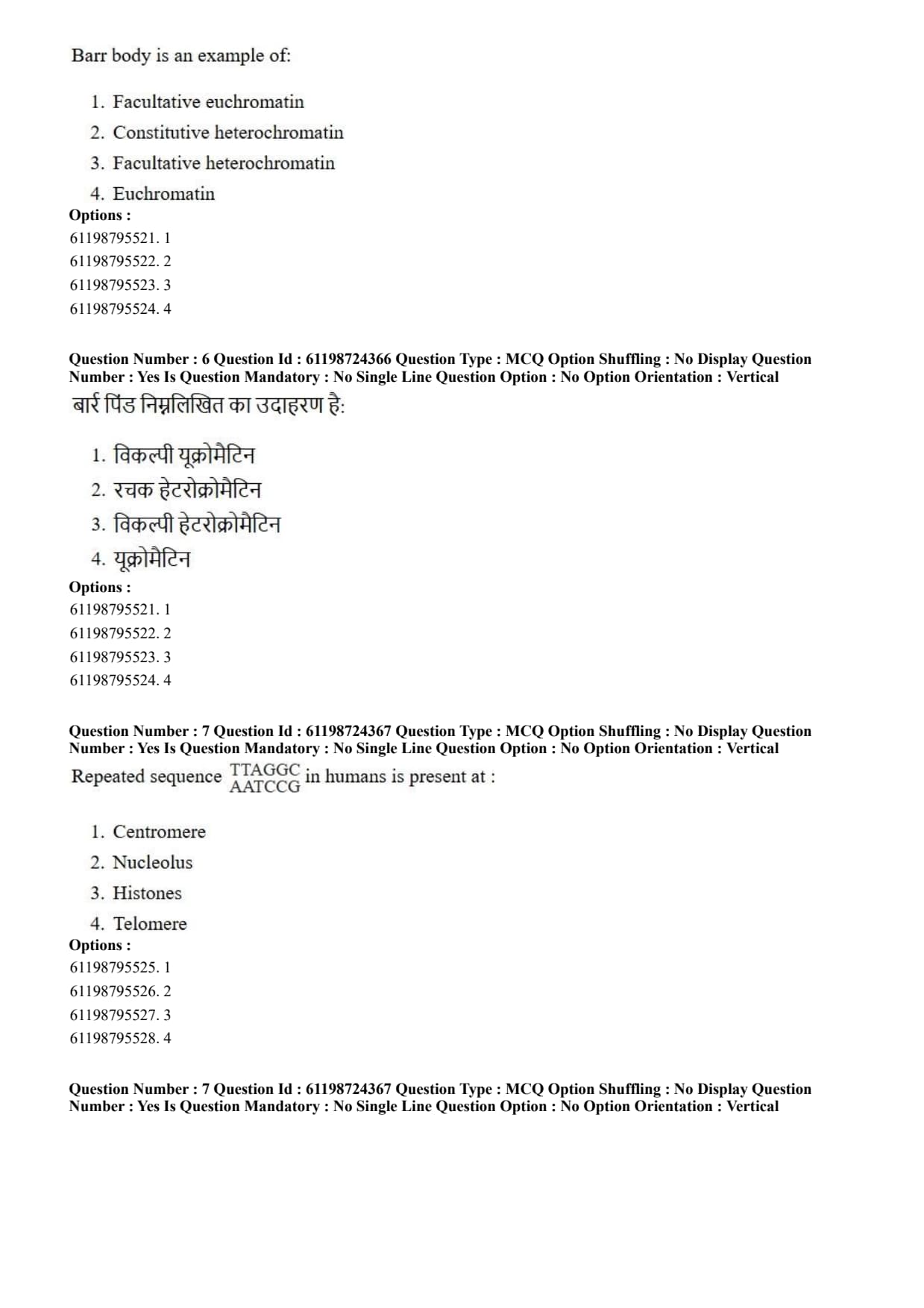
Barr body is an example of:
View Solution
Step 1: Understanding the Question:
The question asks to classify the chromatin state of a Barr body, which is formed during dosage compensation in female mammal somatic cells.
Step 2: Key Formula or Approach:
Chromatin is categorized based on structure and transcriptional activity:
Euchromatin: Uncondensed, transcriptionally active chromatin.
Constitutive Heterochromatin: Permanently condensed and genetically inactive across all cell types (e.g., centromeres and telomeres).
Facultative Heterochromatin: Chromatin that can switch between a condensed (inactive) state and a transcriptionally active state depending on cell development or metabolic context.
Step 3: Detailed Explanation:
Formation of a Barr Body: In female mammals (XX), one of the two X chromosomes is randomly inactivated early in embryonic development to equalize X-linked gene expression with males (XY). This process is known as Lyonization.
Epigenetic Silencing: The inactivated X chromosome (\(X_i\)) condenses into a dense heterochromatic structure called a Barr body, visible against the inner surface of the nuclear envelope during interphase.
Why Facultative? The \(X_i\) chromosome contains functional genes that were transcriptionally active prior to inactivation, and it can become active again in germline cells (oocytes). Because this condensation is reversible across generations and selective per cell, it represents facultative heterochromatin.
Step 4: Final Answer:
A Barr body is a classic biological example of facultative heterochromatin.
Quick Tip: Constitutive heterochromatin = Permanently condensed DNA (e.g., centromeres).
Facultative heterochromatin = Condensation is variable/reversible (e.g., Barr body / inactivated X-chromosome).
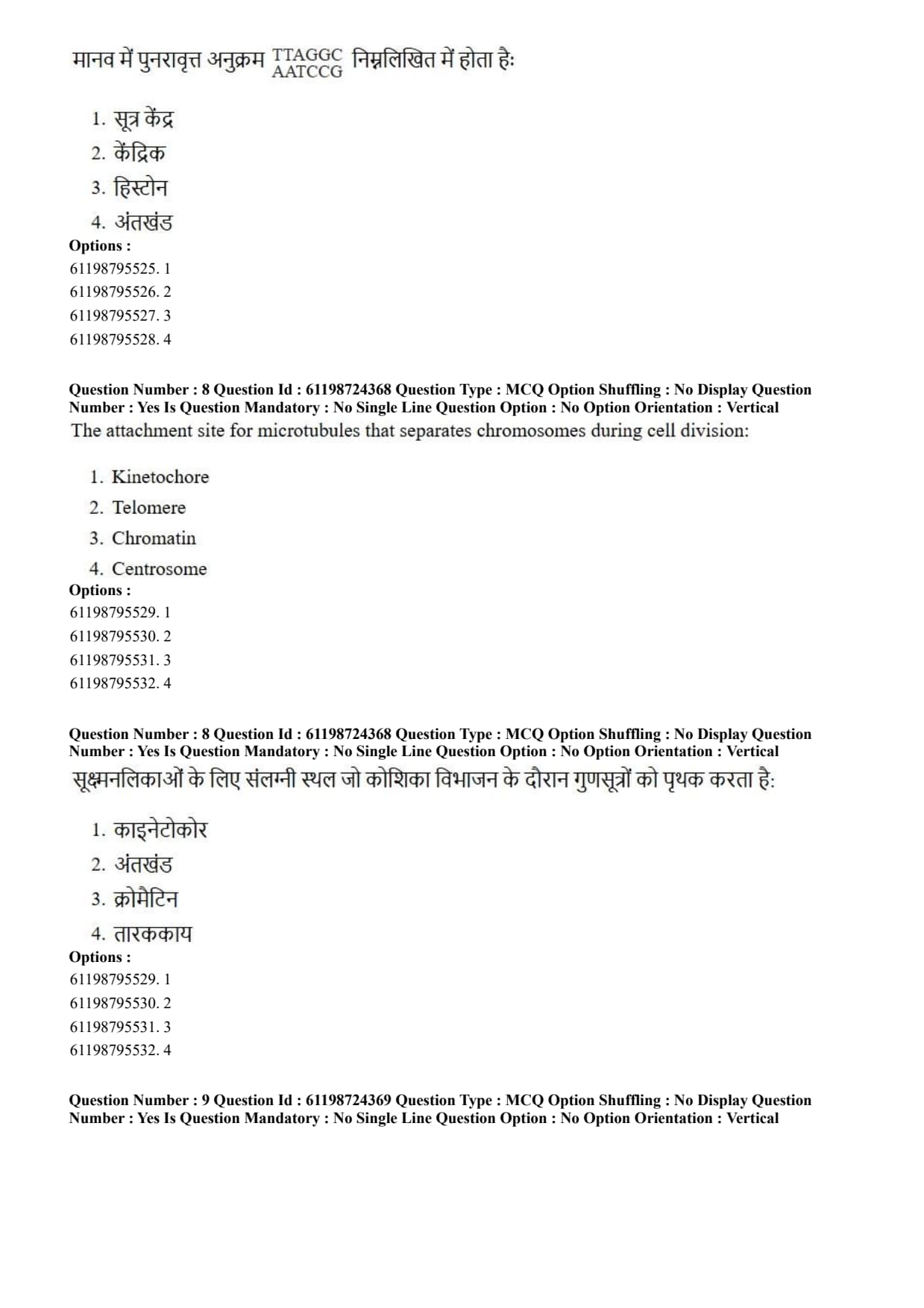
Repeated sequence TTAGGG / AATCCC in humans is present at:
View Solution
Step 1: Understanding the Question:
The question asks to locate the chromosomal region characterized by tandem repeats of the hexanucleotide sequence \(5'\)-TTAGGG-\(3'\) in human DNA.
Step 2: Key Formula or Approach:
Linear chromosome ends require specialized nucleoprotein complexes consisting of short repetitive DNA sequences to protect genomic material from degradation and end-to-end chromosomal fusion.
Step 3: Detailed Explanation:
Structure of Telomeres: Telomeres are specialized nucleoprotein structures situated at the physical termini of eukaryotic linear chromosomes.
Human Telomeric Sequence: Human telomeres consist of hundreds to thousands of tandem repeats of the hexamer motif \(5'\)-TTAGGG-\(3'\) on one strand paired with its complementary strand \(3'\)-AATCCC-\(5'\).
Function: Telomeres resolve the "end-replication problem" caused by the inability of DNA polymerase to fully replicate the extreme \(5'\) end of linear lagging strands.
Telomerase Enzyme: The ribonucleoprotein enzyme telomerase extends these hexameric sequences using an internal RNA template.
Comparison with Other Structural Regions:
Centromere: Contains satellite DNA (e.g., alpha satellite repeats) responsible for kinetochore assembly.
Nucleolus: Nuclear domain surrounding ribosomal RNA (rRNA) gene repeats.
Histones: Basic proteins involved in packaging DNA into nucleosomes, not specific repetitive DNA regions.
Step 4: Final Answer:
The repeating tandem sequence \(5'\)-TTAGGG-\(3'\) is localized specifically at human telomeres.
Quick Tip: Human telomere repeat motif: \(5'\)-TTAGGG-\(3'\).
Telomeres shorten with successive somatic cell divisions, acting as a biological clock for cell aging.
The attachment site for microtubules that separates chromosomes during cell division:
View Solution
Step 1: Understanding the Question:
The question asks to identify the specific protein structure assembled on chromosome centromeres that directly binds spindle microtubules to orchestrate chromosome movement during mitosis and meiosis.
Step 2: Key Formula or Approach:
During cell division, spindle fibers attach to chromosomes to generate mechanical forces necessary for sister chromatid separation. The macromolecular assembly mediating this binding is the kinetochore.
Step 3: Detailed Explanation:
Kinetochore Assembly: The kinetochore is a complex, multiprotein disk-like structure built directly upon centromeric heterochromatin.
Microtubule Binding: It serves as the physical interface connecting spindle microtubules (kinetochore microtubules) to the chromosome.
Force Generation: During metaphase and anaphase, kinetochores interact with motor proteins (such as dynein and kinesin) to regulate chromosome alignment along the metaphase plate and pull separated chromatids toward opposite spindle poles.
Distinction from Other Options:
Telomere: Protects chromosome ends from degradation.
Chromatin: The complex of genomic DNA and histone proteins composing chromosomes.
Centrosome: The main microtubule-organizing center (MTOC) located at the cell poles from which spindle fibers originate, rather than the attachment point on chromosomes.
Step 4: Final Answer:
The kinetochore is the specialized protein complex that attaches spindle microtubules to chromosomes during cell division.
Quick Tip: Centromere = The region of DNA on a chromosome.
Kinetochore = The protein structure that forms ON the centromere to bind microtubules.
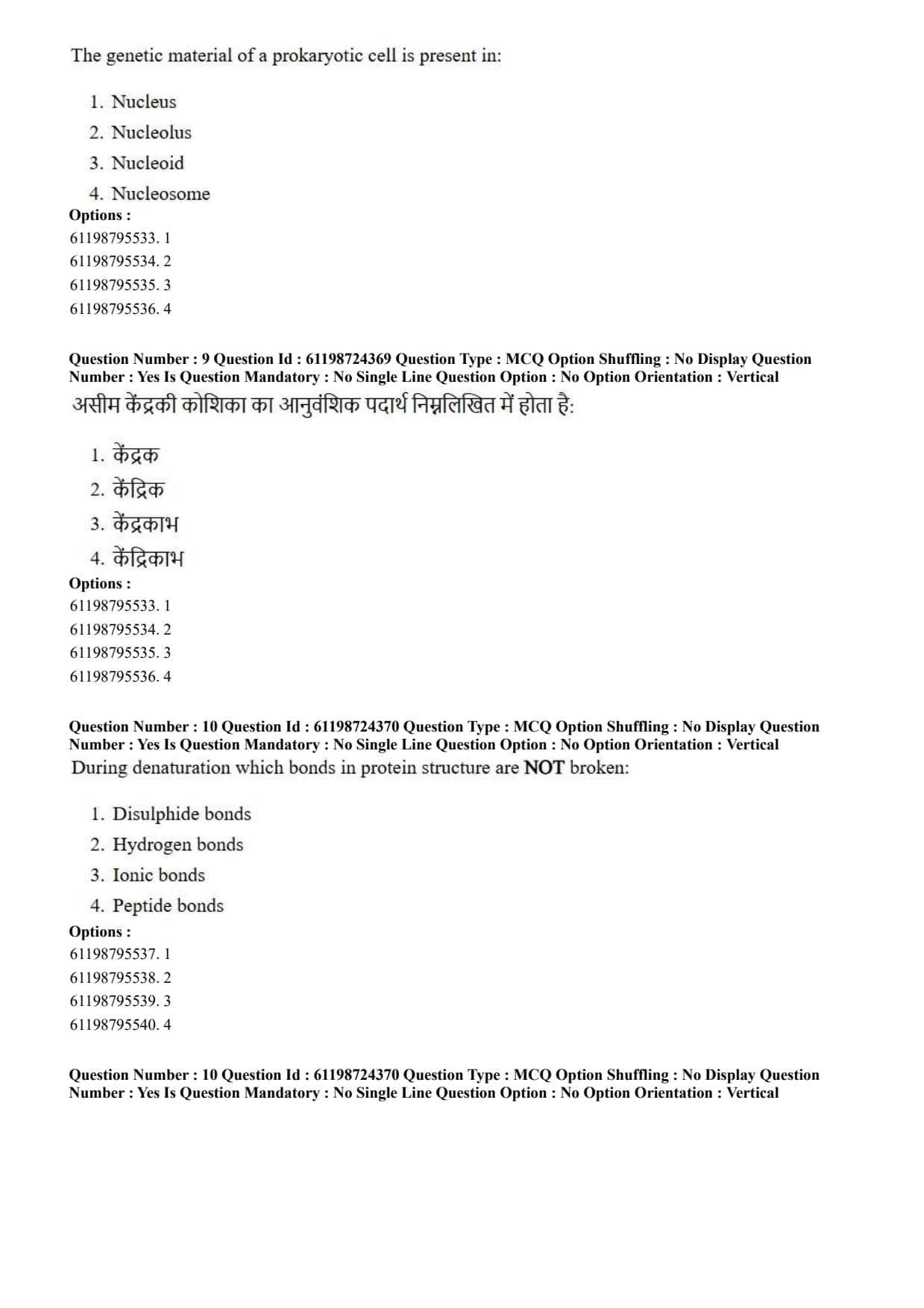
The genetic material of a prokaryotic cell is present in:
View Solution
Step 1: Understanding the Question:
The question asks for the cellular region where genomic DNA is localized within a prokaryotic cell (such as bacteria and archaea).
Step 2: Key Formula or Approach:
Prokaryotes lack internal membrane-bound organelles, including a true membrane-bound nucleus. Their genetic material resides within an irregularly shaped cytoplasmic region known as the nucleoid.
Step 3: Detailed Explanation:
Characteristics of the Nucleoid: The nucleoid is an defined, non-membrane-bound cytoplasmic region that houses the circular double-stranded genomic DNA of prokaryotes.
Prokaryotic DNA Organization: Bacterial DNA is localized in the nucleoid via supercoiling and interactions with nucleoid-associated proteins (NAPs), such as HU and H-NS proteins.
Analysis of Other Structures:
Nucleus: A double-membrane bounded organelle encapsulating genetic material present exclusively in eukaryotic cells.
Nucleolus: A dense region inside the eukaryotic nucleus responsible for ribosome biogenesis.
Nucleosome: The fundamental structural unit of eukaryotic chromatin, consisting of DNA wrapped around an octamer of histone proteins.
Step 4: Final Answer:
In prokaryotic cells, the genetic material is located in the nucleoid region.
Quick Tip: Prokaryotes = No true membrane-bound nucleus; genomic DNA sits directly in the cytoplasm within the \textbf{nucleoid}.
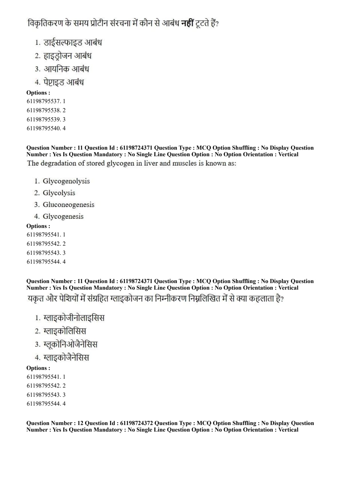
During denaturation which bonds in protein structure are NOT broken:
View Solution
Step 1: Understanding the Question:
The question asks to identify which chemical bonds remain intact during protein denaturation.
Step 2: Key Formula or Approach:
Protein structure is organized into four levels:
Primary structure: Sequence of amino acids linked covalently by peptide bonds.
Secondary, Tertiary, Quaternary structures: Maintained by non-covalent interactions (hydrogen bonds, ionic bonds, hydrophobic interactions) and covalent disulfide bridges.
Denaturation disrupts higher-order structures without breaking primary covalent backbone bonds.
Step 3: Detailed Explanation:
Definition of Denaturation: Denaturation refers to the unfolding of a protein's native three-dimensional tertiary or secondary structure under stress conditions such as high heat, extreme pH, organic solvents, or chaotropic agents (e.g., urea, guanidinium chloride).
Bonds Affected: Denaturation disrupts weak non-covalent interactions (hydrogen bonds, ionic interactions/salt bridges, van der Waals forces) and can reduce disulfide linkages if reducing agents are present.
Preservation of Peptide Bonds: The covalent peptide bonds (amide linkages) forming the primary backbone of the polypeptide chain are robust and require enzymatic cleavage or harsh chemical acid/base hydrolysis to break. Thus, primary structure remains completely intact after denaturation.
Step 4: Final Answer:
Peptide bonds holding together the primary amino acid sequence are NOT broken during protein denaturation.
Quick Tip: Denaturation affects higher-order structures (\(2^\circ, 3^\circ, 4^\circ\)) while keeping the primary structure (\(1^\circ\) peptide bonds) intact.
Proteolysis is required to break peptide bonds, which is distinct from denaturation.
The degradation of stored glycogen in liver and muscles is known as:
View Solution
Step 1: Understanding the Question:
The question asks for the specific biochemical pathway name corresponding to the breakdown of stored glycogen into glucose units in animal tissues.
Step 2: Key Formula or Approach:
Reviewing metabolic terminology for carbohydrate pathways:
Glycogen breakdown \(\to\) Glycogenolysis
Glycogen synthesis \(\to\) Glycogenesis
Glucose oxidation to pyruvate \(\to\) Glycolysis
Synthesis of glucose from non-carbohydrate precursors \(\to\) Gluconeogenesis
Step 3: Detailed Explanation:
Mechanism of Glycogenolysis: Glycogenolysis is the metabolic breakdown of intracellular glycogen reserves into glucose-1-phosphate (G1P) and free glucose.
Key Enzymes Involved:
Glycogen Phosphorylase: Cleaves \(\alpha(1 \to 4)\) glycosidic linkage from non-reducing ends via phosphorolysis to produce glucose-1-phosphate.
Debranching Enzyme: Handles \(\alpha(1 \to 6)\) glycosidic branch points.
Tissue Differences:
Liver: Expresses glucose-6-phosphatase, allowing converted glucose-6-phosphate to yield free glucose released into the bloodstream to maintain glycemia.
Skeletal Muscle: Lacks glucose-6-phosphatase; glucose-6-phosphate enters glycolysis directly to generate ATP for muscle contraction.
Step 4: Final Answer:
The degradation of stored glycogen is termed glycogenolysis.
Quick Tip: Break down the word root:
\textbf{Glycogen} + \textbf{lysis} (breakdown) = Glycogenolysis.
\textbf{Glyco} + \textbf{genesis} (creation) = Glycogenesis.
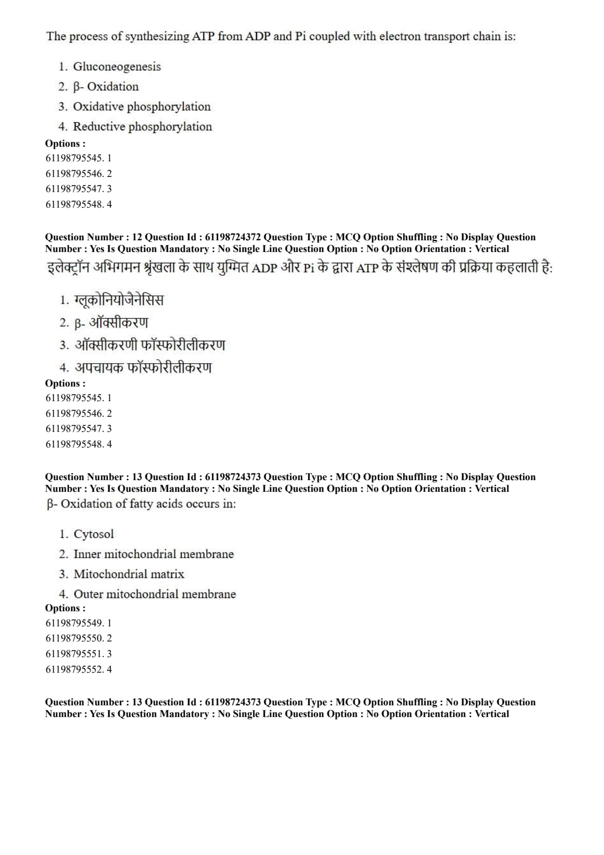
The process of synthesizing ATP from ADP and Pi coupled with electron transport chain is:
View Solution
Step 1: Understanding the Question:
The question asks for the name of the metabolic pathway in which ATP formation from ADP and inorganic phosphate (\(P_i\)) is driven by electron transfer along the mitochondrial respiratory chain.
Step 2: Key Formula or Approach:
Cellular respiration couples exergonic oxidation of electron donors (\(NADH, FADH_2\)) through the Electron Transport Chain (ETC) to the phosphorylation of ADP via ATP synthase, driven by a proton electrochemical gradient (\(\Delta p\)).
Step 3: Detailed Explanation:
Coupled Mechanism: High-energy electrons derived from metabolic substrates are transferred through respiratory complexes I, II, III, and IV embedded in the inner mitochondrial membrane to terminal electron acceptor oxygen (\(O_2\)).
Proton Gradient Generation: As electrons travel down favorable redox gradients, complexes I, III, and IV pump protons (\(H^+\)) from the matrix into the intermembrane space, generating a proton-motive force.
ATP Synthesis: Protons flow back into the matrix down their electrochemical gradient through Complex V (\(F_0F_1\)-ATP Synthase). This mechanical rotation drives the phosphorylation reaction:
\[ ADP + P_i + H^+_{out} \xrightarrow{ATP Synthase} ATP + H_2O + H^+_{in} \]
Definition: This combined electron transfer and coupled ATP synthesis is universally known as oxidative phosphorylation.
Step 4: Final Answer:
The coupling of ATP synthesis to electron transport across the membrane is termed oxidative phosphorylation.
Quick Tip: Oxidative phosphorylation = Electron Transport Chain (oxidation) + ATP Synthase activity (phosphorylation).
Consumes \(O_2\) and produces majority of cellular ATP in aerobic organisms.
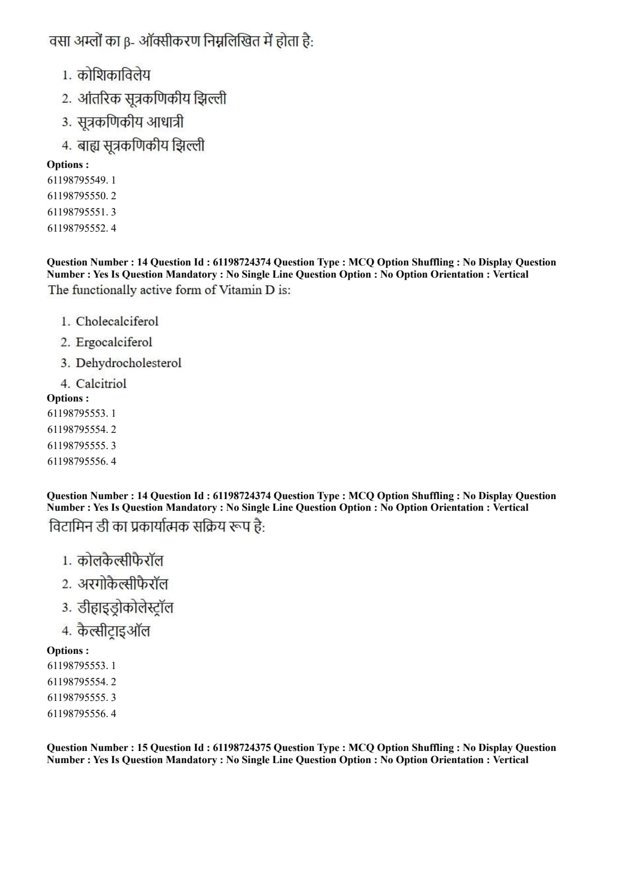
\(\beta\)- Oxidation of fatty acids occurs in:
View Solution
Step 1: Understanding the Question:
The question asks to pinpoint the exact intracellular compartment where \(\beta\)-oxidation of fatty acids takes place in eukaryotic cells.
Step 2: Key Formula or Approach:
Fatty acid breakdown proceeds through four repeating biochemical steps (oxidation, hydration, oxidation, thiolysis) that convert fatty acyl-CoA into acetyl-CoA units inside the mitochondrion.
Step 3: Detailed Explanation:
Fatty Acid Activation: Fatty acids are first activated in the cytosol to form fatty acyl-CoA by acyl-CoA synthetase.
Carnitine Shuttle System: Long-chain fatty acyl-CoA molecules are transported across the inner mitochondrial membrane via the carnitine palmitoyltransferase (CPT-I / CPT-II) shuttle mechanism.
Localization of \(\beta\)-Oxidation Enzymes: Once inside the mitochondrial matrix, soluble enzymes (Acyl-CoA dehydrogenase, Enoyl-CoA hydratase, 3-hydroxyacyl-CoA dehydrogenase, and \(\beta\)-ketoacyl-CoA thiolase) degrade the fatty acyl chain two carbons at a time.
Yield: Each cycle produces 1 molecule of Acetyl-CoA (which enters the TCA cycle located in the matrix), 1 molecule of NADH, and 1 molecule of FADH\(_2\).
Step 4: Final Answer:
The enzymes responsible for \(\beta\)-oxidation of fatty acids reside within the mitochondrial matrix.
Quick Tip: Fatty acid activation = Cytosol / Outer mitochondrial membrane.
Fatty acid transport = Carnitine shuttle across inner membrane.
Fatty acid \(\beta\)-oxidation = Mitochondrial matrix.
The functionally active form of Vitamin D is:
View Solution
Step 1: Understanding the Question:
The question asks to identify the chemically fully active hormonal metabolite derived from Vitamin D in the human body.
Step 2: Key Formula or Approach:
Vitamin D undergoes a series of two sequential hydroxylation reactions in the liver and kidneys to reach its biologically active form: \[ 7-Dehydrocholesterol \xrightarrow{UV Light} Cholecalciferol (Vit D_3) \xrightarrow[Liver]{25-hydroxylase} 25(OH)D_3 \xrightarrow[Kidney]{1\alpha-hydroxylase} 1,25(OH)_2D_3 \]
Step 3: Detailed Explanation:
Cholecalciferol (Vitamin D\(_3\)): Produced in skin exposed to UV-B radiation or ingested via diet; it is biologically inactive precursor.
25-Hydroxycholecalciferol (Calcidiol): Major circulating precursor form produced in the liver by 25-hydroxylase.
1,25-Dihydroxycholecalciferol (Calcitriol): Formed in renal proximal tubules via \(1\alpha\)-hydroxylase. Calcitriol is the fully functional active steroid-like hormone.
Biological Action: Calcitriol binds nuclear Vitamin D Receptors (VDR) to regulate gene expression responsible for intestinal calcium and phosphate absorption and bone mineralization.
Step 4: Final Answer:
The active hormone form of Vitamin D is calcitriol (\(1,25-dihydroxycholecalciferol\)).
Quick Tip: Cholecalciferol (\(D_3\)) = Inactive precursor.
Calcidiol = Main storage/circulating form (\(25-OH D_3\)).
Calcitriol = Active hormonal form (\(1,25-(OH)_2 D_3\)).
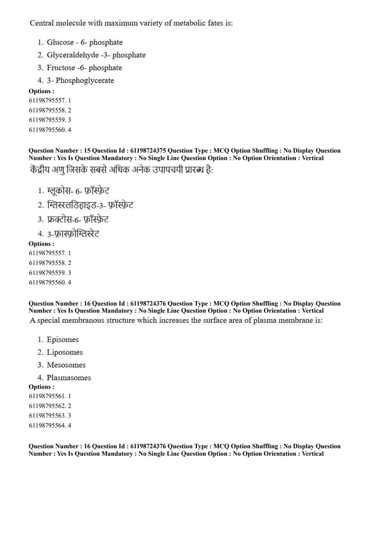
Central molecule with maximum variety of metabolic fates is:
View Solution
Step 1: Understanding the Question:
The question asks to select the metabolite located at a major metabolic crossroads capable of branching into the largest number of essential pathways.
Step 2: Key Formula or Approach:
Glucose-6-phosphate (G6P) sits at the junction of several central carbohydrate pathways:
Glycolysis
Glycogenesis
Pentose Phosphate Pathway (PPP)
Gluconeogenesis / Free Glucose release
Step 3: Detailed Explanation:
Crossroads Position: Upon entry into cells, glucose is phosphorylated by hexokinase or glucokinase to Glucose-6-phosphate, trapping it intracellularly.
Diverse Metabolic Fates of G6P:
Glycolysis: Isomerized to Fructose-6-phosphate by phosphoglucose isomerase to produce ATP and pyruvate.
Glycogenesis: Converted to Glucose-1-phosphate by phosphoglucomutase for glycogen storage.
Pentose Phosphate Pathway (PPP): Oxidized by Glucose-6-phosphate dehydrogenase (G6PD) to generate NADPH and ribose-5-phosphate for nucleotide synthesis.
Free Glucose Formation: Converted back to glucose by glucose-6-phosphatase in liver/kidneys to maintain systemic blood glucose level.
Comparison: None of the other listed triose or hexose intermediates (G3P, F6P, 3-PG) possess such extensive metabolic flexibility across energy, biosynthetic, and storage pathways.
Step 4: Final Answer:
Glucose-6-phosphate is the central metabolic node with the widest variety of metabolic fates.
Quick Tip: Glucose-6-phosphate connects 4 major pathways: Glycolysis, Glycogenesis, Pentose Phosphate Pathway, and Gluconeogenesis.
A special membranous structure which increases the surface area of plasma membrane is:
View Solution
Step 1: Understanding the Question:
The question asks for the name of invaginated cell membrane structures in prokaryotic cells that increase membrane surface area for metabolic processes.
Step 2: Key Formula or Approach:
Prokaryotes lack membrane-bound organelle compartments like mitochondria; invaginations of the cell membrane provide extra surface area for membrane-associated enzymes.
Step 3: Detailed Explanation:
Definition of Mesosomes: Mesosomes are folded invaginations of the plasma membrane in bacteria formed by inward extensions of the cell membrane.
Function: They increase the effective surface area of the plasma membrane and boost enzymatic activity required for cellular processes such as respiration, DNA replication, cell wall synthesis, and secretion.
Analysis of Incorrect Options:
Episomes: Extrachromosomal genetic elements (plasmids) capable of integrating into bacterial host genomic DNA.
Liposomes: Synthetic spherical lipid vesicles used artificially for drug delivery.
Plasmasomes: Non-standard biological terminology not associated with cell membrane surface extension.
Step 4: Final Answer:
Mesosomes are specialized plasma membrane folds in bacteria that increase available membrane surface area.
Quick Tip: Mesosomes = Infoldings of bacterial cell membranes.
They increase surface area for electron transport/respiration and aid in cell division.
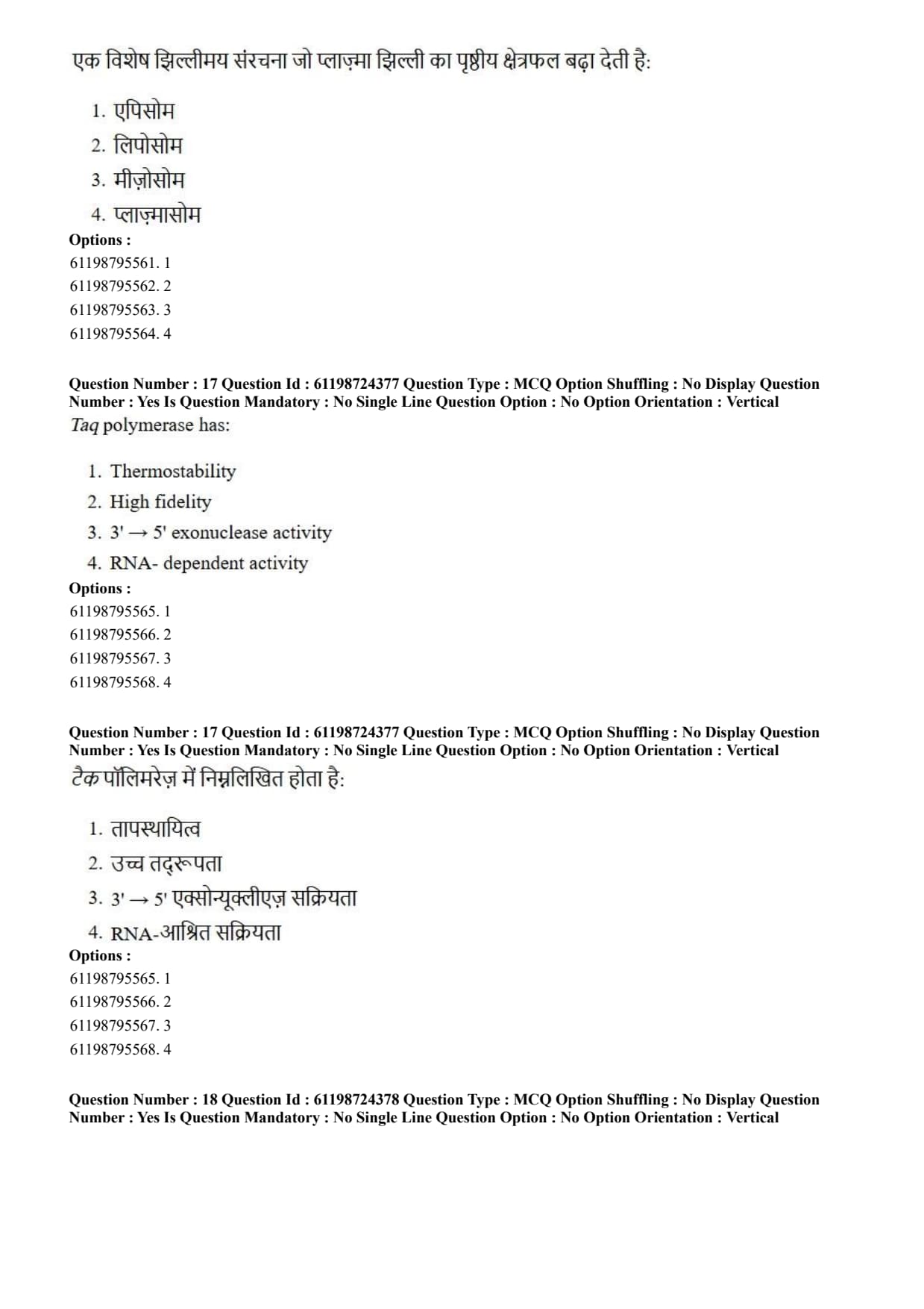
Taq polymerase has:
View Solution
Step 1: Understanding the Question:
The question asks about the primary enzymatic property of \(Taq\) DNA polymerase that enables its widespread utility in PCR reactions.
Step 2: Key Formula or Approach:
PCR requires repeated cycling at high temperatures (\(\sim 95^\circC\)) to denature double-stranded DNA template strands.
Step 3: Detailed Explanation:
Origin of Taq Polymerase: \(Taq\) DNA polymerase is isolated from the thermophilic bacterium \textit{Thermus aquaticus, which naturally thrives in high-temperature hot springs.
Thermostability Property: \(Taq\) polymerase retains structural integrity and enzymatic activity at elevated temperatures, with an optimum activity at \(72^\circC\) and a half-life of over 40 minutes at \(95^\circC\). This thermostability eliminates the need to add fresh enzyme after each denaturation cycle.
Lack of Proofreading: \(Taq\) polymerase naturally lacks \(3' \to 5'\) proofreading exonuclease activity. As a result, it exhibits relatively low replication fidelity compared to proofreading polymerases like \(Pfu\) or \(Vent\).
Template Specificity: \(Taq\) is a DNA-dependent DNA polymerase, not an RNA-dependent enzyme (like reverse transcriptase).
Step 4: Final Answer:
The defining characteristic feature of \(Taq\) polymerase is its high thermostability.
Quick Tip: \(Taq\) Polymerase = Thermostable (\(95^\circC\) resistant), lacks \(3' \to 5'\) proofreading exonuclease activity.
For high fidelity PCR, proofreading polymerases such as \(Pfu\) are preferred.
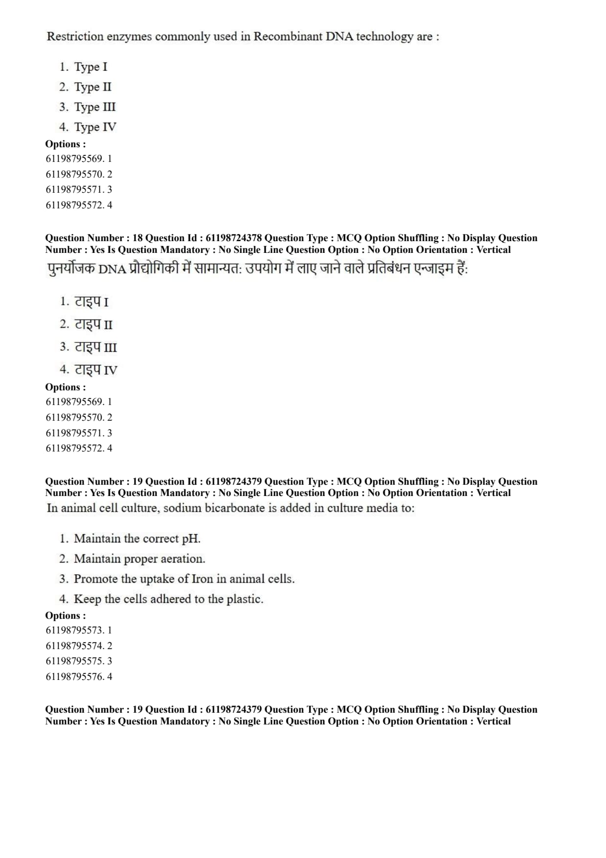
Restriction enzymes commonly used in Recombinant DNA technology are:
View Solution
Step 1: Understanding the Question:
The question asks to identify which class of restriction endonucleases is routinely utilized in molecular cloning and recombinant DNA procedures.
Step 2: Key Formula or Approach:
Restriction endonucleases cleave double-stranded DNA at or near specific recognition sequences. They are classified into Types I, II, III, and IV based on cofactor requirements, subunit composition, and cleavage site specificity.
Step 3: Detailed Explanation:
Type II Restriction Enzymes:
Cleave DNA precisely at or immediately adjacent to specific, palindromic target recognition sites (typically 4–8 bp long).
Do not require ATP or \(S\)-adenosylmethionine (SAM); require only \(Mg^{2+}\) as a cofactor.
Produce predictable, reproducible DNA fragments with sticky (\(5'\) or \(3'\) overhangs) or blunt ends.
Examples include \textit{EcoRI, \textit{HindIII, and \textit{BamHI.
Limitations of Other Types:
Type I and Type III: Cleave DNA at random positions far away from their recognition sites (e.g., Type I cuts \(>1000\ bp\) away), requiring ATP hydrolysis, making them unsuitable for targeted molecular cloning.
Type IV: Recognize modified (methylated/hydroxymethylated) DNA and cut non-specifically.
Step 4: Final Answer:
Type II restriction endonucleases are the standard choice for recombinant DNA technology.
Quick Tip: Type II restriction enzymes cut DNA AT or close to specific palindromic recognition sites without requiring ATP.
This precise cutting makes them indispensable tools for gene cloning.
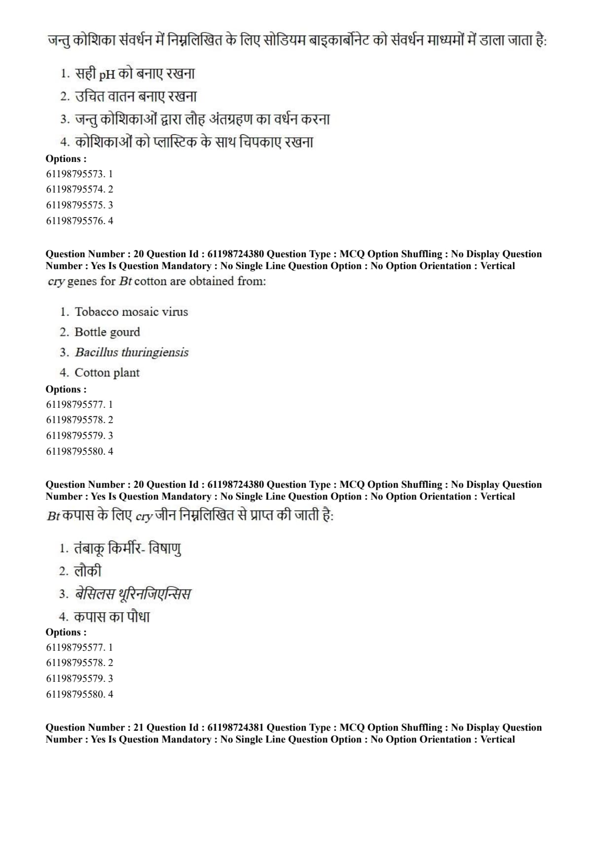
In animal cell culture, sodium bicarbonate is added in culture media to:
View Solution
Step 1: Understanding the Question:
The question asks for the physiological function of sodium bicarbonate (\(NaHCO_3\)) added to cell culture media during animal cell cultivation.
Step 2: Key Formula or Approach:
Animal cells require a strictly maintained physiological pH range (\(pH 7.2 - 7.4\)). This is achieved using a bicarbonate-carbon dioxide buffering equilibrium: \[ CO_2 + H_2O \rightleftharpoons H_2CO_3 \rightleftharpoons H^+ + HCO_3^- \]
Step 3: Detailed Explanation:
Role of \(NaHCO_3\) as Buffer: Dissolved sodium bicarbonate provides bicarbonate ions (\(HCO_3^-\)) in the culture medium.
Interaction with Incubator \(CO_2\): When placed in a humidified incubator containing controlled \(CO_2\) gas (typically 5% \(CO_2\)), the dissolved \(CO_2\) establishes an equilibrium with exogenous \(HCO_3^-\).
pH Stabilization: As cells release metabolic acids (like lactic acid), the equilibrium shifts to neutralize extra protons (\(H^+\)), maintaining stable extracellular physiological pH.
Evaluation of Other Options: Aeration is managed by culture volume/shaking; iron uptake is promoted by transferrin; cell adherence is facilitated by extracellular matrix proteins (e.g., collagen, fibronectin).
Step 4: Final Answer:
Sodium bicarbonate is added to animal culture media primarily to maintain optimal physiological pH.
Quick Tip: \(NaHCO_3\) in media + 5% \(CO_2\) gas in incubator = Bicarbonate Buffer System for maintaining physiological pH (\(\sim 7.4\)).
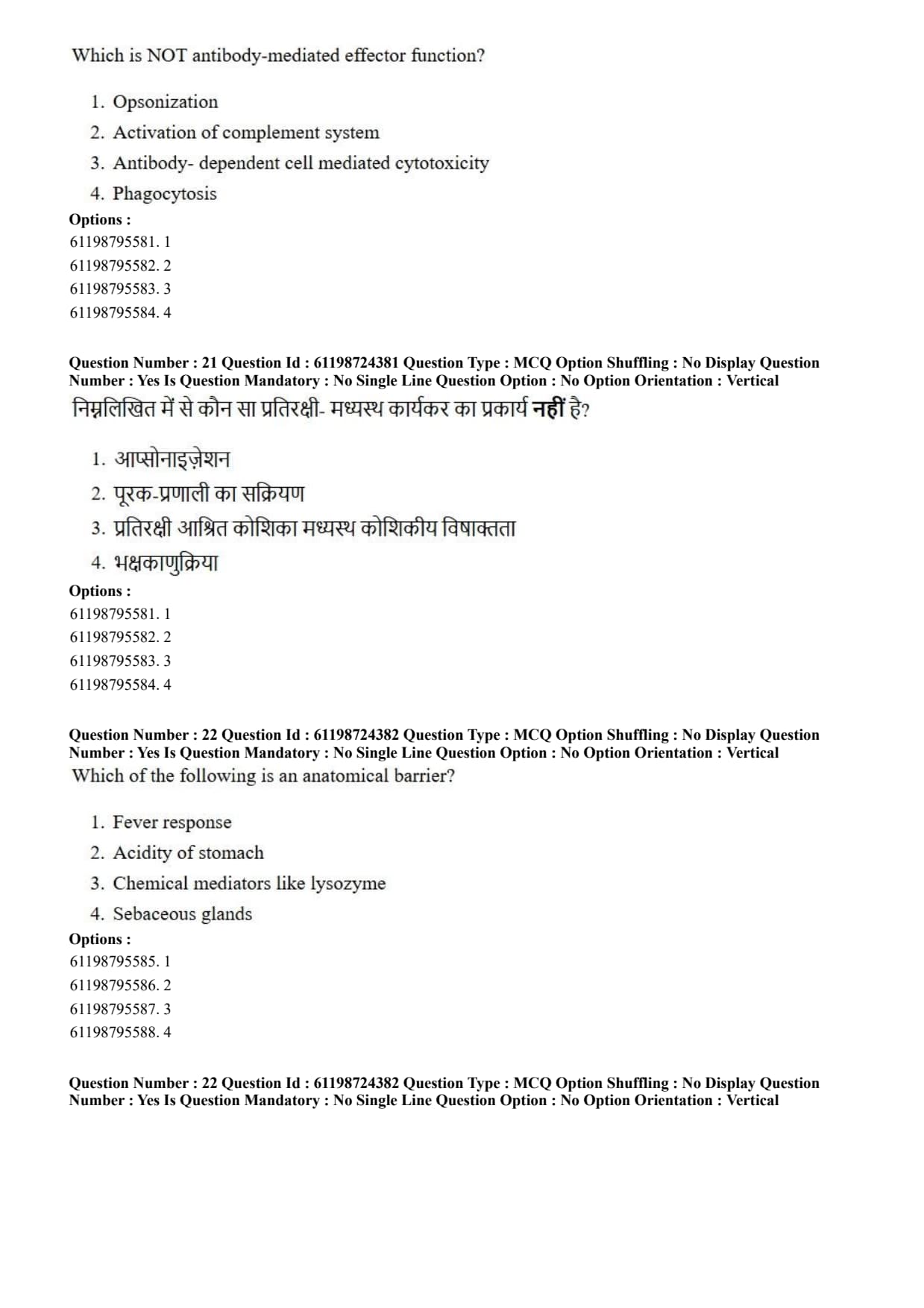
cry genes for Bt cotton are obtained from:
View Solution
Step 1: Understanding the Question:
The question asks to identify the source organism from which the insecticidal *cry* genes expressed in transgenic Bt cotton are isolated.
Step 2: Key Formula or Approach:
Genetic engineering transfers insecticidal endotoxin-encoding genes from soil bacteria into crop plants to grant resistance against target insect pests.
Step 3: Detailed Explanation:
Origin of *cry* Genes: *cry* genes encode crystalline (Cry) endotoxin proteins produced naturally by the Gram-positive soil bacterium Bacillus thuringiensis (hence abbreviated as 'Bt').
Mechanism of Action: Upon ingestion by susceptible insects (such as the cotton bollworm, *Helicoverpa armigera*), inactive protoxins are solubilized in the alkaline gut environment and cleaved by proteases into active toxins.
Pore Formation: Active Cry toxins bind to specific receptors on midgut epithelial cells, forming pores that lead to cell lysis, gut perforation, starvation, and death of the pest.
Transgenic Application: Genes such as *cry1Ac* and *cry2Ab* isolated from \textit{Bacillus thuringiensis were engineered into cotton plants to create pest-resistant transgenic Bt cotton.
Step 4: Final Answer:
The *cry* genes utilized in transgenic Bt cotton are obtained from the bacterium \textit{Bacillus thuringiensis.
Quick Tip: Bt = \textit{Bacillus thuringiensis.
*cry* genes encode crystal proteins that act as specific endotoxins against lepidopteran, coleopteran, and dipteran insect pests.
Which is NOT antibody-mediated effector function?
View Solution
Step 1: Understanding the Question:
The question asks to identify the process that is an intrinsic cellular mechanism performed directly by phagocytic immune cells rather than a specialized effector mechanism mediated by antibody molecules.
Step 2: Key Formula or Approach:
Antibodies (immunoglobulins) act as molecular adaptors that bind antigens via their Fab regions and trigger specific effector functions by interacting with Fc receptors or complement proteins via their Fc regions:
Opsonization: Coating of pathogens with antibodies to enhance recognition by Fc receptors on phagocytes.
Complement Activation: Classical complement pathway initiated by C1q binding to antigen-bound IgM or IgG.
ADCC: NK cells and granulocytes destroying antibody-coated target cells via Fc\(\gamma\)RIII interaction.
Direct Phagocytosis: Engulfment of microbes by macrophages/neutrophils mediated by direct binding of Pattern Recognition Receptors (PRRs) to Pathogen-Associated Molecular Patterns (PAMPs) without requiring antibodies.
Step 3: Detailed Explanation:
Antibody-Mediated Effector Functions: Opsonization, complement activation, and antibody-dependent cell-mediated cytotoxicity (ADCC) strictly require the presence and binding of specific antibodies to function.
Phagocytosis as an Intrinsic Cellular Function: Phagocytosis itself is a fundamental cellular process executed by phagocytes (such as macrophages, neutrophils, and dendritic cells).
Innate Phagocytosis Mechanism: Phagocytes can directly recognize, engulf, and internalize foreign particles or microorganisms via innate surface receptors (e.g., scavenger receptors, mannose receptors, and Toll-like co-receptors) independently of antibodies.
Enhancement vs. Effector Function: While antibodies facilitate phagocytosis through opsonization, the primary mechanism of phagocytosis is a cellular engulfment process rather than an antibody-driven effector cascade.
Step 4: Final Answer:
Direct phagocytosis is an intrinsic cellular uptake mechanism that can occur independently of antibody mediation.
Quick Tip: Antibody effector mechanisms require the Fc region:
1. Opsonization (FcR binding)
2. Complement Activation (C1q binding)
3. ADCC (Fc\(\gamma\)R binding on NK cells)
Phagocytosis itself is a cellular process carried out by innate immune cells.
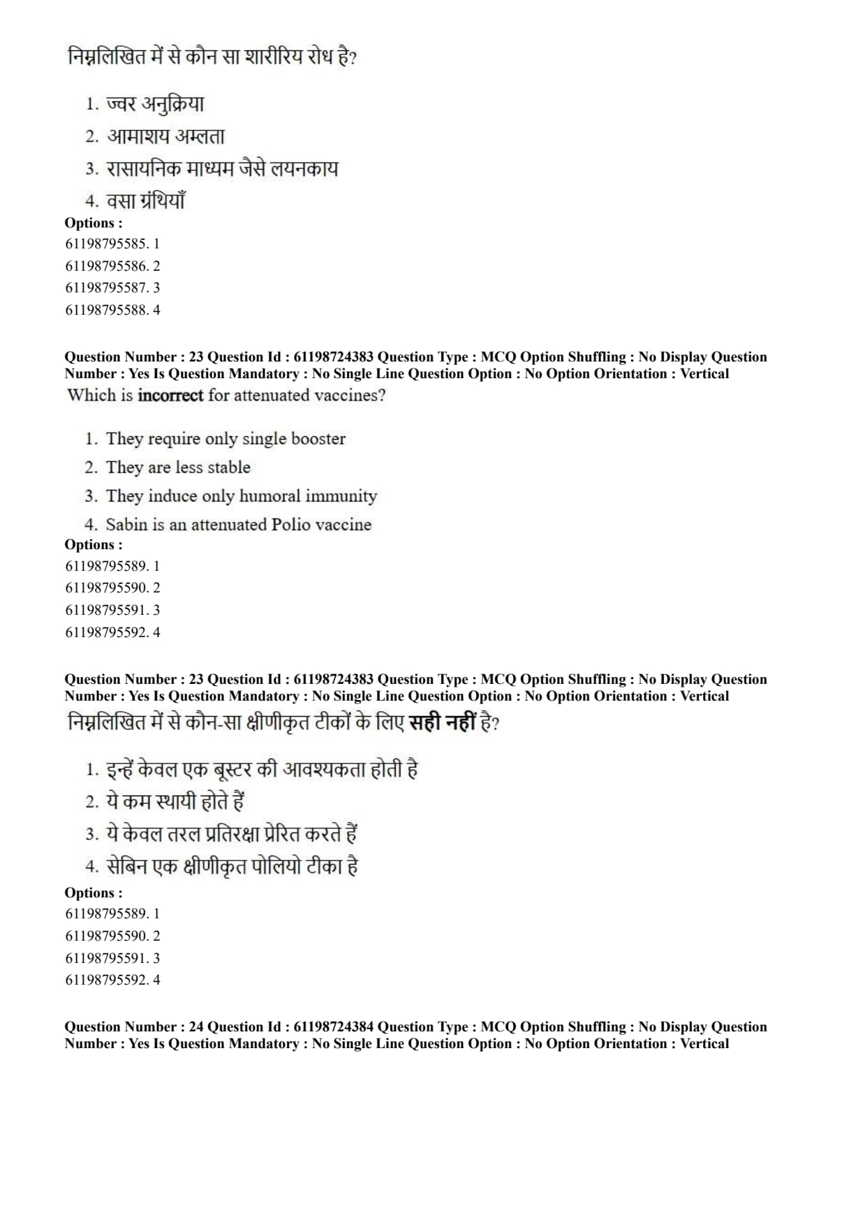
Which of the following is an anatomical barrier?
View Solution
Step 1: Understanding the Question:
The question asks to identify which component represents an anatomical/physical barrier mechanism involved in innate immunity.
Step 2: Key Formula or Approach:
Innate immunity relies on initial protective barriers classified into distinct categories:
Anatomical / Physical Barriers: Skin, mucous membranes, epidermal structures, and associated integumentary glands.
Physiological / Chemical Barriers: Low pH of gastric juice, temperature changes (fever), solubilized enzymes (lysozyme), and antimicrobial peptides.
Step 3: Detailed Explanation:
Sebaceous Glands as Anatomical Components: Sebaceous glands are structural exocrine glands embedded within the dermis layer of the skin (a primary anatomical barrier). They secrete sebum, an oily substance rich in fatty acids, which forms a hydrophobic lipid layer that coats the skin surface to prevent pathogen invasion and microbial colonization.
Evaluation of Other Options:
Fever Response: A systemic physiological response regulated by pyrogens acting on the hypothalamus to elevate body temperature.
Acidity of Stomach: A physiological/chemical barrier created by hydrochloric acid (\(HCl\)) secreted by gastric parietal cells.
Lysozyme: A soluble chemical mediator/enzyme present in tears and saliva that hydrolyzes peptidoglycan linkages in bacterial cell walls.
Step 4: Final Answer:
Sebaceous glands, as part of the integumentary system structure, function as an anatomical barrier.
Quick Tip: Anatomical barriers = Physical tissue structures and integumentary glands (Skin, Mucosa, Sebaceous glands).
Physiological barriers = Temperature (fever), pH (gastric acid), and chemical enzymes (lysozyme).
Which is incorrect for attenuated vaccines?
View Solution
Step 1: Understanding the Question:
The question asks to identify the statement that is false regarding the immunological and biological properties of live-attenuated vaccines.
Step 2: Key Formula or Approach:
Live-attenuated vaccines contain weakened pathogens capable of replicating inside host cells without causing disease. Because they infect cells naturally, they present antigens via both MHC Class I and MHC Class II pathways.
Step 3: Detailed Explanation:
Dual Immune Activation (Humoral and Cell-Mediated): Live-attenuated microbes replicate intracellularly inside host cells. Antigenic peptides are processed and presented on MHC Class I molecules to CD8\(^+\) Cytotoxic T Lymphocytes (CTLs), stimulating robust cell-mediated immunity. Simultaneously, extracellular viral/bacterial antigens are presented on MHC Class II molecules to CD4\(^+\) Helper T cells, driving B cell activation and humoral immunity. Therefore, claiming they induce *only* humoral immunity is incorrect.
Analysis of True Statements:
Booster Requirement: Due to transient replication in the host, live-attenuated vaccines provide strong immunogenic stimulation requiring fewer doses or single boosters compared to inactivated vaccines.
Stability: Live organisms are heat-sensitive and less stable, requiring strict cold-chain storage.
Sabin Vaccine: The Sabin oral polio vaccine (OPV) consists of live-attenuated poliovirus strains.
Step 4: Final Answer:
The statement that attenuated vaccines induce *only* humoral immunity is incorrect because they elicit both strong humoral and cell-mediated immune responses.
Quick Tip: Live Attenuated Vaccines = Replicate inside host cells \(\to\) Activate BOTH Humoral (Antibodies) and Cell-Mediated (CD8\(^+\) T-cell) Immunity!
Killed/Inactivated Vaccines = Elicit mainly Humoral immunity.
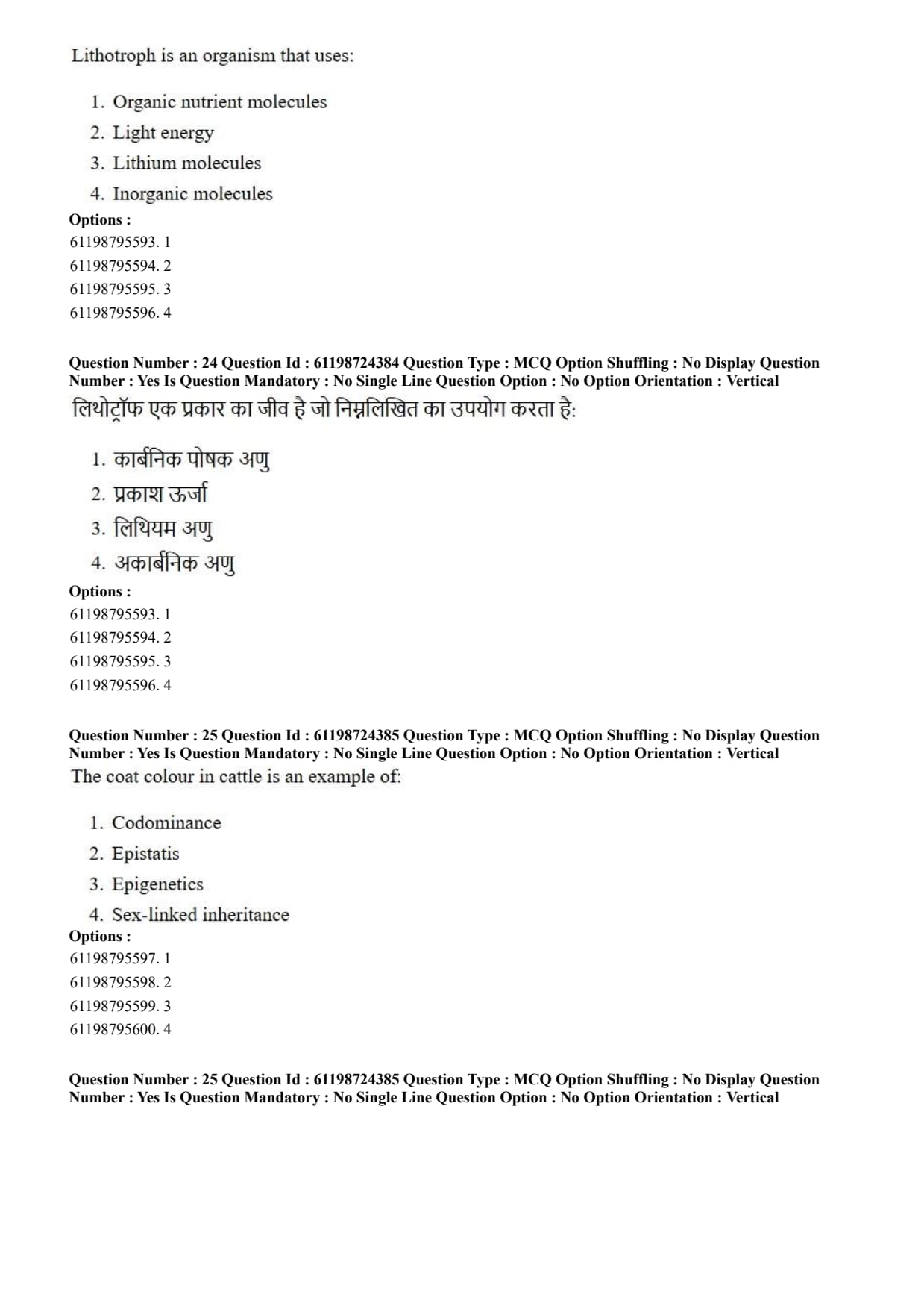
Lithotroph is an organism that uses:
View Solution
Step 1: Understanding the Question:
The question asks for the nutritional/metabolic definition of a lithotrophic organism based on its electron donor source.
Step 2: Key Formula or Approach:
Microorganisms are classified according to their source of electrons for biosynthesis and energy conservation:
Organotrophs: Use reduced organic molecules (e.g., glucose, fatty acids) as electron donors.
Lithotrophs: Use reduced inorganic molecules (e.g., \(H_2, NH_3, NO_2^-, H_2S, Fe^{2+}\)) as electron donors.
Step 3: Detailed Explanation:
Etymology and Definition: The term "lithotroph" derives from the Greek words *lithos* (meaning stone/rock) and *troph* (meaning feeder). Lithotrophs acquire reducing equivalents (electrons) from inorganic compounds.
Metabolic Diversity: Lithotrophs can be chemolithotrophs (obtaining energy from the oxidation of inorganic chemicals) or photolithotrophs (using light energy with inorganic electron donors like \(H_2S\) during anoxygenic photosynthesis).
Examples: Nitrifying bacteria (Nitrosomonas, \textit{Nitrobacter), iron-oxidizing bacteria (\textit{Acidithiobacillus ferrooxidans), and hydrogen-oxidizing bacteria.
Clarification: The term lithotroph does not refer to lithium metal, nor does it refer exclusively to organic compounds or light energy alone.
Step 4: Final Answer:
A lithotroph is defined as an organism that uses inorganic molecules as electron donors.
Quick Tip: Lithotroph = Inorganic electron donor (\(H_2S, NH_3, Fe^{2+\)).
Organotroph = Organic electron donor (Glucose, Amino acids).
Phototroph = Light energy source.
Chemotroph = Chemical energy source.
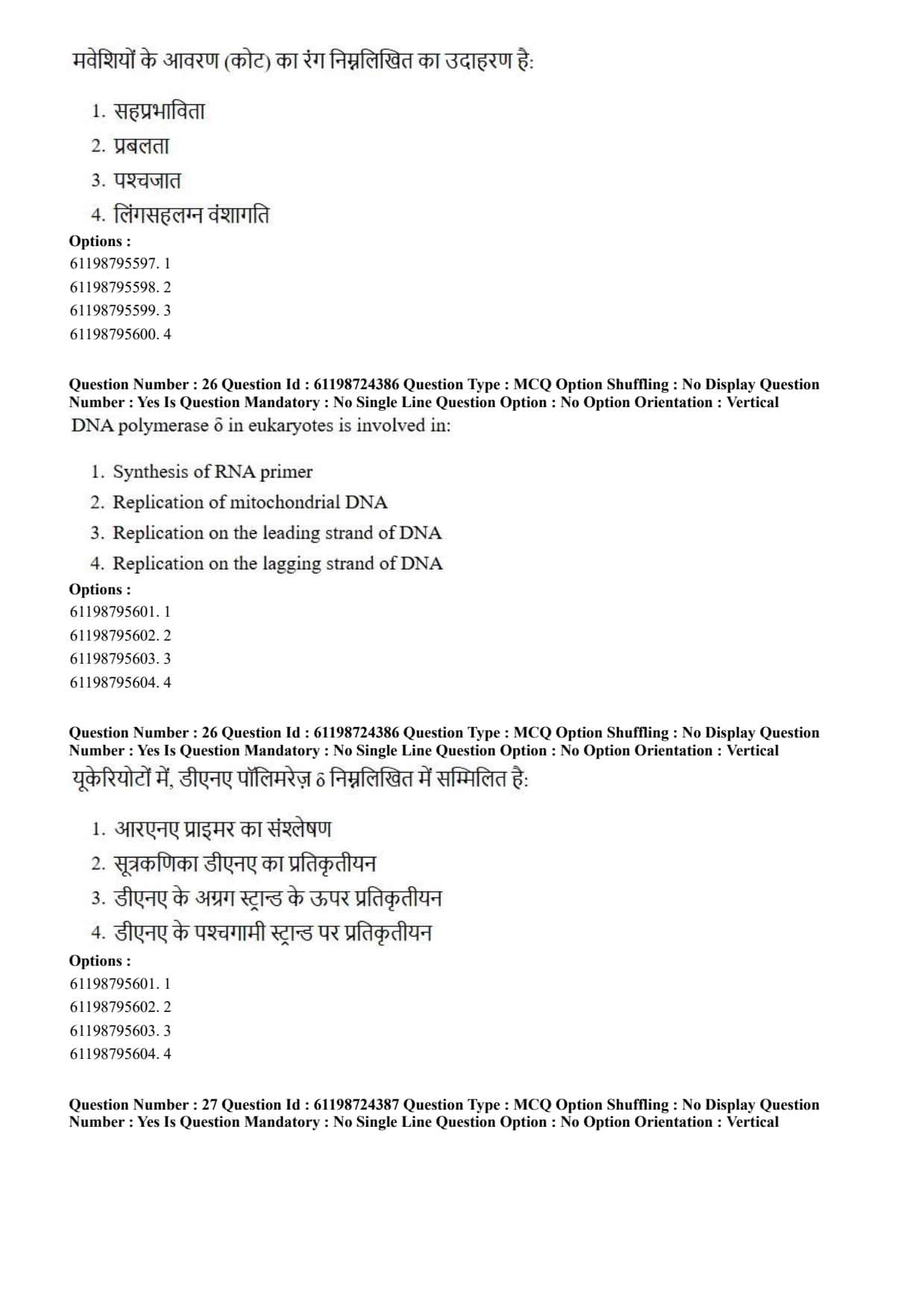
The coat colour in cattle is an example of:
View Solution
Step 1: Understanding the Question:
The question asks to categorize the genetic inheritance pattern responsible for roan coat color determination in cattle.
Step 2: Key Formula or Approach:
In Mendelian inheritance, complete dominance allows one allele to mask another. In non-Mendelian inheritance:
Incomplete Dominance: Heterozygote displays an intermediate, blended phenotype.
Codominance: Both alleles in a heterozygote are fully and simultaneously expressed without blending.
Step 3: Detailed Explanation:
Genetics of Cattle Coat Color: In cattle (such as Shorthorn breeds), coat color is controlled by an autosomal gene with two main alleles: \(R^R\) (red coat) and \(R^W\) (white coat).
Homozygous Genotypes: \(R^R R^R\) cattle express red pigment throughout their coat, while \(R^W R^W\) cattle express white coats.
Heterozygous Genotype (\(R^R R^W\)): Heterozygous cattle display a "roan" phenotype. A roan coat consists of individual red hairs and individual white hairs interspersed together across the animal's body.
Why Codominance? Because both the red allele and the white allele are expressed fully and distinctly side-by-side in the same organism without blending into pink, this serves as a textbook example of codominance.
Step 4: Final Answer:
The coat color in cattle (roan coat) is a classic biological demonstration of codominance.
Quick Tip: Codominance = Both alleles expressed distinctly together (e.g., Roan cattle coat with distinct red and white hairs, ABO blood group \(I^A I^B\)).
Incomplete Dominance = Blended intermediate phenotype (e.g., Red \(\times\) White = Pink flowers in *Snapdragon*).
DNA polymerase \(\delta\) in eukaryotes is involved in:
View Solution
Step 1: Understanding the Question:
The question asks for the primary physiological role of eukaryotic DNA polymerase delta (\(Pol \delta\)) during nuclear DNA replication.
Step 2: Key Formula or Approach:
Eukaryotic nuclear DNA replication utilizes three main replicative DNA polymerases:
Pol \(\alpha\) / Primase: Synthesizes RNA primers and short initiator DNA stretches.
Pol \(\epsilon\): Primary enzyme responsible for continuous leading strand elongation.
Pol \(\delta\): Primary enzyme responsible for discontinuous lagging strand elongation and Okazaki fragment processing.
Pol \(\gamma\): Replicates mitochondrial DNA.
Step 3: Detailed Explanation:
Function of DNA Polymerase \(\delta\): DNA polymerase \(\delta\) is a multi-subunit complex possessing high processivity when bound to Proliferating Cell Nuclear Antigen (PCNA). It carries out synthesis of Okazaki fragments on the lagging strand during nuclear DNA replication.
Proofreading and Repair: Pol \(\delta\) contains intrinsic \(3' \to 5'\) exonuclease activity for proofreading and plays essential roles in nucleotide excision repair and mismatch repair.
Evaluation of Other Options:
Synthesis of RNA primer: Performed by the primase subunit associated with Pol \(\alpha\).
Replication of mitochondrial DNA: Carried out specifically by DNA Polymerase \(\gamma\) (\(Pol \gamma\)).
Leading Strand Replication: Primarily executed by DNA Polymerase \(\epsilon\) (\(Pol \epsilon\)).
Step 4: Final Answer:
DNA polymerase \(\delta\) is predominantly involved in synthesizing the lagging strand during eukaryotic nuclear replication.
Quick Tip: Eukaryotic Replicative Polymerases:
Pol \(\alpha\) = Initiates / Primase.
Pol \(\epsilon\) = Leading strand synthesis.
Pol \(\delta\) = Lagging strand synthesis.
Pol \(\gamma\) = Mitochondrial DNA replication.
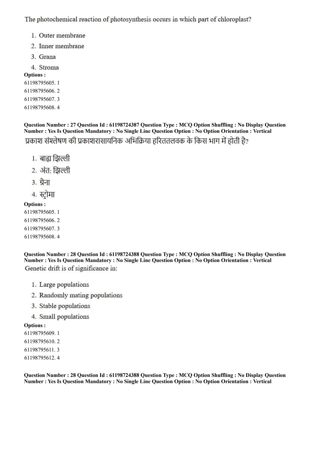
The photochemical reaction of photosynthesis occurs in which part of chloroplast?
View Solution
Step 1: Understanding the Question:
The question asks to identify the structural compartment within the chloroplast where the light-dependent (photochemical) reactions of photosynthesis take place.
Step 2: Key Formula or Approach:
Photosynthesis is divided into two major phases occurring in distinct structural regions of the chloroplast:
Photochemical Phase (Light Reactions): Occurs in the thylakoid membranes / grana.
Biochemical Phase (Dark Reactions / Calvin Cycle): Occurs in the fluid matrix / stroma.
Step 3: Detailed Explanation:
Structure of Grana: Grana (singular: granum) are stacks of disc-like membrane-bound structures called thylakoids suspended within the chloroplast stroma.
Localization of Photochemical Machinery: The thylakoid membranes within grana harbor light-harvesting complex proteins, Photosystem I (PSI), Photosystem II (PSII), cytochromes \(b_6f\), and ATP synthase.
Light Reactions Process: During the photochemical phase, light absorption excites chlorophyll molecules, driving photolysis of water (\(H_2O \to 2H^+ + \frac{1}{2}O_2 + 2e^-\)), non-cyclic/cyclic electron transport, proton pumping, and generation of \(ATP\) and \(NADPH\).
Stroma Function: The stroma houses soluble enzymes like RuBisCO required for \(CO_2\) fixation during the Calvin cycle.
Step 4: Final Answer:
The photochemical reactions of photosynthesis occur within the grana (thylakoid membranes) of the chloroplast.
Quick Tip: Grana / Thylakoids = Light Reaction (Photochemical phase: produces ATP, NADPH, and \(O_2\)).
Stroma = Dark Reaction (Calvin Cycle: consumes ATP and NADPH to fix \(CO_2\) into sugar).
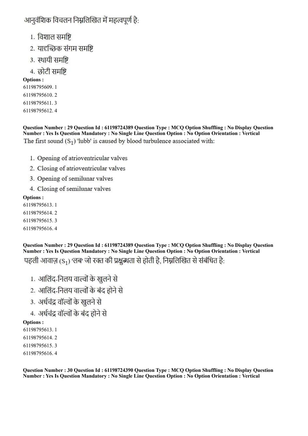
Genetic drift is of significance in:
View Solution
Step 1: Understanding the Question:
The question asks to identify the population size condition under which evolutionary genetic drift exerts a major, statistically significant influence on allele frequencies.
Step 2: Key Formula or Approach:
Genetic drift represents random fluctuations in gene allele frequencies from generation to generation due to sampling errors in gamete sampling.
The variance in allele frequency change (\(\sigma_{\Delta p}^2\)) per generation is inversely proportional to population size (\(N_e\)): \[ \sigma_{\Delta p}^2 = \frac{p(1-p)}{2N_e} \]
Step 3: Detailed Explanation:
Mechanism of Genetic Drift: In finite populations, by random chance alone, certain individuals may produce more offspring or survive better regardless of their adaptative fitness.
Impact of Population Size:
In large populations, random sampling errors average out, making allele frequencies stable unless acted upon by natural selection, mutation, or migration.
In small populations, random sampling error is large (\(2N_e\) in the denominator is small), leading to rapid, unpredictable fluctuations in allele frequencies.
Consequences in Small Populations: Genetic drift can quickly lead to the random fixation of one allele (\(p=1\)) and complete loss of alternative alleles (\(p=0\)), reducing genetic diversity over time.
Classic Phenomena: Founder Effect and Population Bottleneck represent extreme scenarios of genetic drift operating in small isolated populations.
Step 4: Final Answer:
Genetic drift is of paramount evolutionary significance in small populations.
Quick Tip: Genetic Drift = Random change in allele frequency due to chance.
Always most pronounced and significant in \textbf{small populations}!
Large populations are protected from genetic drift according to Hardy-Weinberg equilibrium principles.
The first sound (S\(_1\)) 'lubb' is caused by blood turbulence associated with:
View Solution
Step 1: Understanding the Question:
The question asks to identify the physiological mechanical event in the cardiac cycle responsible for generating the first heart sound (\(S_1\), commonly voiced as 'lubb').
Step 2: Key Formula or Approach:
Heart sounds are produced by blood turbulence resulting from the sudden closure of heart valves during the cardiac cycle:
First Heart Sound (\(S_1\), 'Lubb'): Closure of Atrioventricular (AV) valves (Tricuspid and Mitral/Bicuspid valves).
Second Heart Sound (\(S_2\), 'Dubb'): Closure of Semilunar (SL) valves (Aortic and Pulmonary valves).
Step 3: Detailed Explanation:
Cardiac Cycle Timing of \(S_1\): The first heart sound occurs at the onset of ventricular systole (isovolumetric contraction phase).
Mechanical Event: As ventricles begin to contract, intraventricular pressure rapidly rises above atrial pressure. This pressure differential forces the atrioventricular (AV) valves (mitral and tricuspid) to snap shut sharply.
Sound Generation: The sudden closure of the AV valves causes blood turbulence and vibrations in the ventricular walls and surrounding major blood vessels, creating the characteristic low-pitched, longer-duration \(S_1\) 'lubb' sound.
Comparison with \(S_2\): The second heart sound (\(S_2\), 'dubb') occurs at the beginning of ventricular diastole due to the closure of semilunar valves as intraventricular pressure falls below arterial pressure.
Step 4: Final Answer:
The first heart sound (\(S_1\)) 'lubb' is caused by turbulence created by the closure of atrioventricular valves.
Quick Tip: \(S_1\) ('Lubb') = Closure of Atrioventricular (AV) valves at start of Ventricular Systole.
\(S_2\) ('Dubb') = Closure of Semilunar (SL) valves at start of Ventricular Diastole.
Valves opening does NOT produce normal heart sounds!
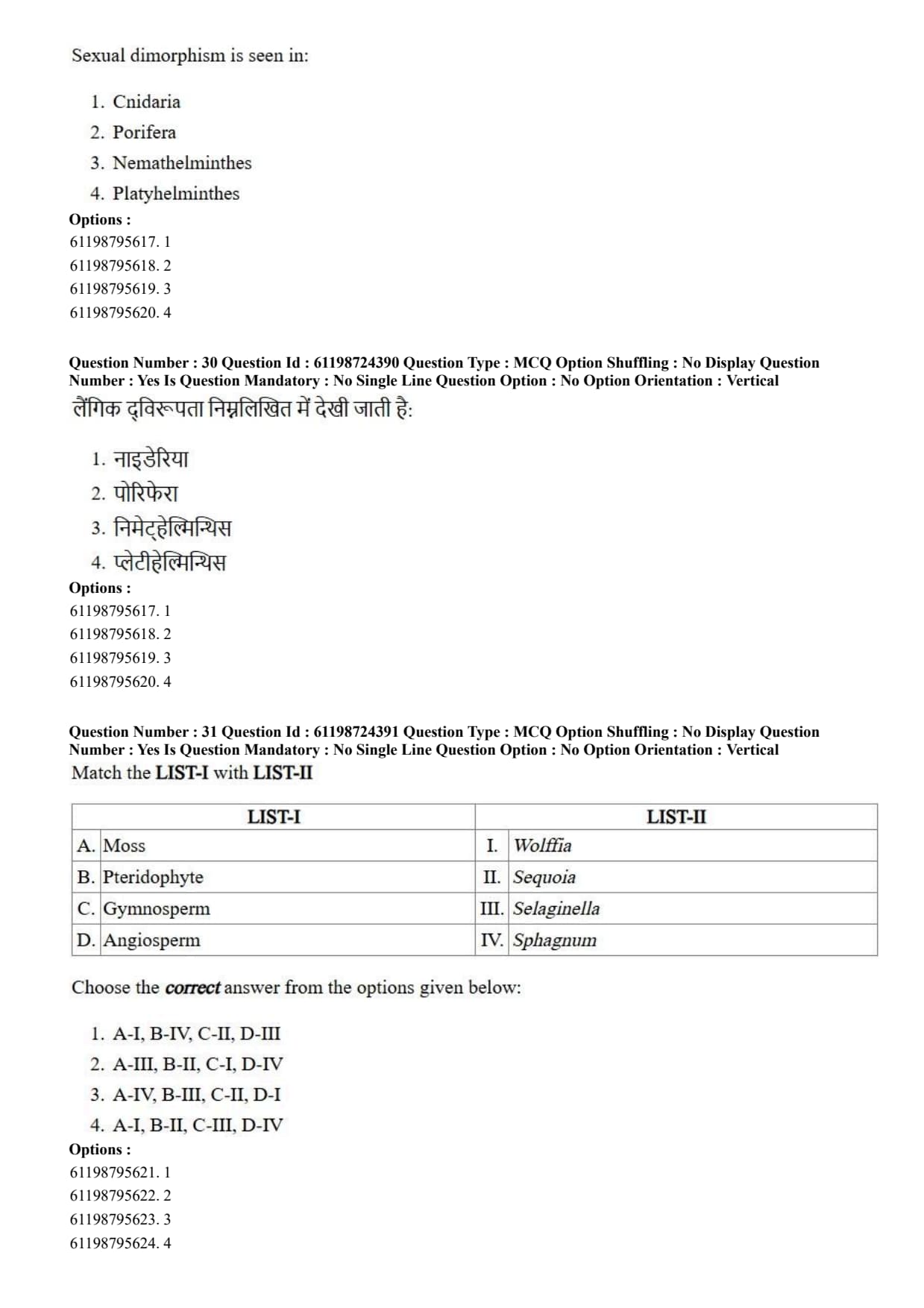
Sexual dimorphism is seen in:
View Solution
Step 1: Understanding the Question:
The question asks to identify the phylum among the given options that exhibits distinct, well-defined sexual dimorphism (morphological differences between male and female individuals).
Step 2: Key Formula or Approach:
Sexual dimorphism refers to the condition where males and females of the same species exhibit obvious phenotypic differences in secondary sexual characters, body size, shape, or anatomy.
Step 3: Detailed Explanation:
Phylum Nemathelminthes (Aschelminthes / Roundworms): Roundworms are predominantly dioecious (unisexual) with prominent sexual dimorphism.
Example in *Ascaris lumbricoides*:
Female: Considerably longer and thicker, with a straight posterior tail end.
Male: Smaller and shorter, with a distinctly curved posterior tail bearing penial setae (spicules) for copulation.
Comparison with Other Phyla:
Porifera (Sponges): Mostly hermaphroditic or monoecious; lack distinct male/female body forms.
Cnidaria: Show metagenesis/polymorphism (polyp and medusa phases), but not true sexual dimorphism.
Platyhelminthes (Flatworms): Predominantly monoecious/hermaphroditic (e.g., *Planaria*, *Tapeworm*, *Liver fluke*), except for *Schistosoma*.
Step 4: Final Answer:
Distinct sexual dimorphism is characteristic of phylum Nemathelminthes.
Quick Tip: Nematodes (*Ascaris*) = Dioecious with marked sexual dimorphism.
Female *Ascaris* is longer with a straight tail; Male *Ascaris* is shorter with a curved tail bearing spicules.
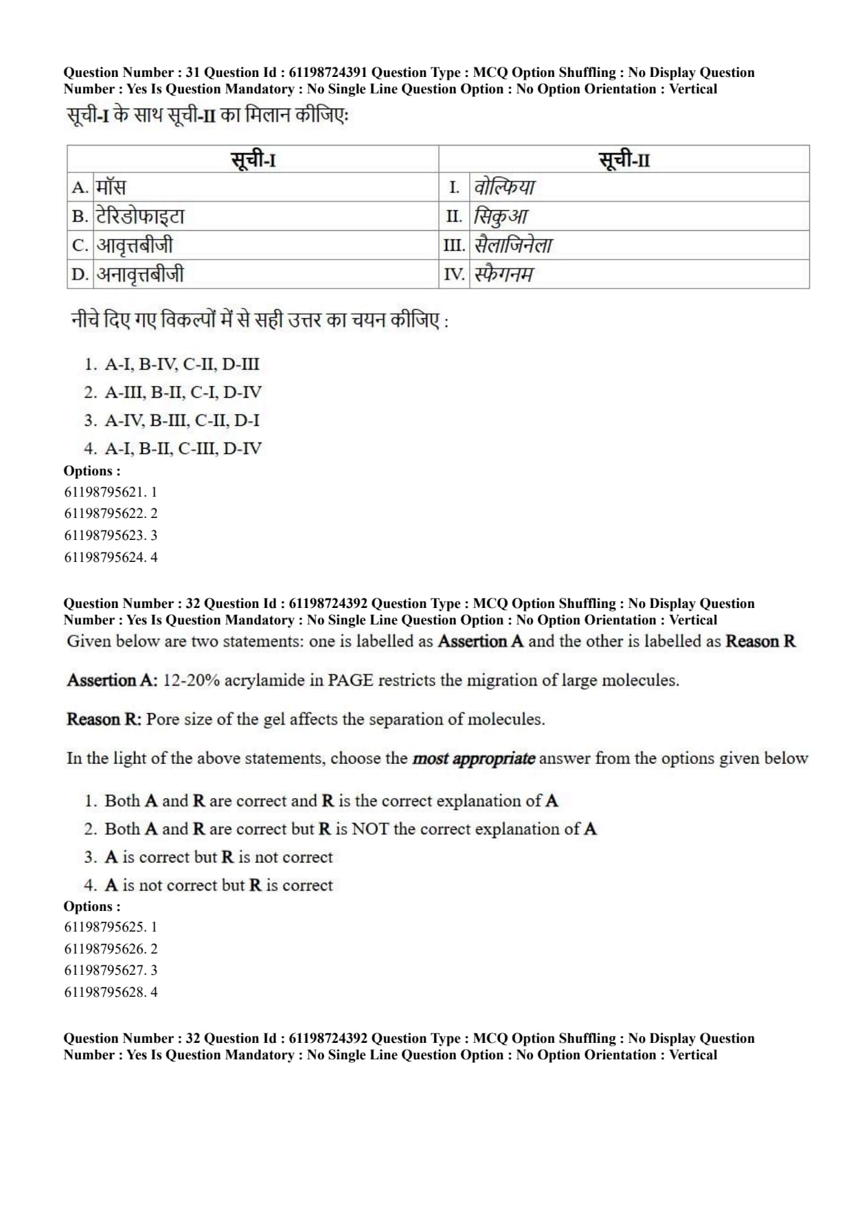
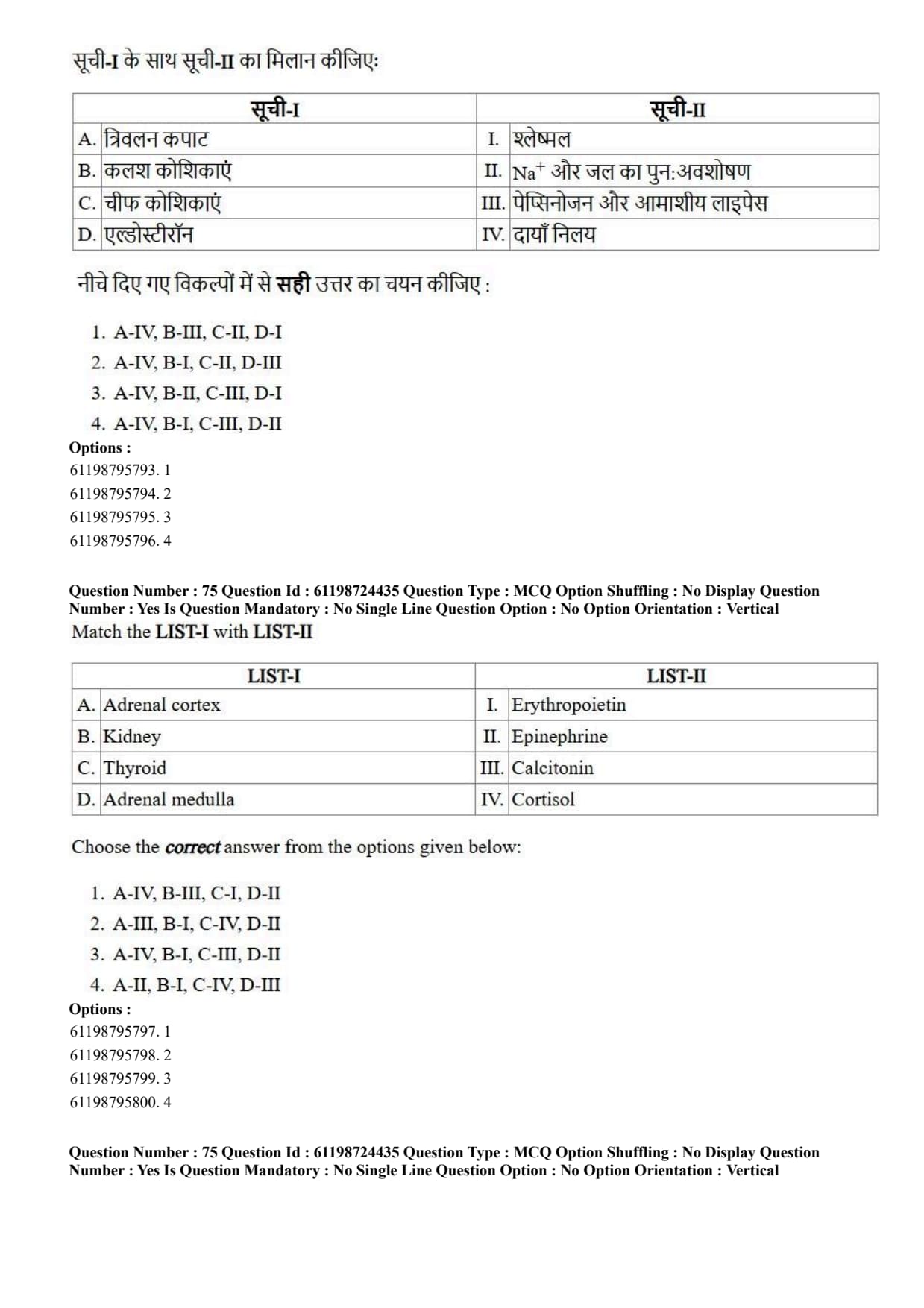
Match the LIST-I with LIST-II

View Solution
Step 1: Understanding the Question:
The question requires matching plant taxonomical groups listed in LIST-I with their corresponding genus examples in LIST-II.
Step 2: Key Formula or Approach:
Systematically evaluate each plant category and match it to its correct representative organism:
Moss (Bryophyte): Sphagnum (Peat moss).
Pteridophyte: \textit{Selaginella (Spike moss/heterosporous pteridophyte).
Gymnosperm: \textit{Sequoia (Giant redwood tree).
Angiosperm: \textit{Wolffia (Smallest flowering plant/duckweed).
Step 3: Detailed Explanation:
A. Moss \(\to\) IV. \textit{Sphagnum: \textit{Sphagnum is a bryophyte belonging to class Bryopsida (mosses), known for building peat bogs due to its high water-retention capacity.
B. Pteridophyte \(\to\) III. \textit{Selaginella: \textit{Selaginella is a vascular seedless plant classified under Lycopsida (pteridophyte) demonstrating heterospory.
C. Gymnosperm \(\to\) II. \textit{Sequoia: \textit{Sequoia sempervirens is a giant coniferous gymnosperm tree species producing exposed seeds.
D. Angiosperm \(\to\) I. \textit{Wolffia: \textit{Wolffia is the smallest known genus of flowering vascular plants (angiosperms).
Step 4: Final Answer:
The correct matching sequence is A-IV, B-III, C-II, D-I.
Quick Tip: Quick mental links:
\textit{Sphagnum = Peat moss (Bryophyte).
Selaginella = Heterosporous Pteridophyte.
Sequoia = Giant Gymnosperm.
Wolffia = Smallest Angiosperm.
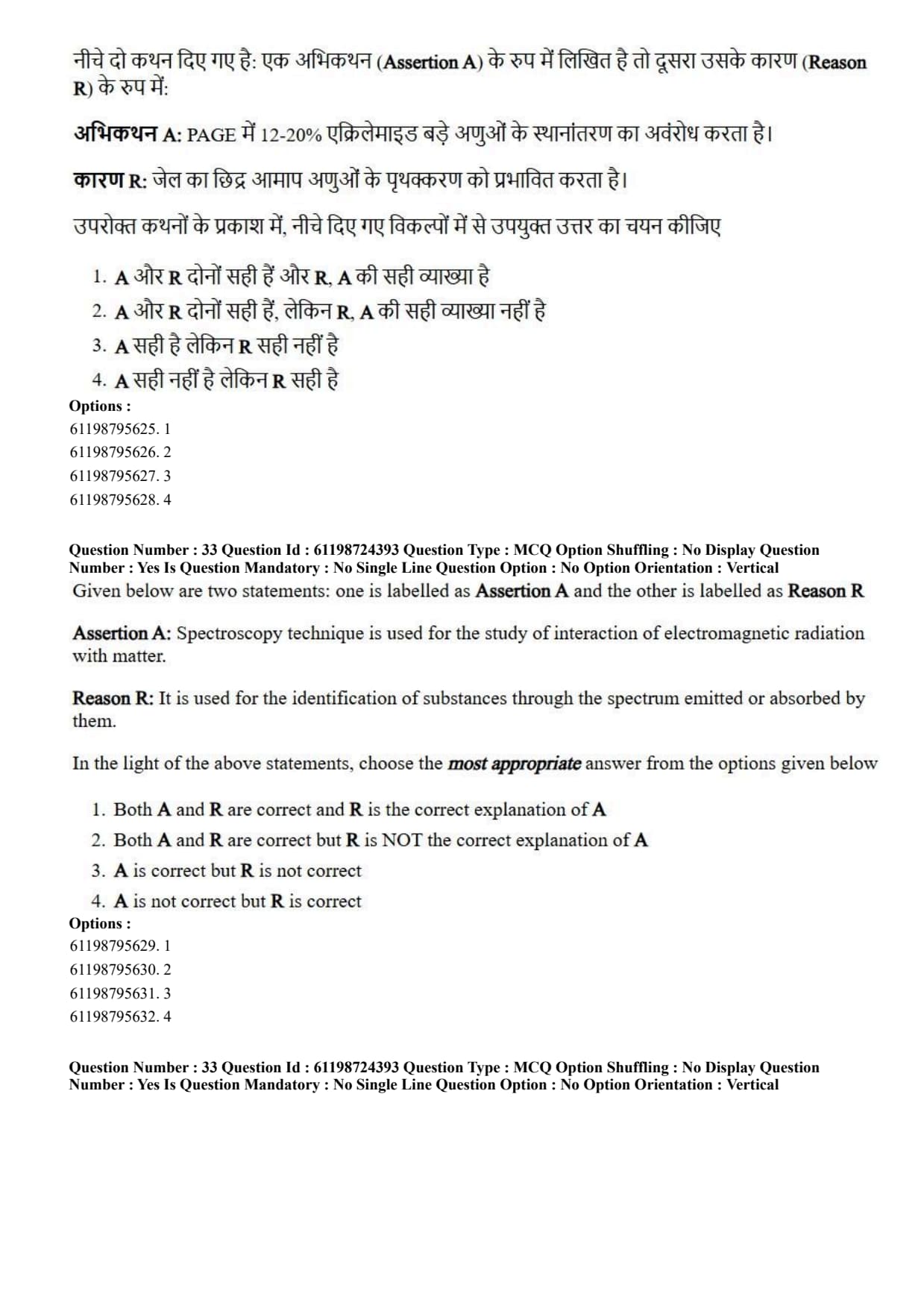
Given below are two statements: one is labelled as Assertion A and the other is labelled as Reason R
Assertion A: 12-20% acrylamide in PAGE restricts the migration of large molecules.
Reason R: Pore size of the gel affects the separation of molecules.
View Solution
Step 1: Understanding the Question:
The question presents an Assertion regarding acrylamide concentration effects in Polyacrylamide Gel Electrophoresis (PAGE) and a Reason regarding gel pore size influence on molecular separation.
Step 2: Key Formula or Approach:
In PAGE, acrylamide polymerizes with bis-acrylamide crosslinkers to form a three-dimensional molecular sieve matrix.
The average gel pore size is inversely proportional to the total concentration of acrylamide (%T). High %T acrylamide gels produce smaller pores.
Step 3: Detailed Explanation:
Analysis of Assertion A: Gels with high total acrylamide concentrations (12–20% PAGE) form dense polymer networks with small average pore diameters. These tight pores physically hinder and restrict the migration of high molecular weight (large) biomolecules, while effectively resolving small proteins or nucleic acids. Thus, Assertion A is correct.
Analysis of Reason R: Molecular separation in gel electrophoresis relies on sieving, where the pore size of the gel dictates the retarding effect experienced by molecules according to their hydrodynamic radius and size. Thus, Reason R is correct.
Causal Relationship: Because the gel's pore size directly dictates molecular sieving (Reason R), increasing the acrylamide concentration to 12–20% reduces the pore size, which directly explains why large molecules are restricted from migrating efficiently (Assertion A). Therefore, R is the correct explanation of A.
Step 4: Final Answer:
Both A and R are correct and R is the correct explanation of A.
Quick Tip: High % Acrylamide (12–20%) = Small pore size \(\to\) Best for separating SMALL molecules (restricts large ones).
Low % Acrylamide (4–7.5%) = Large pore size \(\to\) Best for separating LARGE molecules.
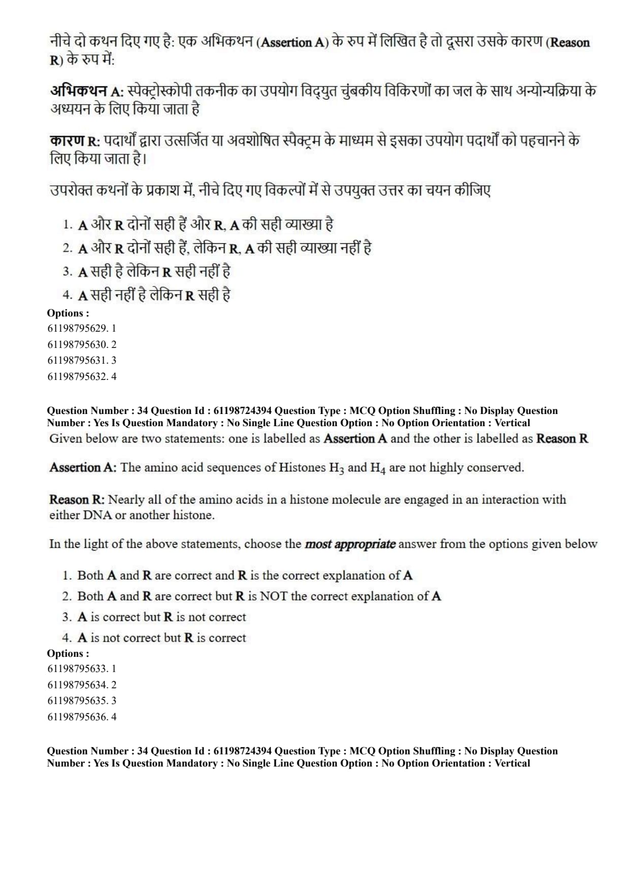
Given below are two statements: one is labelled as Assertion A and the other is labelled as Reason R
Assertion A: Spectroscopy technique is used for the study of interaction of electromagnetic radiation with matter.
Reason R: It is used for the identification of substances through the spectrum emitted or absorbed by them.
View Solution
Step 1: Understanding the Question:
The question evaluates the basic scientific definition and analytical purpose of spectroscopy in chemical and biological sciences.
Step 2: Key Formula or Approach:
Spectroscopy is the branch of science concerned with measuring and interpreting the emission and absorption of electromagnetic radiation (EMR) by matter as a function of wavelength or frequency (\(E = h\nu\)).
Step 3: Detailed Explanation:
Analysis of Assertion A: Spectroscopy is universally defined as the study of interactions between electromagnetic radiation (e.g., UV, visible, IR, microwave) and atomic or molecular matter. Thus, Assertion A is correct.
Analysis of Reason R: When matter absorbs or emits energy transitions, characteristic discrete spectral lines or broad bands are produced. These emission or absorption spectra serve as unique molecular "fingerprints" used to identify and quantify chemical substances. Thus, Reason R is correct.
Causal Relationship: The physical mechanism of measuring interactions between EMR and matter (Assertion A) is implemented analytically by recording absorption or emission spectra to identify target substances (Reason R). Thus, R provides the direct explanation of how spectroscopy functions as an analytical tool.
Step 4: Final Answer:
Both A and R are correct, and R provides the correct explanation of A.
Quick Tip: Spectroscopy = EMR + Matter Interaction.
Absorption/Emission Spectra = Unique chemical fingerprints used for structural identification and quantitative concentration measurement (Beer-Lambert Law).
Given below are two statements: one is labelled as Assertion A and the other is labelled as Reason R
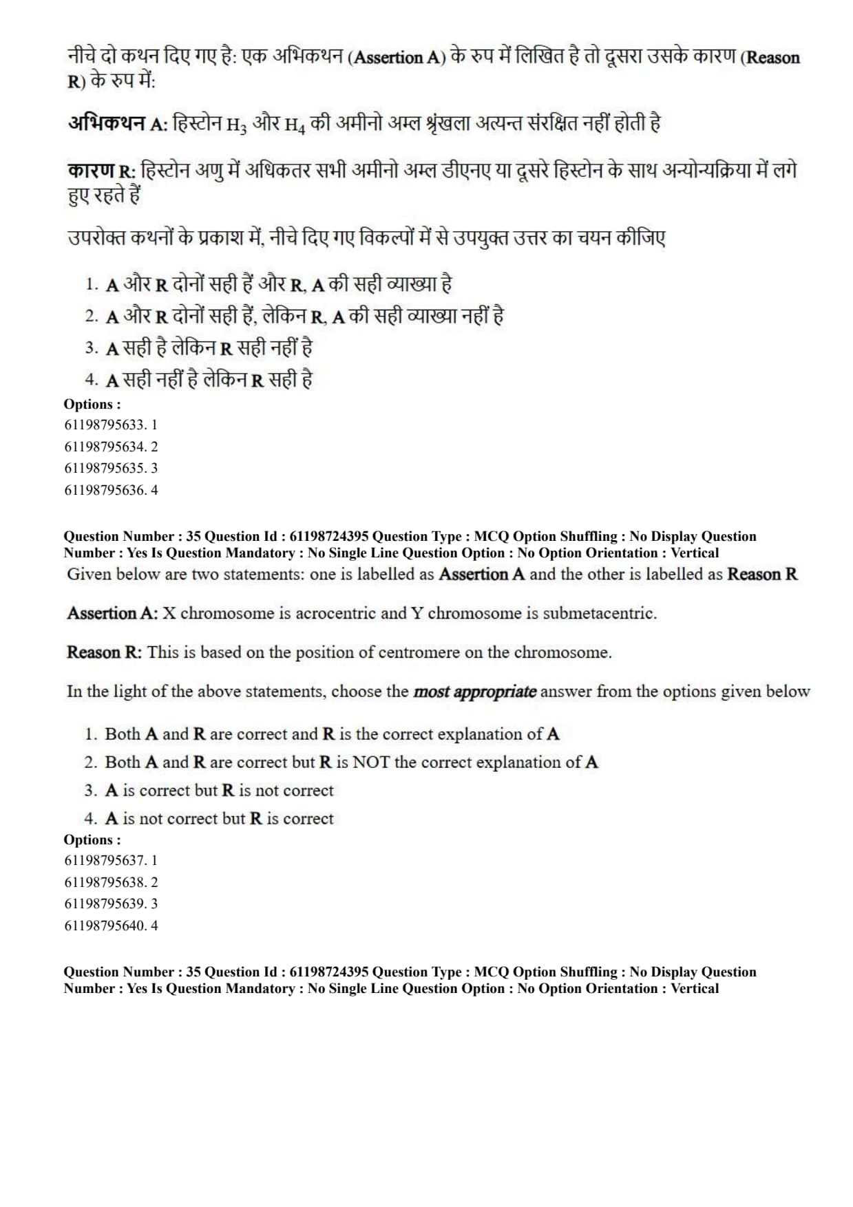
Assertion A: The amino acid sequences of Histones H3 and H4 are not highly conserved.
Reason R: Nearly all of the amino acids in a histone molecule are engaged in an interaction with either DNA or another histone.
View Solution
Step 1: Understanding the Question:
The question examines the evolutionary conservation of histone proteins (H3 and H4) and the structural basis of their molecular interactions within the nucleosome core.
Step 2: Key Formula or Approach:
Histones are basic, positively charged proteins (rich in Lysine and Arginine) that package eukaryotic DNA into nucleosomes. Core histones H3 and H4 undergo extreme selective constraint during evolution due to tight structural interactions.
Step 3: Detailed Explanation:
Analysis of Assertion A: Histone proteins H3 and H4 are among the most extraordinarily conserved proteins known across all eukaryotic evolution. For instance, histone H4 from pea seedlings differs from bovine histone H4 by only 2 amino acid residues out of 102 positions across over a billion years of evolutionary divergence. Thus, stating that H3 and H4 are *not* highly conserved is completely false. Therefore, Assertion A is incorrect.
Analysis of Reason R: Virtually every amino acid side chain in core histones is involved in essential contacts: binding negatively charged DNA phosphodiester backbones or participating in protein-protein interactions within the H3-H4 tetramer and H2A-H2B dimers. Any mutation in these residues typically disrupts nucleosome assembly and is lethal, explaining their high conservation. Thus, Reason R is correct.
Step 4: Final Answer:
Assertion A is incorrect because Histones H3 and H4 are extremely conserved, but Reason R is correct.
Quick Tip: Histones H3 and H4 are among the most evolutionary conserved proteins in all eukaryotic organisms!
Reason: Almost every amino acid residue makes crucial contacts with DNA or other histones within the nucleosome octamer.
Given below are two statements: one is labelled as Assertion A and the other is labelled as Reason R
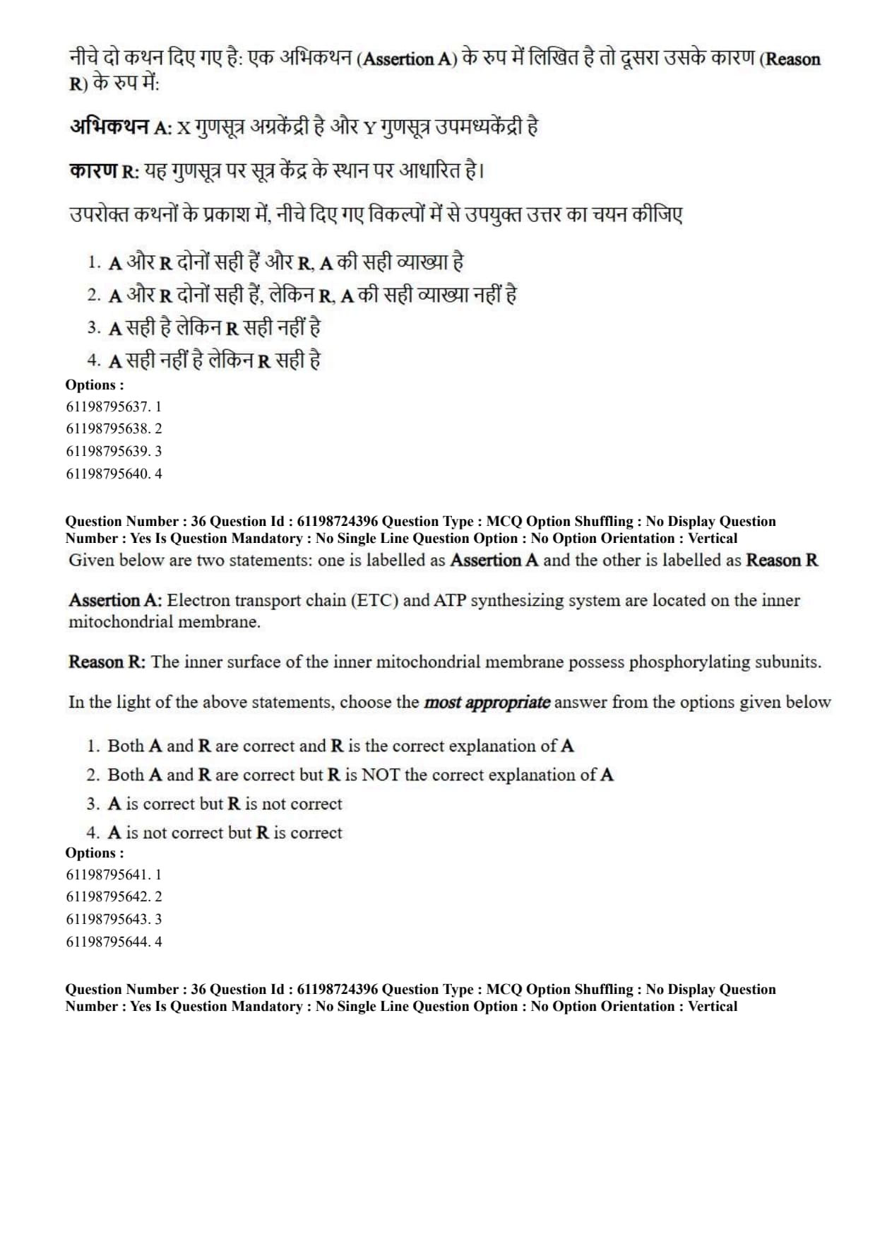
Assertion A: X chromosome is acrocentric and Y chromosome is submetacentric.
Reason R: This is based on the position of centromere on the chromosome.
View Solution
Step 1: Understanding the Question:
The question tests knowledge regarding human sex chromosome morphology (centromere positioning) and chromosome classification definitions.
Step 2: Key Formula or Approach:
Chromosomes are classified according to the relative location of their centromere:
Metacentric: Centromere in the middle; equal \(p\) and \(q\) arms.
Submetacentric: Centromere slightly off-center; unequal arms (\(p < q\)).
Acrocentric: Centromere near one end; very short \(p\) arm.
Telocentric: Centromere at the extreme terminal tip.
Step 3: Detailed Explanation:
Analysis of Assertion A: In human karyotyping:
The X chromosome is a medium-sized submetacentric chromosome (Group C).
The Y chromosome is a small acrocentric chromosome (Group G).
Assertion A claims the exact opposite (stating X is acrocentric and Y is submetacentric), which is factually incorrect. Thus, Assertion A is not correct.
Analysis of Reason R: Chromosomes are universally classified into metacentric, submetacentric, acrocentric, or telocentric categories based directly on the position of the centromere along the chromosome length. Thus, Reason R is correct.
Step 4: Final Answer:
Assertion A is not correct, but Reason R is correct.
Quick Tip: Human Chromosomes:
Human X chromosome = Submetacentric.
Human Y chromosome = Acrocentric.
Human Acrocentric chromosomes = 13, 14, 15, 21, 22, and Y.
Given below are two statements: one is labelled as Assertion A and the other is labelled as Reason R
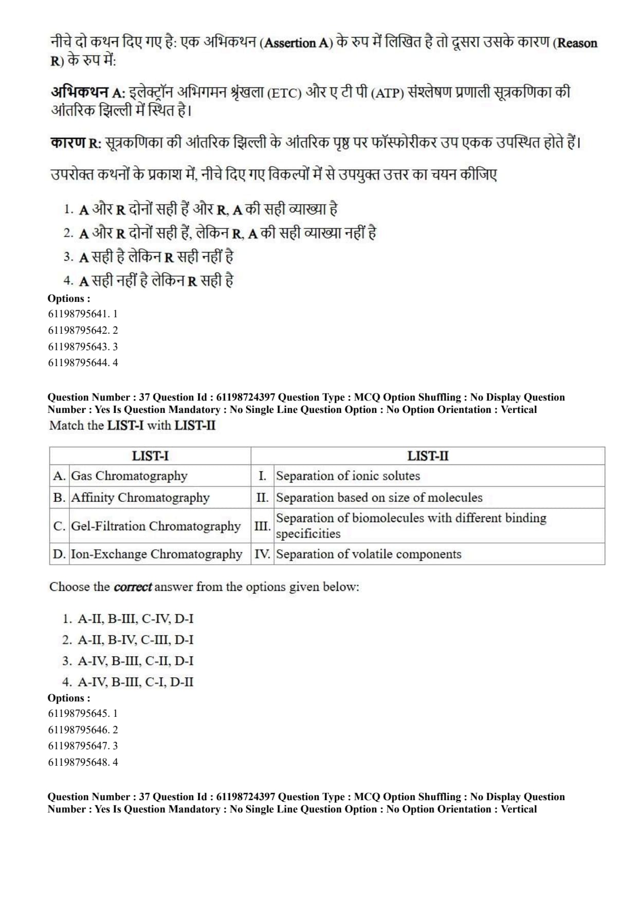
Assertion A: Electron transport chain (ETC) and ATP synthesizing system are located on the inner mitochondrial membrane.
Reason R: The inner surface of the inner mitochondrial membrane possess phosphorylating subunits.
View Solution
Step 1: Understanding the Question:
The question deals with the submitochondrial localization of electron transport chain complexes and ATP synthase subunits involved in oxidative phosphorylation.
Step 2: Key Formula or Approach:
Oxidative phosphorylation requires structural organization within the inner mitochondrial membrane (IMM). Respiratory complexes I–IV pump protons into the intermembrane space, creating a proton gradient that drives ATP synthesis via \(F_0F_1\)-ATP synthase.
Step 3: Detailed Explanation:
Analysis of Assertion A: The enzymes of the Electron Transport Chain (Complexes I, II, III, and IV) and the ATP synthesizing complex (Complex V / ATP synthase) are physically embedded within the inner mitochondrial membrane. Thus, Assertion A is correct.
Analysis of Reason R: The inner surface (matrix-facing side) of the inner mitochondrial membrane contains the spherical \(F_1\) catalytic heads (\(F_1\) phosphorylating subunits) of ATP synthase, connected via a stalk to the membrane-embedded \(F_0\) proton channel. These \(F_1\) subunits carry out the phosphorylation of ADP to ATP. Thus, Reason R is correct.
Causal Relationship: The structural presence and orientation of these catalytic phosphorylating subunits (\(F_1\)) coupled to the electron transport complexes on the inner mitochondrial membrane explains how mitochondrial energy transduction is executed at this membrane site. Thus, R is the correct explanation of A.
Step 4: Final Answer:
Both A and R are correct and R is the correct explanation of A.
Quick Tip: Inner Mitochondrial Membrane = Site of ETC complexes + ATP Synthase (\(F_0F_1\) particles).
\(F_1\) particle projects into the matrix and contains catalytic sites for ADP + \(P_i \to\) ATP.
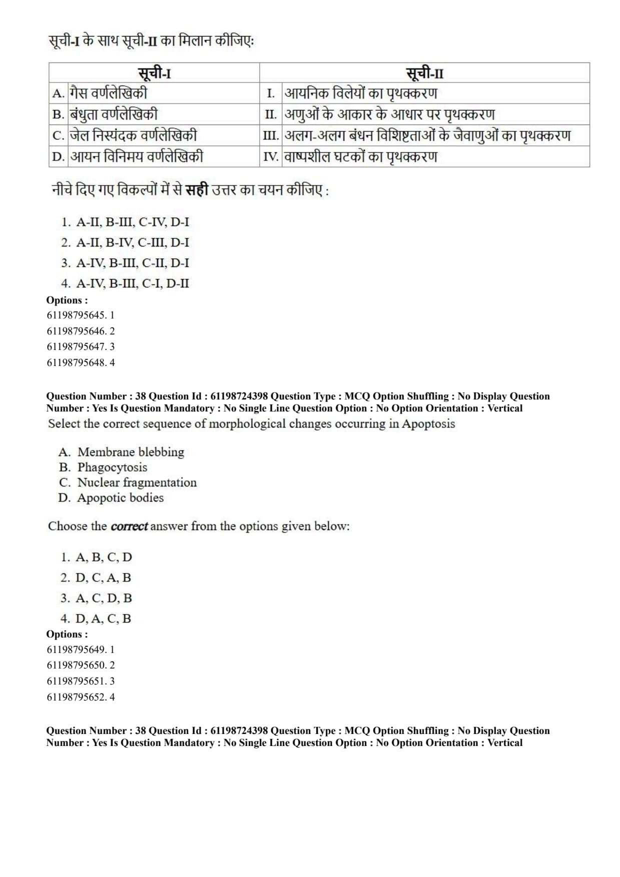
Match the LIST-I with LIST-II

View Solution
Step 1: Understanding the Question:
The question asks to match different physical separation methods of chromatography in LIST-I with their corresponding separation principles in LIST-II.
Step 2: Key Formula or Approach:
Match each chromatographic technique to its defining separation principle:
Gas Chromatography (GC): Separates thermally stable volatile or volatilized compounds.
Affinity Chromatography: Separates molecules based on specific biological ligand-receptor binding interactions.
Gel-Filtration (Size-Exclusion) Chromatography: Separates biomolecules according to molecular size/hydrodynamic volume.
Ion-Exchange Chromatography (IEC): Separates charged molecules based on net electrostatic interactions with charged matrix groups.
Step 3: Detailed Explanation:
A. Gas Chromatography \(\to\) IV. Separation of volatile components: GC uses a gaseous mobile phase to transport volatilized analytes through a coated column.
B. Affinity Chromatography \(\to\) III. Separation of biomolecules with different binding specificities: Relies on specific bioaffinity interactions (e.g., enzyme-substrate, antibody-antigen).
C. Gel-Filtration Chromatography \(\to\) II. Separation based on size of molecules: Uses porous beads; larger molecules elute first as they cannot enter pores, while smaller molecules enter pores and elute later.
D. Ion-Exchange Chromatography \(\to\) I. Separation of ionic solutes: Uses cation/anion exchange resins to separate charged species.
Step 4: Final Answer:
The correct matching sequence is A-IV, B-III, C-II, D-I.
Quick Tip: Chromatography Principles:
Gas = Volatile compounds.
Affinity = Specific biological ligand binding.
Gel-Filtration = Molecular Size.
Ion-Exchange = Charge / Ionic solutes.
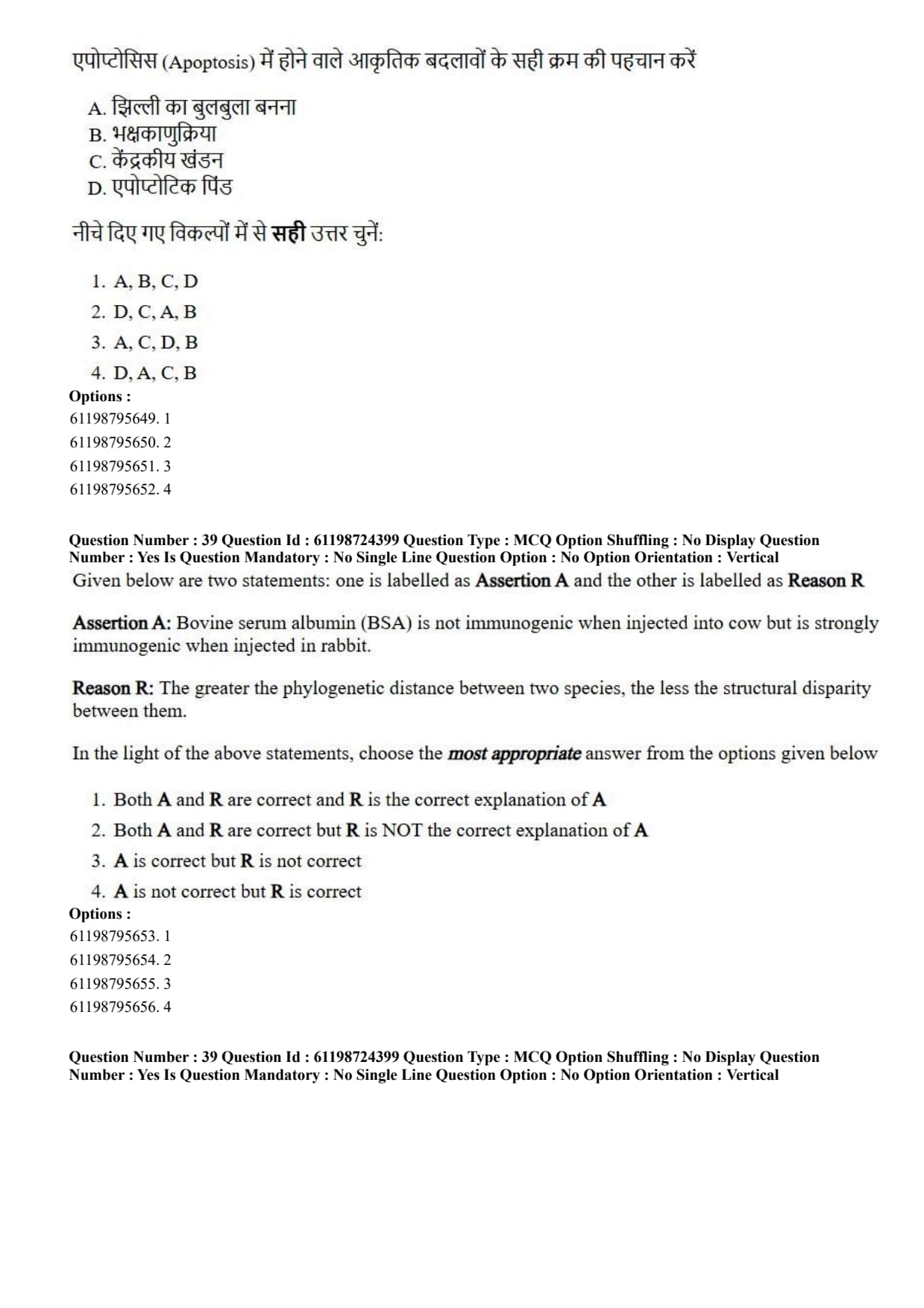
Select the correct sequence of morphological changes occurring in Apoptosis
A. Membrane blebbing
B. Phagocytosis
C. Nuclear fragmentation
D. Apoptotic bodies
View Solution
Step 1: Understanding the Question:
The question asks for the correct temporal order of morphological and cellular events taking place during programmed cell death (apoptosis).
Step 2: Key Formula or Approach:
Apoptosis follows a strictly regulated, orderly sequence of morphological transformations that prevents release of intracellular contents and inflammatory responses:
Membrane Blebbing (A) \(\rightarrow{}\) Nuclear Fragmentation (C) \(\rightarrow{}\) Formation of Apoptotic Bodies (D) \(\rightarrow{}\) Phagocytosis (B)
Step 3: Detailed Explanation:
1. Cell Shrinkage and Membrane Blebbing (A): Early in apoptosis, executioner caspases cleave cytoskeletal proteins, leading to cell shrinkage and dynamic cell membrane bulging known as membrane blebbing.
2. Nuclear Condensation and Fragmentation (C): Pyknosis (chromatin condensation) is followed by karyorrhexis, where endonuclease activation (CAD) fragments nuclear chromatin into nucleosomal units.
3. Formation of Apoptotic Bodies (D): The dying cell pinches off into multiple membrane-bound vesicles containing cytosol, organelles, and nuclear fragments, termed apoptotic bodies.
4. Phagocytosis (B): Apoptotic bodies display "eat-me" signals (such as externalized phosphatidylserine) on their surface and are rapidly recognized and engulfed by phagocytes (macrophages) without inducing inflammation.
Step 4: Final Answer:
The correct sequential order of apoptotic morphological events is A, C, D, B.
Quick Tip: Chronological steps of Apoptosis:
1. Membrane Blebbing \(\to\) 2. Nuclear Fragmentation \(\to\) 3. Apoptotic Bodies formation \(\to\) 4. Phagotic clearance (Phagocytosis).
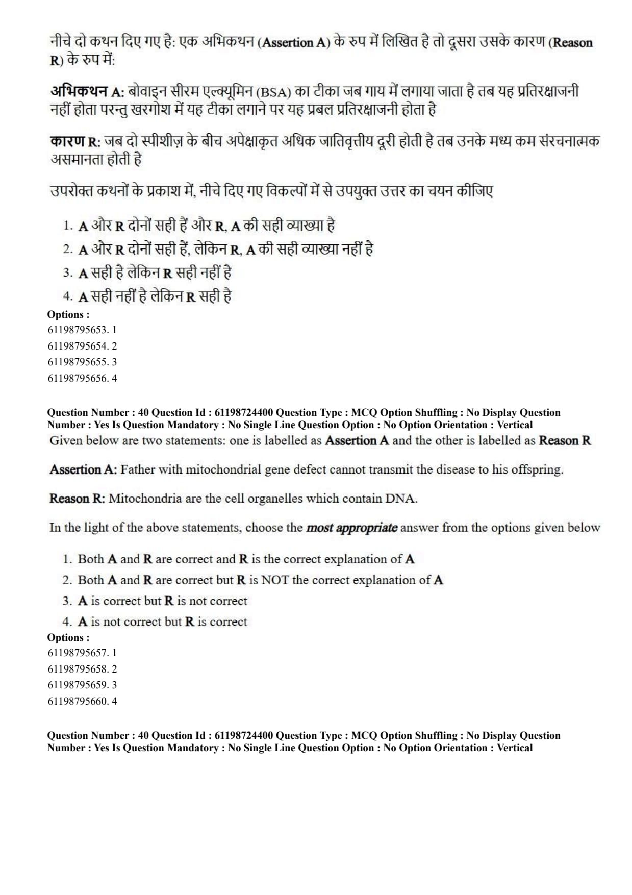
Given below are two statements: one is labelled as Assertion A and the other is labelled as Reason R
Assertion A: Bovine serum albumin (BSA) is not immunogenic when injected into cow but is strongly immunogenic when injected in rabbit.
Reason R: The greater the phylogenetic distance between two species, the less the structural disparity between them.
View Solution
Step 1: Understanding the Question:
The question tests concepts regarding antigenicity, foreignness, immunogenicity, and the relationship between evolutionary phylogenetic distance and protein structural divergence.
Step 2: Key Formula or Approach:
For a molecule to act as a potent immunogen, it must be recognized as "non-self" (foreign) by the recipient immune system.
Greater evolutionary (phylogenetic) distance between species causes greater cumulative sequence and structural divergence in homologous proteins.
Step 3: Detailed Explanation:
Analysis of Assertion A: Bovine Serum Albumin (BSA) is a self-protein in cows; injecting BSA into a cow does not elicit an immune response due to self-tolerance. However, when injected into a rabbit (a phylogenetically distant species), the rabbit immune system recognizes BSA as foreign, mounting a strong primary antibody response. Thus, Assertion A is correct.
Analysis of Reason R: Evolutionary divergence dictates that the *greater* the phylogenetic distance between two species, the *greater* (not less) the structural disparity and sequence differences between their homologous proteins. Therefore, the claim in Reason R is factually incorrect.
Step 4: Final Answer:
Assertion A is correct, but Reason R is incorrect.
Quick Tip: Immunogenicity depends directly on "Foreignness":
Greater Phylogenetic Distance = GREATER Structural Disparity = Higher Immunogenicity.
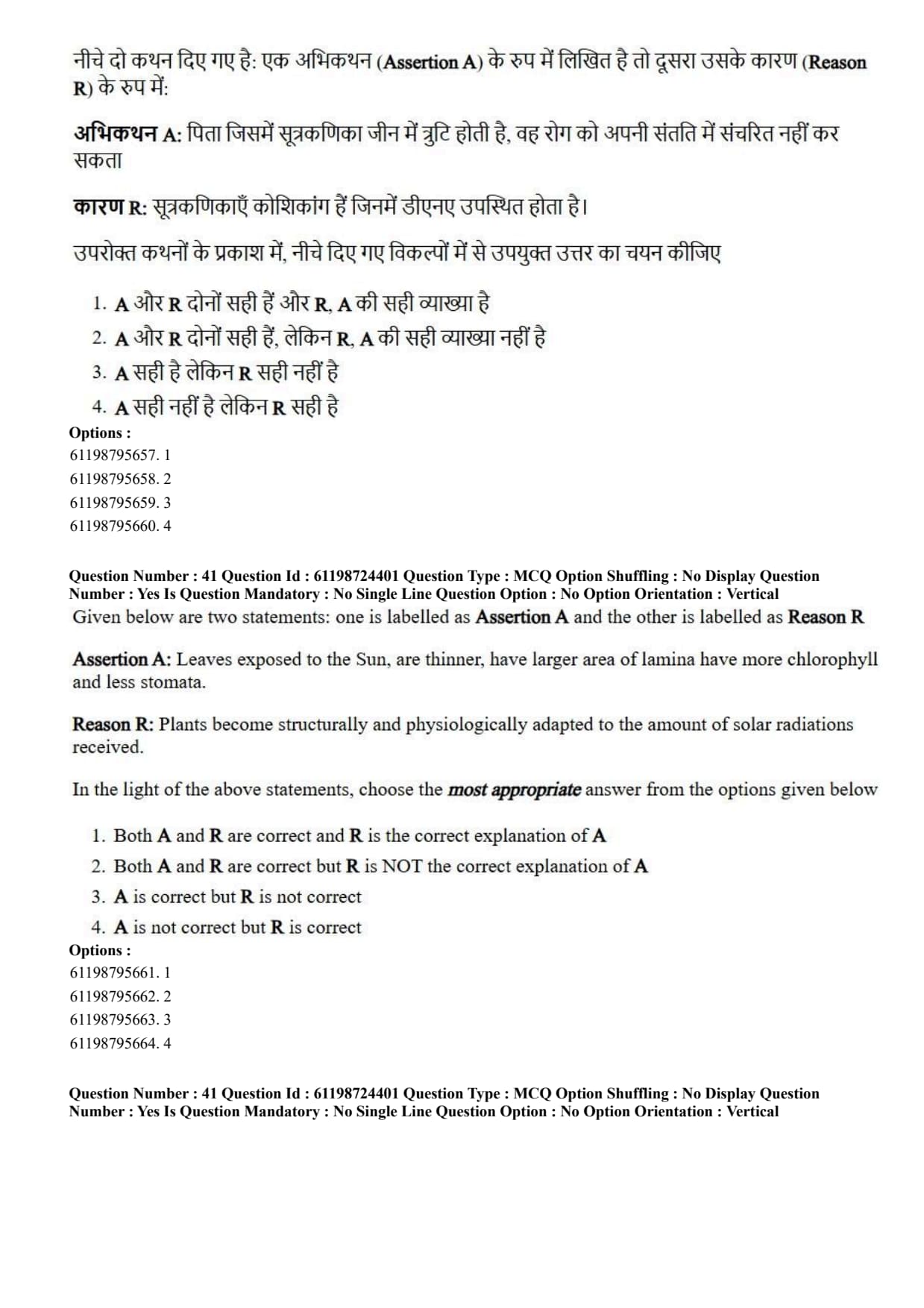
Given below are two statements: one is labelled as Assertion A and the other is labelled as Reason R
Assertion A: Father with mitochondrial gene defect cannot transmit the disease to his offspring.
Reason R: Mitochondria are the cell organelles which contain DNA.
View Solution
Step 1: Understanding the Question:
The question assesses maternal inheritance patterns of mitochondrial diseases and the presence of extranuclear genomic DNA within mitochondria.
Step 2: Key Formula or Approach:
Mitochondrial DNA (mtDNA) exhibits non-Mendelian, non-nuclear maternal inheritance. The zygote receives virtually all of its cytoplasm and organelles from the egg cell (maternal parent), while sperm contributes only a haploid nucleus.
Step 3: Detailed Explanation:
Analysis of Assertion A: Because sperm mitochondria are located in the tail/flagellum and are either excluded during fertilization or targeted for ubiquitin-mediated degradation inside the egg, paternal mtDNA is not passed to offspring. Thus, a father with a mitochondrial gene defect cannot transmit the condition to his children. Thus, Assertion A is correct.
Analysis of Reason R: Mitochondria are semiautonomous endosymbiotic organelles that harbor their own circular genome (mtDNA) encoding essential rRNA, tRNA, and electron transport proteins. Thus, Reason R is correct.
Causal Relationship: While Reason R correctly states that mitochondria contain DNA, it does not explain *why* transmission occurs exclusively through mothers. The actual explanation for non-transmission by fathers is maternal cytoplasmic inheritance (zygote cytoplasm originating from the egg). Thus, R is NOT the correct explanation of A.
Step 4: Final Answer:
Both A and R are correct, but R is NOT the correct explanation of A.
Quick Tip: Mitochondrial diseases are strictly inherited MATERNALLY.
An affected mother passes the trait to ALL her children.
An affected father NEVER passes the trait to any of his children.
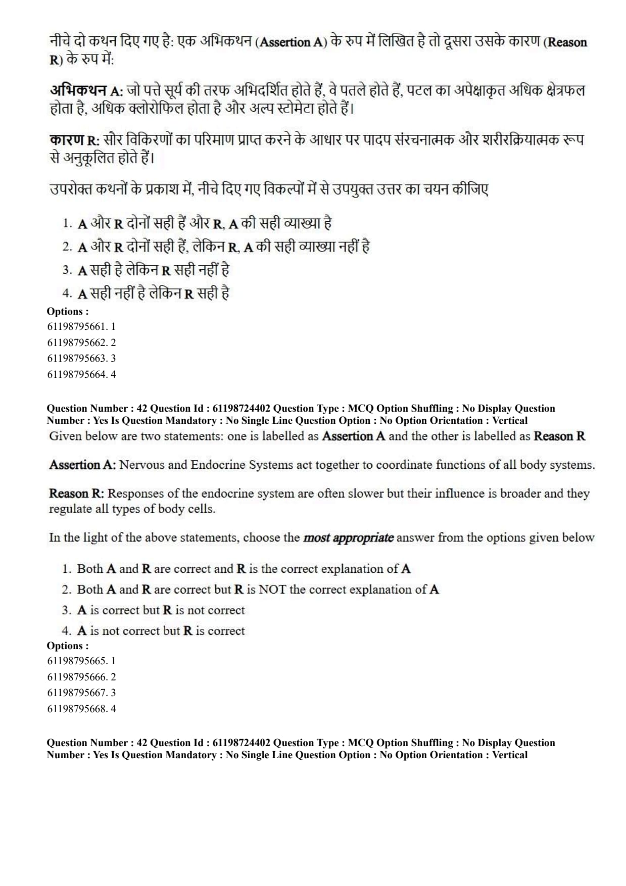
Given below are two statements: one is labelled as Assertion A and the other is labelled as Reason R
Assertion A: Leaves exposed to the Sun, are thinner, have larger area of lamina have more chlorophyll and less stomata.
Reason R: Plants become structurally and physiologically adapted to the amount of solar radiations received.
View Solution
Step 1: Understanding the Question:
The question tests knowledge regarding anatomical and physiological leaf adaptations (sun leaves versus shade leaves) under varying solar irradiance levels.
Step 2: Key Formula or Approach:
Plants adapt leaf structure based on solar exposure:
Sun Leaves: Adapted to high light intensity \(\to\) Thicker lamina, multi-layered palisade mesophyll, smaller surface area, higher stomatal density, lower total chlorophyll per unit weight.
Shade Leaves: Adapted to light capture in dim conditions \(\to\) Thinner lamina, larger surface area (lamina), higher total chlorophyll content to maximize light absorption.
Step 3: Detailed Explanation:
Analysis of Assertion A: Assertion A claims that sun leaves are *thinner*, have *larger area*, *more chlorophyll*, and *fewer stomata*. This description actually corresponds to shade leaves. Sun leaves are thicker (due to well-developed palisade tissue), have smaller leaf area to restrict transpiration, higher stomatal density, and less total chlorophyll per weight. Thus, Assertion A is incorrect.
Analysis of Reason R: Plants demonstrate phenotypic plasticity, adapting both structurally (cuticle thickness, mesophyll layers) and physiologically (photosynthetic capacity, light saturation points) to ambient light levels. Thus, Reason R is correct.
Step 4: Final Answer:
Assertion A is not correct, but Reason R is correct.
Quick Tip: Sun Leaves = THICKER, smaller lamina area, dense stomata, thick cuticle (prevents water loss).
Shade Leaves = THINNER, broader lamina area, higher chlorophyll content (captures maximum light).
Given below are two statements: one is labelled as Assertion A and the other is labelled as Reason R
Assertion A: Nervous and Endocrine Systems act together to coordinate functions of all body systems.
Reason R: Responses of the endocrine system are often slower but their influence is broader and they regulate all types of body cells.
View Solution
Step 1: Understanding the Question:
The question examines the integrative physiological roles and functional differences between the nervous system and the endocrine system in maintaining homeostasis.
Step 2: Key Formula or Approach:
Homeostasis is maintained through neuroendocrine integration:
Nervous System: Delivers rapid, brief, localized point-to-point electrical/chemical impulses along neurons.
Endocrine System: Releases hormones into the bloodstream, producing slower, longer-lasting, and widespread regulatory effects across diverse target tissues.
Step 3: Detailed Explanation:
Analysis of Assertion A: Body systems rely on the combined neuroendocrine system to integrate internal functions. The nervous and endocrine systems work synergistically (e.g., via the hypothalamic-pituitary axis) to control physiological processes. Thus, Assertion A is correct.
Analysis of Reason R: Endocrine signals travel through blood circulation, causing their onset of action to be slower than nerve impulses. However, because hormones circulate systemically to reach all cells possessing complementary receptors, their effects are widespread and long-lasting. Thus, Reason R is correct.
Causal Relationship: The dual nature of neuroendocrine regulation (fast/localized neural responses combined with slow/broad endocrine actions described in Reason R) explains how both systems effectively coordinate all body functions together. Thus, R is the correct explanation of A.
Step 4: Final Answer:
Both A and R are correct, and R provides the correct explanation of A.
Quick Tip: Nervous System = Fast speed, short duration, highly localized targets.
Endocrine System = Slower speed, long duration, broad/widespread targets.
Together, they maintain homeostatic coordination.
Given below are two statements: one is labelled as Assertion A and the other is labelled as Reason R
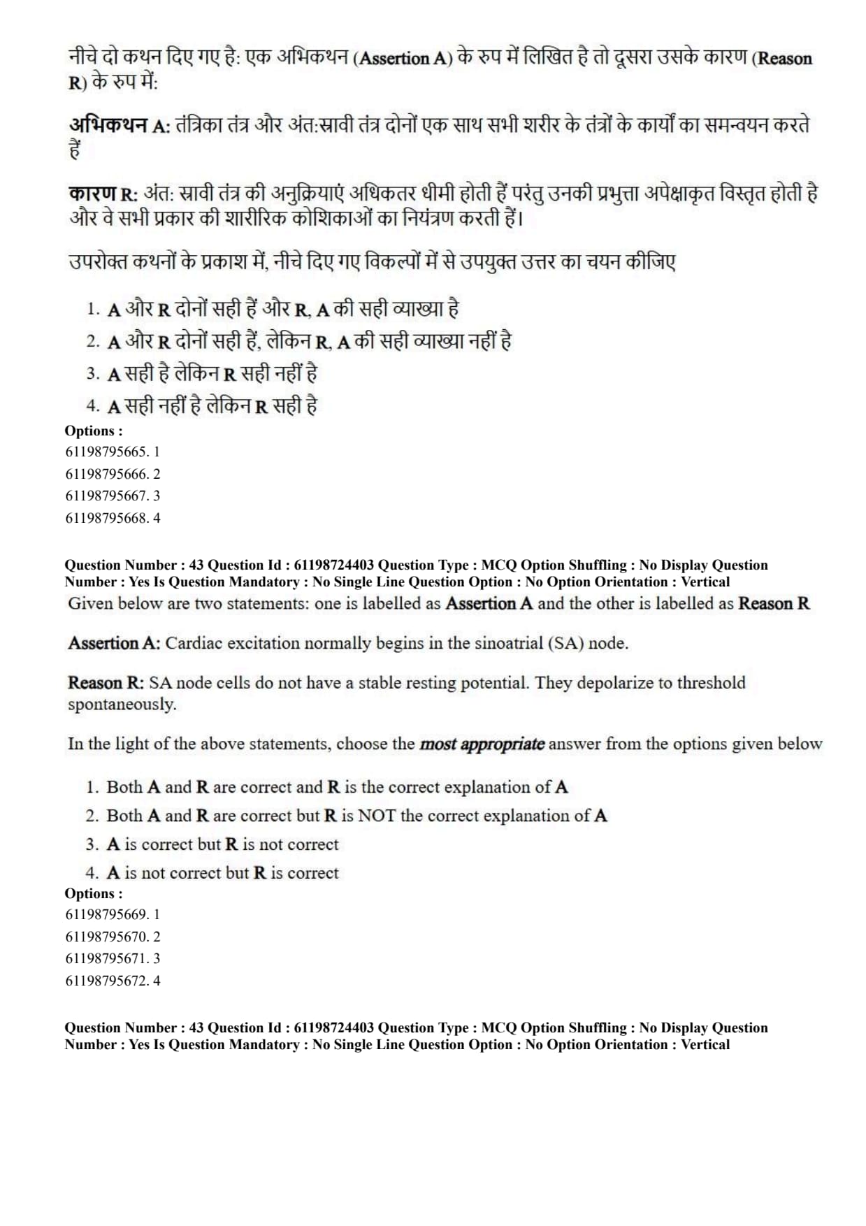
Assertion A: Cardiac excitation normally begins in the sinoatrial (SA) node.
Reason R: SA node cells do not have a stable resting potential. They depolarize to threshold spontaneously.
View Solution
Step 1: Understanding the Question:
The question explores the cardiac conduction system, focusing on why the sinoatrial (SA) node functions as the primary natural pacemaker of the heart.
Step 2: Key Formula or Approach:
Autorhythmic cardiac cells initiate spontaneous action potentials without external neural stimulation due to an unstable resting membrane potential known as the pacemaker potential (prepotential).
Step 3: Detailed Explanation:
Analysis of Assertion A: The sinoatrial (SA) node, situated in the right atrium wall, naturally initiates each heartbeat, establishing the cardiac sinus rhythm. Thus, Assertion A is correct.
Analysis of Reason R: Unlike contractile cardiac myocytes, SA node autorhythmic cells lack a constant, stable resting membrane potential. Inward hyperpolarization-activated cyclic nucleotide-gated channels (\(I_f\) "funny" currents) and T-type \(Ca^{2+}\) channels cause gradual, spontaneous depolarization toward threshold potential (\(\sim -40\ mV\)). Thus, Reason R is correct.
Causal Relationship: Because SA node cells depolarize spontaneously faster than any other autorhythmic region (generating 70–80 action potentials/minute), they reach threshold first and drive the entire cardiac conduction system. Therefore, Reason R directly explains why excitation begins in the SA node.
Step 4: Final Answer:
Both A and R are correct, and R is the correct explanation of A.
Quick Tip: SA Node = Primary Pacemaker of the heart.
Mechanism: Unstable resting potential (Pacemaker Potential driven by \(I_f\) "funny" \(Na^+\) currents) \(\to\) Spontaneous intrinsic depolarization.
For identification of desired DNA by Southern blotting, select the correct order
A. Blotting
B. Probe binding
C. Autoradiogram
D. Electrophoresis
E. Gel treatment
View Solution
Step 1: Understanding the Question:
The question asks for the proper sequential order of experimental steps carried out during Southern blotting for target DNA detection.
Step 2: Key Formula or Approach:
Southern blotting involves digesting target DNA with restriction enzymes, separating fragments by gel electrophoresis, denaturing DNA, transferring single strands to a membrane, hybridizing with labeled probes, and detecting bands via imaging.
Step 3: Detailed Explanation:
Step 1: Electrophoresis (D): Cleaved DNA fragments are separated according to molecular size on an agarose gel.
Step 2: Gel Treatment (E): The gel is treated with alkaline solution (\(NaOH\)) to denature double-stranded DNA into single-stranded DNA.
Step 3: Blotting (A): Single-stranded DNA is transferred from the gel onto a solid support membrane (nitrocellulose or nylon) via capillary or vacuum blotting.
Step 4: Probe Binding (B): The membrane is incubated with a labeled single-stranded probe to allow complementary hybridization.
Step 5: Autoradiogram (C): Unbound probes are washed away, and hybridized probe-target bands are visualized using autoradiography or chemiluminescent imaging.
Step 4: Final Answer:
The correct experimental sequence is D, E, A, B, C.
Quick Tip: Southern Blotting Protocol Steps:
1. Electrophoresis (D) \(\to\) 2. Gel Treatment/Denaturation (E) \(\to\) 3. Membrane Blotting (A) \(\to\) 4. Probe Hybridization (B) \(\to\) 5. Autoradiography Detection (C).
Arrange the following in order 5' - 3' direction
A. Promoter
B. UTR5'
C. ORF
D. Terminator
View Solution
Step 1: Understanding the Question:
The question asks to arrange the functional regulatory and coding elements of a gene transcription unit in sequential order from the \(5'\) end to the \(3'\) end.
Step 2: Key Formula or Approach:
A functional eukaryotic transcription unit displays structural organization along the sense strand in the \(5' \to 3'\) orientation: \[ 5'-Promoter (A) \to 5'-UTR (B) \to ORF (C) \to 3'-Terminator (D)-3' \]
Step 3: Detailed Explanation:
1. Promoter (A): Upstream regulatory sequence located at the extreme \(5'\) end of the transcription unit, serving as the binding site for transcription factors and RNA polymerase.
2. 5'-UTR (B): \(5'\) UnTranslated Region located downstream of the transcription start site but upstream of the translation initiation codon (AUG).
3. ORF (C): Open Reading Frame containing continuous protein-coding codons extending from the start codon to the stop codon.
4. Terminator (D): Downstream sequence located at the extreme \(3'\) end that signals transcription termination and pre-mRNA cleavage/polyadenylation.
Step 4: Final Answer:
The correct \(5' \to 3'\) order of gene elements is A, B, C, D.
Quick Tip: Gene Structure (\(5' \to 3'\) Direction):
Promoter \(\to 5'\)-UTR \(\to\) ORF (Coding Region) \(\to 3'\)-UTR \(\to\) Terminator.
For transport of acyl CoA into mitochondria, select the correct order of steps involved:
A. Acyl- carnitine to acyl CoA
B. Acyl- carnitine transported to mitochondrial matrix
C. Acyl group of acyl CoA transferred to carnitine
D. Carnitine released and return to cytosol
View Solution
Step 1: Understanding the Question:
The question asks for the correct temporal sequence of biochemical steps in the carnitine shuttle system, which transports long-chain fatty acyl-CoA molecules across the impermeable inner mitochondrial membrane into the matrix for \(\beta\)-oxidation.
Step 2: Key Formula or Approach:
The carnitine shuttle operates via a four-step enzymatic sequence across the outer membrane, intermembrane space, and inner mitochondrial membrane:
Transesterification to Carnitine (C) \(\rightarrow{}\) Translocation into Matrix (B) \(\rightarrow{}\) Re-esterification to CoA (A) \(\rightarrow{}\) Carnitine Recycling (D)
Step 3: Detailed Explanation:
1. Transfer of Acyl group to Carnitine (C): Fatty acyl-CoA generated in the cytosol cannot directly cross the inner mitochondrial membrane. The outer membrane enzyme Carnitine Palmitoyltransferase-I (CPT-I) transfers the fatty acyl group from acyl-CoA to carnitine, forming acyl-carnitine and releasing free CoA.
2. Transport across Inner Membrane (B): Acyl-carnitine is translocated across the inner mitochondrial membrane into the mitochondrial matrix by Carnitine-Acylcarnitine Translocase (CACT) via antiport exchange.
3. Conversion back to Acyl-CoA (A): Inside the matrix, Carnitine Palmitoyltransferase-II (CPT-II) transfers the acyl group from acyl-carnitine back onto matrix Coenzyme A, reforming acyl-CoA for \(\beta\)-oxidation.
4. Release and Return of Carnitine (D): The freed carnitine molecule is transported back across the inner membrane into the intermembrane space/cytosol by the translocase to participate in subsequent transport cycles.
Step 4: Final Answer:
The correct step sequence for fatty acyl transport into mitochondria is C, B, A, D.
Quick Tip: Carnitine Shuttle Order:
1. CPT-I attaches carnitine to acyl group (C).
2. Translocase shuttles acyl-carnitine into matrix (B).
3. CPT-II converts acyl-carnitine back to acyl-CoA (A).
4. Free carnitine shuttles back to cytosol (D).
Select the correct sequence of steps involved in TCA cycle:
A. \(\alpha\)-Ketoglutarate
B. Succinyl CoA
C. Fumarate
D. Citrate
E. Oxaloacetate
View Solution
Step 1: Understanding the Question:
The question asks to arrange five key intermediate metabolites of the Tricarboxylic Acid (TCA / Krebs) cycle in their correct chronological order of generation.
Step 2: Key Formula or Approach:
Trace the sequential pathway of the TCA cycle starting from condensation of Acetyl-CoA (\(2C\)) with Oxaloacetate (\(4C\)):
Citrate (6C) \(\rightarrow{\) Isocitrate (6C) \(\rightarrow{\) \alpha-Ketoglutarate (5C) \(\rightarrow{\) Succinyl-CoA (4C) \(\rightarrow{\) Succinate (4C) \(\rightarrow{\) Fumarate (4C) \(\rightarrow{\) Malate (4C) \(\rightarrow{\) Oxaloacetate (4C)
Step 3: Detailed Explanation:
1. Citrate (D): Formed first by Citrate Synthase condensing Oxaloacetate and Acetyl-CoA (\(6\text{C\) compound).
2. \(\alpha\)-Ketoglutarate (A): Produced via oxidative decarboxylation of Isocitrate by Isocitrate Dehydrogenase (\(5C\) compound, releasing \(CO_2\) and \(NADH\)).
3. Succinyl CoA (B): Produced via second oxidative decarboxylation by \(\alpha\)-Ketoglutarate Dehydrogenase complex (\(4C\) compound, releasing \(CO_2\) and \(NADH\)).
4. Fumarate (C): Produced downstream when Succinate is oxidized by Succinate Dehydrogenase (Complex II, generating \(FADH_2\)).
5. Oxaloacetate (E): Regenerated at the final step when Malate is oxidized by Malate Dehydrogenase (generating \(NADH\)).
Step 4: Final Answer:
The correct sequential order of intermediate generation in the TCA cycle is D, A, B, C, E.
Quick Tip: Mnemonic for TCA Cycle Intermediates:
\textbf{C}itrate \(\to\) \textbf{I}socitrate \(\to\) \textbf{\(\alpha\)}-Ketoglutarate \(\to\) \textbf{S}uccinyl-CoA \(\to\) \textbf{S}uccinate \(\to\) \textbf{F}umarate \(\to\) \textbf{M}alate \(\to\) \textbf{O}xaloacetate.
("Can I Keep Selling Substances For Much Money")
Select the correct order of steps of plant tissue culture:
A. Explant sterilization
B. Selection of media
C. Excision of regenerated shoots
D. Inoculation of explants
E. Transfer of plantlets to sterilised soil
View Solution
Step 1: Understanding the Question:
The question requires arranging the basic procedural workflow of plant tissue culture and micropropagation in correct chronological sequence.
Step 2: Key Formula or Approach:
Plant tissue culture progresses sequentially from nutrient preparation to aseptic explant establishment, shoot/root organogenesis, shoot multiplication, and acclimatization:
Media Selection (B) \(\rightarrow{\) Explant Surface Sterilization (A) \to \text{Inoculation (D) \(\rightarrow{\) Shoot Excision (C) \(\rightarrow{\) \text{Acclimatization/Soil Transfer (E)
Step 3: Detailed Explanation:
1. Selection/Preparation of Media (B): Formulating and autoclaving appropriate culture media (e.g., Murashige and Skoog medium) containing essential macronutrients, micronutrients, carbon source, and plant growth regulators (auxins and cytokinins).
2. Explant Sterilization (A): Surface sterilizing plant tissue explants (leaf, stem, or node) using chemical disinfectants (such as sodium hypochlorite or mercuric chloride) under aseptic conditions.
3. Inoculation of Explants (D): Transferring the sterile explants onto the nutrient culture medium inside a laminar air flow cabinet.
4. Excision of Regenerated Shoots (C): Separating and excising regenerated microshoots for subculturing or rooting media treatment once organogenesis occurs.
5. Transfer to Sterilised Soil (E): Hardening off developed plantlets and acclimatizing them in sterilised soil/greenhouse before field transfer.
Step 4: Final Answer:
The correct sequence of plant tissue culture steps is B, A, D, C, E.
Quick Tip: Plant Tissue Culture Workflow:
1. Prepare media (B) \(\to\) 2. Surface sterilize tissue (A) \(\to\) 3. Inoculate onto media (D) \(\to\) 4. Excise shoots (C) \(\to\) 5. Hardening in soil (E).
Select the correct order of steps involved in Cloning:
A. Selection
B. Restriction digestion
C. Ligation
D. Transformation
E. Gene of Interest (Insert) and vector
View Solution
Step 1: Understanding the Question:
The question asks for the standard chronological sequence of steps involved in gene cloning using recombinant DNA technology.
Step 2: Key Formula or Approach:
Recombinant DNA cloning involves isolating target DNA and vector, cutting both with restriction enzymes, joining them covalently, introducing the recombinant construct into host cells, and screening transformed colonies:
Isolate target DNA/Vector (E) \(\rightarrow{\) Restriction Cleavage (B) \to \text{DNA Ligation (C) \(\rightarrow{\) Host Transformation (D) \(\rightarrow{\) \text{Colony Selection (A)
Step 3: Detailed Explanation:
1. Target DNA and Vector Preparation (E): Obtaining the gene of interest (insert DNA) and selecting an appropriate cloning vector (such as pBR322 or pUC plasmid).
2. Restriction Digestion (B): Cleaving both the target DNA insert and vector plasmid using complementary restriction endonucleases to generate matching sticky or blunt ends.
3. Ligation (C): Incubating digested insert and vector DNA together with T4 DNA Ligase to form phosphodiester bonds, generating recombinant DNA molecules.
4. Transformation (D): Introducing recombinant plasmids into competent host bacterial cells (e.g., \textit{E. coli) via heat-shock or electroporation.
5. Selection (A): Plating host bacteria onto selective media (containing antibiotics like ampicillin or X-gal/IPTG for blue-white screening) to identify recombinant transformants.
Step 4: Final Answer:
The correct step sequence in molecular cloning is E, B, C, D, A.
Quick Tip: Cloning Core Steps:
1. DNA/Vector (E) \(\to\) 2. Cut/Digest (B) \(\to\) 3. Paste/Ligate (C) \(\to\) 4. Transform (D) \(\to\) 5. Select (A).
Select the correct order of the steps involved in Gram staining:
A. Counter staining
B. Crystal violet- Iodine (CV-I)
C. Heat Fixation
D. Decolorizing agent
View Solution
Step 1: Understanding the Question:
The question asks for the proper sequential order of procedures involved in performing a Gram stain differential staining procedure on bacterial smears.
Step 2: Key Formula or Approach:
Gram staining differentiates Gram-positive (purple) and Gram-negative (pink/red) bacteria based on cell wall peptidoglycan thickness:
Smear Fixation (C) \(\rightarrow{\) Primary Stain + Mordant (B) \(\rightarrow{\) Decolorization (D) \(\rightarrow{\) \text{Counterstaining (A)
Step 3: Detailed Explanation:
1. Heat Fixation (C): A bacterial smear is air-dried and gently heat-fixed over a flame to adhere cells to the glass slide and kill microbes.
2. Crystal Violet and Iodine Mordant (B): Crystal violet primary stain is applied, followed by Gram's iodine (mordant) application to form large, insoluble Crystal Violet-Iodine (CV-I) complexes inside the peptidoglycan wall.
3. Decolorization (D): Applying decolorizer (95% ethanol or acetone). Ethanol dehydrates thick peptidoglycan in Gram-positive walls (trapping CV-I, retaining purple dye), but dissolves outer lipid membranes of Gram-negative walls, leaching out the CV-I complex (making them colorless).
4. Counterstaining (A): Applying safranin counterstain, which stains decolorized Gram-negative cells pink/red while Gram-positive cells remain purple.
Step 4: Final Answer:
The correct sequential order of Gram staining steps is C, B, D, A.
Quick Tip: Gram Staining Sequence:
1. Heat Fix slide (C) \(\to\) 2. Crystal Violet + Iodine (B) \(\to\) 3. Alcohol Decolorizer (D) \(\to\) 4. Safranin Counterstain (A).
Result: Gram-positive = Purple, Gram-negative = Pink/Red.
Steps involved in Translation. Select the correct order.
A. Formation of peptide bond between charged tRNA
B. Activation of amino acids in the presence of ATP
C. Release of the complete polypeptide
D. Linking of amino acid to their cognate tRNA
View Solution
Step 1: Understanding the Question:
The question asks to arrange the sequential steps of protein translation from amino acid activation to polypeptide termination.
Step 2: Key Formula or Approach:
Translation encompasses charging of tRNAs followed by initiation, elongation, and termination phases:
Amino Acid Activation (B) \(\rightarrow{\) tRNA Charging (D) \(\rightarrow{\)Peptide Bond Formation (A) \(\rightarrow{\) \text{Polypeptide Release (C)
Step 3: Detailed Explanation:
1. Activation of Amino Acids (B): Aminoacyl-tRNA synthetase reacts an amino acid with ATP to form an intermediate aminoacyl-AMP derivative and inorganic pyrophosphate (\(PP_i\)).
2. tRNA Charging / Aminoacylation (D): The activated amino acid moiety is transferred onto the \(3'\)-CCA terminal adenylate residue of its cognate tRNA, releasing AMP and yielding charged aminoacyl-tRNA.
3. Peptide Bond Formation (A): During elongation, peptidyl transferase activity of the large ribosomal subunit forms a peptide bond between the amino group of the A-site charged tRNA and the carboxyl group of the P-site growing polypeptide chain.
4. Release of Polypeptide (C): Upon reaching a stop codon, Release Factors (RFs) promote ester bond hydrolysis, releasing the completed polypeptide chain from the ribosome.
Step 4: Final Answer:
The correct chronological order of translation steps is B, D, A, C.
Quick Tip: Translation Logic:
1. Activate amino acid with ATP (B) \(\to\) 2. Attach to tRNA (D) \(\to\) 3. Synthesize peptide bond on ribosome (A) \(\to\) 4. Terminate and release protein (C).
Select the correct order of steps involved in Nitrogen fixation by microbes:
A. Nitrification
B. Denitrification
C. Organic nitrogen degradation
D. Ammonification
E. Nitrite- formation
View Solution
Step 1: Understanding the Question:
The question asks for the correct functional order of microbial steps converting organic nitrogen compounds back into atmospheric dinitrogen in the global nitrogen cycle.
Step 2: Key Formula or Approach:
Trace nitrogen transformation through microbial decomposition and oxidative/reductive pathways:
Organic N (C) \(\rightarrow{\) Ammonia (D) \(\rightarrow{\) Nitrite (E) \(\rightarrow{\) Nitrate (A) \(\rightarrow{\) Nitrogen Gas (B)
Step 3: Detailed Explanation:
1. Organic Nitrogen Degradation (C): Decomposers break down proteins and nucleic acids from dead plant/animal matter into simpler nitrogenous wastes.
2. Ammonification (D): Ammonifying bacteria convert nitrogenous organic wastes into ammonia (\(\text{NH_3\)) and ammonium ions (\(NH_4^+\)).
3. Nitrite Formation (E): Nitrosifying bacteria (e.g., Nitrosomonas) oxidize ammonium ions into nitrites (\(NO_2^-\)).
4. Nitrification (A): Nitrifying bacteria (e.g., \textit{Nitrobacter) further oxidize nitrites (\(NO_2^-\)) into nitrates (\(NO_3^-\)).
5. Denitrification (B): Denitrifying bacteria (e.g., \textit{Pseudomonas) reduce soil nitrates back into gaseous nitrogen (\(N_2\)), completing the cycle.
Step 4: Final Answer:
The correct order of nitrogen cycle transformations is C, D, E, A, B.
Quick Tip: Nitrogen Cycle Steps:
Organic Nitrogen (C) \(\xrightarrow{Ammonification\) \(NH_4^+\) (D) \(\xrightarrow{Nitrosomonas}\) \(NO_2^-\) (E) \(\xrightarrow{Nitrification}\) \(NO_3^-\) (A) \(\xrightarrow{Denitrification}\) \(N_2\) (B).
The muscle contraction cycle consist of the following steps. Select the correct sequence.
A. ATP hydrolysis
B. Detachment of myosin from actin
C. Power stroke
D. Attachment of myosin to actin to form cross-bridge
View Solution
Step 1: Understanding the Question:
The question asks for the correct sequential order of molecular events during the sliding filament cross-bridge cycle of skeletal muscle contraction.
Step 2: Key Formula or Approach:
The cross-bridge cycle driven by ATP binding and hydrolysis proceeds as follows:
ATP Hydrolysis (A) \(\rightarrow{\) Cross-bridge Formation (D) \(\rightarrow{\) Power Stroke (C) \(\rightarrow{\) Cross-bridge Detachment (B)
Step 3: Detailed Explanation:
1. ATP Hydrolysis (A): Myosin ATPase hydrolyzes ATP bound to the myosin head into ADP and inorganic phosphate (\(\text{P_i\)). This energizes and re-cocks the myosin head into a high-energy position.
2. Attachment / Cross-bridge Formation (D): Activated myosin head binds to exposed myosin-binding sites on actin thin filaments, forming a cross-bridge and releasing \(P_i\).
3. Power Stroke (C): Release of \(P_i\) triggers the power stroke: the myosin head pivots and pulls the thin actin filament toward the M-line of the sarcomere, releasing bound ADP.
4. Detachment (B): A new ATP molecule binds to the myosin head, causing the myosin head to detach from actin, completing one cycle.
Step 4: Final Answer:
The correct sequence of muscle contraction cycle events is A, D, C, B.
Quick Tip: Cross-bridge Cycle Order:
1. ATP Hydrolysis (cocks head) [A] \(\to\) 2. Cross-bridge Attachment [D] \(\to\) 3. Power Stroke (pulls actin) [C] \(\to\) 4. ATP Binds \& Detaches [B].
Rigor mortis occurs when ATP is absent, preventing detachment (step B).
The correct sequence of signal transmission at a chemical synapse:
A. Nerve impulse at synaptic end bulb
B. Neurotransmitter molecules released
C. Binding of neurotransmitter molecules to their receptor
D. Postsynaptic potential
E. Opens voltage gated Ca\(^{2+}\) channels
View Solution
Step 1: Understanding the Question:
The question requires ordering the physiological steps of synaptic transmission across a chemical synapse upon arrival of an action potential.
Step 2: Key Formula or Approach:
Chemical synaptic transmission transfers nerve signals across synaptic clefts via neurotransmitters:
Action Potential (A) \(\rightarrow{\) Ca^{2+\text{ Channel Opening (E) \(\rightarrow{\) Exocytosis of NT (B) \(\rightarrow{\) Receptor Binding (C) \(\rightarrow{\) Postsynaptic Potential (D)
Step 3: Detailed Explanation:
1. Impulse Arrival (A): An action potential propagates along the presynaptic axon and reaches the synaptic end bulb.
2. Calcium Influx (E): Membrane depolarization triggers the opening of presynaptic voltage-gated \(\text{Ca^{2+}\) channels, causing \(Ca^{2+}\) ions to flow into the bulb.
3. Neurotransmitter Release (B): Increased intracellular \(Ca^{2+}\) triggers synaptotagmin/SNARE-mediated exocytosis of synaptic vesicles, releasing neurotransmitters into the synaptic cleft.
4. Receptor Binding (C): Neurotransmitters diffuse across the cleft and bind to ligand-gated ionotropic or metabotropic receptors on the postsynaptic membrane.
5. Postsynaptic Potential (D): Ion channel opening alters postsynaptic membrane permeability, producing excitatory (EPSP) or inhibitory (IPSP) postsynaptic potentials.
Step 4: Final Answer:
The correct sequential order of synaptic transmission is A, E, B, C, D.
Quick Tip: Synaptic Transmission Sequence:
Action Potential (A) \(\to Ca^{2+}\) channels open (E) \(\to\) Exocytosis of Neurotransmitter (B) \(\to\) Receptor binding (C) \(\to\) Postsynaptic potential (D).
Select the correct statements for DNA gel electrophoresis:
A. Small DNA fragments remain near to the well
B. Longer DNA fragments move towards the negative end of the gel
C. Longer DNA fragments remain near to the well
D. Smaller DNA fragments move towards the positive end of the gel
View Solution
Step 1: Understanding the Question:
The question asks to identify the true statements describing DNA migration dynamics during agarose gel electrophoresis.
Step 2: Key Formula or Approach:
DNA possesses a uniform negative charge-to-mass ratio due to its phosphate backbone (\(PO_4^{3-}\)).
When subjected to an electric field, DNA migrates toward the positive anode (\(+\)).
The gel matrix acts as a sieve: smaller fragments experience less frictional resistance and travel faster/farther, whereas larger fragments are retarded and remain close to the loading wells.
Step 3: Detailed Explanation:
Analysis of Statement A: "Small DNA fragments remain near to the well" \(\to\) False. Small fragments migrate quickly through pores toward the bottom (anode) of the gel.
Analysis of Statement B: "Longer DNA fragments move towards the negative end of the gel" \(\to\) False. All DNA fragments move away from the negative cathode towards the positive anode.
Analysis of Statement C: "Longer DNA fragments remain near to the well" \(\to\) True. High molecular weight (longer) DNA fragments migrate slowly due to gel sieving and stay near the loading wells at the top.
Analysis of Statement D: "Smaller DNA fragments move towards the positive end of the gel" \(\to\) True. Smaller fragments migrate rapidly toward the positive electrode (\(+\)).
Step 4: Final Answer:
Statements C and D are correct.
Quick Tip: DNA Gel Electrophoresis Principles:
1. DNA is negative \(\to\) Moves toward Positive Anode (\(+\)).
2. Small fragments = Move faster, migrate farther down gel.
3. Large/Long fragments = Move slower, stay near the top wells.
Features held in common by prokaryotic and eukaryotic cells. Select the incorrect option(s)
A. Plasma membrane of similar construction
B. Cytoskeletal filaments built of proteins similar to actin and tubulin
C. Division of cells into nucleus and cytoplasm
D. Ability to ingest particulate material by phagocytosis
View Solution
Step 1: Understanding the Question:
The question asks to identify which of the listed cellular characteristics are **NOT** shared between prokaryotic and eukaryotic organisms (i.e., features that are unique to eukaryotes).
Step 2: Key Formula or Approach:
Differentiate prokaryotic and eukaryotic cellular features:
Shared features: Phospholipid bilayer cell membranes, ribosomally-mediated translation, metabolic pathways, and homologous skeletal proteins (MreB/Actin, FtsZ/Tubulin).
Eukaryote-exclusive features: Membrane-bound nucleus separating genome from cytoplasm, endomembrane organelle compartmentalization, and vesicular endocytosis/phagocytosis.
Step 3: Detailed Explanation:
Statement A (Shared): Both prokaryotes and eukaryotes possess plasma membranes formed by phospholipid bilayers embedded with proteins.
Statement B (Shared): Prokaryotes possess cytoskeletal protein homologs structurally and functionally similar to eukaryotic cytoskeleton (e.g., MreB is homologous to actin; FtsZ is homologous to tubulin).
Statement C (Not Shared - Incorrect as a common feature): Prokaryotes lack a nuclear envelope; their genome sits directly in the cytoplasm within the nucleoid. Compartmentalization into distinct nuclear and cytoplasmic domains occurs strictly in eukaryotes.
Statement D (Not Shared - Incorrect as a common feature): Phagocytosis requires complex vesicular endocytosis, dynamic membrane remodeling, and molecular motor transport, which are entirely absent in prokaryotes.
Step 4: Final Answer:
Statements C and D represent features exclusive to eukaryotes and are therefore incorrect as shared common features.
Quick Tip: Exclusive Eukaryotic Features:
1. Membrane-bound Nucleus.
2. Membrane-bound Organelles (Mitochondria, ER, Golgi).
3. Phagocytosis and Endocytosis capability.
Prokaryotes lack all of these!
Select the correct options for Telomerase:
A. Reverse Transcriptase
B. Majorly present in germ line cells
C. Add repeats of 5-20 kb length
D. Polymerase
View Solution
Step 1: Understanding the Question:
The question asks to evaluate descriptive statements regarding the enzyme telomerase and identify the correct combination of attributes.
Step 2: Key Formula or Approach:
Telomerase is a specialized ribonucleoprotein complex containing a protein catalytic subunit (TERT: Telomerase Reverse Transcriptase) and an integral RNA component (TERC) that maintains terminal chromosomal telomeres.
Step 3: Detailed Explanation:
Statement A (Correct): Telomerase is an RNA-dependent DNA polymerase (Reverse Transcriptase) that uses its intrinsic RNA template to synthesize complementary single-stranded DNA tandem repeats.
Statement B (Correct): Telomerase activity is prominently active in germline cells, embryonic stem cells, and cancer cells, maintaining chromosome length across generations, whereas it is repressed in most somatic cells.
Statement C (Correct): Telomerase adds tandem hexamer repeats (\(5'\)-TTAGGG-\(3'\) in humans) to extend telomeres, building and maintaining telomeric tracts that measure approximately 5 to 20 kilobases (kb) in human germline cells.
Statement D (Incomplete/Option context): While telomerase functions as a polymerase, the standard combination listed in the evaluation focuses on statements A, B, and C.
Step 4: Final Answer:
Statements A, B, and C are correct descriptions of telomerase.
Quick Tip: Telomerase key features:
1. Ribonucleoprotein enzyme with Reverse Transcriptase activity (TERT).
2. Active in Germline cells, Stem cells, and Cancer cells (inactive in most somatic cells).
3. Extends chromosome ends by adding \(5'\)-TTAGGG-\(3'\) repeats up to 5–20 kb length.
Coenzyme A serves as a carrier of activated acetyl or acyl group. Key enzymes involving Coenzyme A are:
A. Pyruvate dehydrogenase
B. \(\alpha\)- Ketoglutarate dehydrogenase
C. Thiokinase
D. Glyceraldehyde 3 phosphate dehydrogenase
View Solution
Step 1: Understanding the Question:
The question asks to identify which of the listed metabolic enzymes utilize Coenzyme A (CoA-SH) as a required cofactor/substrate to carry acyl or acetyl groups.
Step 2: Key Formula or Approach:
Coenzyme A contains a terminal thiol (\(-SH\)) group that reacts with carboxylic acids to form high-energy thioester bonds (e.g., Acetyl-CoA, Succinyl-CoA, Fatty Acyl-CoA).
Step 3: Detailed Explanation:
A. Pyruvate Dehydrogenase Complex (PDH): Converts pyruvate into acetyl-CoA via oxidative decarboxylation, utilizing CoA-SH as a terminal acceptor substrate. Thus, A is correct.
B. \(\alpha\)-Ketoglutarate Dehydrogenase Complex: Converts \(\alpha\)-ketoglutarate into succinyl-CoA in the TCA cycle, requiring CoA-SH. Thus, B is correct.
C. Thiokinase (Acyl-CoA Synthetase / Succinyl-CoA Synthetase): Catalyzes the activation of fatty acids to acyl-CoA or the cleavage/formation of succinyl-CoA using CoA. Thus, C is correct.
D. Glyceraldehyde-3-Phosphate Dehydrogenase (GAPDH): Glycolytic enzyme that oxidizes glyceraldehyde-3-phosphate using inorganic phosphate (\(P_i\)) and \(NAD^+\) to yield 1,3-bisphosphoglycerate without involving Coenzyme A. Thus, D is incorrect.
Step 4: Final Answer:
Enzymes A, B, and C utilize Coenzyme A, making Option (C) correct.
Quick Tip: Coenzyme A (CoA-SH) participates in thioester bond formation:
1. Pyruvate Dehydrogenase \(\to\) Forms Acetyl-CoA.
2. \(\alpha\)-Ketoglutarate Dehydrogenase \(\to\) Forms Succinyl-CoA.
3. Thiokinase \(\to\) Forms Acyl-CoA / Succinyl-CoA.
GAPDH uses inorganic phosphate (\(P_i\)), NOT Coenzyme A!
Which of the following are reducing disaccharides?
A. Galactose
B. Maltose
C. Lactose
D. Trehalose
View Solution
Step 1: Understanding the Question:
The question asks to identify the carbohydrates that qualify specifically as **reducing disaccharides**.
Step 2: Key Formula or Approach:
A sugar is reducing if it possesses a free hemiacetal or hemiketal anomeric carbon atom capable of opening into an open-chain aldehyde/ketone to reduce Fehling's or Benedict's reagents.
To be a *reducing disaccharide*, it must be composed of two monosaccharide units where at least one anomeric carbon remains free (unbound in the glycosidic linkage).
Step 3: Detailed Explanation:
A. Galactose: A reducing sugar, but it is a **monosaccharide**, not a disaccharide. Thus, A is excluded by the definition in the question.
B. Maltose: Disaccharide composed of two \(D\)-glucose units joined by an \(\alpha(1 \to 4)\) glycosidic bond. The second glucose unit retains a free hemiacetal anomeric carbon (\(C1\)), making maltose a **reducing disaccharide**. Thus, B is correct.
C. Lactose: Disaccharide composed of \(D\)-galactose and \(D\)-glucose joined by a \(\beta(1 \to 4)\) glycosidic bond. The glucose residue retains a free anomeric carbon (\(C1\)), making lactose a **reducing disaccharide**. Thus, C is correct.
D. Trehalose: Disaccharide composed of two glucose units linked via their anomeric carbons \(\alpha(1 \leftrightarrow 1)\alpha\). Because both anomeric carbons are involved in the glycosidic bond, trehalose is a **non-reducing disaccharide**. Thus, D is incorrect.
Step 4: Final Answer:
Maltose and Lactose are reducing disaccharides, corresponding to Option (B).
Quick Tip: Reducing Disaccharides = Maltose, Lactose, Cellobiose (have free anomeric \(C1\)).
Non-Reducing Disaccharides = Sucrose, Trehalose (both anomeric carbons bound in linkage).
Galactose = Reducing MONOSACCHARIDE.
Which of the following are True for modifying enzymes used in rDNA technology?
A. Alkaline phosphatase is used to attach a phosphate group
B. Terminal deoxynucleotidyl transferase can remove terminal phosphate group
C. Reverse transcriptase is used to generate cDNA
D. Polynucleotide kinase is used to attach a phosphate group
View Solution
Step 1: Understanding the Question:
The question asks to select the true functional descriptions of nucleic acid modifying enzymes utilized in recombinant DNA technology.
Step 2: Key Formula or Approach:
Review the biochemical functions of modifying enzymes in molecular cloning:
Alkaline Phosphatase: Removes \(5'\) phosphate groups from DNA/RNA to prevent self-ligation.
Polynucleotide Kinase (T4 PNK): Adds/attaches a phosphate group from ATP to the \(5'\)-OH end of nucleic acids.
Terminal Deoxynucleotidyl Transferase (TdT): Adds template-independent dNTPs to the \(3'\)-OH end of DNA.
Reverse Transcriptase: Synthesizes complementary DNA (cDNA) using an RNA template.
Step 3: Detailed Explanation:
Statement A (False): Alkaline phosphatase *removes* (cleaves) \(5'\) phosphate groups; it does not attach them.
Statement B (False): Terminal deoxynucleotidyl transferase (TdT) catalyzes non-template extension of nucleotides at \(3'\)-OH ends; it does not remove terminal phosphate groups.
Statement C (True): Reverse transcriptase synthesizes a complementary DNA (cDNA) strand from an RNA template, an essential step in RT-PCR and cDNA library construction.
Statement D (True): Polynucleotide kinase transfers the \(\gamma\)-phosphate of ATP to the \(5'\)-hydroxyl terminal of single- or double-stranded DNA/RNA, enabling radio-labeling or subsequent ligation.
Step 4: Final Answer:
Statements C and D are true, corresponding to Option (C).
Quick Tip: DNA Modifying Enzymes Summary:
Alkaline Phosphatase = REMOVES \(5'\) phosphate.
Polynucleotide Kinase = ADDS \(5'\) phosphate.
Terminal Transferase (TdT) = ADDS \(3'\) homopolymer tails.
Reverse Transcriptase = RNA \(\to\) cDNA.
Most commonly used technique(s) for development of transgenic animals are:
A. Microprojectile
B. Retrovirus- mediated gene transfer
C. DNA- pronuclear microinjection
D. Electroporation
View Solution
Step 1: Understanding the Question:
The question asks to identify the principal gene transfer techniques standardly employed to create transgenic animal models.
Step 2: Key Formula or Approach:
Transgenic animals are generated by introducing foreign recombinant DNA into germline cells or early embryos. The primary methods used in animal biotechnology are pronuclear microinjection, viral vectors (retroviruses), and embryonic stem (ES) cell manipulation.
Step 3: Detailed Explanation:
A. Microprojectile (Gene Gun): Uses heavy metal microparticles coated with DNA fired into target cells; predominantly used in plant tissue transformation, rarely in animals. Thus, A is excluded.
B. Retrovirus-mediated Gene Transfer: Recombinant retroviruses infecting early-stage mammalian embryos efficiently integrate foreign DNA into the host genome, making it a major animal transgenic technique. Thus, B is correct.
C. DNA Pronuclear Microinjection: Direct glass-micropipette injection of transgene DNA into the male pronucleus of a fertilized egg (zygote) before nuclear fusion. It remains the gold standard method for producing transgenic mice and livestock. Thus, C is correct.
D. Electroporation: Primarily used to introduce DNA into cultured cell suspensions rather than whole embryos for transgenesis. Thus, D is excluded.
Step 4: Final Answer:
Retrovirus-mediated gene transfer and pronuclear microinjection (B and C) are the standard techniques for generating transgenic animals.
Quick Tip: Transgenic Animal Methods = DNA Pronuclear Microinjection + Retroviral Vectors + ES Cell Mediated Gene Transfer.
Gene Gun (Microprojectile) = Primarily for Transgenic Plants!
Which of the following statements are True?
A. Tetracycline inhibits protein synthesis through interference with the binding of aminoacyl-tRNA to 30S subunit of ribosome
B. Chloramphenicol inhibits protein synthesis by combining with the 50S subunit of ribosome
C. Erythromycin inhibits protein synthesis by binding on the 30S subunit of ribosome
D. Streptomycin inhibits protein synthesis by combining with 30S subunit of ribosome
View Solution
Step 1: Understanding the Question:
The question asks to identify the correct statements regarding the specific ribosomal subunit target sites and mechanisms of action of major antibacterial translation inhibitors.
Step 2: Key Formula or Approach:
Antibiotics target prokaryotic 70S ribosomes (30S and 50S subunits):
30S Subunit Inhibitors: Tetracycline, Aminoglycosides (Streptomycin, Kanamycin).
50S Subunit Inhibitors: Chloramphenicol, Macrolides (Erythromycin), Lincosamides.
Step 3: Detailed Explanation:
Statement A (True): Tetracyclines reversibly bind to the 30S ribosomal subunit, sterically blocking incoming aminoacyl-tRNA from accessing the A-site.
Statement B (True): Chloramphenicol binds to the 50S ribosomal subunit and inhibits peptidyl transferase activity, preventing peptide bond formation.
Statement C (False): Erythromycin (a macrolide) binds to the **50S subunit** (not 30S) near the peptidyl transferase center, blocking ribosome translocation along mRNA. Thus, C is incorrect.
Statement D (True): Streptomycin (an aminoglycoside) binds to the 30S ribosomal subunit (specifically 16S rRNA), causing misreading of genetic code and inhibiting translation initiation.
Step 4: Final Answer:
Statements A, B, and D are true, making Option (C) correct.
Quick Tip: Remember ribosome targets:
\textbf{30S Inhibitors:} \textbf{T}etracycline, \textbf{A}minoglycosides (Streptomycin) [Mnemonic: \textbf{AT} 30].
\textbf{50S Inhibitors:} \textbf{C}hloramphenicol, \textbf{E}rythromycin, \textbf{L}incosamides [Mnemonic: \textbf{CLE}an at 50].
The process of transcription in prokaryotes and eukaryotes differ by. Select the correct option(s):
A. In eukaryotes, there are at least three RNA Polymerase
B. In eukaryotes, the primary transcripts contain exon and intron
C. In prokaryotes, a single DNA dependent DNA polymer is involved
D. In prokaryotes, mRNA does not require processing to become active
View Solution
Step 1: Understanding the Question:
The question tests knowledge of key mechanistic differences in transcription and transcript processing between prokaryotic and eukaryotic organisms.
Step 2: Key Formula or Approach:
Compare prokaryotic and eukaryotic transcription:
Prokaryotes: Single RNA polymerase synthesizes all RNA types; polycistronic mRNA requires no post-transcriptional processing; coupled transcription-translation.
Eukaryotes: Three distinct nuclear RNA polymerases (Pol I, II, III); monocistronic pre-mRNA contains exons and introns requiring \(5'\)-capping, splicing, and \(3'\)-polyadenylation.
Step 3: Detailed Explanation:
Statement A (True): Eukaryotes express three distinct nuclear RNA polymerases: RNA Pol I (rRNA), RNA Pol II (mRNA/snRNA), and RNA Pol III (tRNA/5S rRNA).
Statement B (True): Eukaryotic primary transcripts (pre-mRNA) are interrupted, containing coding sequences (exons) interspersed with non-coding sequences (introns) that must be spliced out.
Statement C (False): In prokaryotes, transcription is mediated by a single DNA-dependent **RNA** polymerase, not a "DNA-dependent DNA polymer(ase)". Thus, C is factually incorrect/a distractor.
Statement D (True): Prokaryotic mRNA lacks introns and is translated directly as it is being synthesized (coupled transcription-translation) without post-transcriptional processing.
Step 4: Final Answer:
Statements A, B, and D correctly state differences between prokaryotic and eukaryotic transcription.
Quick Tip: Transcription Comparisons:
Eukaryotes: 3 RNA Polymerases, Introns present (splicing needed), Post-transcriptional modifications (\(5'\) cap, Poly-A tail).
Prokaryotes: 1 RNA Polymerase, No Introns, No processing needed (Coupled transcription-translation).
Mutation and replication errors may impair the DNA. Repair of DNA can be achieved by which of the following methods?
A. Base- excision repair
B. Nucleotide- excision repair
C. Mismatch repair
D. Single break repair
View Solution
Step 1: Understanding the Question:
The question asks to identify established biological pathways utilized by cells to repair mutated, modified, or mismatched DNA bases.
Step 2: Key Formula or Approach:
Major DNA repair mechanisms include:
Base-Excision Repair (BER): Removes non-bulky damaged single bases (e.g., uracil, 8-oxoG).
Nucleotide-Excision Repair (NER): Removes bulky helix-distorting lesions (e.g., UV-induced pyrimidine dimers).
Mismatch Repair (MMR): Corrects replication slippage and mismatched base pairs escape proofreading.
Double-Strand Break Repair (DSBR): Repairs double-strand breaks via HR or NHEJ.
Step 3: Detailed Explanation:
A. Base-Excision Repair (BER): Initiated by DNA glycosylases that excise damaged individual bases, followed by AP endonuclease cleaving the phosphodiester backbone to allow repair synthesis by DNA polymerase. Thus, A is a primary repair pathway.
B. Nucleotide-Excision Repair (NER): Excises an oligonucleotide fragment containing bulky DNA lesions (e.g., thymine dimers) using dual endonucleases, followed by gap filling. Thus, B is a primary repair pathway.
C. Mismatch Repair (MMR): MutS/MutL system detects mismatched base pairs immediately after replication and corrects the newly synthesized strand. Thus, C is a primary repair pathway.
D. Single break repair: Non-standard nomenclature; single-strand breaks are handled by specialized single-strand break repair (SSBR) or BER enzymes, while major pathways are BER, NER, MMR, and DSBR. Thus, D is excluded from the standard trio.
Step 4: Final Answer:
Base-excision repair, nucleotide-excision repair, and mismatch repair (A, B, and C) are standard DNA repair mechanisms.
Quick Tip: Standard DNA Repair Systems:
1. BER = Small non-bulky damaged bases (Uracil, Deamination).
2. NER = Bulky lesions (UV Thymine Dimers).
3. MMR = Post-replication mismatched base pairs.
4. DSBR (NHEJ / HR) = Double strand break repair.
Plant hormones regulate growth and development. Select the correct statements with reference to them.
A. Auxins regulate fruit ripening
B. Cytokinins promote cell differentiation and division
C. Gibberellins stimulate elongation in dwarf plants
D. Abscisic acid regulates seed dormancy and germination
View Solution
Step 1: Understanding the Question:
The question requires identifying the scientifically accurate functional roles of plant hormones (phytohormones) among the given statements.
Step 2: Key Formula or Approach:
Review major phytohormone functions:
Ethylene: Gaseous hormone primarily responsible for fruit ripening.
Auxin: Promotes cell elongation, apical dominance, and root initiation.
Cytokinin: Promotes cell division (cytokinesis) and shoot differentiation.
Gibberellin: Promotes stem bolting/internode elongation, breaking dwarfism.
Abscisic Acid (ABA): Enforces seed dormancy, stress tolerance, and inhibits premature germination.
Step 3: Detailed Explanation:
Statement A (False): Fruit ripening is regulated primarily by the gaseous hormone **Ethylene**, not Auxin. Auxin prevents pre-harvest fruit drop and promotes fruit set, but does not trigger ripening.
Statement B (True): Cytokinins stimulate cytokinesis (cell division) and work in concert with auxins to promote cell differentiation and shoot morphogenesis.
Statement C (True): Gibberellins (\(GA_3\)) promote internodal cell elongation, overcoming genetic dwarfism in plants like dwarf maize and peas.
Statement D (True): Abscisic acid (ABA) acts as a growth inhibitor, establishing and maintaining seed dormancy until favorable conditions arise.
Step 4: Final Answer:
Statements B, C, and D are correct, corresponding to Option (B).
Quick Tip: Phytohormone Roles:
Fruit Ripening = Ethylene (NOT Auxin!).
Cell Division = Cytokinins.
Overcoming Dwarfism = Gibberellins.
Seed Dormancy = Abscisic Acid (ABA).
Which of the following are True with reference to renal tubules and collecting ducts?
A. Glomerular capsule possesses podocytes
B. PCT carries simple cuboidal epithelial cells with microvilli
C. DCT carries cells with receptors for ADH and aldosterone
D. DCT carries simple cuboidal cells with microvilli
E. PCT carries simple squamous epithelial cells
View Solution
Step 1: Understanding the Question:
The question asks to evaluate histological and physiological statements regarding nephron structures in the mammalian kidney.
Step 2: Key Formula or Approach:
Nephron histology and function:
Bowman's Capsule: Visceral layer composed of podocytes forming filtration slits.
Proximal Convoluted Tubule (PCT): Lined by simple cuboidal epithelium with dense microvilli (brush border) for tubular reabsorption.
Distal Convoluted Tubule (DCT) / Collecting Duct: Lined by simple cuboidal cells lacking a prominent brush border; express receptors for Aldosterone and Antidiuretic Hormone (ADH).
Step 3: Detailed Explanation:
Statement A (True): The visceral layer of Bowman's (glomerular) capsule contains specialized epithelial cells called podocytes with pedicels that wrap around glomerular capillaries.
Statement B (True): The PCT is lined by simple cuboidal epithelial cells with a prominent brush border of microvilli that increases surface area for maximum reabsorption (\(\sim 65-70%\) of filtrate).
Statement C (True): Cells of the late DCT and collecting duct express nuclear/membrane receptors for Aldosterone (increases \(Na^+\) reabsorption) and ADH (increases water permeability via aquaporin-2 channels).
Statement D (False): DCT cells are simple cuboidal but **lack** prominent microvilli/brush border compared to PCT cells.
Statement E (False): The PCT is composed of cuboidal, not simple squamous, epithelium (simple squamous is found in thin limbs of Henle's loop).
Step 4: Final Answer:
Statements A, B, and C are correct, making Option (D) the correct answer.
Quick Tip: Nephron Histology:
Bowman's Visceral Layer = Podocytes.
PCT = Simple cuboidal WITH brush border microvilli.
DCT = Simple cuboidal WITHOUT prominent microvilli (responds to ADH \& Aldosterone).
Loop of Henle Thin Limb = Simple squamous.
Match the LIST-I with LIST-II

View Solution
Step 1: Understanding the Question:
The question requires matching genomic RNA elements and chromatin packaging structures in LIST-I with their definitions or DNA length values in LIST-II.
Step 2: Key Formula or Approach:
Match terms based on molecular biology definitions:
Intron: Non-coding intervening sequence in pre-mRNA.
Exon: Coding expressed sequence retained in mature mRNA.
Core Histones / Core Nucleosome Particle: 147 base pairs of DNA wrapped around histone octamer.
Chromatosome (Core + Linker Histone H1): 166 base pairs of DNA bound to core octamer plus H1 linker histone.
Step 3: Detailed Explanation:
A. Intron \(\to\) IV. Non-coding sequence: Introns are non-coding regions removed during pre-mRNA splicing.
B. Core histones \(\to\) III. 147 base pairs: The core nucleosome particle consists of \(1.65\) turns of double-stranded DNA (\(\sim 147\ bp\)) wrapped around an octamer of core histones (\(H2A, H2B, H3, H4\)).
C. Exon \(\to\) I. Coding sequences: Exons represent protein-coding regions that are spliced together to form mature mRNA.
D. Linker histone \(\to\) II. 166 base pairs: Addition of linker histone H1 locks DNA onto the nucleosome, forming a chromatosome that protects \(\sim 166\ bp\) of DNA.
Step 4: Final Answer:
The correct matching sequence is A-IV, B-III, C-I, D-II.
Quick Tip: Nucleosome Metrics:
Core Nucleosome Particle = \(147\ bp\) DNA + Core Histone Octamer.
Chromatosome (Core + Linker H1) = \(166\ bp\) DNA.
Exon = Coding sequence (I), Intron = Non-coding sequence (IV).
Match the LIST-I with LIST-II

View Solution
Step 1: Understanding the Question:
The question asks to match major metabolic pathways in LIST-I with their primary subcellular compartments or major organ sites in LIST-II.
Step 2: Key Formula or Approach:
Match metabolic pathways to their sub-cellular/organ locations:
Urea Cycle: Partitioned between mitochondrial matrix and cytosol.
Citric Acid Cycle: Matrix of mitochondria.
Gluconeogenesis / Glycogen storage: Liver and muscle tissue contexts.
Glycolysis: Soluble cytosol.
Step 3: Detailed Explanation:
A. Urea Cycle \(\to\) III. Mitochondria and cytosol: First two reactions (CPS-I and OTC) occur in the mitochondrial matrix; subsequent reactions occur in the cytosol.
B. Citric acid cycle \(\to\) I. Mitochondria: All TCA cycle enzymes reside in the mitochondrial matrix (except succinate dehydrogenase embedded in inner membrane).
C. Gluconeogenesis \(\to\) IV. Liver and muscles: Occurs predominantly in the liver (and kidney cortex) to synthesize glucose for extrahepatic tissues.
D. Glycolysis \(\to\) II. Cytosol: All ten glycolytic enzymes are located in the cytoplasm/cytosol.
Step 4: Final Answer:
The correct matching sequence is A-III, B-I, C-IV, D-II.
Quick Tip: Compartmentalization:
Glycolysis = Cytosol (II).
TCA Cycle = Mitochondria (I).
Urea Cycle = Both Mitochondria and Cytosol (III).
Gluconeogenesis = Liver and kidney (IV).
Match the LIST-I with LIST-II

View Solution
Step 1: Understanding the Question:
The question requires matching metabolic pathways listed in LIST-I with their characteristic key enzymes listed in LIST-II.
Step 2: Key Formula or Approach:
Associate each metabolic pathway with its signature enzymatic catalyst:
Urea Cycle: Arginosuccinase (Arginosuccinate lyase).
\(\beta\)-Oxidation: Thiolase (\(\beta\)-Ketoacyl-CoA thiolase).
Citric Acid Cycle: Isocitrate Dehydrogenase.
Electron Transport Chain: Cytochrome c Oxidase (Complex IV).
Step 3: Detailed Explanation:
A. Urea cycle \(\to\) IV. Argino-succinase: Arginosuccinase cleaves arginosuccinate into arginine and fumarate in the fourth step of the urea cycle.
B. \(\beta\)-oxidation \(\to\) I. Thiolase: Thiolase catalyzes the final thiolytic cleavage step of \(\beta\)-oxidation, releasing acetyl-CoA from acyl-CoA.
C. Citric acid cycle \(\to\) II. Isocitrate dehydrogenase: Catalyzes the rate-limiting oxidative decarboxylation of isocitrate to \(\alpha\)-ketoglutarate in the TCA cycle.
D. Electron transport chain \(\to\) III. Cytochrome c oxidase: Complex IV of the respiratory chain transfers electrons from reduced cytochrome c to molecular oxygen.
Step 4: Final Answer:
The correct matching combination is A-IV, B-I, C-II, D-III.
Quick Tip: Key Enzyme Markers:
Arginosuccinase = Urea Cycle.
Thiolase = Fatty acid \(\beta\)-oxidation.
Isocitrate Dehydrogenase = TCA Cycle.
Cytochrome c Oxidase = Complex IV of ETC.
Match the LIST-I with LIST-II

View Solution
Step 1: Understanding the Question:
The question asks to match different cloning vectors in LIST-I with their characteristic DNA insert capacity ranges in LIST-II.
Step 2: Key Formula or Approach:
Cloning vectors carry foreign DNA inserts of varying sizes:
Plasmid: Small insert capacity (\(\le 10\ kb\)).
Bacteriophage (\(\lambda\) phage): Moderate insert capacity (\(8 - 25\ kb\)).
Cosmid: Hybrid plasmid-\(cos\) vector capacity (\(23 - 40\ kb\)).
BAC (Bacterial Artificial Chromosome): High capacity (\(100 - 300\ kb\)).
Step 3: Detailed Explanation:
A. BAC \(\to\) II. 300 kb: Bacterial Artificial Chromosomes derived from the F-factor plasmid can stably carry large genomic inserts up to \(100 - 300\ kb\).
B. Plasmid \(\to\) I. \(\le\) 10 kb: Standard plasmid vectors (e.g., pBR322) carry small foreign DNA fragments typically up to \(10\ kb\).
C. Bacteriophage \(\to\) IV. 8-25 kb: Lambda replacement vectors efficiently package DNA inserts in the range of \(8 - 25\ kb\).
D. Cosmid \(\to\) III. 23-40 kb: Cosmid vectors contain lambda \(\cos\) sites and can be packaged \(in\ vitro\) with inserts ranging from \(23 - 40\ kb\).
Step 4: Final Answer:
The correct matching sequence is A-II, B-I, C-IV, D-III.
Quick Tip: Cloning Capacity Hierarchy:
Plasmid (\(\le 10\ kb\)) \(<\) Bacteriophage (\(8-25\ kb\)) \(<\) Cosmid (\(23-40\ kb\)) \(<\) BAC (\(\sim 300\ kb\)) \(<\) YAC (\(1000\ kb\)).
Match the LIST-I with LIST-II

View Solution
Step 1: Understanding the Question:
The question asks to match immune cell types in LIST-I with their characteristic cell surface markers, functional responses, or categories in LIST-II.
Step 2: Key Formula or Approach:
Match immune cells with their immunological functions:
Neutrophils: Primary phagocytic cells recruited rapidly during acute inflammation.
Natural Killer (NK) cells: Express CD16 (Fc\(\gamma\)RIII) receptors to mediate ADCC.
Lymphocytes: Subdivided into adaptive T cells and B cells.
Eosinophils: Specialized granulocytes providing defense against parasitic helminths.
Step 3: Detailed Explanation:
A. Neutrophils \(\to\) IV. Inflammation: Neutrophils are the most abundant circulating leukocytes and act as first responders during acute inflammatory responses.
B. Natural killer cells \(\to\) III. CD16 receptors: NK cells express CD16 (Fc\(\gamma\)RIII) and CD56 surface receptors, using CD16 to bind antibody-coated target cells for ADCC.
C. Lymphocytes \(\to\) I. T and B cells: Lymphocytes represent agranulocytes that comprise T cells (cell-mediated) and B cells (humoral immunity).
D. Eosinophils \(\to\) II. Parasitic organism: Eosinophils release major basic protein (MBP) and eosinophil cationic protein to destroy large extracellular parasites like helminths.
Step 4: Final Answer:
The correct matching sequence is A-IV, B-III, C-I, D-II.
Quick Tip: Immune Cell Key Matches:
Neutrophils = Acute Inflammation.
NK Cells = CD16 Receptors (ADCC).
Lymphocytes = T and B Cells.
Eosinophils = Helminthic Parasitic infections.
Match the LIST-I with LIST-II

View Solution
Step 1: Understanding the Question:
The question requires matching structural chromosome alterations or genetic disorder classifications in LIST-I with specific disease examples in LIST-II.
Step 2: Key Formula or Approach:
Match genetic terms with disease etiology:
Translocation: Reciprocal chromosomal exchange \(\to\) Burkitt's Lymphoma \(t(8;14)\).
Monogenic Disorder: Single gene mutation \(\to\) Color blindness (X-linked gene mutation).
Duplication: Segmental gene duplication \(\to\) Charcot-Marie-Tooth disease type 1A (PMP22 gene duplication).
Turner's Syndrome: Sex chromosome monosomy \(\to 45, X\).
Step 3: Detailed Explanation:
A. Translocation \(\to\) IV. Burkitt's Lymphoma: Caused by reciprocal translocation \(t(8;14)(q24;q32)\), moving the \(c-MYC\) proto-oncogene next to the immunoglobulin heavy chain (\(IGH\)) enhancer.
B. Monogenic disorder \(\to\) III. Color blindness: Red-green color blindness is a classic single-gene (monogenic) X-linked recessive disorder.
C. Duplication \(\to\) I. Charcot-Marie-Tooth: CMT1A is caused by a \(1.5\ Mb\) tandem duplication of the \(PMP22\) gene on chromosome \(17p11.2\).
D. Turner's Syndrome \(\to\) II. 45, X: A sex chromosome aneuploidy characterized by female monosomy X (\(45, X\)).
Step 4: Final Answer:
The correct matching sequence is A-IV, B-III, C-I, D-II.
Quick Tip: Genetic Disease Mechanisms:
Burkitt's Lymphoma = Reciprocal Translocation \(t(8;14)\).
Charcot-Marie-Tooth 1A = Segmental Duplication (\(PMP22\)).
Color Blindness = Monogenic X-linked disorder.
Turner Syndrome = Monosomy X (\(45, X\)).
Match the LIST-I with LIST-II

View Solution
Step 1: Understanding the Question:
The question asks to match plant genera in LIST-I with their corresponding specialized morphological leaf or stem modifications in LIST-II.
Step 2: Key Formula or Approach:
Identify plant organ adaptations:
Nepenthes: Insectivorous pitcher plant (leaf lamina modified into pitcher).
\textit{Lathyrus: Sweet pea (leaflets modified into climbing tendrils).
\textit{Asparagus: Modified photosynthetic green stem of limited growth (cladode).
\textit{Utricularia: Aquatic insectivorous bladderwort (leaf segments modified into trap bladders).
Step 3: Detailed Explanation:
A. \textit{Nepenthes \(\to\) IV. Pitcher: In Nepenthes, the leaf lamina is modified into a pitcher-shaped trap equipped with digestive enzymes to capture insects.
B. \textit{Lathyrus \(\to\) III. Tendril: In Lathyrus odoratus (sweet pea), the upper leaflets are modified into sensitive coiled tendrils for climbing support.
C. \textit{Asparagus \(\to\) I. Cladode: Asparagus develops flattened, green photosynthetic branch stems of limited growth called cladodes while leaves are reduced to scales.
D. \textit{Utricularia \(\to\) II. Bladder: In Utricularia (bladderwort), aquatic leaf segments form tiny suction bladders to trap small aquatic invertebrates.
Step 4: Final Answer:
The correct matching sequence is A-IV, B-III, C-I, D-II.
Quick Tip: Morphological Modifications:
\textit{Nepenthes = Pitcher leaf.
Lathyrus = Leaf tendril.
Asparagus = Cladode (modified stem).
Utricularia = Bladder leaf.
Match the LIST-I with LIST-II

View Solution
Step 1: Understanding the Question:
The question requires matching anatomical, cellular, or hormonal structures in LIST-I with their functions, secretions, or locations in LIST-II.
Step 2: Key Formula or Approach:
Match physiological structures to their functions:
Tricuspid Valve: Located between right atrium and right ventricle.
Goblet Cells: Secretory epithelial cells producing protective mucus.
Chief Cells (Zymogenic cells): Gastric glands secreting inactive pepsinogen and gastric lipase.
Aldosterone: Mineralocorticoid promoting renal \(Na^+\) and water reabsorption.
Step 3: Detailed Explanation:
A. Tricuspid valve \(\to\) IV. Right ventricle: The right atrioventricular (tricuspid) valve guards the orifice leading into the right ventricle.
B. Goblet cells \(\to\) I. Mucous: Unicellular exocrine goblet cells scattered in respiratory and intestinal epithelia secrete mucin/mucus.
C. Chief cells \(\to\) III. Pepsinogen and gastric lipase: Peptic/chief cells of gastric mucosal glands secrete proenzyme pepsinogen and gastric lipase.
D. Aldosterone \(\to\) II. Reabsorption of Na\(^+\) and water: Mineralocorticoid secreted by adrenal zona glomerulosa that stimulates distal nephron \(Na^+/K^+\) ATPases to reabsorb \(Na^+\) and water.
Step 4: Final Answer:
The correct matching combination is A-IV, B-I, C-III, D-II.
Quick Tip: Anatomy and Physiology Pairings:
Tricuspid Valve = Right Ventricle entrance.
Goblet Cells = Mucus secretion.
Chief Cells = Pepsinogen \& Gastric Lipase.
Aldosterone = Renal \(Na^+\) and Water Reabsorption.
Match the LIST-I with LIST-II

View Solution
Step 1: Understanding the Question:
The question asks to match endocrine glands or organs in LIST-I with their corresponding hormone secretions in LIST-II.
Step 2: Key Formula or Approach:
Match glands/organs with their primary hormone products:
Adrenal Cortex: Cortisol (Glucocorticoid from zona fasciculata).
Kidney: Erythropoietin (EPO).
Thyroid Gland: Calcitonin (from parafollicular C-cells).
Adrenal Medulla: Epinephrine (Adrenaline from chromaffin cells).
Step 3: Detailed Explanation:
A. Adrenal cortex \(\to\) IV. Cortisol: The zona fasciculata layer of the adrenal cortex secretes cortisol in response to pituitary ACTH.
B. Kidney \(\to\) I. Erythropoietin: Interstitial fibroblasts in the kidney secrete erythropoietin (EPO) in response to hypoxia to stimulate red blood cell production in bone marrow.
C. Thyroid \(\to\) III. Calcitonin: Parafollicular cells (C-cells) of the thyroid gland produce calcitonin to lower blood calcium levels.
D. Adrenal medulla \(\to\) II. Epinephrine: Chromaffin cells of the adrenal medulla secrete catecholamines (epinephrine/adrenaline) during fight-or-flight sympathetic activation.
Step 4: Final Answer:
The correct matching sequence is A-IV, B-I, C-III, D-II.
Quick Tip: Endocrine Gland - Hormone Pairs:
Adrenal Cortex = Cortisol.
Kidney = Erythropoietin (EPO).
Thyroid C-cells = Calcitonin.
Adrenal Medulla = Epinephrine (Adrenaline).



Comments