




The National Testing Agency (NTA) conducted the CUET PG 2026 Medical Laboratory Technology (SCQP20) examination on March 25, 2026, during Shift 1 from 09:00 AM to 10:30 AM.
Students who appeared for the exam reported that the overall difficulty level of the paper was moderate. CUET PG 2026 Medical Laboratory Technology Question Paper with Solutions PDF is available here for download. The marking scheme is +4 for correct answers and -1 for wrong answers, totaling 300 marks.
CUET PG 2026 Medical Laboratory Technology Question Paper with Solutions PDF
| CUET PG 2026 Medical Laboratory Technology Question Paper | Download PDF | Check Solutions |
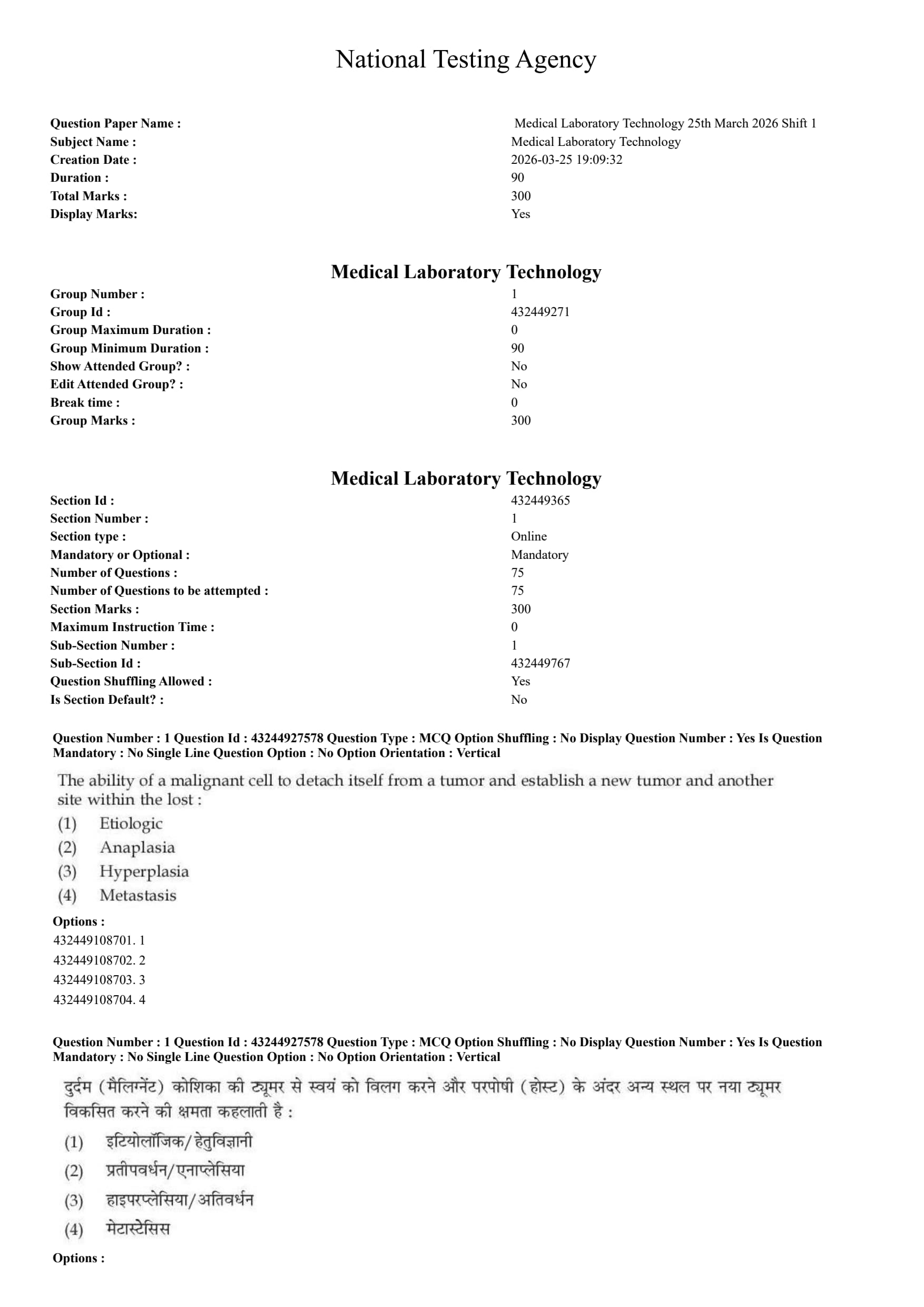




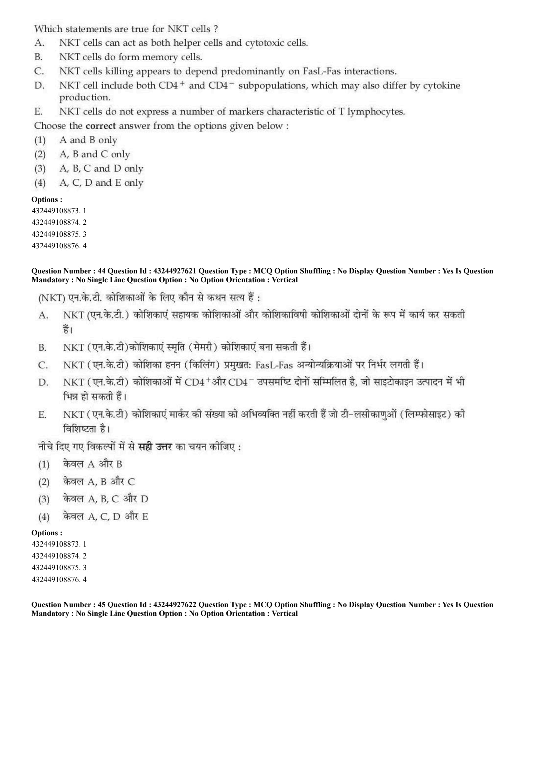


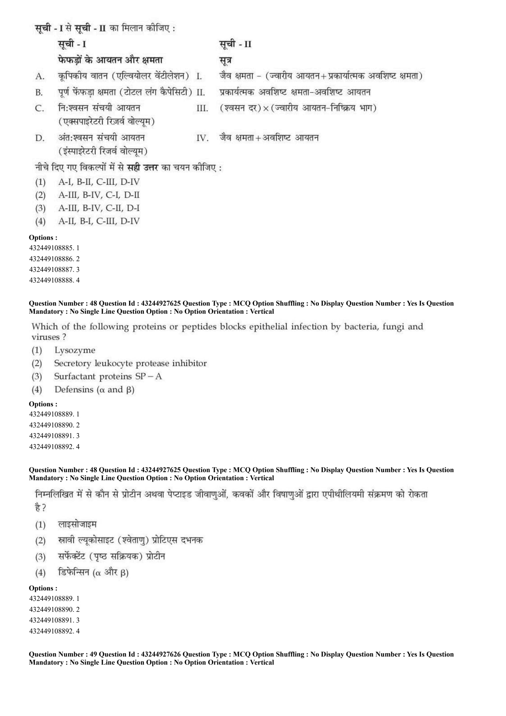

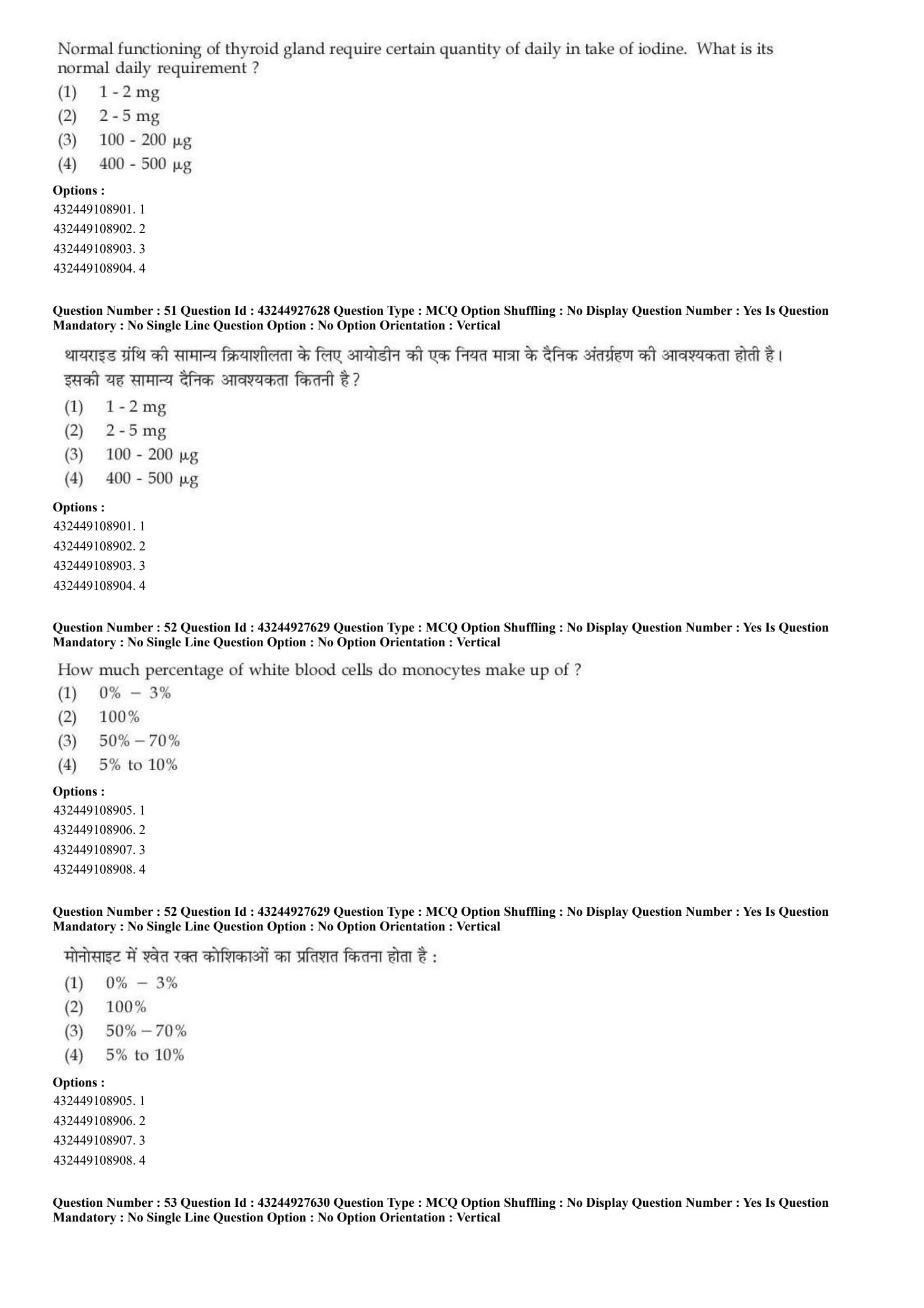

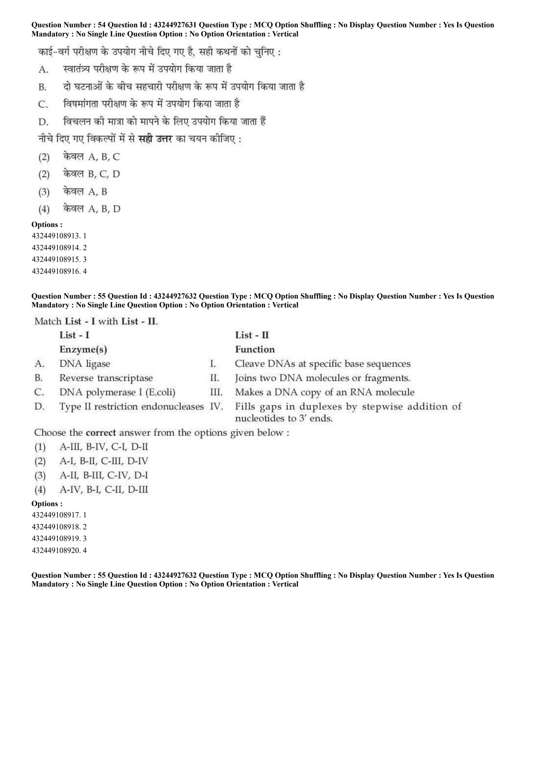
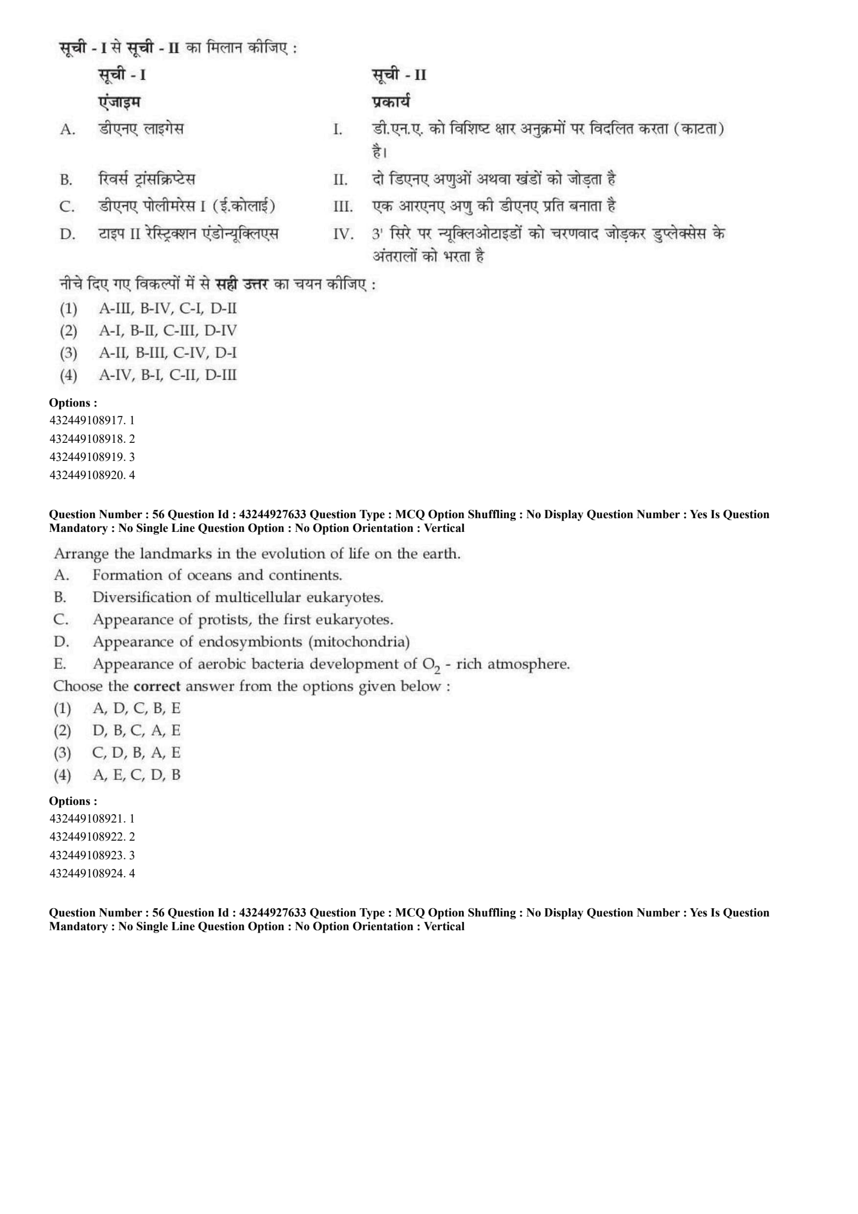


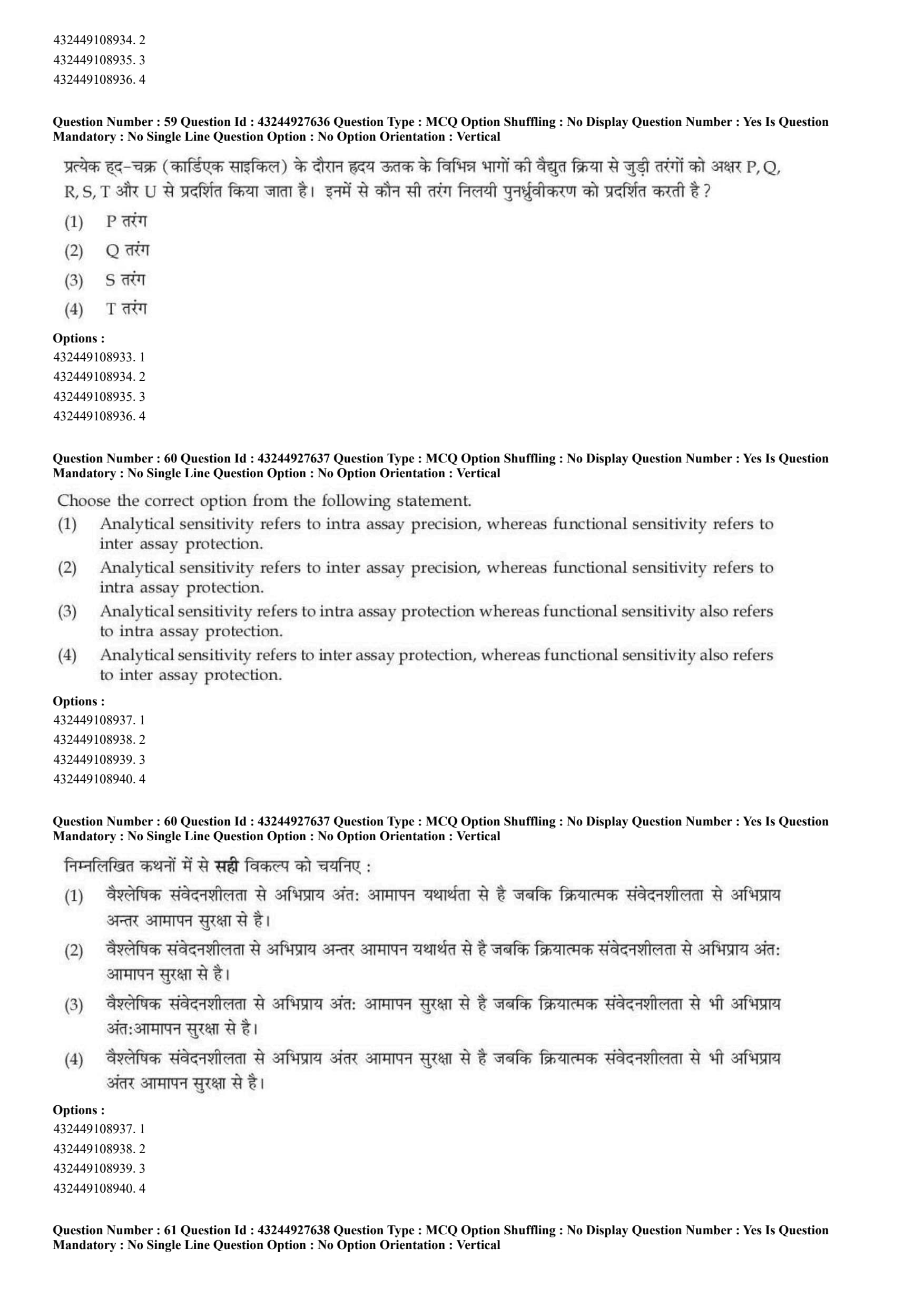
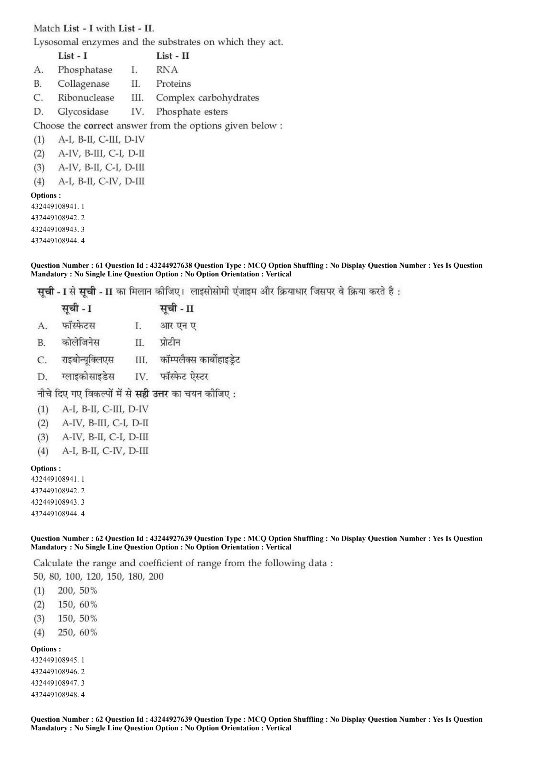


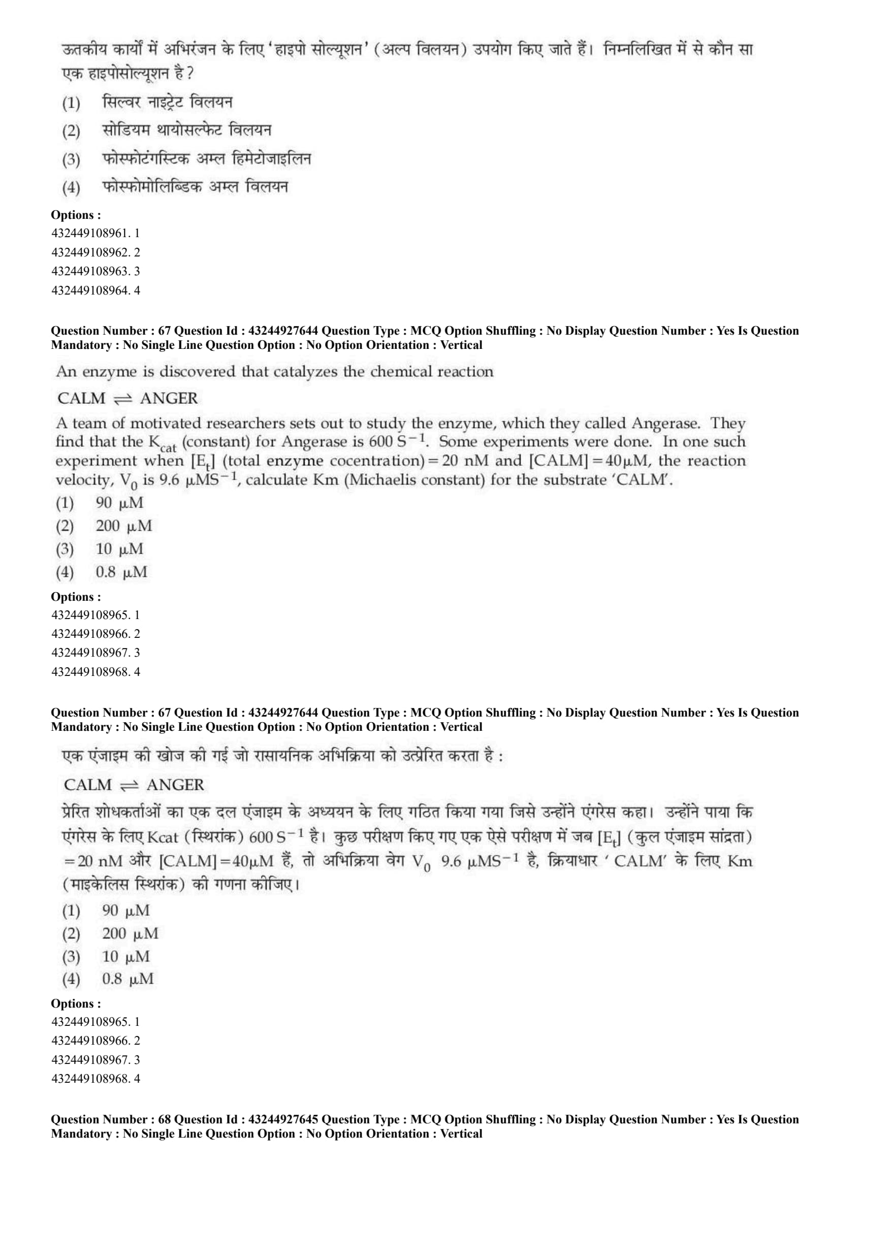




The ability of a malignant cell to detach itself from a tumor and establish a new tumor and another site within the host :
View Solution
Step 1: Concept:
This question tests the knowledge of fundamental cancer biology, specifically the terminology used for the spread of malignant tumors from a primary site to a secondary site within the human body.
Step 2: Step-by-step Explanation:
Etiology refers to the root cause or origin of a disease, such as a genetic mutation, environmental toxin, or a viral infection. It does not describe the physical spread of tumor cells.
Anaplasia is a condition where cells lose their morphological characteristics and mature differentiation. While it is a major hallmark of aggressive cancer cells, it refers to cellular appearance, not the act of spreading.
Hyperplasia is an abnormal physiological increase in the number of cells in an organ or tissue. It is a form of cellular adaptation or early pre-neoplastic growth, but it remains localized.
Metastasis is the complex, multi-step physiological process by which malignant cancer cells break their adhesive bonds, detach from the primary tumor, intravasate into the bloodstream or lymphatic system, travel, and extravasate to establish new, secondary tumors in distant organs.
This ability to metastasize is the primary characteristic that distinguishes malignant tumors from benign tumors.
Step 3: Final Answer:
The correct terminology for this spreading process is Metastasis.
Quick Tip: Remember the "M" in Malignant often leads to the "M" in Metastasis. Benign tumors are strictly encapsulated and do not metastasize, which is a key distinguishing factor in clinical pathology.
Choose the correct sequence for how a type I topoisomerase relaxes DNA.
A. DNA release
B. Cleavage and opening of gate
C. Rejoining of cleaved strand
D. Strand passage
View Solution
Step 1: Concept:
The question focuses on the mechanism of action of Type I topoisomerase, a crucial enzyme that regulates the topological state of DNA by removing supercoils during processes like DNA replication and transcription.
Step 2: Step-by-step Explanation:
Topoisomerase I relaxes supercoiled DNA by creating transient single-strand breaks. This allows the DNA to swivel and relieve torsional strain through a very specific catalytic cycle.
Step B (Cleavage and opening of gate): The enzyme first binds to the DNA duplex. A tyrosine residue in the active site attacks the phosphodiester backbone of one DNA strand, cleaving it and creating a covalent enzyme-DNA intermediate. This creates a transient "gate".
Step D (Strand passage): The intact complementary DNA strand is then passed through this newly opened break (the gate) to relieve the torsional strain, effectively unwinding one supercoil.
Step C (Rejoining of cleaved strand): Once the intact strand has successfully passed through, the enzyme reverses the cleavage reaction, ligating (rejoining) the broken phosphodiester bonds back together.
Step A (DNA release): Finally, the enzyme undergoes a conformational change, releasing the newly relaxed DNA molecule and preparing for another catalytic cycle.
Therefore, the chronologically accurate sequence is B \(\rightarrow\) D \(\rightarrow\) C \(\rightarrow\) A.
Step 3: Final Answer:
The correct sequence is given by option (B).
Quick Tip: Type I topoisomerase cuts ONE strand (Type I = 1 strand), whereas Type II topoisomerase cuts TWO strands. The general mechanism is always: Cut (Cleavage) \(\rightarrow\) Pass (Strand passage) \(\rightarrow\) Seal (Rejoining) \(\rightarrow\) Let go (Release).
Given below are two statements : one is labelled as Assertion (A) and the other is labelled as Reason (R).
Assertion (A) : Cells of different tissues essentially require CO\(_2\) to generate energy and perform metabolic functions.
Reason (R) : Deficiency of oxygen or hypoxia results in failure to carry metabolic functions.
In the light of the above statements, choose the most appropriate answer from the options given below :
View Solution
Step 1: Concept:
This question evaluates the fundamental biochemical requirements for aerobic cellular respiration and energy (ATP) generation within human tissue cells.
Step 2: Step-by-step Explanation:
Analyzing Assertion (A): The assertion claims that cells essentially require carbon dioxide (CO\(_2\)) to generate energy. This is a biologically inaccurate statement for mammalian cells. Human tissue cells absolutely require oxygen (O\(_2\)) for aerobic cellular respiration to generate ATP. Carbon dioxide is actually a metabolic waste byproduct produced during the Krebs cycle, which must be expelled from the body. Therefore, Assertion (A) is false.
Analyzing Reason (R): The reason states that a deficiency of oxygen, clinically termed hypoxia, leads to a failure in carrying out metabolic functions. This is completely true. Oxygen serves as the final electron acceptor in the mitochondrial electron transport chain (ETC).
Without oxygen, the ETC halts, a proton gradient cannot be maintained, and oxidative phosphorylation ceases. This forces the cell into inefficient anaerobic glycolysis, rapidly depleting ATP and ultimately causing cellular metabolic failure and death.
Since Assertion (A) is false and Reason (R) is true, the correct logical relationship is identified.
Step 3: Final Answer:
Option (D) correctly identifies that (A) is not correct but (R) is correct.
Quick Tip: Always carefully read Assertion statements for swapped biological terms. Here, substituting the requirement of O\(_2\) with the waste product CO\(_2\) instantly makes the statement factually incorrect.
Depending upon the antigen how much lag period is required for memory B cell after antigen administration.
View Solution
Step 1: Concept:
The question requires understanding the kinetics of the adaptive immune system, specifically the duration of the lag (or latent) phase during a secondary immune response mediated by memory B cells.
Step 2: Step-by-step Explanation:
The immune response is categorized into primary and secondary responses based on prior exposure to a specific antigen.
During a primary immune response (the first time the body encounters an antigen), naive B cells take significant time to recognize the antigen, become activated by T-helper cells, undergo clonal expansion, and differentiate into antibody-producing plasma cells. This primary lag phase is relatively long, typically spanning 7 to 10 days.
After this initial infection is cleared, a small subset of these specifically tailored cells differentiates into long-lived memory B cells.
Upon subsequent exposure to the exact same antigen, a secondary immune response is triggered. Memory B cells are already primed, circulating, and ready to act immediately.
Because they skip the initial prolonged activation steps, the lag period is drastically reduced, usually taking only 1 to 3 days before a massive and rapid surge of high-affinity antibodies (predominantly IgG) is produced in the bloodstream.
Step 3: Final Answer:
The required lag period for memory B cells is 1 - 3 days.
Quick Tip: Primary Immune Response = Long lag phase (7-10 days), predominantly IgM antibodies initially.
Secondary Immune Response = Short lag phase (1-3 days), predominantly IgG antibodies, much faster and stronger due to primed Memory B cells.
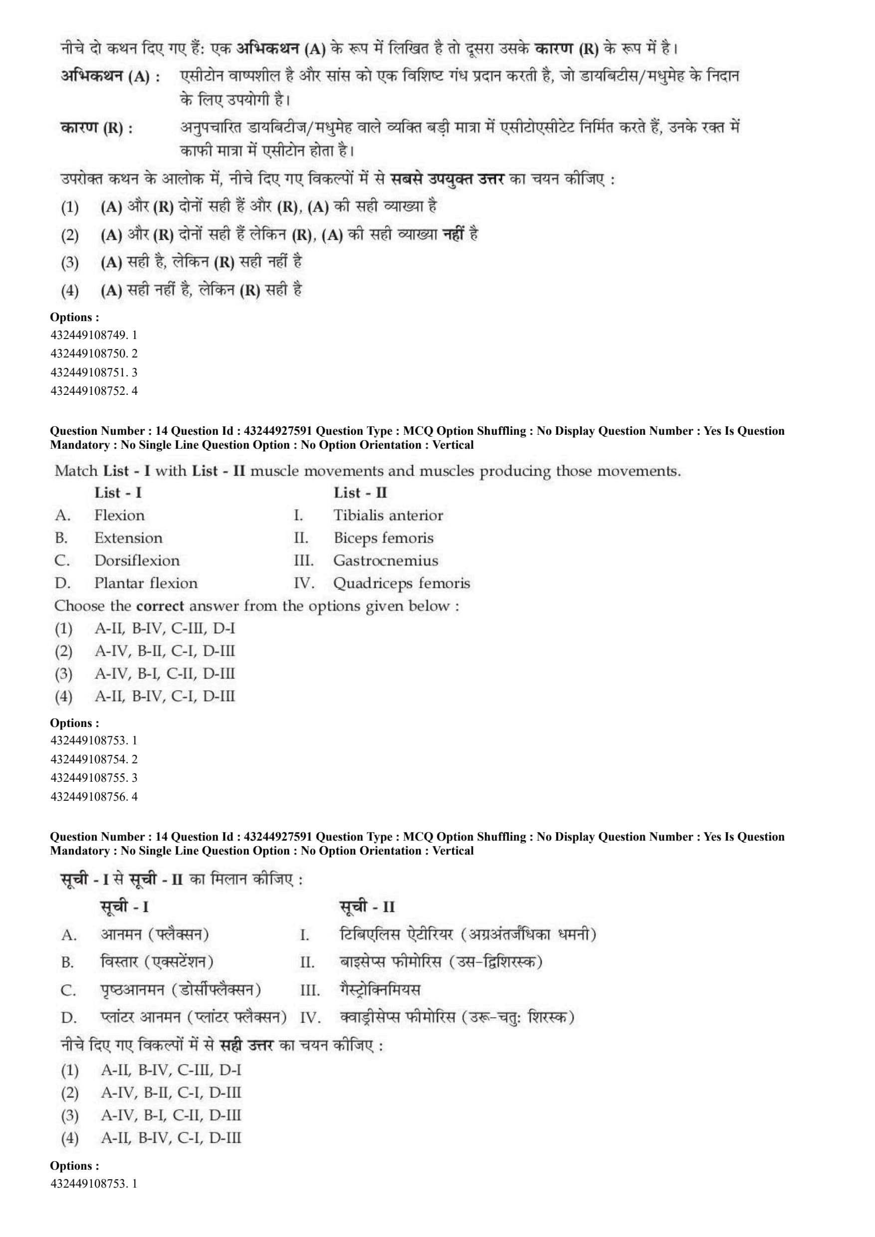
Given below are two statements : one is labelled as Assertion (A) and the other is labelled as Reason (R).
Assertion (A) : The conduction in the Atrio-ventricular (AV) node is slow i.e. 0.05 m/sec, there is a delay of approx. 0.1 second before excitation spreads to the ventricles.
Reason (R) : AV node delay enables the atria to empty the blood present within them into the ventricles before these contract.
In the light of the above statements, choose the most appropriate answer from the options given below :
View Solution
Step 1: Concept:
This question focuses on the electrophysiology of the cardiac conduction system, specifically highlighting the physiological significance of the decreased conduction velocity at the Atrio-ventricular (AV) node.
Step 2: Step-by-step Explanation:
Analyzing Assertion (A): Electrical impulses generated by the Sinoatrial (SA) node travel rapidly through the atrial syncytium and reach the AV node. The AV node fibers have smaller diameters and significantly fewer gap junctions compared to other cardiac tissues. This structural difference causes a marked decrease in conduction velocity (down to about 0.05 m/sec), resulting in a physiological delay of roughly 0.09 to 0.1 seconds. The assertion is factually accurate.
Analyzing Reason (R): If the electrical impulse traveled instantly from the atria to the ventricles, the entire heart would contract simultaneously. This would be highly inefficient, as the ventricles would attempt to pump blood while still mostly empty.
The critical 0.1-second delay imposed by the AV node ensures that atrial systole (contraction) is fully completed, allowing the atria to pump their remaining blood volume into the ventricles (active ventricular filling) before ventricular systole initiates. The reason is factually correct.
Because the necessity of complete ventricular filling is the exact biological evolutionary reason why the AV node slows down the electrical signal, Reason (R) perfectly explains Assertion (A).
Step 3: Final Answer:
Both statements are true, and (R) correctly explains (A).
Quick Tip: Think of the AV node as an electrical "speed bump" or "traffic light" in the heart. It deliberately pauses the electrical signal (delay) so the atria can finish their job of topping off the ventricles with blood before the main pumping action begins.
Choose the correct sequence of the structure of the ear starting from external ear (entry of sound) to inner ear.
A. Helix
B. Oval window
C. Pinna
D. Cochlae
E. Tympanic membrane
View Solution
Step 1: Concept:
The question requires tracing the precise anatomical pathway that acoustic sound waves take as they travel from the external environment, through the outer and middle ear, and finally into the inner ear for sensory processing.
Step 2: Step-by-step Explanation:
Sound waves in the environment first encounter the most lateral structure of the external ear, the prominent cartilaginous rim known as the Helix (A).
The waves are then captured and directed inward by the broader, bowl-like cartilaginous structure of the external ear, collectively referred to as the auricle or Pinna (C).
Funneled through the external auditory canal, the acoustic waves strike the Tympanic membrane (E), commonly known as the eardrum, causing it to vibrate. This marks the boundary between the outer and middle ear.
These vibrations are mechanically amplified by the three small ossicle bones (malleus, incus, and stapes) in the middle ear. The stapes bone pushes against the Oval window (B), a membrane-covered opening that serves as the entry portal to the inner ear.
Once the vibrations pass through the oval window, they create fluid waves within the Cochlea (D), the snail-shaped, fluid-filled organ where mechanical waves are finally transduced into electrical neural signals by hair cells.
Therefore, the sequential anatomical pathway is A \(\rightarrow\) C \(\rightarrow\) E \(\rightarrow\) B \(\rightarrow\) D.
Step 3: Final Answer:
Option (D) provides the correct anatomical sequence.
Quick Tip: A helpful mnemonic for the major boundaries of the auditory system: \textbf{P}inna (Outer receiver) \(\rightarrow\) \textbf{T}ympanic Membrane (Middle ear border) \(\rightarrow\) \textbf{O}val Window (Inner ear border) \(\rightarrow\) \textbf{C}ochlea (Neural processing center).
The correct order of systemic circulation in a heart :
A. Aorta
B. Aortic valve
C. Left ventricle
D. Superior and inferior venae cavae
E. Right atrium
View Solution
Step 1: Concept:
This question requires mapping out the systemic circulation pathway, which is the extensive portion of the cardiovascular system responsible for delivering oxygenated blood to all body tissues and returning deoxygenated blood back to the heart.
Step 2: Step-by-step Explanation:
The systemic circuit officially begins its powerful outward journey from the Left ventricle (C). This chamber has the thickest myocardial wall to generate the high pressure needed to pump blood throughout the entire body.
During ventricular contraction (systole), the high pressure forces oxygenated blood to eject through the semilunar Aortic valve (B), which opens outward and subsequently snaps shut to prevent regurgitation (backflow) into the ventricle.
The blood immediately enters the Aorta (A), the largest and most elastic artery in the human body. The aorta branches extensively into smaller arteries, arterioles, and capillaries, delivering oxygen and nutrients to systemic tissues.
After cellular respiration occurs at the tissue level, the now deoxygenated, carbon dioxide-rich blood travels back toward the heart through venules and veins, eventually draining into the two largest veins in the body: the Superior and inferior venae cavae (D).
These massive veins empty their deoxygenated blood load directly into the Right atrium (E) of the heart, successfully completing the systemic circulatory loop.
Consequently, the correct chronological sequence is C \(\rightarrow\) B \(\rightarrow\) A \(\rightarrow\) D \(\rightarrow\) E.
Step 3: Final Answer:
Option (D) correctly outlines this physiological pathway.
Quick Tip: Remember the two main loops. Systemic circuit: Left side of Heart \(\rightarrow\) Body tissues \(\rightarrow\) Right side of Heart. Pulmonary circuit: Right side of Heart \(\rightarrow\) Lungs \(\rightarrow\) Left side of Heart.
Given below are two statements : one is labelled as Assertion (A) and the other is labelled as Reason (R).
Assertion (A) : Lysosomes are called suicidal bags.
Reason (R) : When a cell dies, lysosomal enzymes cause autolysis of the remanant.
In the light of the above statements, choose the most appropriate answer from the options given below :
View Solution
Step 1: Concept:
This question tests the functional biology of lysosomes, focusing specifically on their internal enzymatic contents and their critical role in cellular breakdown, recycling, and programmed cell death.
Step 2: Step-by-step Explanation:
Assertion (A): Lysosomes are membrane-bound organelles that are indeed widely referred to in biological literature as the "suicidal bags" or "suicide sacs" of the cell. Therefore, this assertion statement is completely correct.
Reason (R): The interior of a lysosome is highly acidic and packed with potent hydrolytic enzymes (such as proteases, lipases, nucleases, and glycosidases). Under normal conditions, these enzymes digest food particles or damaged organelles (autophagy).
However, if a cell undergoes severe irreversible damage, disease, or initiates programmed cell death (apoptosis), the lysosomal membranes can intentionally rupture.
Upon this rupture, the highly destructive enzymes spill out into the cytoplasm, where they rapidly digest and break down the cell's own internal components. This destructive process of self-digestion is termed autolysis.
Because the release of these internal enzymes effectively destroys the host cell from the inside out, Reason (R) provides the exact, scientifically sound explanation for why lysosomes earned the nickname "suicidal bags" mentioned in Assertion (A).
Step 3: Final Answer:
Both statements are correct, and the reason perfectly explains the assertion.
Quick Tip: Understand the biological prefixes and suffixes: "Auto-" means self, and "-lysis" means breakdown or destruction. Autolysis = Self-breakdown, which perfectly describes the function of lysosomal "suicide bags."
Which of the following wavelength (nm) correspond to visible range of humans ?
A. 20 nm
B. 200 nm
C. 400 nm
D. 600 nm
E. 900 nm
View Solution
Step 1: Concept:
This question requires identifying the specific portion of the broad electromagnetic (EM) spectrum that is perceivable by the photoreceptors in the human eye, measured in units of nanometers (nm).
Step 2: Step-by-step Explanation:
The human visual system is biologically adapted to detect electromagnetic radiation only within a very specific, narrow band known as the visible light spectrum.
This visible spectrum generally ranges from approximately 400 nanometers (nm) at the violet end to about 700 nanometers (nm) at the red end.
Let us evaluate the given wavelength options against this standard biological range:
- A. 20 nm: This very short wavelength falls in the extreme Ultraviolet (UV) or soft X-ray region, which is completely invisible to human eyes and is highly ionizing.
- B. 200 nm: This falls in the UV-C region (used for germicidal irradiation), which is also invisible and harmful to humans.
- C. 400 nm: This sits exactly at the lower boundary of the visible spectrum, corresponding to violet or deep blue light.
- D. 600 nm: This wavelength sits comfortably in the middle-upper section of the visible spectrum, corresponding to orange or reddish light.
- E. 900 nm: This long wavelength falls in the near-Infrared (IR) region. While we might feel it as heat, our eyes cannot see it.
Therefore, only 400 nm and 600 nm fall within the human visible range.
Step 3: Final Answer:
Options C and D are correct, matching choice (B).
Quick Tip: Always memorize the visible spectrum as strictly the 400-700 nm range. Anything lower (like 200 nm) is UV (causes sunburns, invisible), and anything higher (like 900 nm) is IR (heat signatures, invisible).
Which of the following advantages of chemiluminescence technology are true :
A. Low cost
B. Non linearity
C. Stability
D. Sensitivity
View Solution
Step 1: Concept:
This question tests the fundamental characteristics, operational benefits, and analytical advantages of Chemiluminescence Immunoassay (CLIA) technology when compared to older conventional methods like Radioimmunoassay (RIA) or ELISA.
Step 2: Step-by-step Explanation:
Chemiluminescence relies on the emission of light as a byproduct of a chemical reaction, without requiring an external excitation light source.
Sensitivity (D): CLIA is exceptionally sensitive. Because there is no incident light required, there is virtually zero background scatter or noise. Photomultiplier tubes can detect minute flashes of light, allowing for the detection of extremely low concentrations of analytes (often down to picograms or femtograms). This is a true advantage.
Stability (C): The luminescent reagents used in CLIA (such as luminol derivatives or acridinium esters) are highly stable and possess a long shelf life. This is a massive improvement over RIA, which relies on radioactive isotopes that decay rapidly and pose safety hazards. This is a true advantage.
Low Cost (A): While the initial capital investment for automated CLIA analyzers is high, the routine operational and per-test costs are generally low, especially because it avoids the massive regulatory, disposal, and safety costs associated with handling radioactive waste. This is considered a true advantage in modern labs.
Non-linearity (B): This statement is false. A major analytical advantage of chemiluminescence is its exceptionally wide linear dynamic range. The intensity of emitted light is directly and linearly proportional to the analyte concentration across several orders of magnitude. Non-linearity is an analytical drawback, not an advantage.
Therefore, the true advantages among the options are A, C, and D.
Step 3: Final Answer:
The correct combination is A, C, and D only.
Quick Tip: In clinical laboratory instrumentation, "linearity" is highly prized because it makes calibration accurate over a broad range of patient samples. "Non-linearity" is almost always an undesirable trait or an incorrect distractor in multiple-choice questions.
When the number of frequencies are put in a cell in a contingency table, the degree of freedom will be _________
View Solution
Step 1: Concept:
The question asks for the standard statistical formula used to calculate the "degrees of freedom" when performing a chi-square test of independence on categorical data organized within a contingency table.
Step 2: Key Formulas and approach:
For any two-way contingency table with \( R \) representing the total number of rows and \( C \) representing the total number of columns, the degrees of freedom (\( df \)) is universally given by the formula:
\[ df = (R - 1) \times (C - 1) \]
Step 3: Step-by-step Explanation:
A contingency table is a matrix format that displays the frequency distribution of two categorical variables simultaneously (e.g., studying the relationship between gender and blood type).
In statistics, the "degrees of freedom" represents the number of independent values or quantities in the final calculation of a statistic that are completely free to vary without violating any constraints.
In a chi-square contingency table, the marginal totals (the sum of each individual row and the sum of each individual column) are fixed quantities.
Because these totals are fixed, if you know the values of \( (C - 1) \) cells in a specific row, the value of the very last cell in that row is strictly predetermined mathematically to ensure the row sum is correct.
Similarly, knowing \( (R - 1) \) cells in a column strictly dictates the final cell in that column to meet the column total constraint.
By multiplying these independent dimensions together, we arrive at the standard formula for the degrees of freedom: \( (R - 1) (C - 1) \).
Step 4: Final Answer:
The correct mathematical expression is \( (R - 1) (C - 1) \).
Quick Tip: Always remember to subtract 1 from BOTH the number of rows and the number of columns before multiplying! For a standard \(2 \times 2\) table (like testing sick vs healthy against treatment vs placebo), the degrees of freedom is simply \((2-1) \times (2-1) = 1\).
From the options given below choose the weakest bonds in biological systems.
View Solution
Step 1: Concept:
The question requires ordering various types of chemical interactions based on their relative bond strengths and identifying the weakest interaction among the given options, specifically in the context of aqueous biological systems.
Step 2: Step-by-step Explanation:
Biological macromolecules rely on a variety of chemical bonds to maintain their complex three-dimensional structures. We evaluate them from strongest to weakest:
Covalent bonds (not listed) are the strongest, involving the actual sharing of electron pairs between atoms (e.g., peptide bonds).
Ionic bonds occur due to electrostatic attraction between fully charged opposite ions (e.g., Na\(^+\) and Cl\(^-\)). While water weakens them significantly compared to a dry salt crystal, they still remain relatively strong biological interactions.
Hydrogen bonds involve the electrostatic attraction between a hydrogen atom covalently bonded to a highly electronegative atom (like Oxygen or Nitrogen) and another nearby electronegative atom. They provide crucial, moderate-strength stability to structures like the DNA double helix and protein alpha-helices.
Hydrophobic bonds (interactions) are not true bonds, but thermodynamic forces that dictate the folding of proteins as non-polar molecules aggregate closely to minimize their contact with water. This driving force is quite significant.
Van der Waals bonds (forces) are extremely weak, highly distance-dependent, transient electrostatic attractions. They arise from temporary, rapidly fluctuating dipoles in the electron clouds around atoms. They only become significant when molecules are exceptionally close together.
Because Van der Waals forces possess the absolute lowest bond energy (usually roughly 0.4 to 4 kJ/mol) compared to all the others, they are definitively the weakest.
Step 3: Final Answer:
Van der Waals bonds are the weakest.
Quick Tip: Keep this general hierarchy of bond strength in biological (aqueous) environments memorized: Covalent (strongest) \(>\) Ionic \(>\) Hydrogen \(>\) Hydrophobic \(>\) Van der Waals (weakest).
Given below are two statements : one is labelled as Assertion (A) and the other is labelled as Reason (R).
Assertion (A) : Acetone is volatile and imparts a characteristic odor to the breath, which is useful in diagnosis of diabetes.
Reason (R) : Individuals with untreated diabetes produces large quantities of acetoacetate, their blood contains significant amount of acetone.
In the light of the above statements, choose the most appropriate answer from the options given below :
View Solution
Step 1: Concept:
This question tests clinical knowledge of Diabetic Ketoacidosis (DKA), a severe metabolic complication commonly seen in untreated Type 1 diabetes, focusing on its biochemical pathways and associated diagnostic clinical symptoms.
Step 2: Step-by-step Explanation:
Analyzing Assertion (A): Patients presenting with severe, uncontrolled diabetes often exhibit a distinct "fruity" or "sweet" odor on their breath. Clinically, this is widely recognized as the smell of acetone, a highly volatile organic compound. Detecting this odor is a rapid, classic bedside diagnostic clue for ketoacidosis. Thus, Assertion (A) is factually correct.
Analyzing Reason (R): In untreated diabetes (particularly due to absolute insulin deficiency), the body's cells cannot utilize blood glucose for energy. To survive, the body shifts to massively breaking down fat (lipolysis), releasing free fatty acids into the blood.
These fatty acids travel to the liver, where excessive beta-oxidation overloads the metabolic pathways, leading to ketogenesis—the heavy production of ketone bodies. The primary ketone body produced is acetoacetate.
Acetoacetate is inherently chemically unstable. In the blood, it undergoes spontaneous, non-enzymatic decarboxylation to form acetone. Because acetone is highly volatile (turns to gas easily), it diffuses from the pulmonary capillaries into the alveoli of the lungs and is physically exhaled.
Therefore, Reason (R) perfectly details the biochemical mechanism that results in the clinical symptom described in Assertion (A).
Step 3: Final Answer:
Both statements are correct, and (R) is the correct explanation for (A).
Quick Tip: Remember the three main metabolic ketone bodies are Acetoacetate, \(\beta\)-hydroxybutyrate, and Acetone. Among these, only Acetone is exhaled through the respiratory system because of its high volatility, generating the classic "fruity breath" hallmark of DKA.
Match List - I with List - II muscle movements and muscles producing those movements.

Choose the correct answer from the options given below :
View Solution
Step 1: Concept:
This question requires matching basic anatomical muscle actions and movements of the human lower limb to the specific primary skeletal muscles responsible for generating those actions.
Step 2: Step-by-step Explanation:
A. Flexion (of the knee joint): Bending the knee (decreasing the angle of the joint) is primarily driven by the hamstring muscle group located on the posterior aspect of the thigh. The Biceps femoris is one of the major muscles in this hamstring group. Thus, A matches with II.
B. Extension (of the knee joint): Straightening the knee (increasing the angle) is powerfully driven by the large muscle group on the anterior aspect of the thigh, collectively known as the Quadriceps femoris. Thus, B matches with IV.
C. Dorsiflexion (of the ankle joint): Lifting the foot upwards so that the toes point toward the shin is performed by the anterior compartment muscles of the lower leg. The primary muscle for this action is the Tibialis anterior. Thus, C matches with I.
D. Plantar flexion (of the ankle joint): Pointing the foot and toes downward (similar to pressing a gas pedal in a car) is performed by the large posterior calf muscles, mainly the Gastrocnemius and the underlying soleus. Thus, D matches with III.
Compiling all the correct matches together yields: A-II, B-IV, C-I, D-III.
Step 3: Final Answer:
Option (D) provides the perfectly aligned pairs for these muscle actions.
Quick Tip: Visual mnemonics work best here: Dorsiflexion = imagine a dolphin's "Dorsal" fin pointing up (toes point up \(\rightarrow\) Tibialis anterior). Plantar flexion = "Planting" your foot flat on the ground or pressing down (toes point down \(\rightarrow\) Calf muscles/Gastrocnemius).
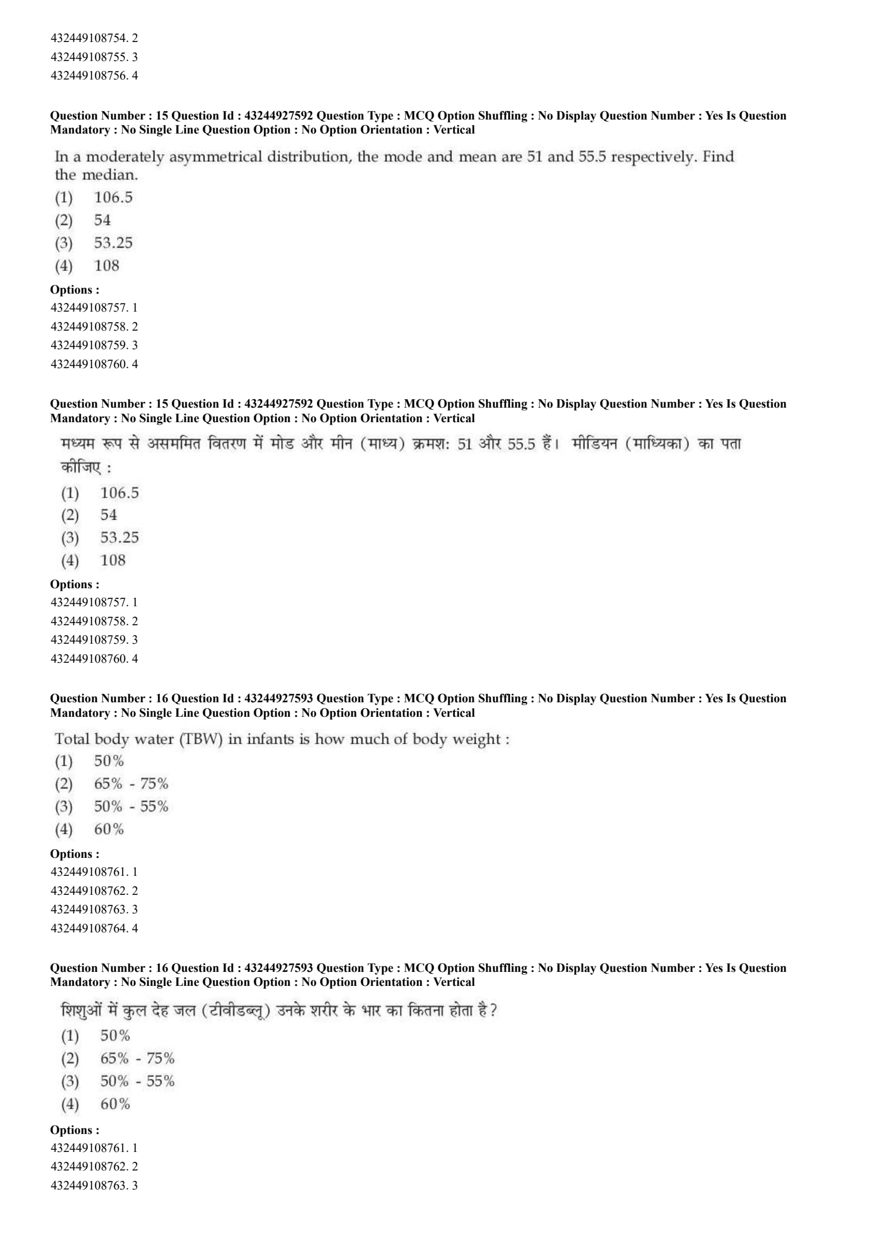
In a moderately asymmetrical distribution, the mode and mean are 51 and 55.5 respectively. Find the median.
View Solution
Step 1: Concept:
This problem requires calculating the median of a statistical dataset when the mean and mode are known. Since the distribution is explicitly described as "moderately asymmetrical" (skewed), we can apply Karl Pearson's empirical relationship to find the missing central tendency value.
Step 2: Key Formulas and approach:
Karl Pearson's empirical formula relating the three measures of central tendency for a moderately skewed distribution is:
\[ Mode = 3 \times Median - 2 \times Mean \]
Step 3: Step-by-step Explanation:
Given variables from the question:
Mode = 51
Mean = 55.5
Substitute the known numerical values directly into Pearson's empirical formula:
\[ 51 = 3 \times Median - 2 \times (55.5) \]
Calculate the multiplication product on the right side of the equation:
\[ 2 \times 55.5 = 111 \]
Update the equation with this new value:
\[ 51 = 3 \times Median - 111 \]
Isolate the Median term by algebraically adding 111 to both sides of the equation:
\[ 51 + 111 = 3 \times Median \]
\[ 162 = 3 \times Median \]
Solve for the final Median by dividing the total by 3:
\[ Median = \frac{162}{3} \]
\[ Median = 54 \]
Step 4: Final Answer:
The calculated median of the distribution is 54, which exactly corresponds to option (B).
Quick Tip: To memorize Karl Pearson's empirical formula easily, think of the words alphabetically backwards: "Mean, Median, Mode" and assign the coefficients 1, 3, and 2 respectively: 1 \(\times\) Mode = 3 \(\times\) Median - 2 \(\times\) Mean.
Total body water (TBW) in infants is how much of body weight :
View Solution
Step 1: Concept:
The question tests clinical physiological knowledge regarding the percentage of total body water (TBW) relative to total body weight, and how this composition specifically changes during human development, particularly in infants.
Step 2: Step-by-step Explanation:
Total body water is a dynamic physiological metric that fluctuates significantly throughout a human's lifespan. It is inversely correlated with the amount of adipose (fat) tissue in the body, because fat tissue contains very little water compared to muscle and organs.
In a healthy, average adult male, TBW makes up approximately 60% of their total body weight.
In a healthy adult female, due to a naturally higher proportion of essential adipose tissue, TBW is comparatively lower, usually sitting around 50% to 55%.
Infants, particularly newborns and neonates, possess a drastically different body composition. They have very little body fat, a lower proportion of dense bone mass, and a remarkably high extracellular fluid volume.
Because of this unique composition, full-term healthy infants have a much higher TBW percentage, typically ranging from 65% to 75% of their total body weight. Premature infants can exhibit an even higher water composition, sometimes reaching up to 80%.
Step 3: Final Answer:
Infants contain roughly 65% - 75% body water relative to their weight.
Quick Tip: A solid rule of thumb for clinical exams: As humans age, their body fat percentage generally increases, and therefore their TBW progressively decreases. The hierarchy is: Infants (highest, ~70%) \(>\) Adult Males (~60%) \(>\) Adult Females (~55%) \(>\) Elderly individuals (~50%).
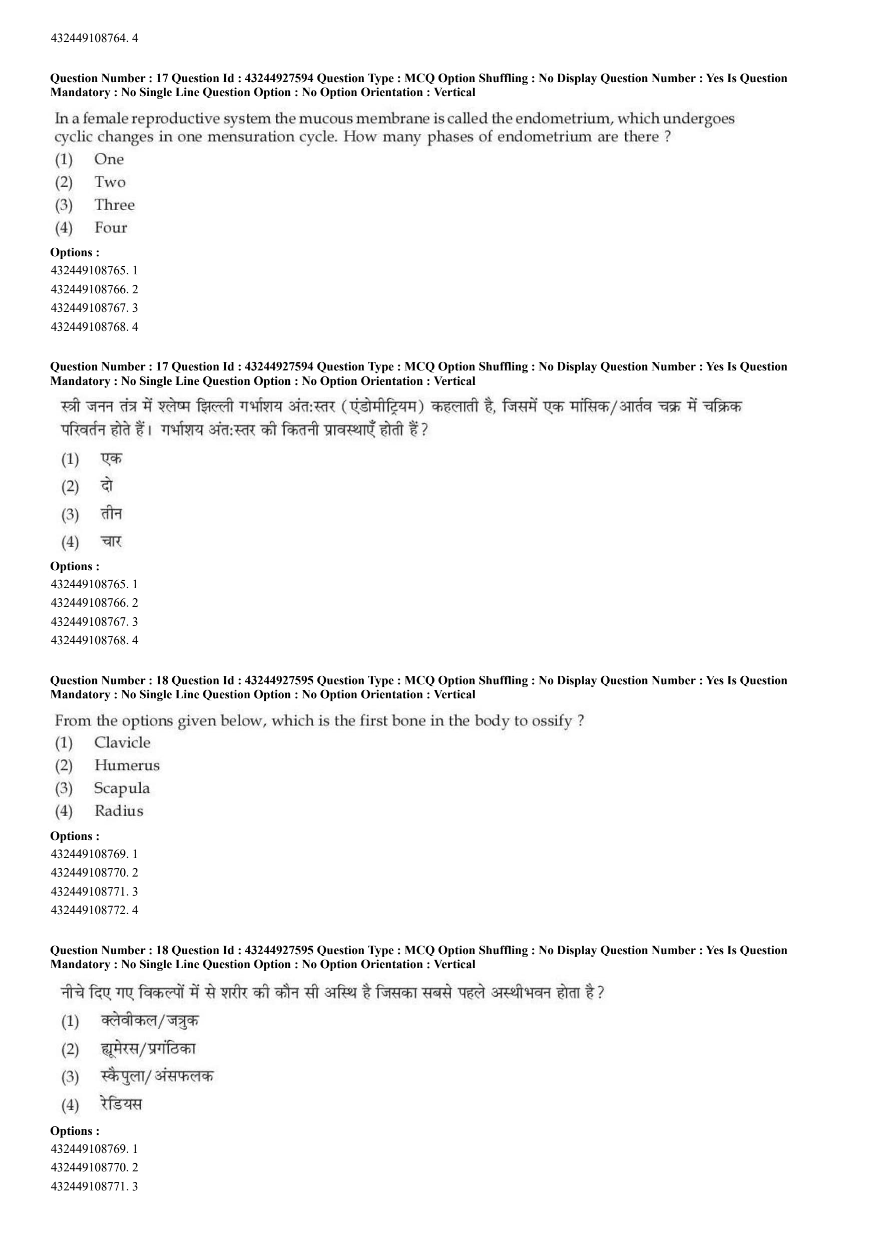
In a female reproductive system the mucous membrane is called the endometrium, which undergoes cyclic changes in one mensuration cycle. How many phases of endometrium are there ?
View Solution
Step 1: Concept:
This question asks for the standard biological and anatomical classification regarding the phases of the uterine (endometrial) cycle that occur during a typical healthy menstrual cycle in females.
Step 2: Step-by-step Explanation:
The endometrium is the highly dynamic, inner mucosal lining of the uterus. It undergoes significant structural, vascular, and glandular changes driven cyclically by the ovarian hormones estrogen and progesterone, all to prepare a hospitable environment for a potential embryo.
Histologically and clinically, the uterine cycle is classically divided into exactly three distinct phases:
1. Menstrual Phase (Typically Days 1-5): This marks the beginning of the cycle. If fertilization does not occur, hormone levels drop, leading to the shedding and sloughing off of the thick functional layer of the endometrium (stratum functionalis), which presents as menstrual bleeding.
2. Proliferative Phase (Typically Days 6-14): Driven by rapidly rising estrogen levels secreted from the developing ovarian follicles, the thin remaining endometrial layer begins to rebuild, thicken, and rapidly re-vascularize to restore its structure.
3. Secretory Phase (Typically Days 15-28): Following ovulation, high levels of progesterone secreted by the corpus luteum cause the newly rebuilt endometrial glands to enlarge, coil, and begin secreting nutrient- and glycogen-rich fluids. This perfectly preps the lining for the potential implantation of a blastocyst.
Therefore, summarizing the physiology, there are three primary structural phases of the endometrium.
Step 3: Final Answer:
There are precisely three phases in the endometrial cycle.
Quick Tip: Do not confuse the Uterine (endometrial) cycle with the Ovarian cycle! While they happen simultaneously, they have different names. The Ovarian cycle is: Follicular, Ovulatory, and Luteal. Both systems, however, are conveniently divided into three main phases.
From the options given below, which is the first bone in the body to ossify ?
View Solution
Step 1: Concept:
The question tests specific knowledge of human embryology and osteology (skeletal development), specifically identifying which single bone initiates the biological process of ossification (bone tissue formation) the earliest during fetal development in the womb.
Step 2: Step-by-step Explanation:
Ossification is the process where mesenchymal tissue or cartilage is replaced by hardened bone. In a human embryo, this process begins remarkably early, acting as a crucial structural scaffold for the developing organism.
Among all the 206 bones in the adult human body, the clavicle (commonly known as the collarbone) is well-documented in anatomical texts as the very first bone to begin the ossification process.
The clavicle initiates ossification incredibly early, starting around the 5th to 6th week of embryonic development.
It is biologically unique because it ossifies primarily via intramembranous ossification. This means bone develops directly from sheets of primitive mesenchymal connective tissue.
This is in stark contrast to long bones like the humerus, radius, and femur, which develop via \textit{endochondral ossification (where a hyaline cartilage model forms first and is slowly replaced by bone later in development).
While other bones like the mandible follow very shortly after, the clavicle is unequivocally the pioneer in this developmental stage.
Step 3: Final Answer:
The clavicle is the first bone to undergo ossification.
Quick Tip: A classic embryology trivia fact: While the clavicle is the very \textit{first bone to begin ossification in utero (at week 5), it is ironically one of the absolute last bones in the human body to completely finish growing and fuse its medial growth plates (which occurs around age 21 to 25).
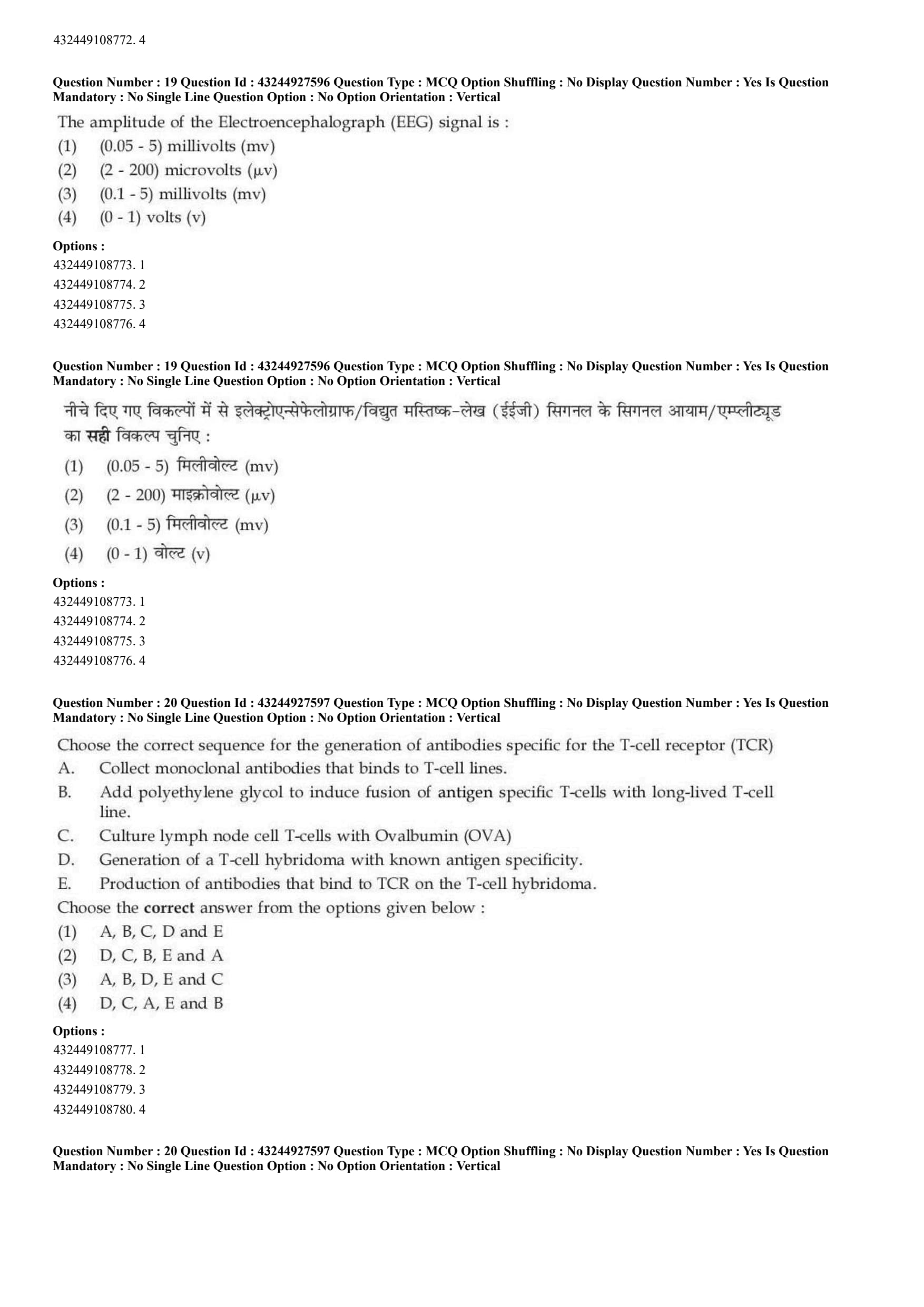
The amplitude of the Electroencephalograph (EEG) signal is :
View Solution
Step 1: Concept:
The question tests knowledge of clinical biomedical instrumentation, specifically asking for the typical electrical voltage range of signals produced by the human brain as measured non-invasively on the scalp by an Electroencephalograph (EEG).
Step 2: Step-by-step Explanation:
An Electroencephalograph (EEG) is designed to measure the synchronized summation of electrical post-synaptic activity originating from millions of pyramidal neurons located in the cerebral cortex.
Because these delicate electrical signals originate deep within the brain, they must physically travel through several highly resistive biological layers—including the meninges, the thick cerebrospinal fluid (CSF), the dense bone of the skull, and the skin of the scalp—before reaching the surface recording electrodes.
Due to this massive resistance, the signals are heavily attenuated (weakened) by the time they are recorded.
Consequently, surface EEG signals are extremely small in amplitude. Typical normal adult human EEG signals range from approximately 10 to 100 microvolts (\(\mu\)V). Depending on the state of arousal, age, or the presence of intense pathology (like epileptic seizures), they can peak up to about 200 \(\mu\)V.
Let's evaluate the options to confirm:
- Volts (v): This is far too massive. Measuring whole volts on the scalp would imply lethal electrocution levels.
- Millivolts (mV): This range (e.g., 1 to 5 mV) represents much stronger bio-signals, specifically the heart's electrocardiogram (ECG).
- Microvolts (\(\mu\)V): This is the correct, highly sensitive measurement scale required for brain waves.
Therefore, Option (B) provides the precise and accurate clinical range for these signals.
Step 3: Final Answer:
The typical EEG amplitude ranges from (2 - 200) microvolts.
Quick Tip: Scale associations for biomedical signals are common exam topics: Brain (EEG) = Microvolts (\(\mu\)V, extremely weak, needs high amplification). Heart (ECG) = Millivolts (mV, moderately weak, roughly 1-2 mV). Muscle (EMG) = Millivolts (mV). Always remember the brain's signals are the hardest to detect through the skull!
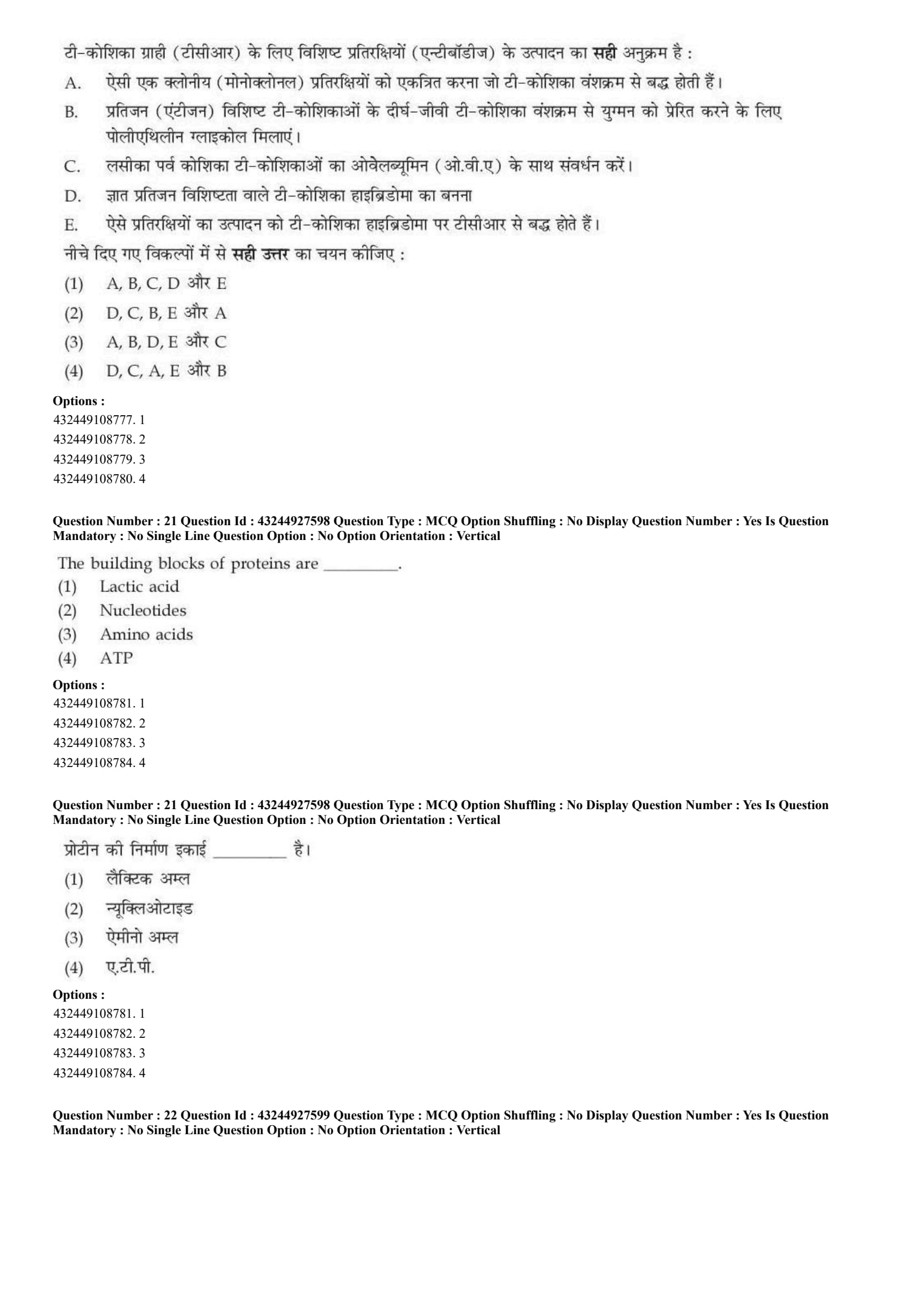
Choose the correct sequence for the generation of antibodies specific for the T-cell receptor (TCR)
A. Collect monoclonal antibodies that binds to T-cell lines.
B. Add polyethylene glycol to induce fusion of antigen specific T-cells with long-lived T-cell line.
C. Culture lymph node cell T-cells with Ovalbumin (OVA)
D. Generation of a T-cell hybridoma with known antigen specificity.
E. Production of antibodies that bind to TCR on the T-cell hybridoma.
View Solution
Step 1: Concept:
This complex question outlines the standard biotechnological and immunological procedure for creating custom monoclonal antibodies specifically targeted against a particular T-Cell Receptor (TCR) using adapted hybridoma technology.
Step 2: Step-by-step Explanation:
To successfully generate antibodies against a specific TCR, a researcher must first isolate, expand, and immortalize a pure clonal population of T-cells bearing that specific receptor.
Step C: The protocol begins by activating the desired cells. Lymph node cells are harvested and cultured in vitro with a known, specific antigen, such as Ovalbumin (OVA). This stimulates the activation and proliferation of only those T-cells with a TCR specific to OVA.
Step B: Primary T-cells die quickly in standard culture. Therefore, they must be immortalized. A chemical agent called Polyethylene glycol (PEG) is added to the culture to induce the physical cell membrane fusion of these specific primary T-cells with a cancerous, long-lived T-cell tumor line (lymphoma line).
Step D: If the fusion is successful and selected properly, this results in the generation of a T-cell hybridoma. This hybrid cell now possesses both the desired OVA-specific TCR (from the normal cell) and the ability to divide infinitely (from the tumor cell).
Step E: This stable hybridoma is now used as an immunogen. It is typically injected into a host animal (like a mouse) to trigger the animal's immune system into the production of antibodies that recognize and bind directly to the unique TCR on the surface of the injected hybridoma cells.
Step A: Finally, B-cells from the immunized animal are harvested, run through standard antibody-hybridoma generation, allowing researchers to isolate, purify, and collect monoclonal antibodies that specifically bind to the original T-cell line.
Following this strict biological logic, the chronologically accurate sequence is absolutely C \(\rightarrow\) B \(\rightarrow\) D \(\rightarrow\) E \(\rightarrow\) A.
\textit{Note: Upon reviewing the given multiple-choice options (A, B, C, and D), none of them present this correct sequence. The question likely contains a typographical error from the examiners, but the sequence explained above is the only scientifically valid method.
Step 3: Final Answer:
The scientifically correct sequence is C, B, D, E, A, though this is absent from the provided options.
Quick Tip: For any hybridoma or monoclonal antibody generation protocol, always follow this fundamental logic: 1. Sensitize/Stimulate (expose to antigen) \(\rightarrow\) 2. Fuse (using PEG chemical) \(\rightarrow\) 3. Select/Generate stable Hybridoma \(\rightarrow\) 4. Induce Production of Antibodies \(\rightarrow\) 5. Harvest/Collect final product.
The building blocks of proteins are _________
View Solution
Step 1: Concept:
This question tests fundamental biochemistry, specifically the monomeric subunits that polymerize to form complex biological macromolecules like proteins.
Step 2: Step-by-step Explanation:
Proteins are large, complex biopolymers that play crucial structural, enzymatic, and regulatory roles in all living organisms. The fundamental, repeating structural units (monomers) of all proteins are amino acids.
During the cellular process of translation, amino acids are covalently linked together in a specific linear sequence dictated by mRNA. The bond connecting them is called a peptide bond, forming a polypeptide chain that eventually folds into a functional protein.
Let's review the other options to understand why they are incorrect:
Lactic acid (A) is a metabolic byproduct of anaerobic glycolysis in muscle tissue, not a structural monomer for macromolecules.
Nucleotides (B) are the building blocks of nucleic acids, specifically DNA (Deoxyribonucleic acid) and RNA (Ribonucleic acid), not proteins.
ATP (D) stands for Adenosine Triphosphate, which is the primary energy currency of the cell, not a structural building block for proteins.
Step 3: Final Answer:
The building blocks of proteins are amino acids.
Quick Tip: Always associate the major macromolecules with their monomers: Proteins \(\rightarrow\) Amino acids; Nucleic Acids (DNA/RNA) \(\rightarrow\) Nucleotides; Carbohydrates \(\rightarrow\) Monosaccharides (like glucose); Lipids \(\rightarrow\) Fatty acids and glycerol.
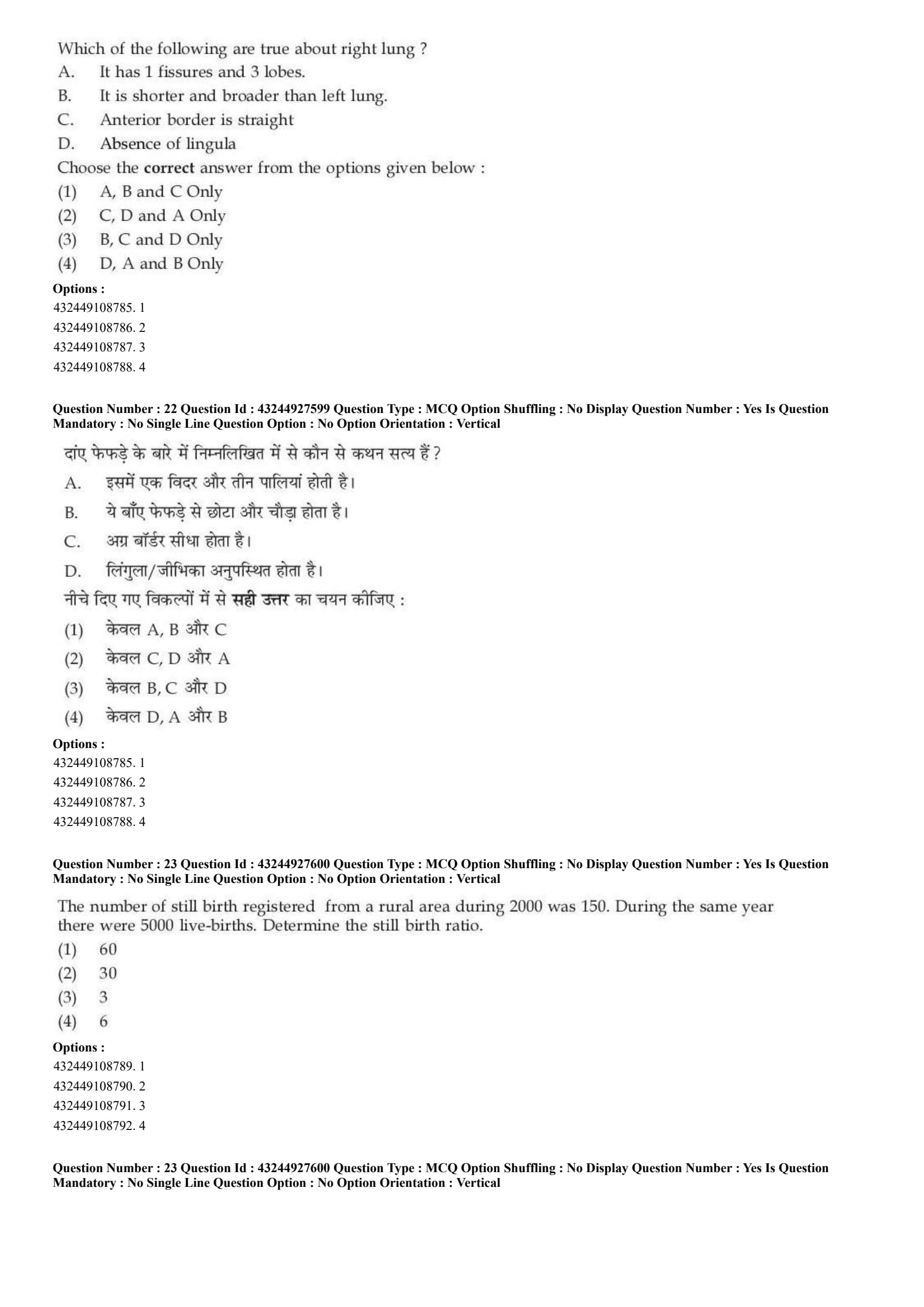
Which of the following are true about right lung ?
A. It has 1 fissures and 3 lobes.
B. It is shorter and broader than left lung.
C. Anterior border is straight.
D. Absence of lingula.
View Solution
Step 1: Concept:
This question requires a detailed anatomical understanding of the human respiratory system, specifically contrasting the physical features of the right lung against the left lung.
Step 2: Step-by-step Explanation:
Statement A is False: The right lung is divided into 3 lobes (superior, middle, and inferior) by 2 fissures (the oblique fissure and the horizontal fissure). The statement incorrectly claims it has only 1 fissure.
Statement B is True: The right lung is anatomically shorter than the left lung because the right dome of the diaphragm sits higher to accommodate the large liver underneath. However, it is also broader (wider) and has a larger total volume because the heart bulges more into the left thoracic cavity.
Statement C is True: The anterior border of the right lung is relatively straight and vertical. In contrast, the anterior border of the left lung features a distinct indentation called the cardiac notch to make room for the apex of the heart.
Statement D is True: The lingula is a small, tongue-like projection of tissue found exclusively on the superior lobe of the left lung (just below the cardiac notch). It is absent in the right lung.
Since B, C, and D are factually correct anatomical statements, this combination forms the correct answer.
Step 3: Final Answer:
The correct combination of true statements is B, C, and D Only.
Quick Tip: Remember the "Rule of 2s and 3s": The Right lung has 3 lobes and 2 fissures. The Left lung has 2 lobes and 1 fissure (plus the Lingula and Cardiac notch).
The number of still birth registered from a rural area during 2000 was 150. During the same year there were 5000 live-births. Determine the still birth ratio.
View Solution
Step 1: Concept:
This question involves public health biostatistics, specifically requiring the calculation of the "Stillbirth Ratio" from raw demographic data.
Step 2: Key Formulas and approach:
The Stillbirth Ratio is a vital statistical measure defined as the number of stillbirths per 1,000 live births during a given year in a specific population.
The formula is:
\[ Stillbirth Ratio = \left( \frac{Number of Stillbirths}{Number of Live Births} \right) \times 1000 \]
Step 3: Step-by-step Explanation:
Given Data:
Number of stillbirths = 150
Number of live births = 5000
Substitute these values directly into the formula:
\[ Stillbirth Ratio = \left( \frac{150}{5000} \right) \times 1000 \]
Simplify the expression by handling the multiplier first for easier mental math:
\[ Stillbirth Ratio = 150 \times \left( \frac{1000}{5000} \right) \]
\[ Stillbirth Ratio = 150 \times \left( \frac{1}{5} \right) \]
Calculate the final division:
\[ Stillbirth Ratio = \frac{150}{5} = 30 \]
The calculated ratio means there were 30 stillbirths for every 1,000 live births in that rural area.
Step 4: Final Answer:
The stillbirth ratio is 30, matching option (B).
Quick Tip: Do not confuse Stillbirth Ratio with Stillbirth Rate!
Ratio denominator = Live births only.
Rate denominator = Total births (Live births + Stillbirths). Always check which term the examiner uses.
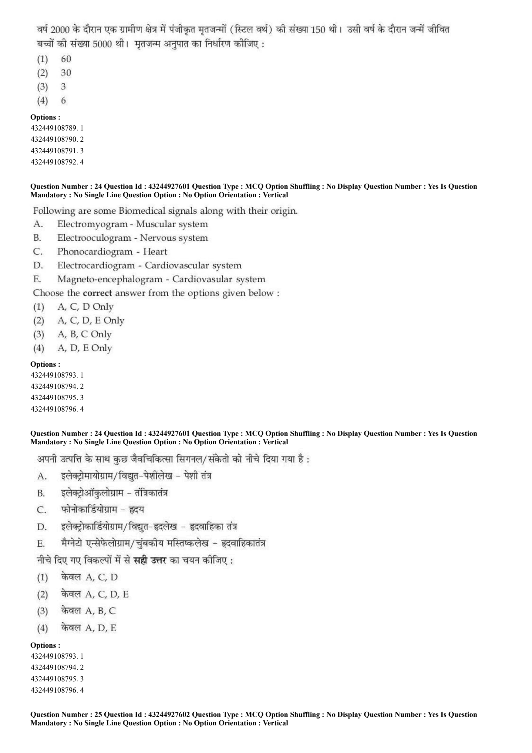
Following are some Biomedical signals along with their origin.
A. Electromyogram - Muscular system
B. Electrooculogram - Nervous system
C. Phonocardiogram - Heart
D. Electrocardiogram - Cardiovascular system
E. Magneto-encephalogram - Cardiovascular system
Choose the correct answer from the options given below :
View Solution
Step 1: Concept:
This question tests knowledge of various diagnostic biomedical instrumentation signals and accurately matching them to their physiological source systems within the human body.
Step 2: Step-by-step Explanation:
A. Electromyogram (EMG) - Muscular system (True): An EMG measures the electrical activity produced by skeletal muscles during rest and voluntary contraction.
B. Electrooculogram (EOG) - Nervous system (Debatable/Often classed separately): An EOG measures the resting corneo-retinal potential (the dipole between the front and back of the eye). While the retina is neural tissue, clinical questions often separate the visual/ocular system from the general "nervous system." Given the option choices, this statement is treated as excluded.
C. Phonocardiogram (PCG) - Heart (True): A PCG is a high-fidelity diagnostic recording of the acoustic sounds and murmurs produced by the mechanical pumping and valve closures of the heart.
D. Electrocardiogram (ECG/EKG) - Cardiovascular system (True): An ECG records the electrical conduction pathways (depolarization and repolarization) traversing the heart muscle during each cardiac cycle.
E. Magneto-encephalogram (MEG) - Cardiovascular system (False): An MEG maps brain activity by recording extremely weak magnetic fields produced by electrical currents occurring naturally in the brain. Its origin is strictly the central nervous system, not the cardiovascular system.
Because E is definitively false, any option containing E is incorrect. Based on the remaining definitive truths, A, C, and D are the most accurate pairing.
Step 3: Final Answer:
The correct valid combinations are A, C, and D Only.
Quick Tip: Break down the root words to never miss these:
"Myo" = Muscle (EMG)
"Oculo" = Eye (EOG)
"Phono" = Sound (PCG)
"Cardio" = Heart (ECG)
"Encephalo" = Brain (EEG/MEG).
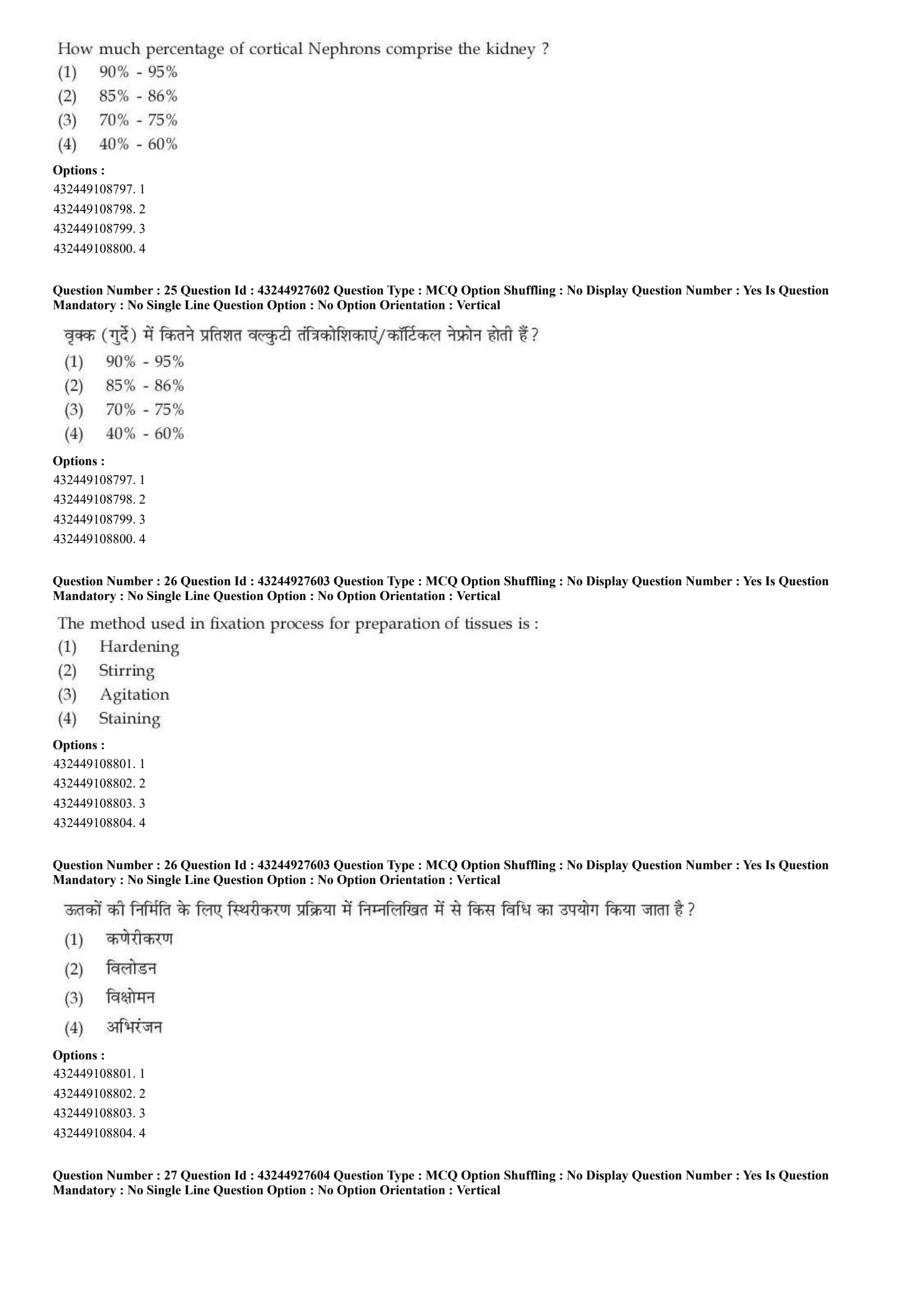
How much percentage of cortical Nephrons comprise the kidney ?
View Solution
Step 1: Concept:
The question tests microscopic renal anatomy, specifically the population distribution of the two distinct types of nephrons (the functional units of the kidney) found within the human renal system.
Step 2: Step-by-step Explanation:
The human kidney contains approximately 1 to 1.5 million nephrons, which are broadly classified into two categories based on their anatomical location and the length of their Loop of Henle.
1. Cortical Nephrons: These constitute the vast majority of nephrons. Their renal corpuscles (glomeruli) are located high up in the outer renal cortex. They possess relatively short Loops of Henle that barely penetrate into the outer zone of the renal medulla. Cortical nephrons are primarily responsible for the bulk reabsorption of water and solutes, and they represent roughly 80% to 85% (up to 86%) of all nephrons in humans.
2. Juxtamedullary Nephrons: These constitute the minority. Their renal corpuscles are located deep in the cortex, adjacent to the medulla (juxta = next to). They have extremely long Loops of Henle that dive deep into the inner medulla. These specialized nephrons are crucial for establishing the medullary osmotic gradient required to concentrate urine, representing the remaining 15% to 20%.
Among the provided options, 85% - 86% is the most medically accurate representation of the cortical nephron population.
Step 3: Final Answer:
Cortical nephrons comprise approximately 85% - 86% of the kidney.
Quick Tip: Always remember: Cortical = Common \& Short (85%). Juxtamedullary = Rare \& Long (15%). The long loops of the juxtamedullary nephrons are the secret to highly concentrated, yellow urine!
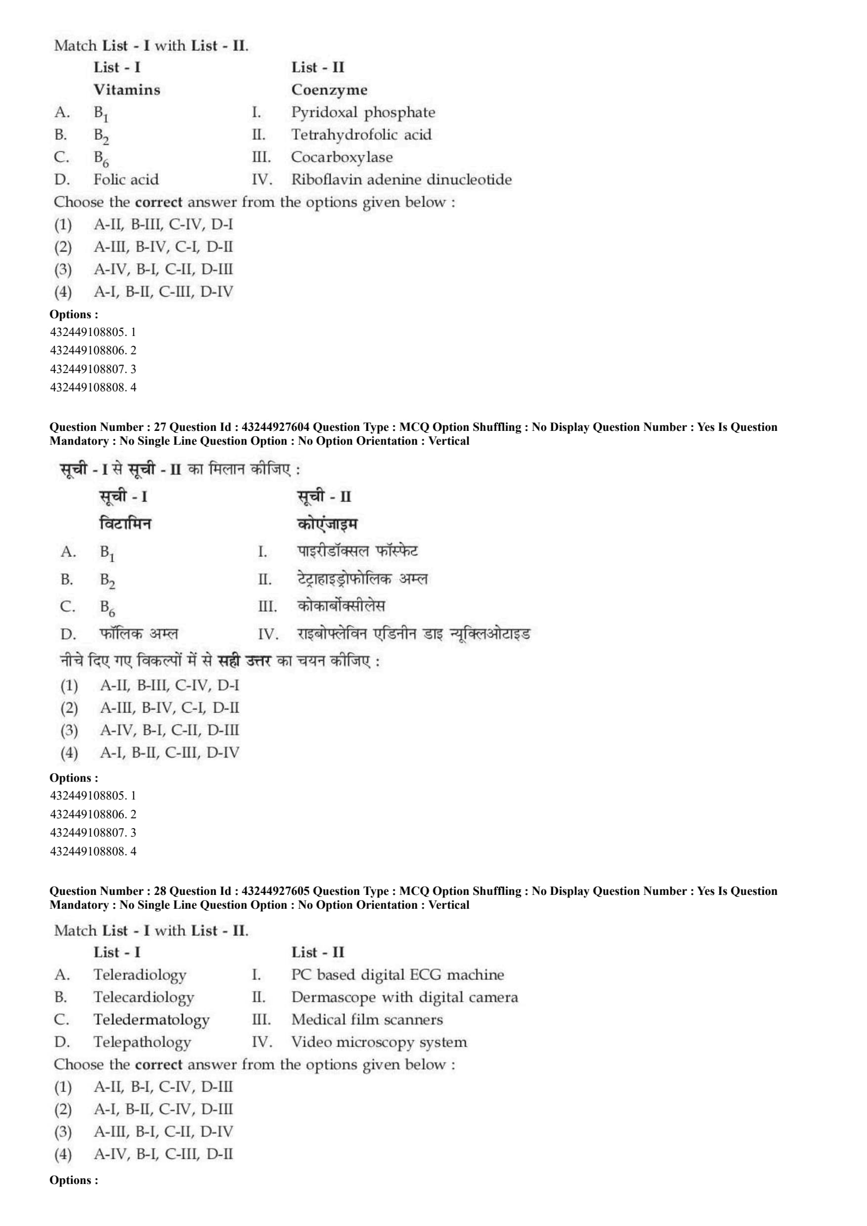
The method used in fixation process for preparation of tissues is :
View Solution
Step 1: Concept:
This question delves into histopathology techniques, specifically assessing the primary physical characteristics and goals achieved during the "fixation" step of tissue processing.
Step 2: Step-by-step Explanation:
Fixation is the critical first step in tissue processing for histopathological examination. When fresh tissue is removed from the body, it is soft, fragile, and immediately subject to autolysis (self-digestion by its own enzymes) and putrefaction (bacterial decay).
To prevent this, tissue is submerged in a chemical fixative (most commonly 10% Neutral Buffered Formalin).
The primary biochemical effect of fixatives is cross-linking proteins. This chemical cross-linking achieves two major outcomes: it permanently preserves the cellular architecture, and it induces significant physical hardening of the tissue.
This hardening is entirely intentional and necessary; without it, the soft tissue would simply crush or tear during the subsequent steps of grossing and microtome sectioning (cutting into ultra-thin slices).
\textit{Analyzing other options: "Stirring" and "Agitation" are mechanical actions used to speed up the penetration of fluids, but they are not the defining mechanism of fixation itself. "Staining" is a completely different, later step used to add contrast to the slides using dyes (like H\&E).
Step 3: Final Answer:
Hardening is the primary functional method/result achieved during the tissue fixation process.
Quick Tip: The three main goals of Fixation: 1. Prevent Autolysis (kill enzymes). 2. Prevent Putrefaction (kill bacteria). 3. Harden the tissue (to allow for ultra-thin microtome sectioning later).
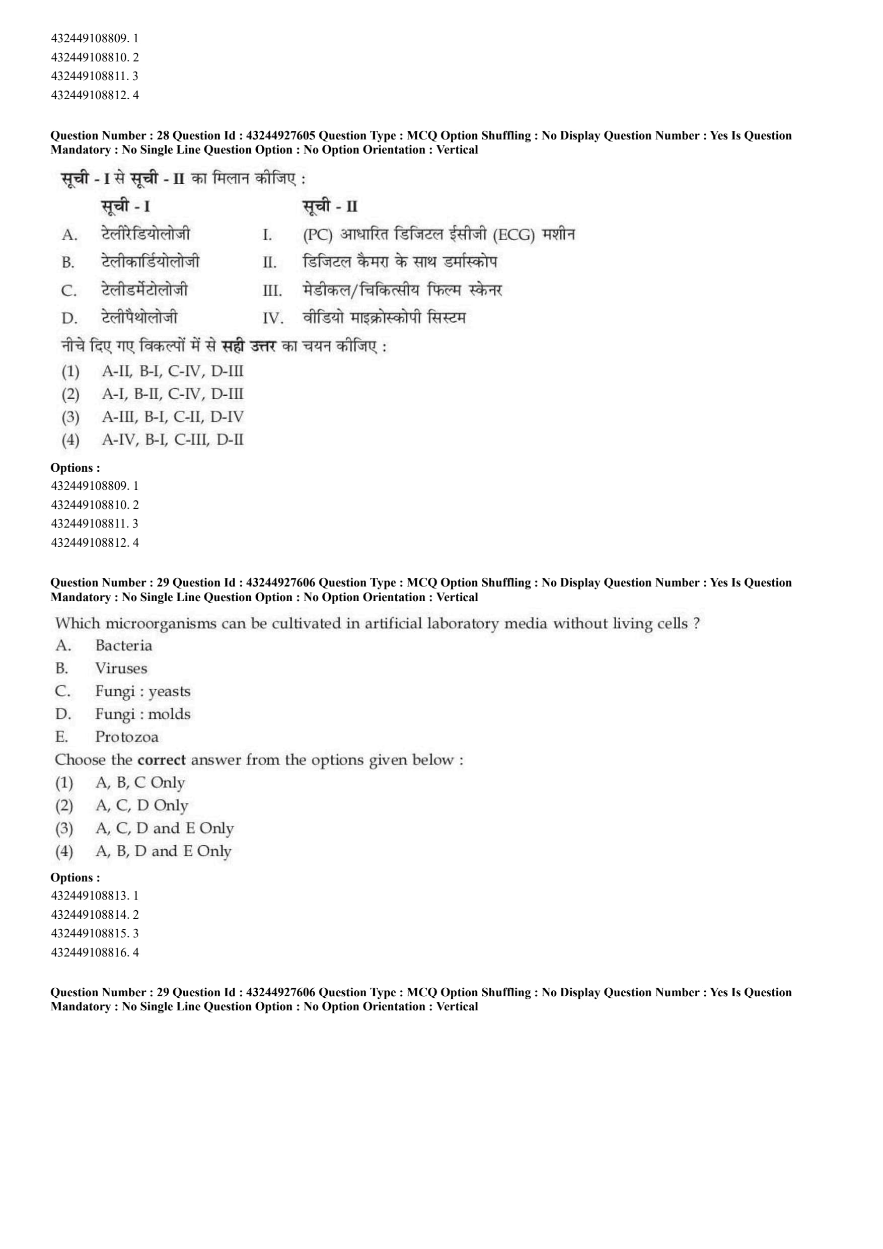
Match List - I with List - II.

Choose the correct answer from the options given below :
View Solution
Step 1: Concept:
This question tests nutritional biochemistry, requiring you to correctly match essential B-complex vitamins with their biologically active coenzyme forms used in cellular metabolism.
Step 2: Step-by-step Explanation:
A. Vitamin B\(_1\) (Thiamine): Its active coenzyme form is Thiamine Pyrophosphate (TPP). TPP is historically and clinically also known as Cocarboxylase because it acts as an essential cofactor for decarboxylation reactions (like Pyruvate dehydrogenase). Thus, A matches with III.
B. Vitamin B\(_2\) (Riboflavin): Its active coenzyme forms are Flavin Mononucleotide (FMN) and Riboflavin adenine dinucleotide (FAD). These are crucial electron carriers in the Krebs cycle and Electron Transport Chain. Thus, B matches with IV.
C. Vitamin B\(_6\) (Pyridoxine): Its biologically active coenzyme form is Pyridoxal phosphate (PLP). PLP is vital for amino acid metabolism, particularly in transamination reactions. Thus, C matches with I.
D. Folic acid (Vitamin B\(_9\)): Within the body, folic acid is reduced to its metabolically active form, Tetrahydrofolic acid (THF), which is essential for one-carbon metabolism and DNA synthesis. Thus, D matches with II.
Compiling the correct pairs yields: A-III, B-IV, C-I, D-II.
Step 3: Final Answer:
Option (B) represents the correct sequence of matches.
Quick Tip: Vitamin Coenzyme memory anchors:
B1 (Thiamine) \(\rightarrow\) TPP / Cocarboxylase (Energy metabolism).
B2 (Riboflavin) \(\rightarrow\) FAD/FMN (Flavin = Riboflavin).
B6 (Pyridoxine) \(\rightarrow\) Pyridoxal Phosphate (PLP).
B9 (Folic Acid) \(\rightarrow\) Tetrahydrofolate (THF).
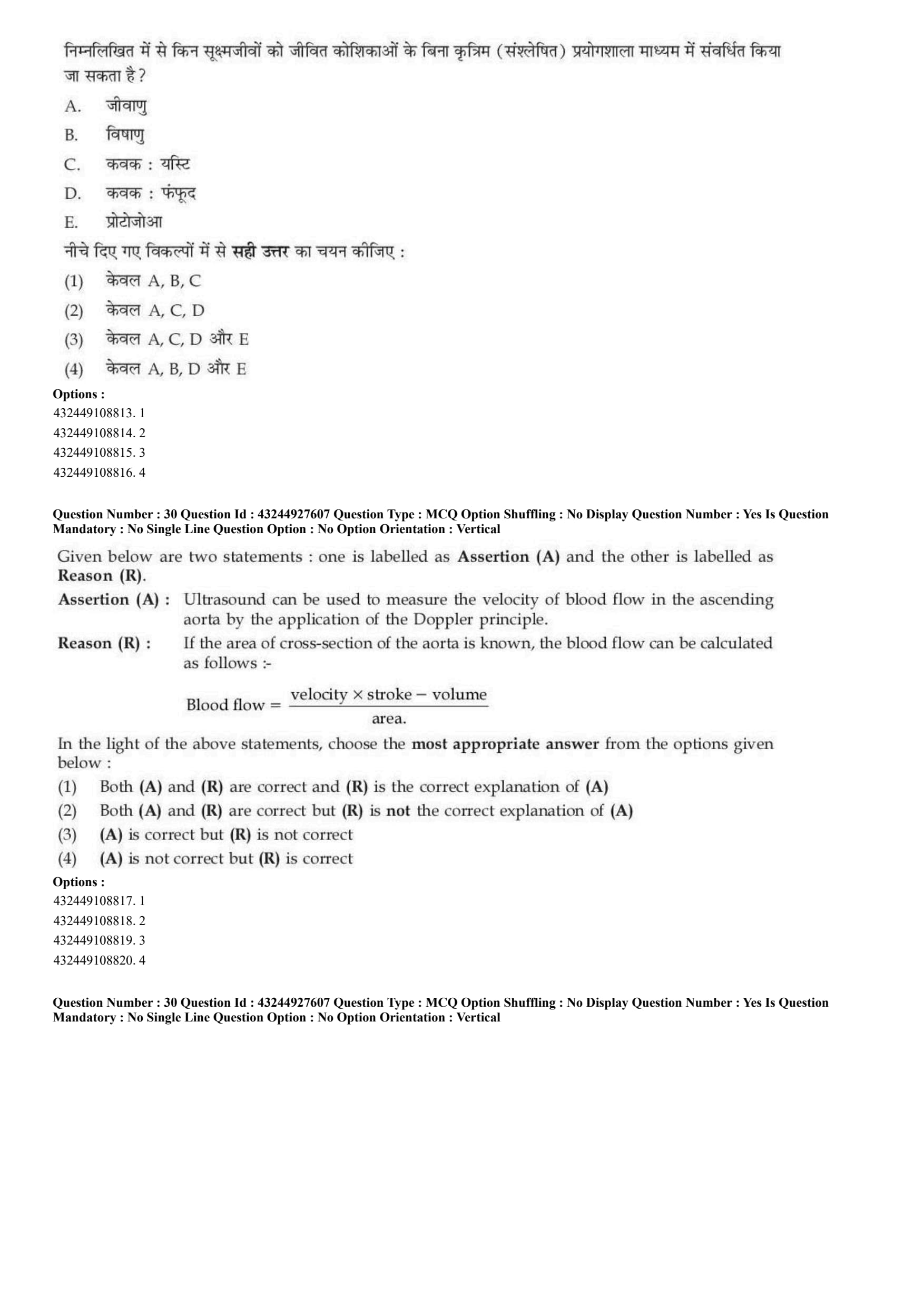
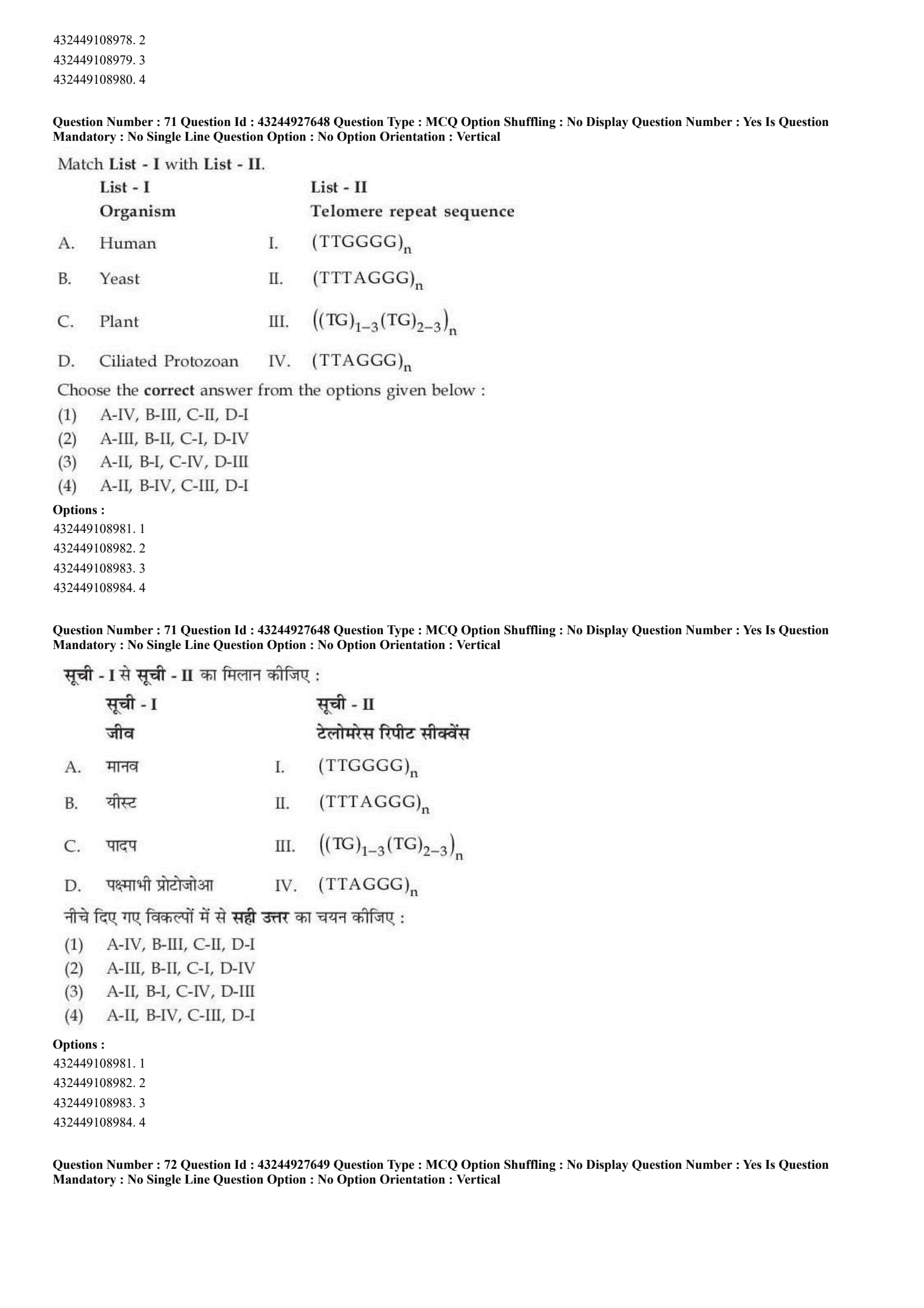
Match List - I with List - II.

Choose the correct answer from the options given below :
View Solution
Step 1: Concept:
This question evaluates knowledge of modern Telemedicine applications, requiring the matching of specialized tele-diagnostic fields with the specific digital instrumentation required to perform them remotely.
Step 2: Step-by-step Explanation:
A. Teleradiology: This field involves the remote transmission of radiological patient images (like X-rays, CTs, and MRIs). To transmit physical X-ray films, specialized high-resolution Medical film scanners are used to digitize them. Thus, A matches with III.
B. Telecardiology: This focuses on remote cardiovascular diagnostics. The most common tool used is a PC based digital ECG machine, which allows a patient's electrical heart rhythm to be recorded locally and transmitted instantly to a remote cardiologist for interpretation. Thus, B matches with I.
C. Teledermatology: This involves remote consultation for skin lesions. Practitioners use a Dermascope with a digital camera to take highly magnified, illuminated pictures of moles or rashes and send them to a dermatologist. Thus, C matches with II.
D. Telepathology: This deals with the remote practice of pathology (studying disease at the microscopic cellular level). It heavily relies on a Video microscopy system or whole-slide imaging scanners to allow a remote pathologist to view tissue biopsies live or digitally. Thus, D matches with IV.
Compiling the correct pairs yields: A-III, B-I, C-II, D-IV.
Step 3: Final Answer:
Option (C) provides the correct alignment of telemedicine fields to their instruments.
Quick Tip: Match the root words to the technology:
Radio (X-rays) \(\rightarrow\) Film Scanners.
Cardio (Heart) \(\rightarrow\) ECG.
Dermato (Skin) \(\rightarrow\) Dermascope.
Patho (Tissue/Cells) \(\rightarrow\) Microscopy.
Which microorganisms can be cultivated in artificial laboratory media without living cells ?
A. Bacteria
B. Viruses
C. Fungi : yeasts
D. Fungi : molds
E. Protozoa
View Solution
Step 1: Concept:
This question tests basic microbiology, specifically distinguishing between free-living microbes that can synthesize their own requirements from chemical nutrients versus obligate intracellular parasites that completely rely on a living host cell machinery to replicate.
Step 2: Step-by-step Explanation:
Artificial cell-free media (like nutrient agar, blood agar, or broth) contain various chemical nutrients, amino acids, and sugars, but absolutely no living host cells.
A. Bacteria: The vast majority of bacteria are free-living and easily cultivated on standard artificial media (with rare exceptions like Chlamydia or \textit{Rickettsia).
B. Viruses: Viruses are strictly obligate intracellular parasites. They lack their own metabolic machinery and ribosomes, meaning they cannot synthesize proteins or replicate without hijacking a living host cell (like a tissue culture or embryonated egg). They can \textit{never be cultivated on cell-free artificial media.
C \& D. Fungi (yeasts and molds): Fungi are completely free-living, eukaryotic organisms. They grow exceptionally well on specialized cell-free artificial media, such as Sabouraud Dextrose Agar (SDA).
E. Protozoa: While some advanced techniques (axenic cultivation) exist for certain protozoa (like \textit{Entamoeba), many medically important protozoa (like \textit{Plasmodium causing malaria) strictly require living host cells (like red blood cells). Because of this strict requirement for many clinical species, they are generally excluded in classic textbook definitions of routine cell-free cultivation alongside bacteria and fungi.
Therefore, Bacteria, Yeasts, and Molds are the classic group routinely grown on cell-free artificial media.
Step 3: Final Answer:
The correct combination is A, C, and D Only.
Quick Tip: The most important rule in introductory microbiology: \textbf{Viruses cannot grow on agar! They are essentially just genetic material wrapped in a protein coat; they need a living factory to copy themselves.
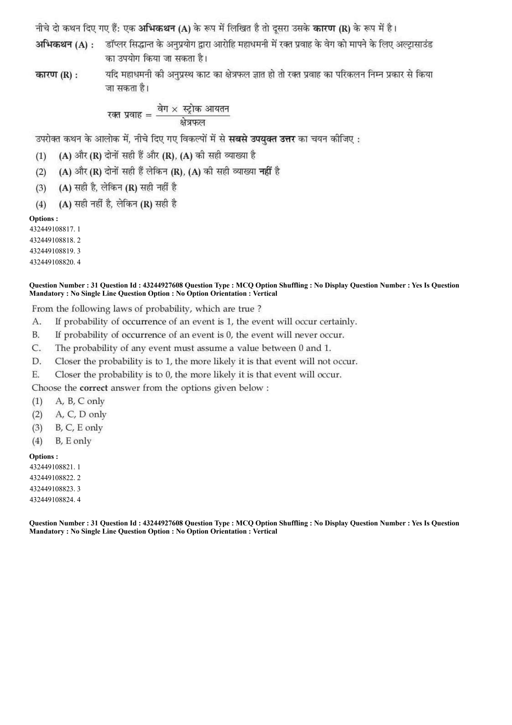
Given below are two statements : one is labelled as Assertion (A) and the other is labelled as Reason (R).
Assertion (A) : Ultrasound can be used to measure the velocity of blood flow in the ascending aorta by the application of the Doppler principle.
Reason (R) : If the area of cross-section of the aorta is known, the blood flow can be calculated as follows :-
Blood flow = \(\frac{velocity \times stroke - volume}{area}\)
In the light of the above statements, choose the most appropriate answer from the options given below :
View Solution
Step 1: Concept:
This question evaluates the physics of biomedical ultrasound, specifically Doppler flowmetry, and the fundamental hemodynamic formulas used to calculate blood flow and stroke volume.
Step 2: Step-by-step Explanation:
Analyzing Assertion (A): Doppler ultrasound is a standard, non-invasive clinical tool. By bouncing high-frequency sound waves off moving red blood cells, the change in frequency (Doppler shift) allows computers to accurately calculate the velocity of the blood flow in major vessels like the ascending aorta. Therefore, Assertion (A) is completely correct.
Analyzing Reason (R): The reason provides a mathematical formula for calculating blood flow. However, the formula provided is physically and mathematically nonsensical.
The correct physiological principle of fluid dynamics (Continuity Equation) states that Blood Flow (Volume Flow Rate, Q) is simply the product of the flow velocity and the cross-sectional area of the vessel:
\[ Q \ (Flow) = V \ (Velocity) \times A \ (Cross-sectional Area) \]
Alternatively, to calculate Stroke Volume (SV) using echocardiography, the formula used is:
\[ SV = VTI \ (Velocity Time Integral) \times CSA \ (Cross-sectional Area) \]
The formula presented in Reason (R) places Area in the denominator and subtracts volume, which breaks dimensional analysis and is entirely incorrect. Thus, Reason (R) is false.
Step 3: Final Answer:
(A) is correct but (R) is not correct.
Quick Tip: Always check the units in formulas! Flow rate (\(Q\)) is measured in \(cm^3/sec\) (or \(mL/sec\)). Velocity is \(cm/sec\). Area is \(cm^2\). Therefore, \(cm/sec \times cm^2 = cm^3/sec\). Dividing by area as shown in (R) would yield \(1/sec\), proving the formula is fake.
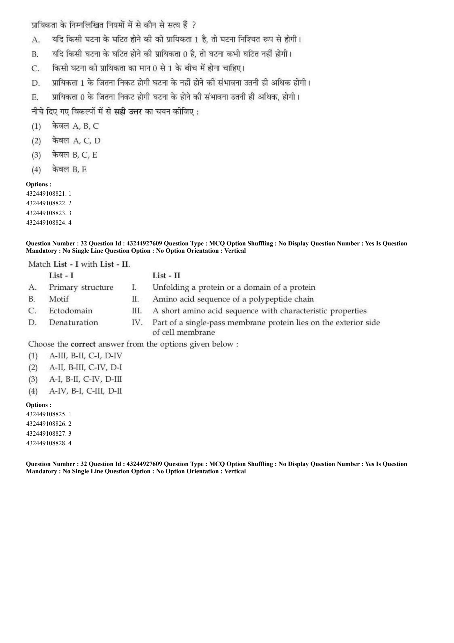
From the following laws of probability, which are true ?
A. If probability of occurrence of an event is 1, the event will occur certainly.
B. If probability of occurrence of an event is 0, the event will never occur.
C. The probability of any event must assume a value between 0 and 1.
D. Closer the probability is to 1, the more likely it is that event will not occur.
E. Closer the probability is to 0, the more likely it is that event will occur.
Choose the correct answer from the options given below :
View Solution
Step 1: Concept:
This question tests the fundamental, axiomatic rules of probability theory, which are mathematically established foundations for all statistical analysis.
Step 2: Step-by-step Explanation:
Probability (\(P\)) is a mathematical measure of the likelihood that a specific event will occur.
Statement C is True: According to Kolmogorov's First Axiom of probability, the probability of any event \(E\) is a real number bounded exactly between zero and one (inclusive). Expressed mathematically: \(0 \leq P(E) \leq 1\).
Statement A is True: If \(P(E) = 1\), the event is considered an "absolute certainty" or a "sure event." It is guaranteed to happen 100% of the time.
Statement B is True: If \(P(E) = 0\), the event is considered an "impossible event." There is a 0% chance of it occurring, meaning it will never happen.
Statement D is False: The closer a probability moves toward 1, the \textit{more likely the event is to occur, not less likely.
Statement E is False: The closer a probability moves toward 0, the \textit{less likely the event is to occur, not more likely.
Therefore, only statements A, B, and C represent valid mathematical laws of probability.
Step 3: Final Answer:
The correct options are A, B, and C only.
Quick Tip: Probability acts exactly like a percentage divided by 100. \(0 = 0%\), \(0.5 = 50%\), and \(1 = 100%\). The higher the number, the more likely it happens!
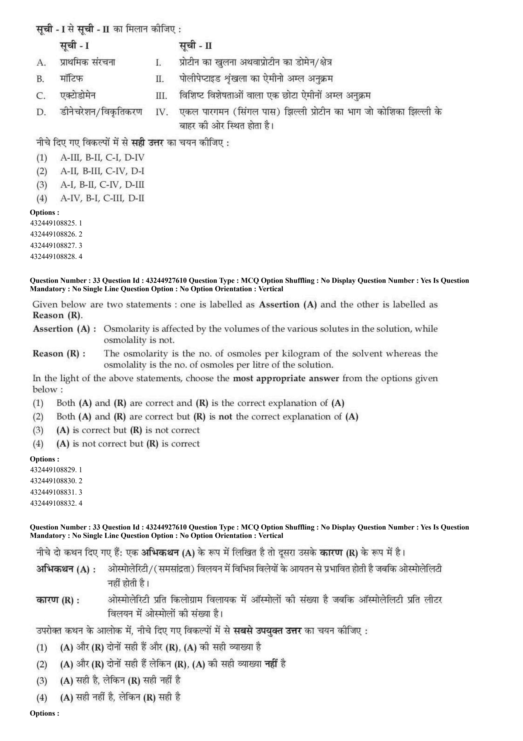
Match List - I with List - II.

Choose the correct answer from the options given below :
View Solution
Step 1: Concept:
This question requires matching specific biochemical terminology related to protein architecture, folding, and cellular localization to their correct definitions.
Step 2: Step-by-step Explanation:
A. Primary structure: This is the most fundamental level of protein architecture. It simply refers to the specific linear Amino acid sequence of a polypeptide chain linked by peptide bonds, dictated directly by DNA. Thus, A matches with II.
B. Motif: In structural biology, a motif (or supersecondary structure) is a recognizable folding pattern involving a short amino acid sequence with characteristic properties that appears in many different proteins (e.g., a zinc finger or a helix-turn-helix motif). Thus, B matches with III.
C. Ectodomain: Proteins embedded in the cell membrane have different functional parts. The "ecto" (meaning outside) domain is the Part of a single-pass membrane protein that lies on the exterior (extracellular) side of the cell membrane, often involved in binding ligands. Thus, C matches with IV.
D. Denaturation: This is the physical or chemical process of Unfolding a protein or a domain, destroying its complex 3D shape (secondary, tertiary, and quaternary structures) without breaking the primary sequence peptide bonds, causing loss of function. Thus, D matches with I.
Compiling the correct pairs yields: A-II, B-III, C-IV, D-I.
Step 3: Final Answer:
Option (B) represents the correct sequence of matches.
Quick Tip: Break down complex biological words: "Ecto" = Outside (Extracellular). "Endo" = Inside (Intracellular). Denature = To take away its natural shape (unfolding).
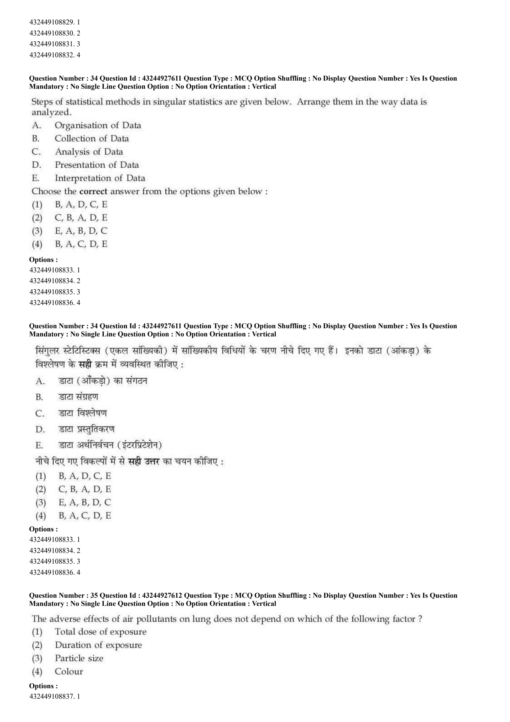
Given below are two statements : one is labelled as Assertion (A) and the other is labelled as Reason (R).
Assertion (A) : Osmolarity is affected by the volumes of the various solutes in the solution, while osmolality is not.
Reason (R) : The osmolarity is the no. of osmoles per kilogram of the solvent whereas the osmolality is the no. of osmoles per litre of the solution.
In the light of the above statements, choose the most appropriate answer from the options given below :
View Solution
Step 1: Concept:
This question tests the critical physical chemistry distinction between two closely related concentration metrics used constantly in clinical fluid therapy: Osmolarity and Osmolality.
Step 2: Step-by-step Explanation:
Analyzing Reason (R): Let's establish the definitions first.
- OsmolaRity (with an 'R') is officially defined as the number of osmoles of solute per Litre of solution (volume).
- OsmolaLity (with an 'L') is defined as the number of osmoles of solute per Kilogram of solvent (mass).
- Look closely at Reason (R): It completely swaps these two definitions! It claims osmolarity is per kg, and osmolality is per litre. Therefore, Reason (R) is fundamentally FALSE.
Analyzing Assertion (A): Because OsmolaRity relies on the total volume of the solution (litres), it is directly affected by anything that changes volume—including temperature fluctuations, pressure changes, and the physical space (volumes) taken up by large solutes (like proteins or lipids).
- Conversely, OsmolaLity relies strictly on the \textit{mass of the solvent (kg). Mass does not expand or contract with temperature, nor is it displaced by the volume of other solutes. Therefore, it remains constant regardless of these factors.
- Therefore, Assertion (A) is a perfectly accurate scientific statement.
Step 3: Final Answer:
Assertion (A) is correct, but Reason (R) is not correct.
Quick Tip: A simple mnemonic to never mix these up:
Osmola\textbf{Rity = per Lit\textbf{R}e (Volume).
Osmola\textbf{L}ity = per ki\textbf{L}ogram (Mass).
Clinical labs prefer measuring osmolality because it is temperature-independent!
Steps of statistical methods in singular statistics are given below. Arrange them in the way data is analyzed.
A. Organisation of Data
B. Collection of Data
C. Analysis of Data
D. Presentation of Data
E. Interpretation of Data
Choose the correct answer from the options given below :
View Solution
Step 1: Concept:
This question requires identifying the standard, chronological stages involved in a complete statistical investigation, from raw information gathering to final conclusions.
Step 2: Step-by-step Explanation:
A scientific statistical inquiry follows a strict, logical sequence to ensure data integrity and accurate conclusions.
1. Collection of Data (B): This is the obligatory first step. Researchers must gather the raw raw, unorganized data using tools like surveys, experiments, or hospital records.
2. Organisation of Data (A): Raw data is messy. It must be cleaned, edited, classified, and tabulated into a manageable format (e.g., grouping ages into brackets).
3. Presentation of Data (D): Once organized, the data is converted into visual formats like graphs, charts (pie/bar), or formal tables so patterns become visible to the human eye.
4. Analysis of Data (C): Mathematical tools are now applied. Researchers calculate averages, standard deviations, correlations, or run hypothesis tests (like t-tests or chi-square) to extract hard mathematical facts.
5. Interpretation of Data (E): The final step. The mathematical results are translated into real-world meaning, conclusions are drawn, and decisions are made based on the findings.
The correct chronological sequence is therefore B \(\rightarrow\) A \(\rightarrow\) D \(\rightarrow\) C \(\rightarrow\) E.
Step 3: Final Answer:
The sequence matching option (A) is correct.
Quick Tip: Remember the acronym \textbf{COPAI}: \textbf{C}ollect, \textbf{O}rganize, \textbf{P}resent, \textbf{A}nalyze, \textbf{I}nterpret. You can't organize what you haven't collected, and you can't interpret what you haven't mathematically analyzed.
The adverse effects of air pollutants on lung does not depend on which of the following factor ?
View Solution
Step 1: Concept:
This question tests basic principles of environmental toxicology and respiratory pathology, specifically identifying which physical or chemical parameters dictate how much damage an inhaled pollutant will cause.
Step 2: Step-by-step Explanation:
The toxicity and adverse physiological effects of any inhaled airborne pollutant are governed by several strict physical and biological factors:
Total dose of exposure (A): Toxicology dictates that "the dose makes the poison." Inhaling a massive concentration of a chemical causes more severe and rapid damage than a tiny amount.
Duration of exposure (B): Chronic (long-term) exposure, even at lower doses, leads to cumulative lung damage (like pneumoconiosis or COPD) compared to brief, transient exposures.
Particle size (C): This is a critical aerodynamic factor. Large particles (\(>10 \mu m\)) get trapped in the nose and throat. Fine particles (PM 2.5) bypass upper defenses and lodge deep in the pulmonary alveoli, causing severe systemic damage. Thus, particle size is highly relevant.
Colour (D): The visual color of a gas or particulate cloud (whether it is white smoke, black soot, or a colorless gas like Carbon Monoxide) has absolutely zero biological bearing on its chemical toxicity or its physical mechanism of damaging lung tissue.
Step 3: Final Answer:
The adverse effects do not depend on the colour of the pollutant.
Quick Tip: Some of the most dangerous and deadly respiratory toxins (like Carbon Monoxide and Radon gas) are completely invisible and colorless. Biological damage is entirely based on chemistry and physics (dose, time, size), never optics.
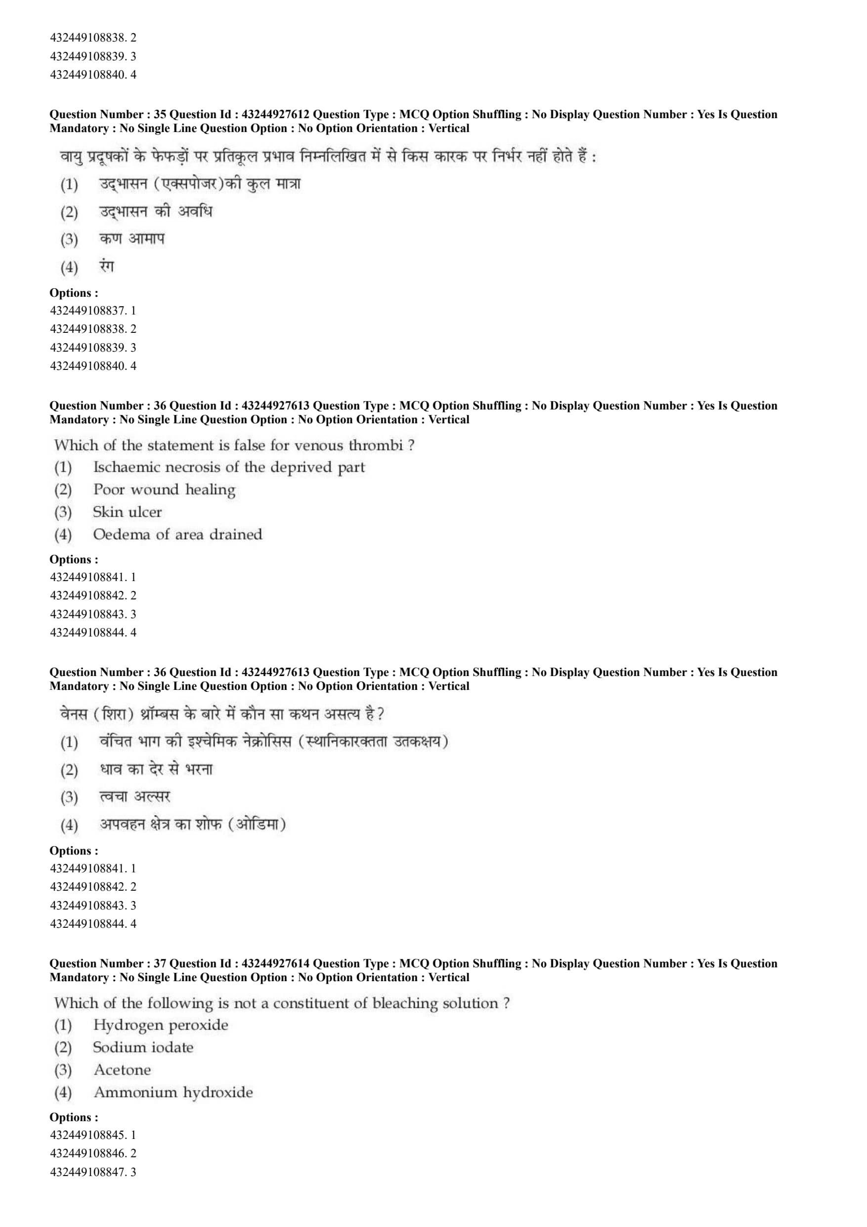
Which of the statement is false for venous thrombi ?
View Solution
Step 1: Concept:
This question evaluates the pathophysiological differences between arterial thrombosis (blockage of blood flowing \textit{to a tissue) and venous thrombosis (blockage of blood draining \textit{away from a tissue).
Step 2: Step-by-step Explanation:
A venous thrombus (like a DVT in the leg) blocks the return of deoxygenated blood back to the heart. This creates a massive "traffic jam" or back-pressure in the venous system.
This back-pressure forces plasma out of the capillaries into the surrounding tissues, leading to severe Oedema (swelling) of the area drained (Option D is true).
Because the venous blood is stagnating and pooling (venous stasis), tissues struggle to clear metabolic waste, leading to chronically Poor wound healing (Option B is true) and the eventual breakdown of tissue forming Skin ulcers (stasis ulcers) (Option C is true).
Ischaemic necrosis (infarction) (Option A) happens when a tissue is completely starved of fresh, oxygenated arterial blood. This is the classic hallmark of an arterial thrombus (e.g., causing a heart attack or stroke). While severe venous blockages can occasionally lead to gangrene (phlegmasia cerulea dolens), ischemic necrosis is fundamentally defined as an arterial failure issue, making it the textbook "false" statement for venous thrombi.
Step 3: Final Answer:
Ischaemic necrosis is false for venous thrombi; it is an arterial characteristic.
Quick Tip: Arterial Block = Ischemia, Pallor (pale), Cold, Necrosis/Infarct (Tissue starves of oxygen).
Venous Block = Congestion, Oedema (swelling), Red/Blue, Ulcers (Waste products pool up).
Which of the following is not a constituent of bleaching solution ?
View Solution
Step 1: Concept:
This question tests knowledge of chemical reagents used in histopathology laboratories, specifically focusing on the composition of solutions used for histological tissue bleaching (such as melanin depigmentation).
Step 2: Step-by-step Explanation:
In histopathology, highly pigmented tissues (like skin containing dense melanin) can obscure cellular details when stained. To fix this, technologists use bleaching solutions to oxidize and remove the pigment before applying routine stains like H\&E.
Hydrogen peroxide (A) is one of the most common, powerful oxidizing agents used globally as a primary bleaching agent in laboratories.
Ammonium hydroxide (D) is frequently added in drops to the hydrogen peroxide solution. The strong alkaline environment accelerates the oxidative bleaching process significantly.
Sodium iodate (B) is a strong oxidizing agent. While primarily known for chemically "ripening" hematoxylin stains by oxidizing it to hematein, oxidizing agents by their chemical nature have bleaching properties.
Acetone (C) is a simple, highly volatile organic ketone. In the lab, it acts purely as a non-polar solvent, a dehydrating agent, or a rapid fixative. It possesses absolutely zero oxidative properties and therefore cannot chemically bleach pigments.
Step 3: Final Answer:
Acetone is not a constituent of bleaching solutions.
Quick Tip: Bleaching requires a chemical reaction called \textbf{Oxidation}. Always look for strong oxidizers (like peroxides, permanganates, or iodates) as bleaches. Acetone and alcohol are just solvents/dehydrators.
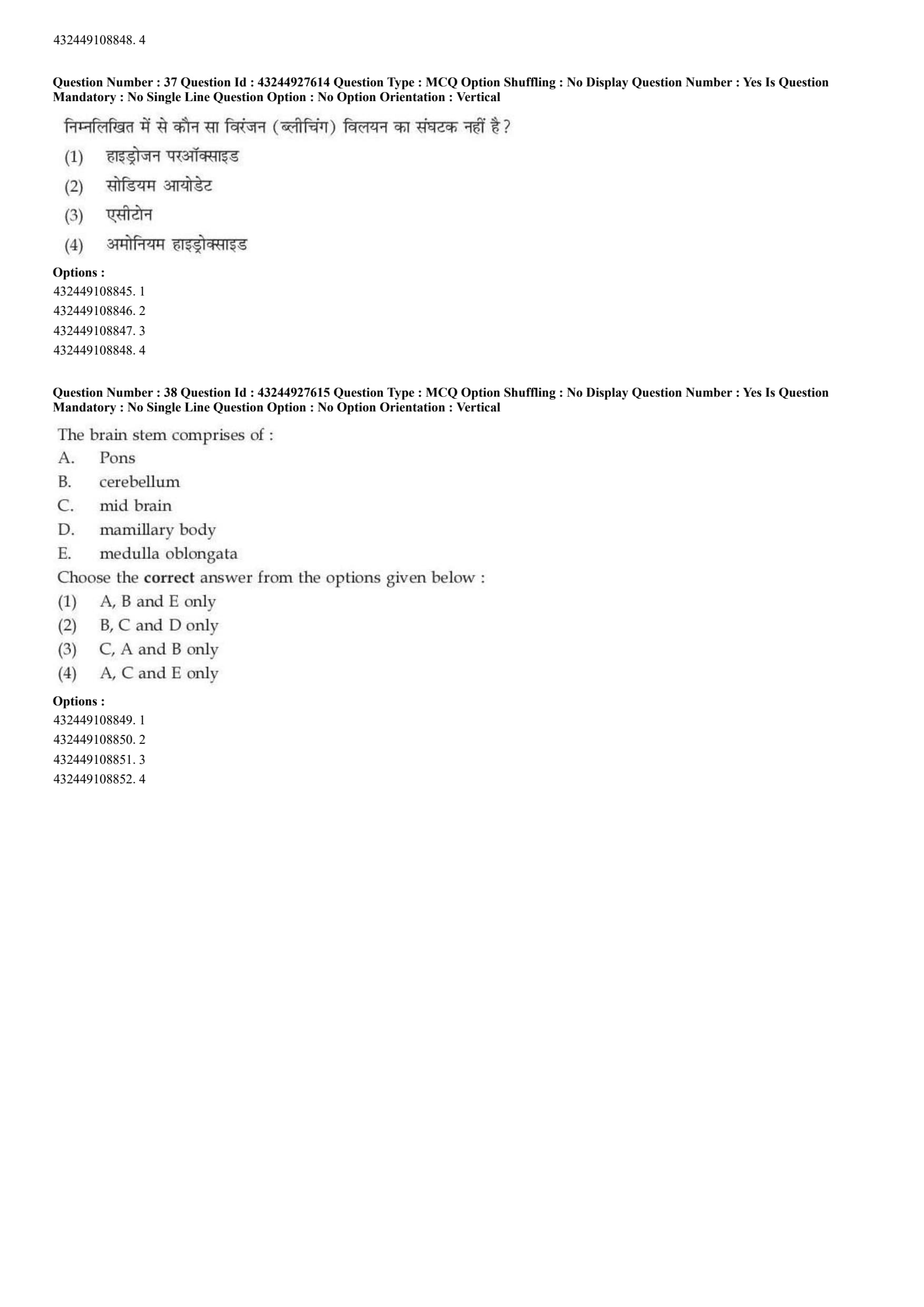
The brain stem comprises of :
A. Pons
B. cerebellum
C. mid brain
D. mamillary body
E. medulla oblongata
Choose the correct answer from the options given below :
View Solution
Step 1: Concept:
This question requires strict anatomical identification of the structures that structurally and functionally compose the human brainstem.
Step 2: Step-by-step Explanation:
The brainstem is the stalk-like lower portion of the brain that physically connects the higher cerebral hemispheres to the spinal cord. It houses critical autonomic control centers (like breathing and heart rate).
Anatomically, the brainstem is rigidly defined as consisting of exactly three structures, arranged from top to bottom:
1. Midbrain (Mesencephalon) (C) - The uppermost segment.
2. Pons (A) - The bulging middle segment.
3. Medulla oblongata (E) - The lowest segment that continuously merges into the spinal cord.
Let's review the distractors:
- Cerebellum (B): While it sits right behind the brainstem and connects to the pons via peduncles, it is a separate structural organ responsible for motor coordination, not a part of the brainstem stalk itself.
- Mamillary bodies (D): These are small round structures on the undersurface of the brain involved in memory. They are anatomically part of the diencephalon (limbic system), sitting just above the midbrain.
Therefore, only A, C, and E belong to the brainstem.
Step 3: Final Answer:
The correct option is (D) containing A, C, and E only.
Quick Tip: To remember the brainstem from top to bottom, think \textbf{M.P.M.}: \textbf{M}idbrain, \textbf{P}ons, \textbf{M}edulla. The cerebellum just "hangs on" to the back of the Pons for the ride!
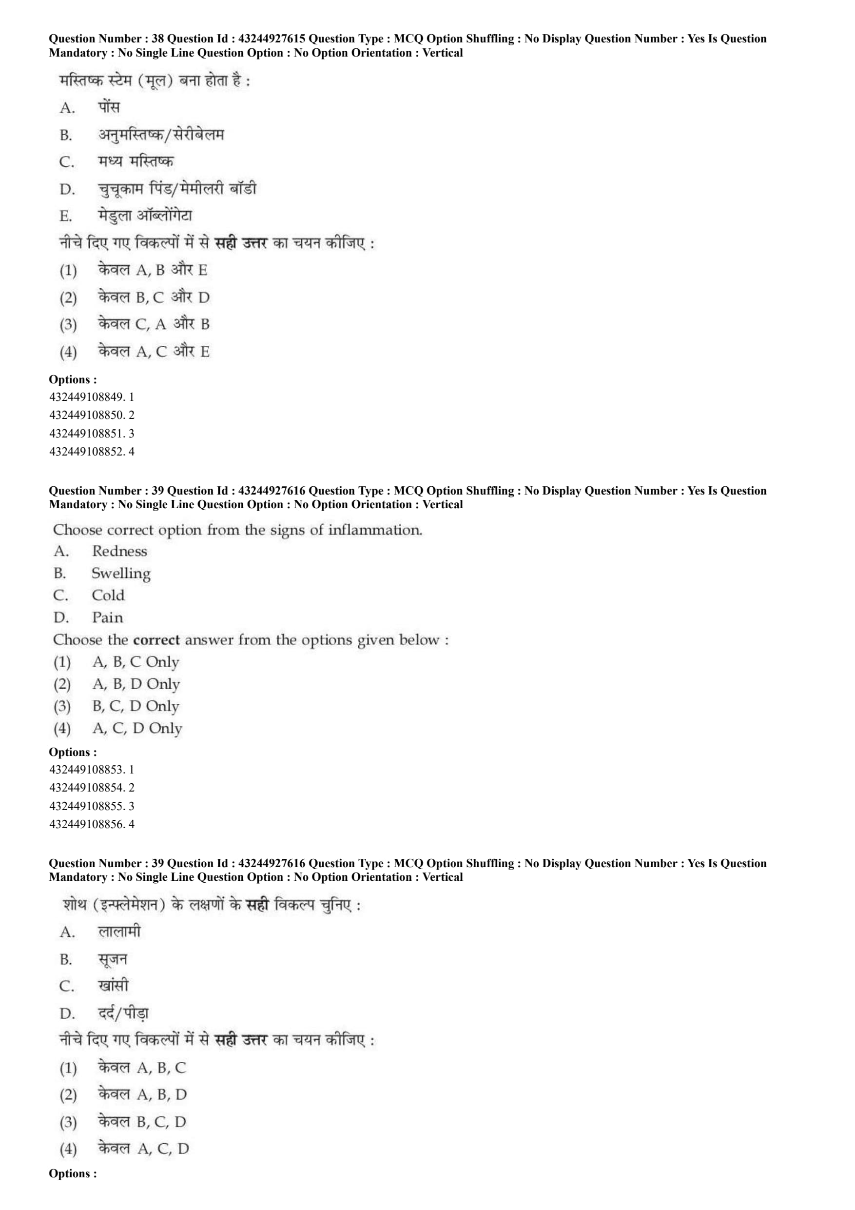
Choose correct option from the signs of inflammation.
A. Redness
B. Swelling
C. Cold
D. Pain
Choose the correct answer from the options given below :
View Solution
Step 1: Concept:
This question tests foundational general pathology, specifically asking for the classic cardinal signs of acute localized inflammation originally described by the Roman encyclopedist Celsus.
Step 2: Step-by-step Explanation:
When tissue suffers an injury or infection, the body mounts an acute inflammatory response characterized by local vascular dilation and increased capillary permeability.
This process manifests in four classic, universally recognized physical signs (often taught in their Latin forms):
1. Rubor (Redness - A): Caused by massive vasodilation and increased blood flow to the damaged area.
2. Tumor (Swelling - B): Caused by the leakage of protein-rich plasma fluid (exudate) into the surrounding interstitial tissues.
3. Calor (Heat): Also caused by the heavy influx of warm, core-body blood to the surface area. Therefore, Cold (C) is completely false. An inflamed area is remarkably warm to the touch.
4. Dolor (Pain - D): Caused by physical swelling pressing on nerve endings, coupled with the release of chemical pain mediators like bradykinin and prostaglandins.
(A fifth sign, \textit{Functio laesa or Loss of Function, was later added by Rudolf Virchow).
Based on the true signs, A, B, and D are correct.
Step 3: Final Answer:
The correct combination is A, B, and D Only.
Quick Tip: Memorize the 4 classic Latin signs of Celsus: Rubor (Redness), Tumor (Swelling), Calor (Heat - not cold!), and Dolor (Pain).
Which of the following are EEG signal bands ?
A. Alpha (\(\alpha\))
B. Beta (\(\beta\))
C. Lambda (\(\lambda\))
D. Theta (\(\theta\))
E. Delta (\(\delta\))
Choose the correct answer from the options given below :
View Solution
Step 1: Concept:
This question tests clinical neurophysiology, specifically asking the student to identify the standard frequency bands (rhythms) used to classify brainwaves recorded by an Electroencephalogram (EEG).
Step 2: Step-by-step Explanation:
The electrical activity of the brain is highly rhythmic. Clinically, these rhythms are categorized into specific "bands" based strictly on their frequency (measured in Hertz, Hz). The major physiological bands are:
1. Delta (\(\delta\)) (E): 0.5 - 4 Hz. Deep, slow-wave sleep.
2. Theta (\(\theta\)) (D): 4 - 8 Hz. Drowsiness or light sleep.
3. Alpha (\(\alpha\)) (A): 8 - 13 Hz. Awake, relaxed state with eyes closed.
4. Beta (\(\beta\)) (B): 14 - 30 Hz. Awake, alert, active concentration.
5. Gamma (\(\gamma\)): \(>30\) Hz. High-level cognitive processing (not listed in options).
What about Lambda (\(\lambda\)) (C)? While "lambda waves" do exist in clinical EEG terminology, they are sharp, transient waveform \textit{events seen over the occipital lobe when scanning visual patterns. They are not a continuous frequency "band" like the others.
Therefore, the classic frequency bands listed are Alpha, Beta, Theta, and Delta.
Step 3: Final Answer:
The correct options are A, B, D, and E Only.
Quick Tip: Learn the bands in order of increasing frequency: Delta (slowest, deep sleep) \(\rightarrow\) Theta (drowsy) \(\rightarrow\) Alpha (relaxed, eyes closed) \(\rightarrow\) Beta (awake, thinking) \(\rightarrow\) Gamma (fastest, intense focus).
Molecules that bind to a protein (or any other target) in a defined way are known as ________.
View Solution
Step 1: Concept:
The question asks for the general scientific term used to describe molecules that specifically bind to a target protein or receptor to form a complex.
Step 2: Step-by-step Explanation:
Ligands (Option C): In biochemistry and pharmacology, a ligand is defined as any molecule, ion, or complex that binds reversibly or irreversibly to a specific structural site on a target protein or receptor.
The binding between a ligand and its target protein is generally characterized by high structural specificity and specific binding affinity.
This interaction often induces a three-dimensional conformational change in the target protein, which subsequently alters its physiological functional state or enzymatic activity.
The strength of ligand-protein binding is quantitatively described by the dissociation constant (\(K_d\)), where a lower \(K_d\) signifies a stronger binding affinity.
The intermolecular forces governing this binding usually include non-covalent interactions such as hydrogen bonds, ionic bonds, van der Waals forces, and hydrophobic interactions.
Operator (Option A): An operator is a specific segment of DNA to which a transcription factor (often a repressor) binds to regulate the transcription of adjacent structural genes.
Strand (Option B): In molecular biology, a strand typically refers to a single contiguous chain of nucleotides, such as one half of the DNA double helix or a single-stranded RNA molecule.
Inducer (Option D): An inducer is a specific type of regulatory molecule that binds to a repressor or activator protein, disabling a repressor or enabling an activator to increase gene transcription.
While an inducer is technically a type of ligand, the term "ligand" is the universal and broadly applicable term for any molecule binding a protein target.
Step 3: Final Answer:
Based on the definitions, molecules that bind to a protein in a defined functional way are universally known as ligands.
Quick Tip: Always remember that 'Ligand' comes from the Latin word 'ligare', which means 'to bind'.
It acts as a signaling trigger for receptors, similar to a specific key fitting into a targeted lock.
Match List - I with List - II. Static characteristics of transducer with their definition.

View Solution
Step 1: Concept:
The question requires matching the fundamental static characteristics of measurement instruments (transducers) with their correct standard definitions.
Step 2: Step-by-step Explanation:
Span (A): Span is defined as the algebraic difference between the upper and lower limits of the measurement range of the transducer.
Therefore, it represents the total operating range of the transducer over which it can accurately measure the physical variable.
Thus, A matches with II.
Noise (B): Noise refers to any random, erratic, and unwanted electrical or physical signal that interferes with the primary desired signal at the output of the instrument.
In transducer applications, minimizing noise is critical to maintaining a high signal-to-noise ratio.
Thus, B matches with IV.
Precision (C): Precision indicates the degree of repeatability or reproducibility of a measurement when the same input quantity is repeatedly measured under identical conditions.
It is important to note that high precision does not necessarily imply high accuracy.
Thus, C matches with I.
Offset (D): Offset, also known as zero error or zero drift, is defined as the baseline output signal that exists even when the true input variable is identically zero.
Calibration is often required to eliminate this offset error before actual measurements are taken.
Thus, D matches with III.
Compiling all the correct matches: A \(\rightarrow\) II, B \(\rightarrow\) IV, C \(\rightarrow\) I, and D \(\rightarrow\) III.
Step 3: Final Answer:
The matched sequence perfectly aligns with Option (A).
Quick Tip: Differentiate clearly between 'Precision' and 'Accuracy'.
Precision refers to how close multiple measurements are to each other (repeatability), whereas Accuracy refers to how close the measurement is to the absolute true value.
Given below are two statements : one is labelled as Assertion (A) and the other is labelled as Reason (R).
Assertion (A) : Iodine is highly effective bactericidal agent and it is effective against all kinds of bacteria.
Reason (R) : Iodines are protein denaturants, and this property may to a large extent, account for their antimicrobial activity.
In the light of the above statements, choose the most appropriate answer from the options given below :
View Solution
Step 1: Concept:
The question evaluates the biological mechanism of iodine as a broad-spectrum antimicrobial agent and checks whether its protein-denaturing capability justifies its bactericidal efficiency.
Step 2: Step-by-step Explanation:
Analyzing the Assertion (A): Iodine has long been used in clinical settings as a highly potent and fast-acting bactericidal agent.
It exhibits a broad spectrum of activity and is highly effective against Gram-positive bacteria, Gram-negative bacteria, bacterial endospores (with sufficient exposure time), mycobacteria, and even many fungi and viruses.
Therefore, the statement that it is a highly effective bactericidal agent effective against almost all kinds of bacteria is factually correct.
Analyzing the Reason (R): The primary mechanism of action of elemental iodine involves penetrating the cell wall of microorganisms.
Once inside, iodine acts as a strong oxidizing agent. It oxidizes the sulfhydryl (-SH) groups of amino acids (like cysteine) present in essential cellular enzymes.
This massive oxidation leads to the irreversible denaturation and precipitation of cellular proteins and enzymes.
Furthermore, iodine rapidly halogenates the phenolic rings of tyrosine residues within proteins, rendering them non-functional.
Because the destruction of essential enzymatic proteins inherently kills the bacterial cell, this denaturing property directly accounts for its widespread antimicrobial activity.
Thus, Reason (R) is correct, and it fundamentally provides the biological mechanism explaining Assertion (A).
Step 3: Final Answer:
Both statements are accurate, and R is the correct and logical explanation for why A occurs.
Quick Tip: Iodine-based disinfectants (like Betadine/Povidone-iodine) are known as iodophors.
They slowly release free iodine, which minimizes tissue irritation while maintaining powerful protein-denaturing and oxidizing capabilities against pathogens.
Which statements are true for NKT cells ?
A. NKT cells can act as both helper cells and cytotoxic cells.
B. NKT cells do form memory cells.
C. NKT cells killing appears to depend predominantly on FasL-Fas interactions.
D. NKT cell include both CD4\(^+\) and CD4\(^-\) subpopulations, which may also differ by cytokine production.
E. NKT cells do not express a number of markers characteristic of T lymphocytes.
Choose the correct answer from the options given below :
View Solution
Step 1: Concept:
The question tests knowledge regarding the unique characteristics, cellular subsets, and effector mechanisms of Natural Killer T (NKT) cells in the immune system.
Step 2: Step-by-step Explanation:
Statement A: NKT cells function uniquely at the intersection of innate and adaptive immunity. Upon activation, they rapidly secrete huge amounts of cytokines (like IFN-\(\gamma\) and IL-4), acting as helper cells. They can also directly lyse target cells, acting as cytotoxic cells. Thus, A is true.
Statement B: Traditionally, innate-like cells were thought to lack memory. However, recent immunological research has identified that specific subsets of NKT cells can undergo clonal expansion and form long-lived memory populations that mount an enhanced response upon re-challenge. Thus, B is generally considered true in modern contexts.
Statement C: The cytotoxic killing mechanism of NKT cells primarily relies on the extrinsic apoptotic pathway, predominantly mediated by the interaction between Fas Ligand (FasL) on the NKT cell and the Fas receptor on the target cell. Thus, C is true.
Statement D: NKT cells are phenotypically heterogeneous. In humans and mice, they can be categorized into distinct subpopulations, such as CD4\(^+\) and CD4\(^-\) (which are mostly CD4\(^-\)CD8\(^-\) double negative). These subsets distinctly differ in their cytokine secretion profiles. Thus, D is true.
Statement E: By definition, NKT cells express a semi-invariant T-cell receptor (TCR), which is a definitive hallmark of T lymphocytes. They heavily express CD3 and other standard T-cell markers alongside NK cell markers (like NK1.1). Therefore, stating they "do not express" T-cell markers is false.
Since Statements A, B, C, and D are true, and E is false, the correct grouping corresponds to Option (C).
Step 3: Final Answer:
The combination of true statements is A, B, C, and D only.
Quick Tip: Unlike conventional T cells that recognize peptide antigens presented by MHC molecules, NKT cells specifically recognize lipid and glycolipid antigens presented by the non-polymorphic CD1d molecule.
Given below are two statements : one is labelled as Assertion (A) and the other is labelled as Reason (R).
Assertion (A) : The ulnar nerve is also known as the 'musician's nerve'.
Reason (R) : It controls fine movements of the fingers.
In the light of the above statements, choose the most appropriate answer from the options given below :
View Solution
Step 1: Concept:
The question requires matching a common anatomical colloquialism for the ulnar nerve with its functional physiological role in the human body.
Step 2: Step-by-step Explanation:
Analyzing the Assertion (A): The ulnar nerve originates from the brachial plexus (specifically nerve roots C8 and T1).
It travels down the arm, passing behind the medial epicondyle of the humerus (often colloquially referred to as the 'funny bone').
In medical anatomy, the ulnar nerve is indeed famously referred to as the "musician's nerve". Therefore, Assertion (A) is entirely correct.
Analyzing the Reason (R): The rationale behind this specific moniker stems from the nerve's extensive innervation of the intrinsic muscles of the hand.
It supplies motor function to the hypothenar muscles, the interossei (both palmar and dorsal), the medial two lumbricals, and the adductor pollicis.
These specific intrinsic muscles are completely responsible for executing intricate, highly coordinated, and fine-tuned micro-movements of the fingers.
Playing complex musical instruments (like a piano, guitar, or violin) relies heavily on these exact fine motor movements.
Because the ulnar nerve dictates these critical fine movements, it rightfully earns its title. Thus, Reason (R) correctly explains Assertion (A).
Step 3: Final Answer:
Both statements are true, and the ability to control fine finger movements is precisely why it is termed the musician's nerve.
Quick Tip: Damage or compression of the ulnar nerve (often at the cubital tunnel in the elbow) results in a characteristic clinical presentation known as 'Claw Hand' deformity.
It predominantly affects the 4th and 5th digits due to the loss of intrinsic muscle function.
From the following components, choose correct sequence of any medical instrumentation system.
A. Signal conditioner
B. Microcontroller
C. Display
D. Sensor/transducer
Choose the correct answer from the options given below :
View Solution
Step 1: Concept:
The question asks to arrange the generic block diagram components of a standard medical instrumentation and data acquisition system in their logical chronological order of signal processing.
Step 2: Step-by-step Explanation:
A generalized medical instrumentation system captures biological signals from a patient and processes them into a readable format. The systemic progression happens step-by-step.
1st Stage - Sensor/transducer (D): This is the very first component interfacing with the patient. It detects physical physiological parameters (like temperature, pressure, or biochemical activity) and converts them into a proportional raw electrical signal.
2nd Stage - Signal conditioner (A): The raw electrical signal generated by the transducer is typically extremely weak and heavily contaminated with noise. The signal conditioner amplifies the signal and applies specialized electronic filters to remove noise and isolate the target frequency band.
3rd Stage - Microcontroller (B): Once the signal is clean and amplified, it enters the microcontroller unit. Here, an Analog-to-Digital Converter (ADC) digitizes the signal so that the microcontroller's CPU can perform complex computational processing and algorithmic analysis.
4th Stage - Display (C): After processing is complete, the final computed data is outputted to an interface for the clinician to interpret. This is typically a graphical display or an alarm system.
The proper sequential flow of the signal is thus: Sensor \(\rightarrow\) Signal Conditioner \(\rightarrow\) Microcontroller \(\rightarrow\) Display.
Translating this to the given letters yields the sequence: D, A, B, C.
Step 3: Final Answer:
The chronological processing sequence logically corresponds to Option (D).
Quick Tip: Always trace the path of the 'data' or 'signal' from the physical source (patient) to the final output (physician).
Physical Source \(\rightarrow\) Detection (Sensor) \(\rightarrow\) Processing (Conditioning/Compute) \(\rightarrow\) Output (Display).
Match List - I with List - II. Volumes and capacities of lungs with their formulas.

Choose the correct answer from the options given below :
View Solution
Step 1: Concept:
The question tests physiological definitions related to respiratory volumes and their corresponding mathematical relationships.
Step 2: Step-by-step Explanation:
Let us systematically deduce the correct mathematical relations for each parameter.
A. Alveolar Ventilation: Alveolar ventilation represents the actual volume of fresh air reaching the respiratory zone per minute.
It is calculated by subtracting the anatomical dead space from the tidal volume, and multiplying by the respiratory rate.
Formula: \( (Breathing rate) \times (Tidal Volume - Dead Space) \).
Thus, A matches closely with III.
B. Total Lung Capacity (TLC): This is the maximum volume of air the lungs can hold after a maximal inhalation.
It is the sum of Vital Capacity (the maximal mobilizable volume) and the Residual Volume (the air left in lungs that cannot be exhaled).
Formula: \text{Vital Capacity + \text{Residual Volume.
Thus, B matches exactly with IV.
C. Expiratory Reserve Volume (ERV): This is the extra amount of air that can be forcefully exhaled after a normal resting exhalation.
The Functional Residual Capacity (FRC) is the amount of air left in the lungs after a normal breath, encompassing both ERV and RV (\(FRC = ERV + RV\)).
Rearranging this: \(ERV = FRC - RV\).
Thus, C matches exactly with II.
D. Inspiratory Reserve Volume (IRV): This is the extra air that can be inhaled forcefully after a normal resting inhalation.
Vital Capacity (VC) is the sum of IRV, Tidal Volume (TV), and ERV (\(VC = IRV + TV + ERV\)).
Rearranging for IRV: \(IRV = VC - (TV + ERV)\).
Note: In List-II, statement I says "Vital Capacity - (Tidal Volume + Functional Residual Capacity)". This is a slight misprint in the exam paper. It likely intended to say "Tidal Volume + Expiratory Reserve Volume". Regardless, by the process of elimination of the flawless matches for A, B, and C, D undeniably pairs with I.
The consolidated correct matching sequence is: A \(\rightarrow\) III, B \(\rightarrow\) IV, C \(\rightarrow\) II, D \(\rightarrow\) I.
Step 3: Final Answer:
The matched sequence clearly aligns with Option (C).
Quick Tip: To memorize basic lung capacities, remember that a 'Capacity' is always the sum of two or more 'Volumes'.
For example: Vital Capacity (VC) = Inspiratory Reserve Volume (IRV) + Tidal Volume (TV) + Expiratory Reserve Volume (ERV).
Which of the following proteins or peptides blocks epithelial infection by bacteria, fungi and viruses ?
View Solution
Step 1: Concept:
The question asks us to identify the specific class of antimicrobial peptides that provide broad-spectrum barrier immunity on epithelial surfaces against a wide array of pathogens including bacteria, fungi, and viruses.
Step 2: Step-by-step Explanation:
Epithelial surfaces (like the skin, respiratory tract, and gastrointestinal tract) serve as the body's first line of defense. They secrete various biochemical agents to neutralize invading pathogens.
Lysozyme (Option A): Lysozyme is an enzyme found in tears, saliva, and mucus. Its primary function is to target and cleave the peptidoglycan linkages present specifically in bacterial cell walls, primarily acting against Gram-positive bacteria. It is not highly effective against fungi or viruses.
Secretory leukocyte protease inhibitor (Option B): SLPI primarily protects local tissues from the detrimental effects of excessive proteolytic enzymes released during inflammation. While it has some antibacterial and antiviral properties, it is not the classic broad-spectrum blocker described.
Surfactant proteins SP-A (Option C): SP-A is an opsonin found primarily in the alveoli of the lungs. It binds to pathogens to enhance their phagocytosis by alveolar macrophages. Its role is more about clearance than direct broad-spectrum killing.
Defensins (\(\alpha\) and \(\beta\)) (Option D): Defensins are a major family of small, cysteine-rich, cationic antimicrobial peptides.
Because they are cationic and amphipathic, they possess a strong affinity for the negatively charged lipid membranes characteristic of most bacteria, fungi, and enveloped viruses.
Once attached, defensins embed themselves into the pathogen's lipid bilayer and form massive pores. This disrupts membrane integrity, leading to rapid leakage of essential cellular contents and subsequent pathogen death.
Their broad-spectrum efficacy precisely fits the criteria of blocking infection by all three classes of pathogens mentioned.
Step 3: Final Answer:
Defensins are the correct multifaceted antimicrobial peptides.
Quick Tip: Remember that Defensins form 'pores' to 'deflate' pathogens.
\(\alpha\)-defensins are typically produced by neutrophils and Paneth cells (in the gut), while \(\beta\)-defensins are broadly secreted by diverse epithelial cells.
Interferon-\(\alpha\)-2a protein is used for the treatment of :
View Solution
Step 1: Concept:
The question requires identifying the primary clinical application of the recombinant therapeutic protein Interferon-\(\alpha\)-2a.
Step 2: Step-by-step Explanation:
Interferons are a group of signaling proteins (cytokines) made and released by host cells in response to the presence of several viruses. They fundamentally "interfere" with viral replication.
Interferon-\(\alpha\)-2a (Option A): This is a specifically engineered recombinant variant of Type I interferon. Clinically, it is heavily prescribed as an antiviral and antineoplastic agent.
Its most prominent application is in the treatment of chronic viral hepatitis, specifically Hepatitis B virus (HBV) and Hepatitis C virus (HCV) infections.
It acts by binding to specific cell surface receptors, triggering a complex intracellular cascade that induces the expression of enzymes like Protein Kinase R (PKR) and RNase L, which collectively halt viral RNA/DNA replication and degrade viral genomes.
Rheumatoid arthritis (Option B): This is an autoimmune inflammatory condition. Treatments typically include immunosuppressants and TNF-alpha inhibitors (like Infliximab or Adalimumab), not antiviral interferons.
Stimulates production of platelets (Option C): The protein used to stimulate platelet production (megakaryopoiesis) is primarily Thrombopoietin or drugs like Oprelvekin (IL-11).
Stimulates red-blood-cell production (Option D): The hormone responsible for erythropoiesis is Erythropoietin (EPO), widely used in managing anemia.
Therefore, only Option A correctly correlates with the medical use of Interferon-\(\alpha\)-2a.
Step 3: Final Answer:
Interferon-\(\alpha\)-2a is used clinically to treat Hepatitis B.
Quick Tip: To remember interferon therapies: Type I interferons (\(\alpha\) and \(\beta\)) are generally Antiviral.
IFN-\(\alpha\) is used for Hepatitis B and C. IFN-\(\beta\) is famously used to treat Multiple Sclerosis.
Which vitamin is required for growth simulation of 'Bacillus anthracis' ?
View Solution
Step 1: Concept:
The question identifies a specific nutritional requirement acting as an essential growth factor for the cultivation and robust growth of the bacterium \textit{Bacillus anthracis.
Step 2: Step-by-step Explanation:
\textit{Bacillus anthracis is a Gram-positive, spore-forming, rod-shaped bacterium that is the causative agent of the deadly disease anthrax.
While it can grow on basic nutrient agars, optimizing its growth in laboratory settings or in specific pathogenic states requires certain accessory growth factors that the organism cannot synthesize in sufficient quantities autonomously.
Scientific studies on the minimal nutritional requirements of \textit{B. anthracis have demonstrated an absolute dependency on specific vitamins for robust metabolic function.
The critical requirement for \textit{Bacillus anthracis is Thiamine, which is also known as Vitamin B\(_1\).
Inside the bacterial cell, Thiamine is converted to its biologically active coenzyme form, Thiamine pyrophosphate (TPP).
TPP is an absolutely indispensable cofactor for several key enzymatic reactions involved in central carbon metabolism and cellular energy generation.
Specifically, it is required for the function of the pyruvate dehydrogenase complex (converting pyruvate into acetyl-CoA) and the \(\alpha\)-ketoglutarate dehydrogenase complex (a crucial step in the vital Krebs/TCA cycle).
Without Vitamin B\(_1\), the bacterium's central metabolic pathways stall, preventing cellular respiration and ultimately restricting growth.
Step 3: Final Answer:
Vitamin B\(_1\) (Thiamine) is the crucial vitamin required for its growth.
Quick Tip: For many heterotrophic bacteria, if a specific B-vitamin is required, it is usually Thiamine (B\(_1\)) or Niacin (B\(_3\)) because they form the foundational coenzymes (TPP and NAD\(_+\)) necessary for basic ATP-generating catabolism.
Normal functioning of thyroid gland require certain quantity of daily in take of iodine. What is its normal daily requirement ?
View Solution
Step 1: Concept:
The question asks for the clinically recommended daily dietary intake of the trace element iodine necessary for maintaining healthy thyroid function.
Step 2: Step-by-step Explanation:
The thyroid gland is a vital endocrine organ located in the neck. Its primary physiological role is the synthesis and secretion of thyroid hormones, primarily thyroxine (T4) and triiodothyronine (T3).
Iodine is an irreplaceable structural component of these hormones. T4 contains four iodine atoms, and T3 contains three iodine atoms.
Since the human body cannot synthesize iodine endogenously, it must be absorbed entirely from the diet.
To synthesize adequate amounts of these hormones to maintain a eumetabolic state, the thyroid gland continuously traps circulating iodide from the bloodstream.
According to major health organizations, including the World Health Organization (WHO), the standard recommended dietary allowance (RDA) of iodine for healthy human adults is \( 150 \, \mug \) per day.
This requirement increases slightly during periods of rapid growth or heightened metabolic demand, such as during pregnancy and lactation, where the recommendation ranges between \( 220 \) to \( 290 \, \mug \) per day.
When evaluating the provided options, the range of \( 100 - 200 \, \mug \) perfectly brackets the standard adult requirement of \( 150 \, \mug \).
The other options present values that are either massively excessive (milligram range, which could induce iodine toxicity or Wolff-Chaikoff effect) or too high for baseline daily intake (\(400 - 500 \, \mug\)).
Step 3: Final Answer:
The normal daily dietary requirement is 100 - 200 \(\mu\)g.
Quick Tip: A simple way to remember micronutrient units: Iodine is a 'trace' mineral, meaning it is required in micrograms (\(\mu\)g), unlike 'macrominerals' like calcium or magnesium which are needed in milligrams (mg) or grams.
How much percentage of white blood cells do monocytes make up of ?
View Solution
Step 1: Concept:
The question asks for the normal relative percentage of a specific leukocyte subtype (monocytes) within the total white blood cell (WBC) population in a healthy human differential blood count.
Step 2: Step-by-step Explanation:
White blood cells (leukocytes) are categorized broadly into granulocytes (neutrophils, eosinophils, basophils) and agranulocytes (lymphocytes, monocytes).
In a standard complete blood count (CBC) with differential, the relative proportions of these cells are rigorously analyzed.
Neutrophils: These are the most abundant WBCs, rapidly responding to acute bacterial infections, and normally constitute 50% - 70% of the total WBC count. (Option C represents neutrophils).
Lymphocytes: These mediate the adaptive immune response (T-cells, B-cells) and typically make up 20% - 40% of the count.
Monocytes: Monocytes are the largest circulating white blood cells. They circulate in the bloodstream for several days before migrating into tissues, where they differentiate into highly phagocytic tissue-resident macrophages or dendritic cells.
In a healthy individual, monocytes typically comprise 2% to 8% (or frequently cited up to 10%) of the total leukocyte population.
Eosinophils and Basophils: Eosinophils generally make up 1% - 4% (Option A is close to this), and basophils are the rarest at \(<\)1%.
Matching these physiological facts with the options, Option (D) "5% to 10%" is the accepted standard reference range for circulating monocytes.
Step 3: Final Answer:
Monocytes normally account for 5% to 10% of total white blood cells.
Quick Tip: Use the popular mnemonic "Never Let Monkeys Eat Bananas" to remember the order of abundance of white blood cells:
\textbf{N}eutrophils (highest), \textbf{L}ymphocytes, \textbf{M}onocytes, \textbf{E}osinophils, \textbf{B}asophils (lowest).
Choose correct statements about cancer cells.
A. They disobey the growth controlling signals in the body.
B. They escape death signals and achieve immortality.
C. They do not lose properties of differentiation.
D. Due to gain of growth controls, they are genetically stable.
Choose the correct answer from the options given below :
View Solution
Step 1: Concept:
The question tests fundamental knowledge regarding the hallmarks and pathological characteristics of malignant cancer cells.
Step 2: Step-by-step Explanation:
Statement A: Normal cells require external mitogenic signals to divide and halt division upon receiving contact inhibition or anti-growth signals. Cancer cells acquire mutations (e.g., in oncogenes or tumor suppressor genes) that allow them to autonomously sustain proliferative signaling. They effectively disobey systemic growth-controlling signals. Thus, Statement A is True.
Statement B: Apoptosis (programmed cell death) is a natural safeguard against damaged cells. Cancer cells frequently mutate pathways (like the p53 tumor suppressor pathway) to evade these apoptotic death signals. Furthermore, they often upregulate the enzyme telomerase, which maintains telomere length, conferring unlimited replicative potential (immortality). Thus, Statement B is True.
Statement C: Normal cells are highly specialized (differentiated) to perform specific physiological functions. As cancer cells progress, they typically undergo 'anaplasia' or dedifferentiation, losing their specialized features to become more primitive and exclusively focused on rapid replication. Therefore, they \textit{do lose properties of differentiation. Thus, Statement C is False.
Statement D: Cancer cells are characterized by a profound \textit{loss of growth control, not a gain. This rampant, unchecked division combined with defective DNA repair mechanisms leads to massive genomic instability, rampant mutations, and chromosomal abnormalities. They are highly genetically unstable. Thus, Statement D is False.
The only true statements are A and B.
Step 3: Final Answer:
Since only A and B correctly describe cancer hallmarks, Option (C) is the right choice.
Quick Tip: Review the "Hallmarks of Cancer" by Hanahan and Weinberg. Key features include: Evading apoptosis, self-sufficiency in growth signals, insensitivity to anti-growth signals, tissue invasion/metastasis, limitless replicative potential, and sustained angiogenesis.
The uses of chi-square test are given below, choose the correct statements.
A. Used as test of Independence.
B. Used as a test of association between two events.
C. Used as a test of heterogeneity.
D. To measure degree of deviation.
Choose the correct answer from the options given below :
View Solution
Step 1: Concept:
The question asks to identify the statistically valid applications of the Chi-square (\(\chi^2\)) test among the given options.
Step 2: Step-by-step Explanation:
The Chi-square test is a non-parametric statistical tool heavily utilized to analyze categorical (qualitative) data.
Statement A: The most common application of this test is the "Chi-square test for Independence". It evaluates whether two categorical variables from a single population are significantly associated or entirely independent of one another. Thus, Statement A is correct.
Statement B: This is effectively synonymous with the test of independence. When the test determines that two variables are not independent, it implies there is a statistically significant association or correlation between those two events/variables. Thus, Statement B is correct.
Statement C: The "Chi-square test of Homogeneity/Heterogeneity" is used to determine whether different subpopulations share the same distribution of a single categorical variable. If the distributions are significantly different, the populations are deemed heterogeneous. Thus, Statement C is correct.
Statement D: The term "deviation" in strict statistical terms generally refers to measures of dispersion (like standard deviation or mean absolute deviation) for continuous numerical data. While the \(\chi^2\) formula calculates the difference between expected and observed frequencies (\(\sum \frac{(O - E)^2}{E}\)), its primary designated statistical purpose is not generically termed a "measure of degree of deviation" in standard statistical taxonomy compared to A, B, and C.
The most universally accepted and rigidly defined primary uses in standard textbook curricula encompass A, B, and C.
Step 3: Final Answer:
Statements A, B, and C accurately reflect the standard primary applications of the test.
Quick Tip: Remember the three primary applications of Chi-square:
1. Goodness of Fit (1 variable, 1 population)
2. Test of Independence (2 variables, 1 population)
3. Test of Homogeneity (1 variable, 2+ populations).
Match List - I with List - II.

Choose the correct answer from the options given below :
View Solution
Step 1: Concept:
The question tests the fundamental biochemical functions of vital enzymes used heavily in molecular biology and recombinant DNA technology.
Step 2: Step-by-step Explanation:
A. DNA Ligase: This enzyme facilitates the joining of DNA strands together by catalyzing the formation of a critical phosphodiester bond. It essentially acts as molecular glue to seal nicks or join Okazaki fragments.
Therefore, A correctly pairs with II.
B. Reverse Transcriptase: This is an RNA-dependent DNA polymerase uniquely found in retroviruses (like HIV). It utilizes a single-stranded RNA template to synthesize a complementary DNA (cDNA) strand.
Therefore, B correctly pairs with III.
C. DNA polymerase I (E.coli): Discovered by Arthur Kornberg, this enzyme possesses 5'\(\rightarrow\)3' polymerase activity alongside a unique 5'\(\rightarrow\)3' exonuclease activity. In vivo, its primary role is to remove RNA primers and fill in the resultant gaps with DNA nucleotides during replication.
Therefore, C correctly pairs with IV.
D. Type II restriction endonucleases: These are standard molecular scissors that recognize highly specific, short palindromic nucleotide sequences in double-stranded DNA and cleave the phosphodiester backbone at defined positions within or near that specific sequence.
Therefore, D correctly pairs with I.
Combining these logic derivations gives the sequence A-II, B-III, C-IV, D-I.
Step 3: Final Answer:
The matched sequence clearly identifies Option (C) as the correct choice.
Quick Tip: To avoid confusion between polymerases: DNA Pol III is the main workhorse for replication in E.coli. DNA Pol I is specifically for 'gap filling' and 'primer removal' due to its unique 5'\(\rightarrow\)3' exonuclease capability.
Arrange the landmarks in the evolution of life on the earth.
A. Formation of oceans and continents.
B. Diversification of multicellular eukaryotes.
C. Appearance of protists, the first eukaryotes.
D. Appearance of endosymbionts (mitochondria).
E. Appearance of aerobic bacteria development of O\(_2\) - rich atmosphere.
Choose the correct answer from the options given below :
View Solution
Step 1: Concept:
The question asks for the correct chronological sequence of major geological and biological milestones during the evolutionary history of Earth.
Step 2: Step-by-step Explanation:
First Event (A): Earth formed roughly 4.5 billion years ago. As the molten planet cooled, water vapor condensed to form the primordial oceans and early tectonic plates formed the continents. This abiotic geological event forms the foundation for life and happens first.
Second Event (E): Early life consisted entirely of anaerobic prokaryotes. Around 2.4 billion years ago, a massive evolutionary leap occurred with the evolution of photosynthetic cyanobacteria. They produced oxygen as a metabolic byproduct, leading to the "Great Oxidation Event," thereby establishing an O\(_2\)-rich atmosphere and paving the way for aerobic respiration.
Third Event (C): With oxygen readily available, more complex single-celled organisms evolved. These were the first primitive eukaryotes, broadly categorized as protists. They possessed distinct membrane-bound nuclei, differentiating them from their prokaryotic ancestors.
Fourth Event (D): According to the widely accepted Endosymbiotic Theory (proposed by Lynn Margulis), these early ancestral eukaryotic cells engulfed free-living aerobic bacteria (alpha-proteobacteria). Instead of digesting them, a symbiotic relationship was formed. These engulfed endosymbionts eventually evolved into modern mitochondria, granting the host cells immense energy-generating capabilities.
Fifth Event (B): Armed with massive energetic advantages provided by mitochondria, eukaryotic cells eventually began congregating and cooperating, leading to the massive diversification of complex multicellular eukaryotes (like early plants, fungi, and animals).
Thus, the chronological order is: Oceans (A) \(\rightarrow\) Oxygen atmosphere (E) \(\rightarrow\) First Eukaryotes (C) \(\rightarrow\) Mitochondria endosymbiosis (D) \(\rightarrow\) Multicellularity (B).
Step 3: Final Answer:
The correctly arranged sequence corresponds to Option (D).
Quick Tip: A simple evolutionary timeline mnemonic: Abiotic (Oceans) \(\rightarrow\) Prokaryotes \(\rightarrow\) Oxygen Crisis \(\rightarrow\) Eukaryotes (Nucleus then Mitochondria) \(\rightarrow\) Multicellularity.
Arrange the components of membrane-based rapid diagnostic test in the order of specimen flow
A. Soak pad
B. Test band
C. Control band
D. Sample pad
E. Conjugate pad
Choose the correct answer from the options given below :
View Solution
Step 1: Concept:
The question asks for the sequential arrangement of physical structural components in a standard Lateral Flow Immunoassay (LFIA) based on the direction the liquid specimen travels.
Step 2: Step-by-step Explanation:
A Lateral Flow Immunoassay (like a rapid pregnancy or COVID-19 antigen test) relies entirely on the principle of capillary action to draw a liquid sample across a series of reactive zones.
1. Sample pad (D): This is the initial port of entry. The patient specimen (blood, urine, saliva) is dropped here. It acts as a sponge, filtering out large particulates and uniformly distributing the fluid to the next stage.
2. Conjugate pad (E): The liquid flows immediately into the conjugate pad, which contains freeze-dried, artificially labeled antibodies (often conjugated with colored gold nanoparticles). If the target antigen is present in the sample, it binds to these colored antibodies here to form a mobile complex.
3. Test band (B): The fluid then wicks across the nitrocellulose membrane, reaching the Test line first. This band contains immobilized capture antibodies. If the target antigen is present, the colored complex binds here, generating a visible colored line indicating a positive result.
4. Control band (C): The fluid continues past the test line to the Control line. This band contains secondary antibodies that bind to the unbound, excess labeled antibodies migrating from the conjugate pad. A visible line here proves the fluid successfully migrated across the entire membrane, confirming the test is functionally valid.
5. Soak pad / Absorbent pad (A): Finally, the fluid reaches the absorbent pad at the very end. This acts as a reservoir to aggressively draw the remaining liquid forward, ensuring continuous unidirectional capillary flow and preventing backflow.
Sequence of flow: D \(\rightarrow\) E \(\rightarrow\) B \(\rightarrow\) C \(\rightarrow\) A.
Step 3: Final Answer:
The correct order of specimen flow is strictly Option (C).
Quick Tip: Remember the flow logic: Put the sample in (Sample pad), mix with detectors (Conjugate), read the result (Test), confirm it worked (Control), and soak up the trash (Soak pad).
Arrange different stages of protein synthesis :
A. A specific Amino Acid initiates protein synthesis
B. Aminoacyl-tRNA synthetases attach the correct Amino Acids to their tRNAs
C. Termination of polypeptide synthesis requires a special signal
D. Peptide bonds are formed in the elongation stage
E. Newly synthesized polypeptide chains undergo folding and processing
Choose the correct answer from the options given below :
View Solution
Step 1: Concept:
The question asks for the proper sequential timeline of molecular events that occur during cellular translation (protein synthesis).
Step 2: Step-by-step Explanation:
Step 1: Activation of Amino Acids (B) - Before translation on the ribosome can even begin, the transfer RNA (tRNA) molecules must be "charged" with their appropriate amino acids. This crucial preparatory step is catalyzed by specific enzymes called aminoacyl-tRNA synthetases in the cytoplasm.
Step 2: Initiation (A) - The actual ribosomal synthesis begins when the small ribosomal subunit binds to the mRNA. The process is always initiated by a specific amino acid dictated by the start codon (usually AUG, coding for Methionine in eukaryotes or formyl-methionine in prokaryotes).
Step 3: Elongation (D) - Once the initiation complex is fully assembled, the ribosome translocates along the mRNA. The enzyme peptidyl transferase iteratively catalyzes the formation of peptide bonds between sequentially delivered amino acids, actively elongating the growing polypeptide chain.
Step 4: Termination (C) - Elongation continues continuously until the ribosome encounters a "special signal"—specifically, a nonsense/stop codon (UAA, UAG, or UGA) positioned in the mRNA sequence. Release factors bind, forcing the ribosome to dissemble and release the newly completed raw polypeptide chain.
Step 5: Post-translational modifications (E) - A linear chain of amino acids is rarely functional immediately. The newly synthesized, nascent polypeptide chain must undergo spontaneous or chaperone-mediated 3D folding and potentially other chemical processing (like glycosylation or cleavage) in the ER/Golgi to achieve its final, functional conformation.
Therefore, the strictly ordered chronological sequence of events is B \(\rightarrow\) A \(\rightarrow\) D \(\rightarrow\) C \(\rightarrow\) E.
Step 3: Final Answer:
Option (D) accurately represents the biological sequence of translation.
Quick Tip: Translation steps mirror reading a book: Get your materials ready (charging tRNA), open the book (Initiation), read page by page (Elongation), reach the end (Termination), and then understand the plot (Folding into functional protein).
The waves associated with the electrical activity of the various parts of the heart tissue during each cardiac cycle are represented by letters P, Q, R, S, T and U. Which of these wave represent ventricular repolarization ?
View Solution
Step 1: Concept:
The question asks to identify the specific waveform on an Electrocardiogram (ECG/EKG) trace that corresponds to the physiological event of ventricular repolarization.
Step 2: Step-by-step Explanation:
An Electrocardiogram (ECG) is a graphical representation of the sequential electrical activity traversing the heart muscle during a single heartbeat.
P wave: This is the first small upward deflection. It is generated by the depolarization of the atria initiated by the Sinoatrial (SA) node, which immediately precedes atrial contraction.
QRS complex (comprising Q, R, and S waves): This is the largest, sharpest spike on the trace. It signifies the rapid and massive electrical depolarization of the right and left ventricles just prior to powerful ventricular systole (contraction). It physically masks the tiny electrical signal of atrial repolarization occurring simultaneously.
T wave: Following the QRS complex, there is a slightly slower, broader upward deflection called the T wave. This wave graphically represents the phase of ventricular repolarization. During this phase, the ventricular muscle fibers electrically reset (restore their resting membrane potential) in preparation for the next subsequent heartbeat.
U wave: Sometimes a small U wave follows the T wave, which is thought to represent the repolarization of the Purkinje fibers or papillary muscles, though it is often absent.
Since the question specifically identifies ventricular repolarization, the answer is definitively the T wave.
Step 3: Final Answer:
The T wave uniquely corresponds to ventricular repolarization on an ECG trace.
Quick Tip: A handy summary for ECGs:
P wave = Atrial Depolarization.
QRS complex = Ventricular Depolarization.
T wave = Ventricular Repolarization.
Choose the correct option from the following statement.
(1) Analytical sensitivity refers to intra assay precision, whereas functional sensitivity refers to inter assay protection.
(2) Analytical sensitivity refers to inter assay precision, whereas functional sensitivity refers to intra assay protection.
(3) Analytical sensitivity refers to intra assay protection whereas functional sensitivity also refers to intra assay protection.
(4) Analytical sensitivity refers to inter assay protection, whereas functional sensitivity also refers to inter assay protection.
View Solution
Step 1: Concept:
The question tests the definitions of analytical and functional sensitivity within the context of clinical immunoassay validation protocols.
Step 2: Step-by-step Explanation:
In laboratory medicine and diagnostics, determining the lowest measurable amount of an analyte is critical for assay reliability. This is evaluated using two distinct metrics:
Analytical Sensitivity: Also known as the Limit of Detection (LOD). It is defined statistically as the lowest concentration of a target analyte that can be reliably distinguished from the background noise (the zero calibrator or blank). It is fundamentally determined by the intra-assay precision (the variance seen when running the same blank sample multiple times within the exact same batch/run).
Functional Sensitivity: Also known as the Limit of Quantitation (LOQ). It represents the lowest concentration of the analyte that can be quantitatively measured with an acceptable degree of clinical reliability and reproducibility over time. It is rigorously determined by measuring the inter-assay precision (the coefficient of variation, usually capped at 20%, obtained by analyzing samples over many different runs, across multiple days).
Therefore, conceptually, Analytical Sensitivity aligns with intra-assay metrics, and Functional Sensitivity aligns with inter-assay metrics.
Looking closely at the provided text, Option (1) states: "Analytical sensitivity refers to intra assay precision, whereas functional sensitivity refers to inter assay protection."
Despite the glaring typographical error in the examination paper where "precision" was mistakenly printed as "protection" in the latter half of the sentence, Option (1) is the only statement that correctly pairs the intra/inter prefixes with their respective sensitivity types.
Analytical \(\rightarrow\) Intra-assay. Functional \(\rightarrow\) Inter-assay.
Step 3: Final Answer:
Accounting for the typographical error in the source material, the first statement conceptually correctly differentiates the two terminologies.
Quick Tip: Analytical sensitivity is theoretically what the machine can barely "see" within a single controlled run (intra-assay). Functional sensitivity is what you can consistently "trust" day after day in a real-world clinical setting (inter-assay).
Match List - I with List - II. Lysosomal enzymes and the substrates on which they act.

Choose the correct answer from the options given below :
View Solution
Step 1: Concept:
The question asks us to accurately match various hydrolytic lysosomal enzymes with their specific biological substrates.
Step 2: Step-by-step Explanation:
A. Phosphatase: Phosphatases are enzymes that cleave phosphate groups from their substrates by hydrolyzing phosphoric acid monoesters into a phosphate ion and a molecule with a free hydroxyl group. Therefore, their specific substrate is Phosphate esters.
Thus, A matches with IV.
B. Collagenase: This is a specialized proteolytic enzyme (protease) that breaks the peptide bonds in collagen, which is a structural protein. Therefore, its primary substrate broadly falls under Proteins.
Thus, B matches with II.
C. Ribonuclease (RNase): This enzyme catalyzes the degradation of RNA into smaller components by cleaving the phosphodiester bonds of ribonucleotides. Therefore, its substrate is unequivocally RNA.
Thus, C matches with I.
D. Glycosidase: Glycosidases (or glycoside hydrolases) are enzymes that catalyze the hydrolysis of glycosidic bonds in complex sugars. Their primary targets are Complex carbohydrates (like glycogen or glycoproteins).
Thus, D matches with III.
Combining all matches: A-IV, B-II, C-I, D-III.
Step 3: Final Answer:
The matching pairs correspond to Option (C).
Quick Tip: Enzyme nomenclature typically takes the name of the substrate and adds the suffix "-ase".
Ribonucleic acid (RNA) + ase = Ribonuclease. Glycoside + ase = Glycosidase.
Calculate the range and coefficient of range from the following data :
50, 80, 100, 120, 150, 180, 200
View Solution
Step 1: Concept:
The question asks for two fundamental measures of statistical dispersion: the Range and the Coefficient of Range for a given numerical dataset.
Step 2: Key Formula or Approach:
The formulas for these measures are:
\[ Range = L - S \]
\[ Coefficient of Range = \frac{L - S}{L + S} \times 100% \]
where \( L \) is the largest value and \( S \) is the smallest value in the dataset.
Step 3: Step-by-step Explanation:
First, identify the extreme values in the given sorted dataset (50, 80, 100, 120, 150, 180, 200).
Smallest value (\( S \)) = 50.
Largest value (\( L \)) = 200.
Calculating Range:
\[ Range = 200 - 50 = 150 \]
Calculating Coefficient of Range:
\[ Coefficient of Range = \frac{200 - 50}{200 + 50} \]
\[ Coefficient of Range = \frac{150}{250} \]
\[ Coefficient of Range = 0.60 \]
To express this as a percentage (as seen in the options), multiply by 100:
\[ 0.60 \times 100% = 60% \]
Step 4: Final Answer:
The Range is 150 and the Coefficient of Range is 60%, which corresponds to Option (B).
Quick Tip: The Coefficient of Range is a relative measure of dispersion. It is unitless and allows for comparing the variability of different datasets, unlike the absolute Range.
Arrange different parts of microscope from Eyepiece onwards.
A. Focus knob
B. Specimen stage
C. Objectives
D. Condenser
E. Base with built-in light source
Choose the correct answer from the options given below :
View Solution
Step 1: Concept:
The question asks us to sequentially trace the structural layout of a standard compound light microscope, starting top-down from the eyepiece (where the observer looks) toward the bottom.
Step 2: Step-by-step Explanation:
Top (Starting point): The Eyepiece (Ocular lens) is at the very top.
Next Level (C): Directly below the eyepiece and the body tube is the revolving nosepiece holding the Objectives (objective lenses). These are positioned directly over the specimen to magnify the image.
Next Level (B): Right beneath the objective lenses is the Specimen stage (mechanical stage), the flat platform where the glass slide holding the specimen is placed.
Next Level (D): Positioned immediately beneath an aperture in the specimen stage is the Condenser. Its role is to focus the incoming light cone directly onto the specimen.
Next Level (A): Structurally on the arm of the microscope, extending downward toward the base, are the coarse and fine Focus knobs. They physically move the stage up and down. Structurally, they are located below the level of the stage/condenser.
Bottom (E): At the very bottom of the entire apparatus is the Base with built-in light source (illuminator), resting on the table.
Therefore, the logical top-to-bottom structural sequence is: Objectives (C) \(\rightarrow\) Stage (B) \(\rightarrow\) Condenser (D) \(\rightarrow\) Focus knob mechanism (A) \(\rightarrow\) Base (E).
Step 3: Final Answer:
The correct order is C, B, D, A, E, matching Option (A).
Quick Tip: Trace the path of light in reverse! Light goes from Base \(\rightarrow\) Condenser \(\rightarrow\) Specimen Stage \(\rightarrow\) Objectives \(\rightarrow\) Eyepiece. The structural components essentially follow this optical path.
Match List - I with List - II. Proteins and Function.

Choose the correct answer from the options given below :
View Solution
Step 1: Concept:
The question tests knowledge of the complement system, specifically the regulatory proteins that tightly control the complement cascade to prevent unnecessary damage to host cells.
Step 2: Step-by-step Explanation:
A. CR1 (Complement Receptor 1 / CD35): CR1 acts as an essential cofactor for Factor I-mediated cleavage of C3b and C4b. By doing so, it aggressively accelerates the decay of C3 convertases (C4b2a in classical, C3bBb in alternative pathways) and inherently blocks their formation.
Thus, A matches with II.
B. C1 inhibitor (C1INH): This is a classic Serine protease inhibitor (serpin). It binds to and irreversibly inactivates the serine proteases C1r and C1s of the classical pathway (as well as MASPs in the lectin pathway), halting cascade initiation.
Thus, B matches with IV.
C. CD59 (Protectin): CD59 is a membrane-bound regulatory protein on host cells. It acts late in the complement cascade by specifically binding to the C5b678 complex and sterically hindering the recruitment and polymerization of C9, thereby preventing the formation of the lethal Membrane Attack Complex (MAC) on host tissues.
Thus, C matches with I.
D. S protein (Vitronectin): The S protein acts as a fluid-phase regulatory protein. It binds to the soluble C5b67 complex as it is forming in the serum, preventing this hydrophobic complex from randomly inserting into the lipid bilayers of nearby innocent bystander host cells.
Thus, D matches with III.
Consolidating the matches yields A-II, B-IV, C-I, D-III.
Step 3: Final Answer:
The matching corresponds exactly to Option (A).
Quick Tip: To differentiate late-stage regulators: CD59 works on the cell surface to block C9. S protein works in the fluid phase (blood serum) to block C5b67 membrane insertion.
Match List - I with List - II.

Choose the correct answer from the options given below :
View Solution
Step 1: Concept:
The question asks to match fundamental data collection and statistical terminology with their formal definitions.
Step 2: Step-by-step Explanation:
A. Precision: Precision defines the degree of reproducibility or repeatability. It is defined as the closeness of repeated measurements of the same quantity to each other, regardless of whether those measurements are close to the actual true value.
Thus, A matches with IV.
B. Accuracy: Accuracy defines correctness. It is universally defined as the closeness of a measured or computed value to its actual true (or accepted) value.
Thus, B matches with II.
C. Reliability: In research methodology, reliability refers to the overall consistency of a measure. It heavily depends on the preciseness of the procedure used for data collection; a highly precise and standardized procedure yields highly reliable (repeatable) data.
Thus, C matches with I.
D. Sampling: Sampling is the fundamental statistical practice of selecting a few representative units from all the observational units (the entire population) to estimate population parameters without taking a complete census.
Thus, D matches with III.
The consolidated matching sequence is: A-IV, B-II, C-I, D-III.
Step 3: Final Answer:
The correct matching sequence is given in Option (C).
Quick Tip: A classic analogy: Think of a dartboard.
Accuracy is hitting the bullseye.
Precision is throwing 5 darts and hitting the exact same spot 5 times (even if it's far away from the bullseye).
Hypo solution is used in histological work for staining. Which of the following is hypo solution ?
View Solution
Step 1: Concept:
The question requires the chemical identification of the laboratory reagent commonly referred to as "hypo," frequently utilized in histology and traditional photography.
Step 2: Step-by-step Explanation:
In traditional darkroom photography and certain silver-based histological staining protocols (like the Grocott's Methenamine Silver stain or Von Kossa stain), a specific chemical is required as a "fixer."
The purpose of this fixer is to chemically dissolve and wash away any unreduced, unexposed silver halides (like silver bromide or silver nitrate) from the tissue section or photographic film.
If these unreduced silver salts are not removed, they will eventually react with ambient light over time, turning the entire background of the tissue slide pitch black.
The chemical exclusively used for this purpose is Sodium Thiosulfate (\(Na_2S_2O_3\)).
Historically, sodium thiosulfate was originally (and incorrectly) named "hyposulfite of soda." Because of this historical naming, laboratory technicians and photographers universally abbreviated it as "Hypo".
Silver Nitrate (Option A) is the actual stain that deposits the silver, not the hypo. Options C and D are mordants/stains for muscle and collagen, entirely unrelated to the term hypo.
Step 3: Final Answer:
The term "hypo solution" strictly refers to Sodium Thiosulfate.
Quick Tip: Whenever you see a silver-based staining procedure (reticulin stain, fungal silver stains), expect Sodium Thiosulfate (hypo) to be used as the final fixing step to 'clear' the background.
An enzyme is discovered that catalyzes the chemical reaction CALM \(\rightleftharpoons\) ANGER. A team of motivated researchers sets out to study the enzyme, which they called Angerase. They find that the \( K_{cat} \) (constant) for Angerase is \( 600 \, s^{-1} \). Some experiments were done. In one such experiment when [\( E_t \)] (total enzyme concentration) \( = 20 \, nM \) and [CALM] \( = 40 \, \mu M \), the reaction velocity, \( V_0 \) is \( 9.6 \, \mu M s^{-1} \), calculate Km (Michaelis constant) for the substrate 'CALM'.
View Solution
Step 1: Concept:
We are given enzyme kinetics parameters (\(K_{cat}\), Total Enzyme \([E_t]\), Substrate concentration \([S]\), and initial velocity \(V_0\)) and asked to calculate the Michaelis-Menten constant (\(K_m\)).
Step 2: Key Formula or Approach:
This requires two foundational equations of Michaelis-Menten kinetics:
1) Maximum velocity: \( V_{max} = K_{cat} \times [E_t] \)
2) Michaelis-Menten equation: \( V_0 = \frac{V_{max} \times [S]}{K_m + [S]} \)
Step 3: Step-by-step Explanation:
First, identify the given variables and standardize units:
\( K_{cat} = 600 \, s^{-1} \)
\( [E_t] = 20 \, nM = 20 \times 10^{-9} \, M \)
\( [S] = [CALM] = 40 \, \muM \)
\( V_0 = 9.6 \, \muM s^{-1} \)
Calculate \( V_{max} \):
\[ V_{max} = K_{cat} \times [E_t] \]
\[ V_{max} = 600 \, s^{-1} \times (20 \times 10^{-9} \, M) \]
\[ V_{max} = 12000 \times 10^{-9} \, M s^{-1} = 12 \times 10^{-6} \, M s^{-1} \]
Convert to \(\muM\) to match the other units:
\[ V_{max} = 12 \, \muM s^{-1} \]
Substitute into the Michaelis-Menten equation:
\[ V_0 = \frac{V_{max} \times [S]}{K_m + [S]} \]
\[ 9.6 = \frac{12 \times 40}{K_m + 40} \]
Solve for \( K_m \):
\[ 9.6 \times (K_m + 40) = 480 \]
\[ 9.6 \, K_m + 384 = 480 \]
\[ 9.6 \, K_m = 480 - 384 \]
\[ 9.6 \, K_m = 96 \]
\[ K_m = \frac{96}{9.6} = 10 \, \muM \]
Step 4: Final Answer:
The calculated \( K_m \) for the substrate CALM is \( 10 \, \muM \), corresponding to Option (C).
Quick Tip: Always ensure concentration units match before plugging them into the Michaelis-Menten equation. Converting nM (Total Enzyme) to \(\mu\)M prevents massive calculation errors.
\( 20 \, nM = 0.02 \, \muM \). Then \( V_{max} = 600 \times 0.02 = 12 \, \muM s^{-1} \). Much faster!
What is the concentration of OH\(^-\) in a solution with an H\(^+\) concentration of \( 2 \times 10^{-4} \, M \) ? The ion product of water at 25\(^\circ\)C is \( 1.0 \times 10^{-14} \, M^2 \).
View Solution
Step 1: Concept:
The question asks for the hydroxide ion \([OH^-]\) concentration in an aqueous solution given the hydrogen ion \([H^+]\) concentration and the constant ion product of water (\(K_w\)).
Step 2: Key Formula or Approach:
At standard temperature (25\(^\circ\)C), the auto-ionization constant of water, \(K_w\), establishes an inverse relationship between \([H^+]\) and \([OH^-]\):
\[ K_w = [H^+] \times [OH^-] \]
To find \([OH^-]\), rearrange the formula:
\[ [OH^-] = \frac{K_w}{[H^+]} \]
Step 3: Step-by-step Explanation:
Identify Given Values:
\( K_w = 1.0 \times 10^{-14} \)
\( [H^+] = 2.0 \times 10^{-4} \, M \)
Perform the Calculation:
\[ [OH^-] = \frac{1.0 \times 10^{-14}}{2.0 \times 10^{-4}} \]
\[ [OH^-] = \left( \frac{1.0}{2.0} \right) \times \left( \frac{10^{-14}}{10^{-4}} \right) \]
\[ [OH^-] = 0.5 \times 10^{-14 - (-4)} \]
\[ [OH^-] = 0.5 \times 10^{-10} \, M \]
Standardize into Scientific Notation:
To convert \( 0.5 \times 10^{-10} \) into standard scientific notation, move the decimal one place to the right, which decreases the exponent by 1:
\[ [OH^-] = 5.0 \times 10^{-11} \, M \]
Step 4: Final Answer:
The calculated concentration is exactly Option (B).
Quick Tip: An alternative check: \( pH + pOH = 14 \).
\( pH = -\log(2 \times 10^{-4}) \approx 4 - 0.3 = 3.7 \).
\( pOH = 14 - 3.7 = 10.3 \).
\( [OH^-] = 10^{-10.3} \approx 5 \times 10^{-11} \). Both methods yield the exact same result!
Choose the correct sequence from the option for evolution of pathologic hypertensive arteriolosclerosis.
A. Increase in Extra cellular matrix (ECM), smooth muscle proliferation
B. Vascular injury from malignant hypertension
C. Stress (haemodynamic, metabolic)
D. Increase in Permeability, leakage of plasma components
Choose the correct answer from the options given below :
View Solution
Step 1: Concept:
The question asks us to order the pathological steps underlying the development of hypertensive arteriolosclerosis, charting the progression from initial physical stress to severe malignant vascular damage.
Step 2: Step-by-step Explanation:
Arteriolosclerosis refers to the thickening and hardening of the walls of small arteries (arterioles). It fundamentally occurs as an adaptive (and ultimately destructive) response to chronic high blood pressure.
Step 1 (C - Stress): The initiating event is chronic hemodynamic stress caused by high hydrostatic pressure inside the blood vessel, sometimes coupled with metabolic stress (e.g., in diabetes).
Step 2 (D - Permeability): This persistent mechanical stress physically damages the delicate endothelial cells lining the vessels. This endothelial dysfunction causes an increase in vascular permeability, allowing plasma components (like proteins and lipids) to leak into and accumulate within the vessel wall (intima).
Step 3 (A - Proliferation): In response to the endothelial injury and plasma leakage, the body attempts to repair the vessel. Smooth muscle cells migrate into the intima, rapidly proliferate, and synthesize large amounts of Extracellular Matrix (ECM) (collagen and proteoglycans). This causes the walls to aggressively thicken, narrowing the lumen.
Step 4 (B - Malignant Injury): If the hypertension continues unabated and reaches extreme, malignant levels, this narrowed, stiffened vessel suffers catastrophic acute failure. This leads to profound vascular injury characterized by fibrinoid necrosis (hyperplastic arteriolosclerosis), typical of malignant hypertension.
Therefore, the sequential biological progression is completely logical: C \(\rightarrow\) D \(\rightarrow\) A \(\rightarrow\) B.
Step 3: Final Answer:
The correct pathophysiological sequence matches Option (B).
Quick Tip: Pathology sequences follow cause and effect:
1. The Cause (Stress)
2. The Immediate Damage (Leakage)
3. The Body's Response (ECM Proliferation)
4. The Final Pathological Outcome (Malignant Injury/Necrosis).
Given below are two statements : one is labelled as Assertion (A) and the other is labelled as Reason (R).
Assertion (A) : If the device is left out on the worktable "faint ghost band" appears at the test region.
Reason (R) : The unbound conjugate deposited on the test band results in appearance of a faint ghost band.
In the light of the above statements, choose the most appropriate answer from the options given below :
View Solution
Step 1: Concept:
The question tests practical laboratory knowledge regarding the troubleshooting and interpretation of rapid diagnostic lateral flow assay (LFA) devices.
Step 2: Step-by-step Explanation:
Analyzing Assertion (A): In lateral flow tests (like rapid antigen tests or pregnancy strips), instructions explicitly state to read the results within a specific time window (usually 15-30 minutes). If the device is left out on the worktable for hours, a faint line often appears at the test region regardless of whether the sample was actually positive. This non-specific line is known as an evaporation line or "ghost band". Therefore, Assertion (A) is correct.
Analyzing Reason (R): The mechanism behind this phenomenon relies on fluid dynamics. During a test, colored antibody conjugates (usually gold nanoparticles) sweep across the nitrocellulose membrane. In a negative test, they do not bind and continue flowing into the absorbent pad. However, as the device is left out to completely dry, the capillary flow stops, and any residual fluid remaining in the membrane evaporates.
This evaporation causes whatever unbound conjugate remains suspended over the test line to precipitate and physically deposit non-specifically onto the nitrocellulose membrane. This creates the optical illusion of a faint positive line (the ghost band).
Therefore, Reason (R) is correct, and it perfectly explains the biochemical/physical mechanism causing the event described in Assertion (A).
Step 3: Final Answer:
Both statements are true, and R correctly explains A.
Quick Tip: Never interpret a lateral flow assay outside the manufacturer's recommended time window! Evaporation lines (ghost bands) are the most common cause of false-positive interpretations in at-home testing.
Match List - I with List - II.

Choose the correct answer from the options given below :
View Solution
Step 1: Concept:
The question tests knowledge of the specific, highly conserved DNA nucleotide sequences that make up the protective telomeres at the ends of linear chromosomes across different eukaryotic kingdoms.
Step 2: Step-by-step Explanation:
Telomeres consist of tandem repeats of short, G-rich DNA sequences. While highly conserved, slight variations exist among major phylogenetic groups.
A. Human (Vertebrates): The universal telomeric repeat sequence for humans and all other vertebrates is (TTAGGG)\(_n\).
Thus, A matches with IV.
B. Yeast: In budding yeast like Saccharomyces cerevisiae, the telomere sequence is notably more irregular and heterogeneous compared to higher eukaryotes. It is characterized by variable repeats commonly denoted as ((TG)\(_{1-3\)(TG)\(_{2-3}\))\(_n\) (often simplified as TG\(_{1-3}\) repeats).
Thus, B matches with III.
C. Plant: The vast majority of higher plants, including the model organism \textit{Arabidopsis thaliana, possess a slightly longer telomeric repeat sequence consisting of seven nucleotides: (TTTAGGG)\(_n\).
Thus, C matches with II.
D. Ciliated Protozoan: Telomeres were first discovered and sequenced by Elizabeth Blackburn (who won a Nobel Prize for it) in the ciliated protozoan \textit{Tetrahymena thermophila. Its characteristic telomere sequence is (TTGGGG)\(_n\).
Thus, D matches with I.
Compiling the sequence gives: A-IV, B-III, C-II, D-I.
Step 3: Final Answer:
The matched sequence clearly aligns with Option (A).
Quick Tip: Notice the evolutionary pattern!
Ciliates: TTGGGG
Vertebrates (Humans): TTAGGG (one G becomes an A)
Plants: TTTAGGG (adds an extra T). All are heavily 'G'-rich on the 3' overhanging strand to form G-quadruplex structures.
The amount of blood in an average adult human is ________.
View Solution
Step 1: Concept:
The question asks for the standard physiological total blood volume of an average healthy human adult.
Step 2: Step-by-step Explanation:
Blood constitutes roughly 7% to 8% of a person's total body weight.
For an average adult male weighing approximately 70 kg (154 lbs), the total blood volume is calculated as roughly 70 kg \(\times\) 75 mL/kg, which equals about 5.25 liters.
Generally, standard medical textbooks state that the average adult female has about 4.5 to 5.5 liters of blood, while the average adult male has about 5.0 to 6.0 liters of blood.
Options A (1-2 L) and B (2-3 L) are vastly insufficient for maintaining adult hemodynamic stability, aligning closer to the blood volumes of infants or small children.
Option D (6-8 L) is overly excessive and would typically only be seen in unusually large individuals or hypervolemic pathological states.
Therefore, the universally accepted standard reference range for an average adult is 5 to 6 liters.
Step 3: Final Answer:
The correct volume is 5 to 6 liters.
Quick Tip: When donating blood, a standard pint collected is about 450-500 mL. This represents less than 10% of your total blood volume (5-6 Liters), which is why healthy adults can donate without suffering adverse hemodynamic effects.
Match List - I with List - II.

Choose the correct answer from the options given below :
View Solution
Step 1: Concept:
The question requires matching fundamental cellular organelles and structural components with their primary physiological functions.
Step 2: Step-by-step Explanation:
A. Mitochondria: Universally known as the "powerhouse of the cell," the primary function of mitochondria is oxidative phosphorylation, producing the cell's vital energy currency. Therefore, its function is Energy generation by ATP synthesis.
Thus, A matches with IV.
B. Cytoskeleton: This is a dynamic network of interlinking protein filaments (microtubules, actin filaments, intermediate filaments) present in the cytoplasm. It acts like a cellular scaffold, providing structural Shape and support to the cell.
Thus, B matches with III.
C. Lysosomes: These are membrane-bound organelles containing an array of acidic hydrolytic enzymes. They function as the cell's waste disposal system, digesting obsolete components and participating in the Breakdown of macromolecules for catabolism.
Thus, C matches with I.
D. Ribosomes: Composed of RNA and proteins, these macromolecular machines translate messenger RNA (mRNA) into amino acid chains. They are the Main site for synthesis of new proteins or polypeptides.
Thus, D matches with II.
Putting it together: A-IV, B-III, C-I, D-II.
Step 3: Final Answer:
The matching pairs correspond to Option (A).
Quick Tip: Organelle functions are standard textbook biology. Always match the easiest, most iconic pair first (e.g., Mitochondria = ATP). Here, identifying A = IV instantly eliminates options B, C, and D, saving valuable time during the exam!
Arrange DNA crossover between two identical circular molecules from recombination site to final stage.
A. DNA replication
B. Complete DNA replication
C. Site-specific recombination
D. Homologous recombination
Choose the correct answer from the options given below :
View Solution
Step 1: Concept:
The question asks to chronologically order the biological events surrounding genetic crossing over between two identical circular DNA molecules (such as bacterial chromosomes or replicating plasmids) from initiation to resolution.
Step 2: Step-by-step Explanation:
In organisms possessing circular chromosomes (like bacteria), the physical topology of the DNA causes unique issues during reproduction that must be managed by sequential recombination mechanisms.
Phase 1 (A - DNA Replication): The process naturally begins when the circular DNA molecule initiates duplication at the origin of replication to create two identical sister chromatids.
Phase 2 (D - Homologous Recombination): As the replication forks progress, they can frequently stall or encounter breaks. To rescue the stalled fork and ensure genetic fidelity, the cell utilizes homologous recombination between the two identical newly replicating circular DNA strands. Because the DNA is circular, an odd number of homologous crossover events physically links the two sister chromatids together into a single, massive continuous loop called a "circular dimer."
Phase 3 (B - Complete DNA Replication): The replication machinery eventually finishes copying the entire genome. However, the two identical circular chromosomes remain physically tethered together as a dimeric cointegrate.
Phase 4 (C - Site-specific recombination): Before the cell can safely divide, this dimeric chromosome must be untangled (resolved) back into two independent circular monomers. The cell utilizes specialized enzymes (like the XerCD recombinase complex in \textit{E. coli) that perform site-specific recombination at specialized target sequences (like the \textit{dif site). This neatly separates the identical circular molecules so they can be segregated into daughter cells.
Therefore, the biological sequence is: DNA Replication initiates (A) \(\rightarrow\) Homologous Recombination forms a dimer (D) \(\rightarrow\) Replication finishes (B) \(\rightarrow\) Site-specific Recombination resolves the dimer (C).
Step 3: Final Answer:
The sequence matching this logic is A, D, B, C, which is Option (D).
Quick Tip: Remember the "Chromosome Dimer Resolution" paradox. Homologous recombination (general) accidentally tangles circular chromosomes during replication, and Site-Specific recombination (precise) is strictly required at the very end to untangle them!
The ability to construct recombinant DNA molecules and maintain them in cells is called ________.
View Solution
Step 1: Concept:
The question asks for the overarching scientific terminology that encompasses the entire experimental process of creating recombinant DNA and propagating it within living host cells.
Step 2: Step-by-step Explanation:
Cloning (Option B): In molecular biology, DNA cloning (or gene cloning) is defined as the experimental process of isolating a specific target DNA sequence, ligating it into a vector (constructing a recombinant DNA molecule), and introducing it into a host cell (like bacteria or yeast) where it can be actively maintained and replicated. This is the exact definition provided in the question.
Libraries (Option A): A genomic or cDNA library is a large collection of cloned DNA fragments representing the entire genome or transcriptome of an organism. It is a resulting product of cloning, not the name of the methodology itself.
Plasmids (Option C): Plasmids are the small, circular, extrachromosomal DNA molecules used as "vectors" or vehicles to carry the recombinant DNA into the cell. They are merely the tools used during the process, not the process itself.
Fusion (Option D): Fusion typically refers to cell fusion (combining two cells) or creating fusion proteins (joining two genes together). It is a specific technique, not the broad term for constructing and maintaining recombinant DNA.
Step 3: Final Answer:
The complete process described is formally known as DNA cloning.
Quick Tip: The basic steps of DNA Cloning to memorize:
1. Cut (Restriction Enzymes)
2. Paste (Ligase to make Recombinant DNA)
3. Insert (Transformation into host)
4. Maintain/Multiply (Propagation in culture).



Comments