




The National Testing Agency (NTA) conducted the CUET PG 2026 Zoology (SCQP28) examination on March 16, 2026, during Shift 2 from 12:30 PM to 02:00 PM.
Students who appeared for the exam reported that the overall difficulty level of the paper was moderate. CUET PG 2026 Zoology Question Paper with Solutions PDF is available here for download. The marking scheme is +4 for correct answers and -1 for wrong answers, totaling 300 marks.
CUET PG 2026 Zoology Question Paper with Solutions PDF
| CUET PG 2026 Zoology Question Paper with Answer Key | Download PDF | Check Solutions |
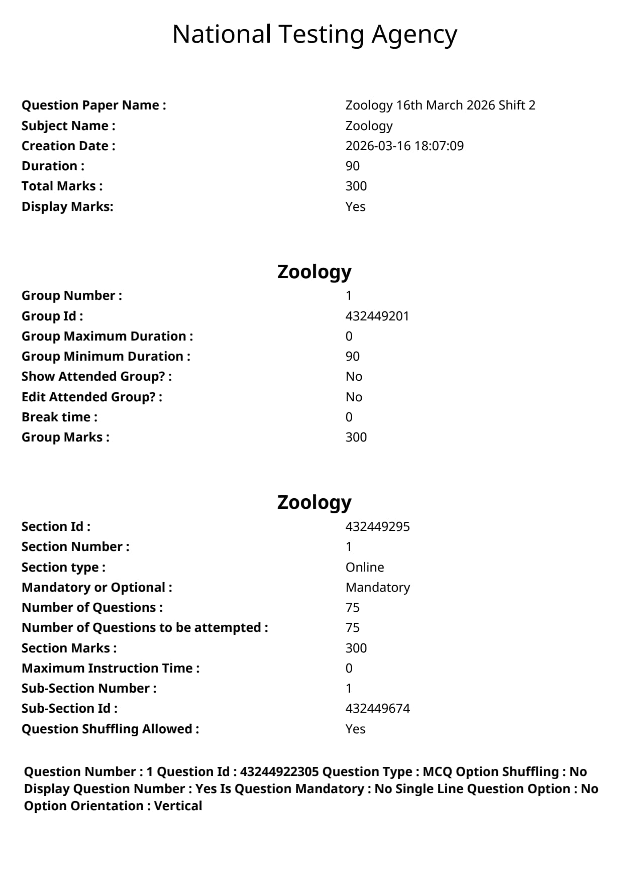
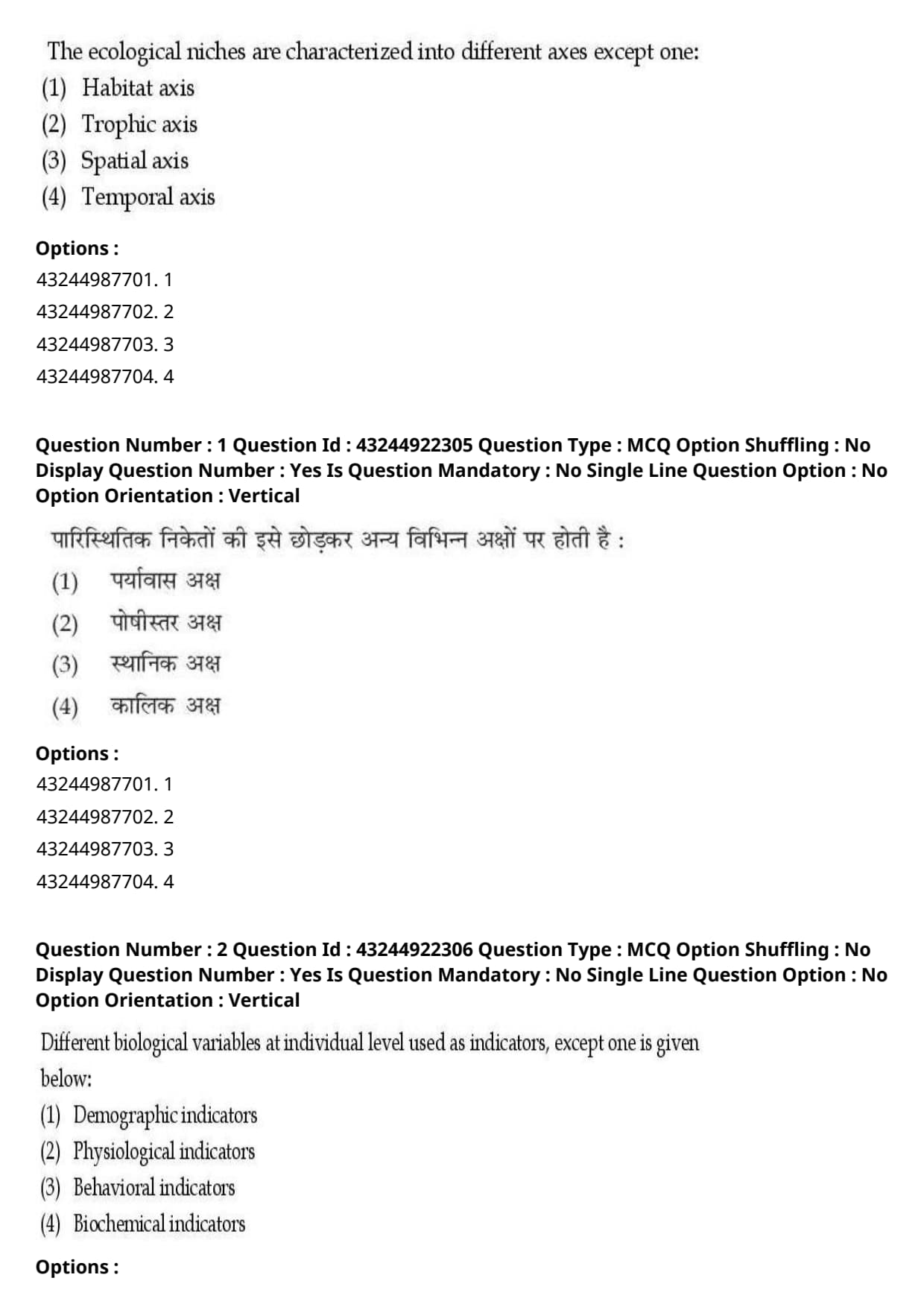
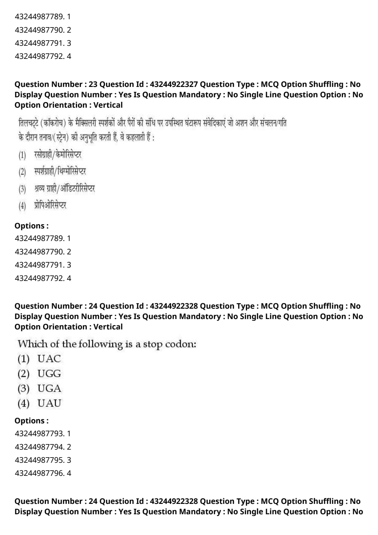
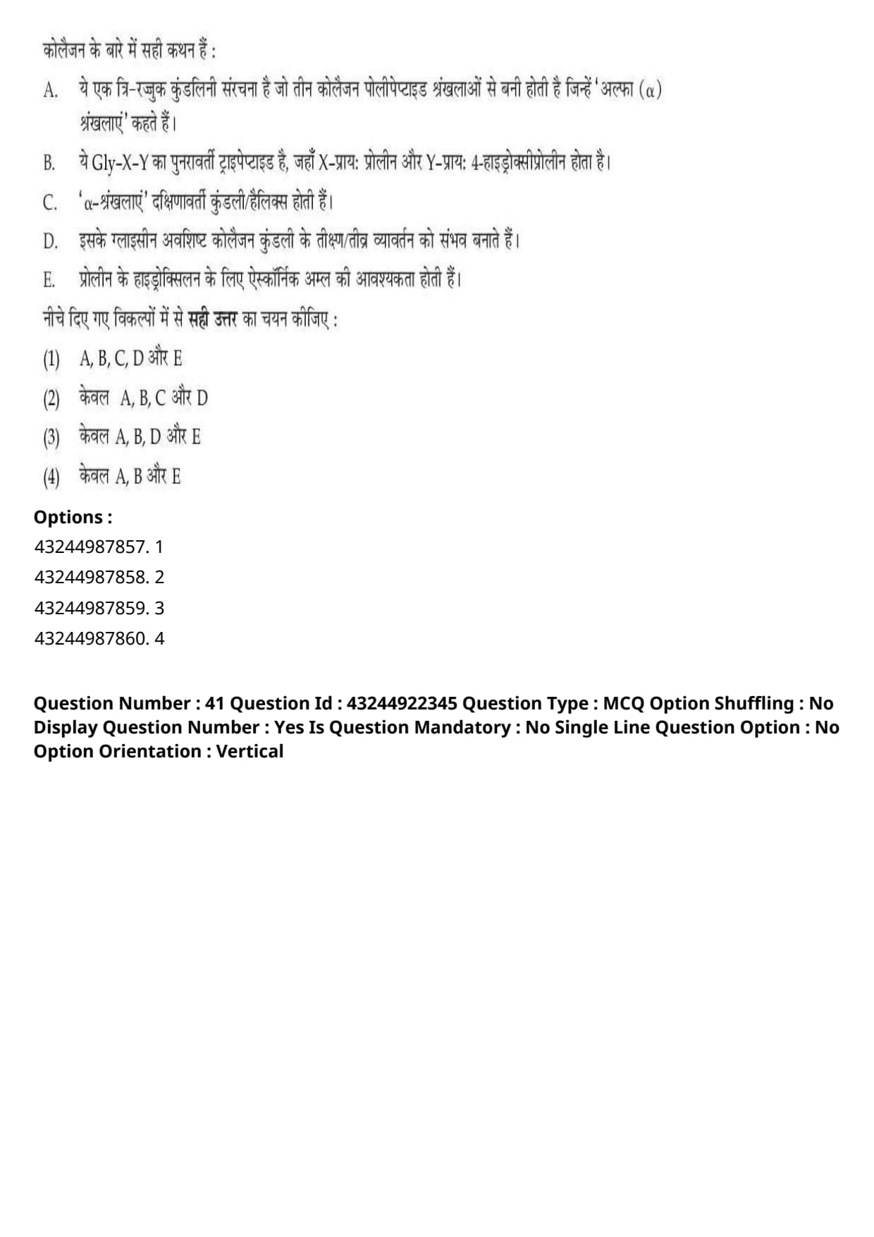
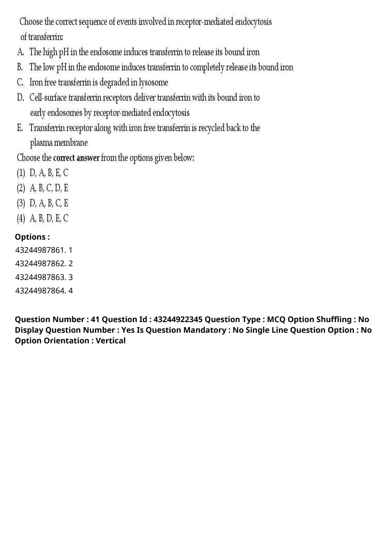
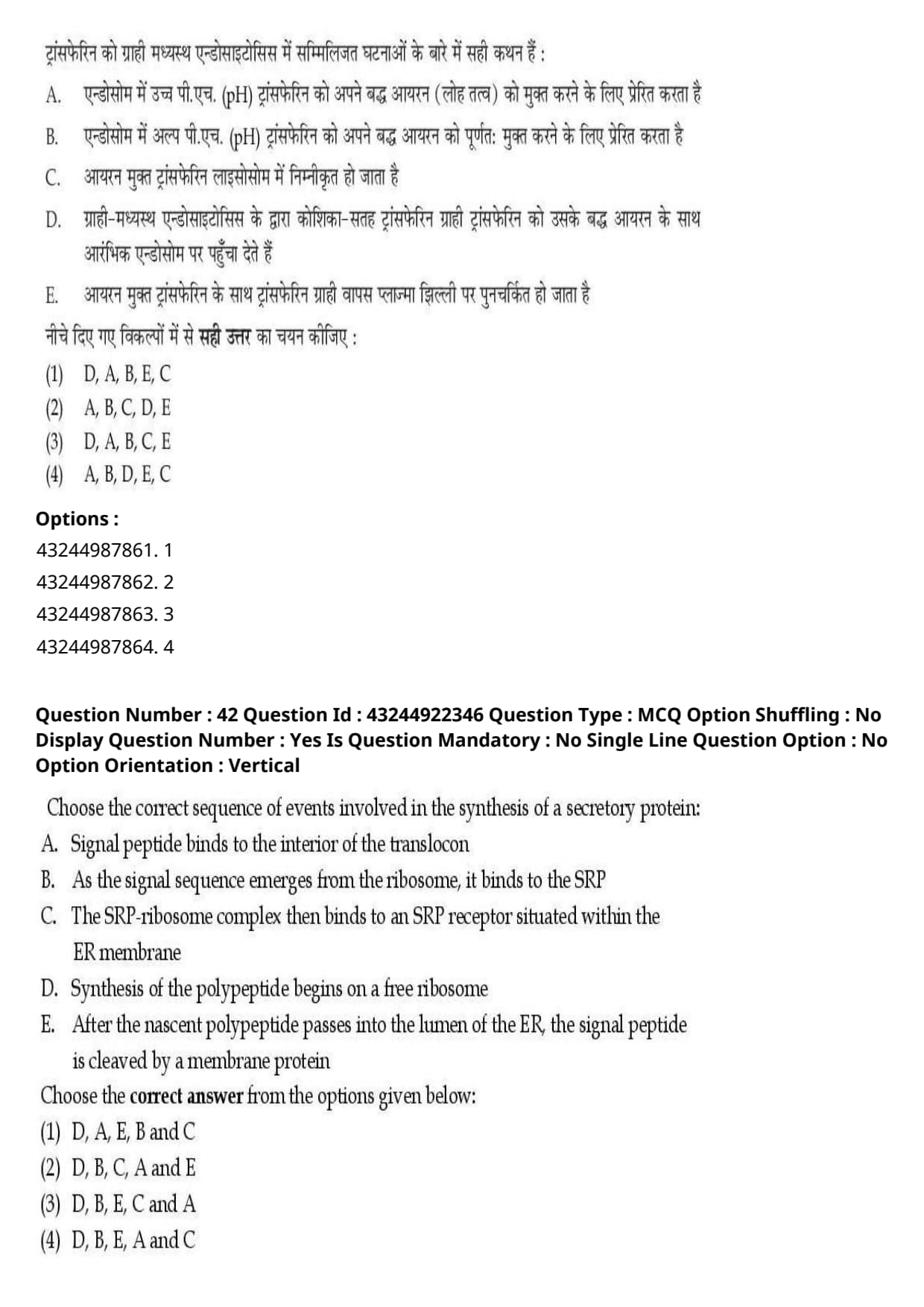
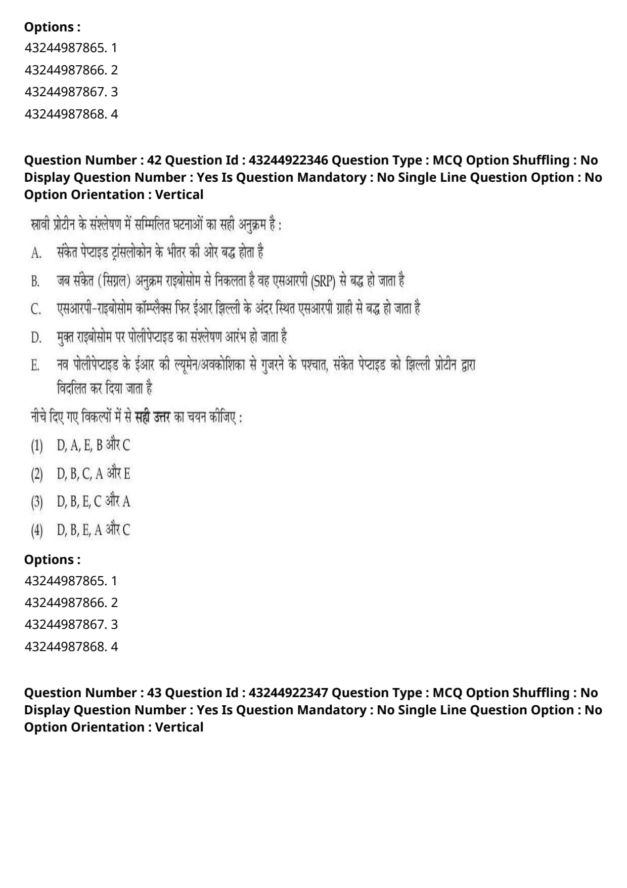
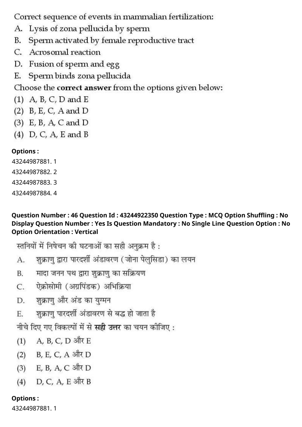
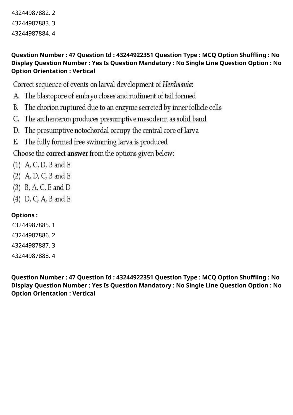
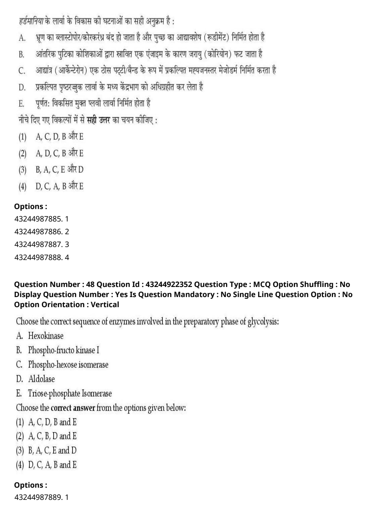
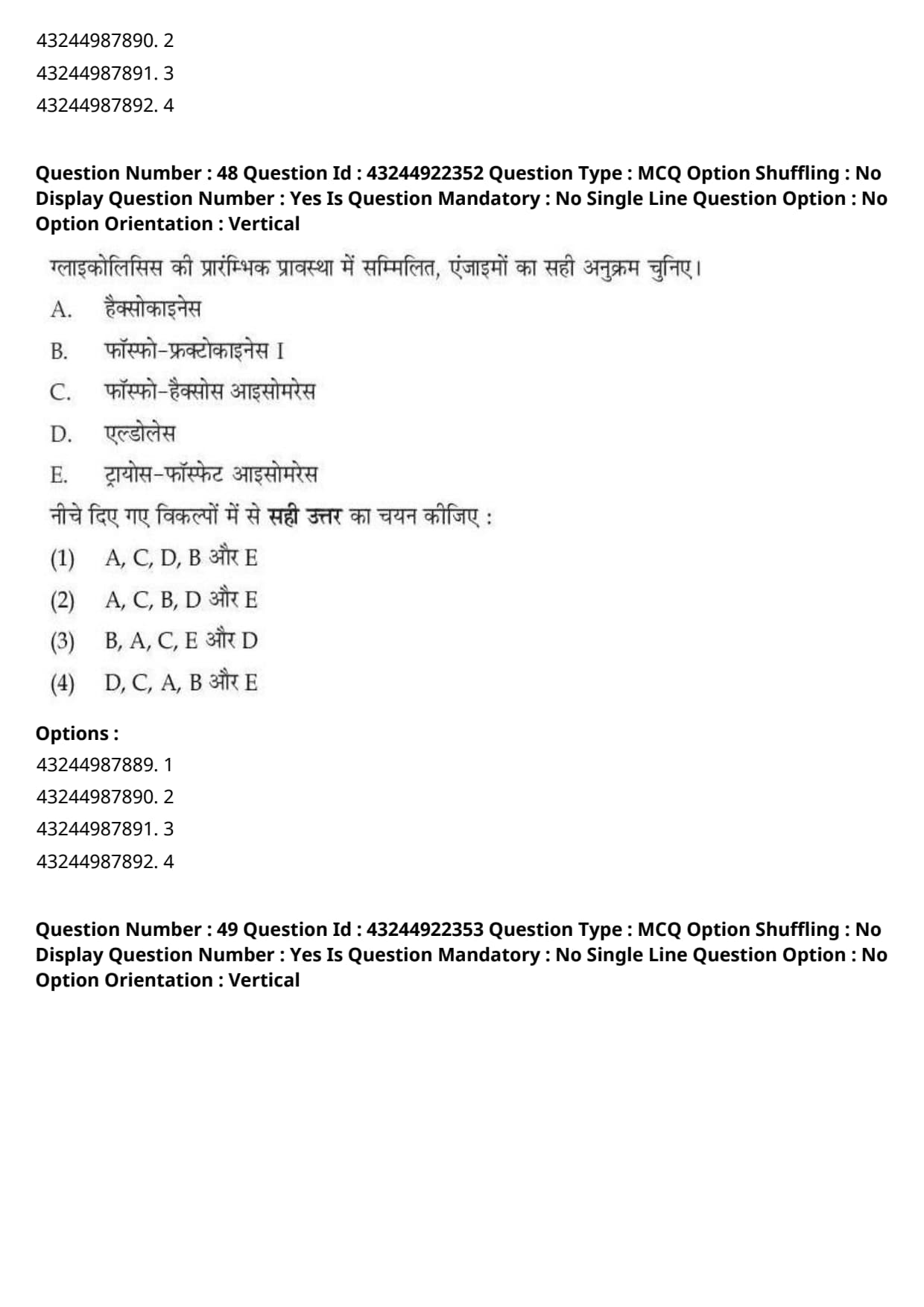
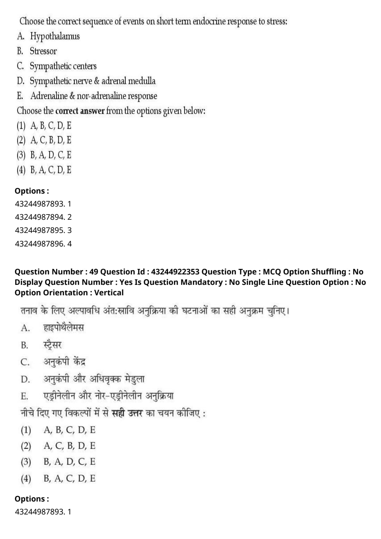
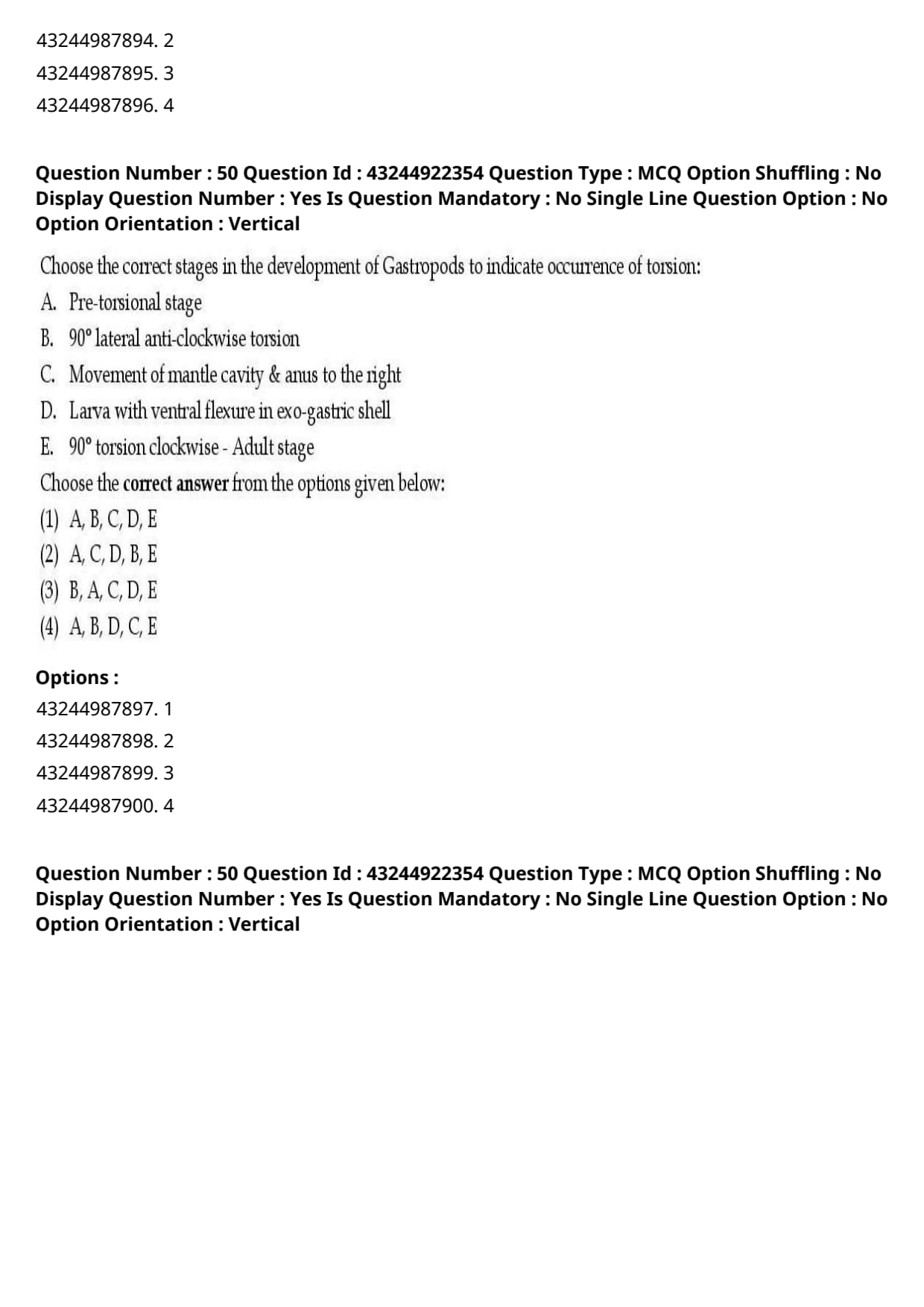
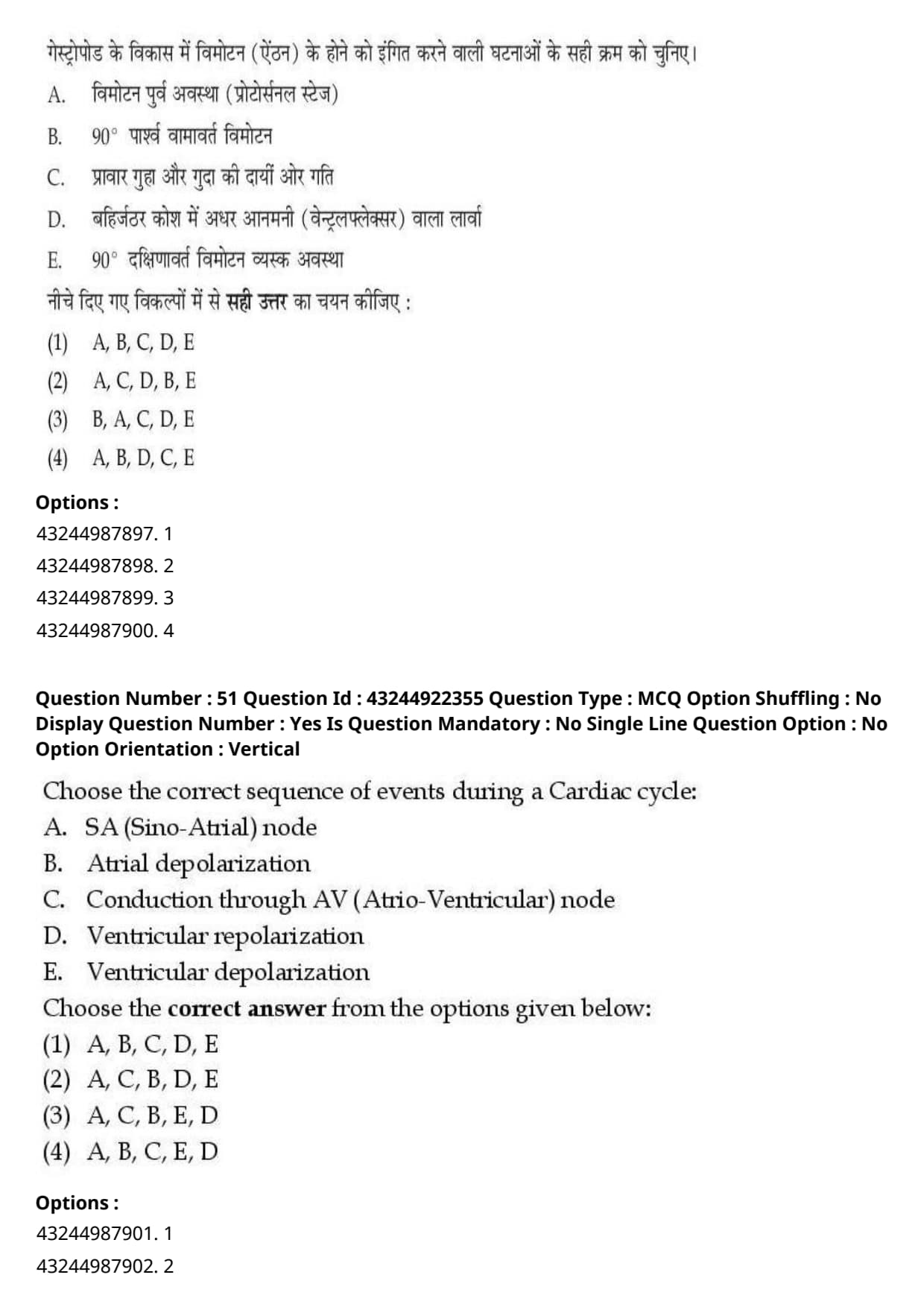
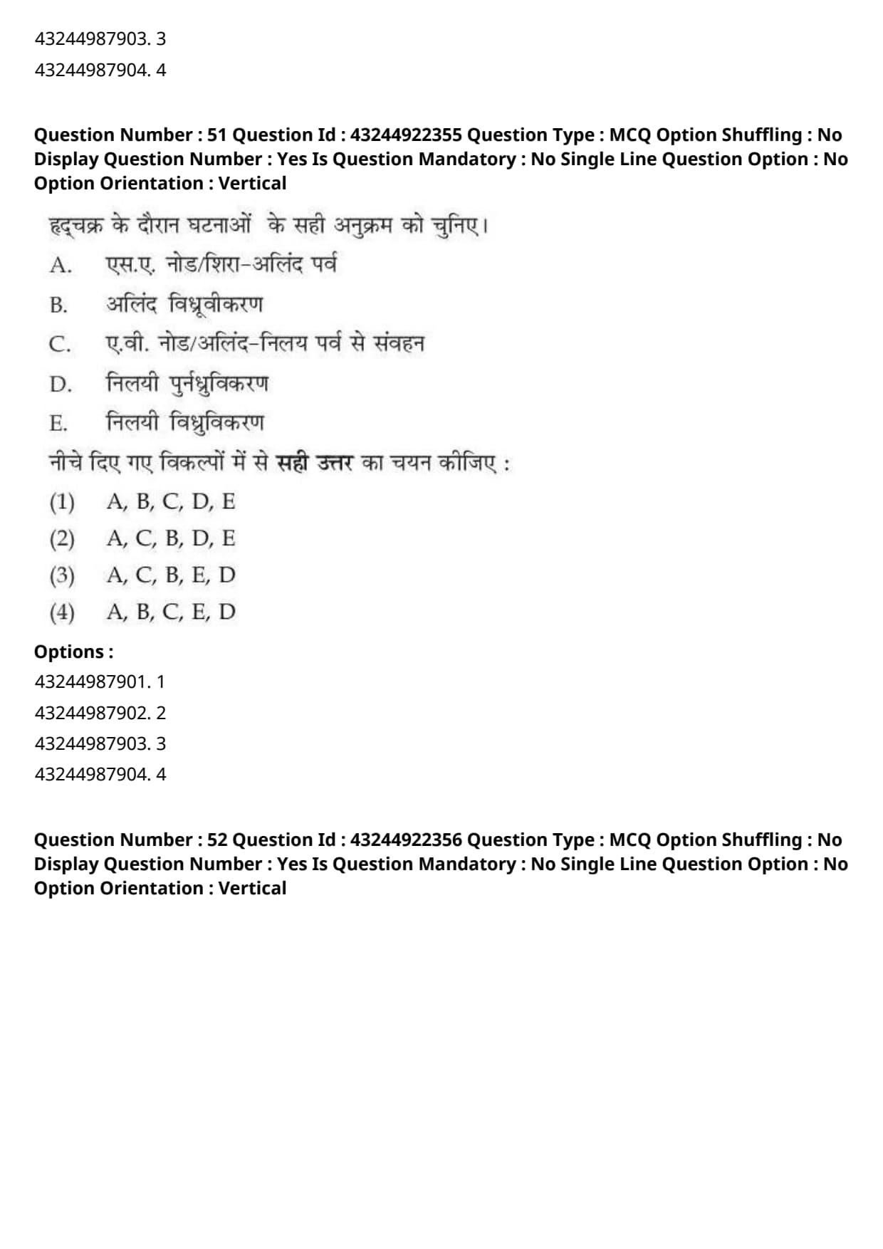
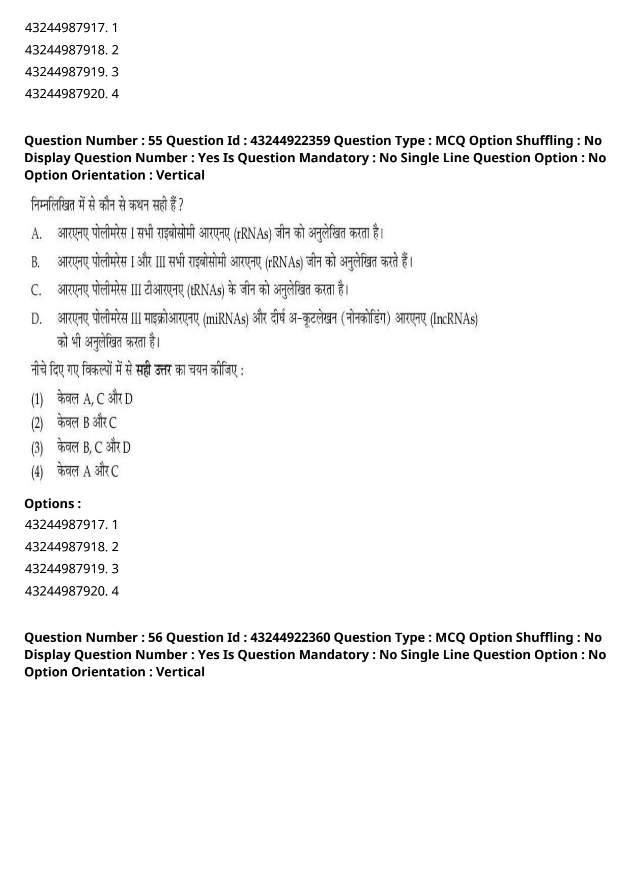
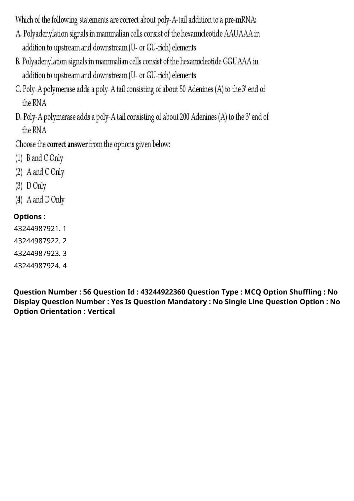
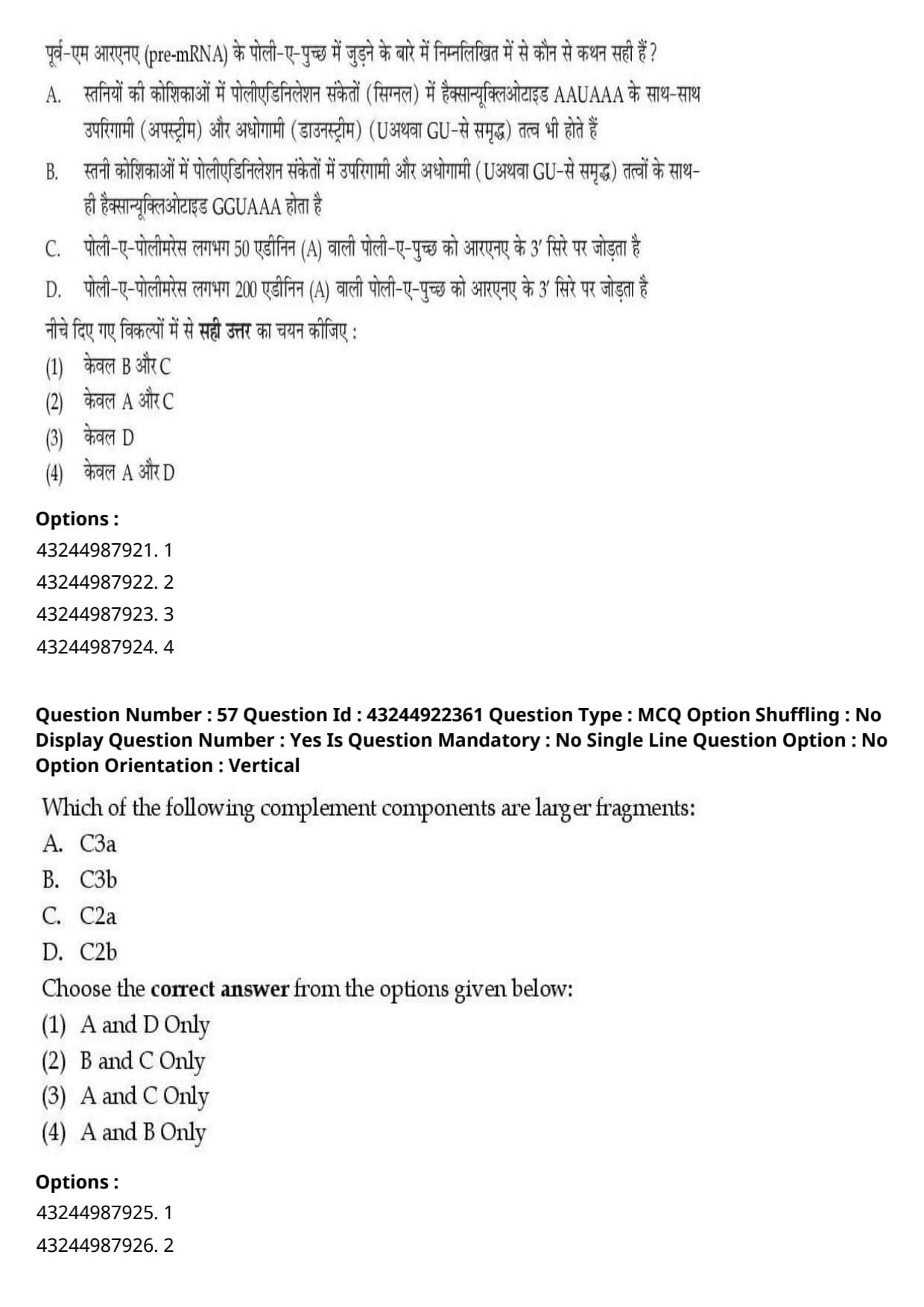
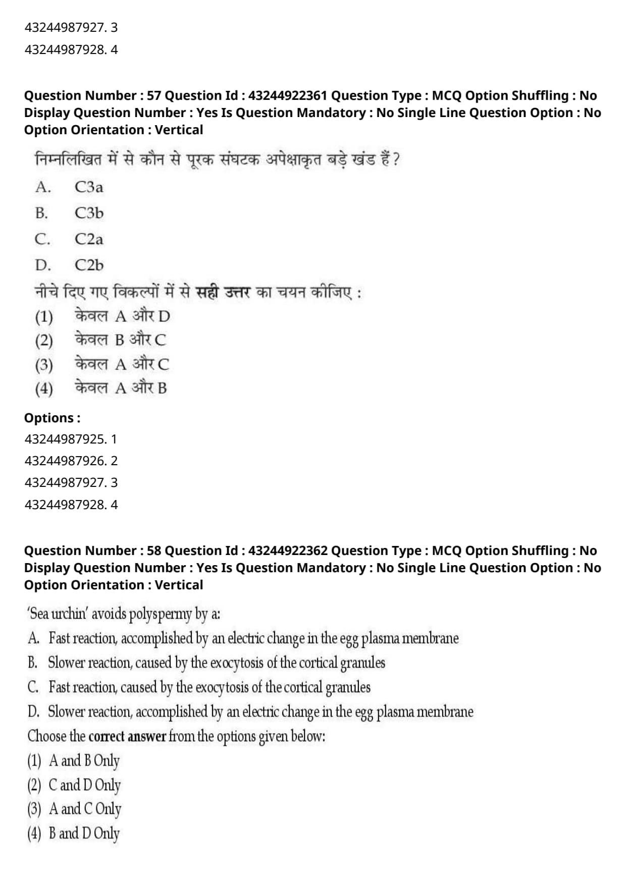
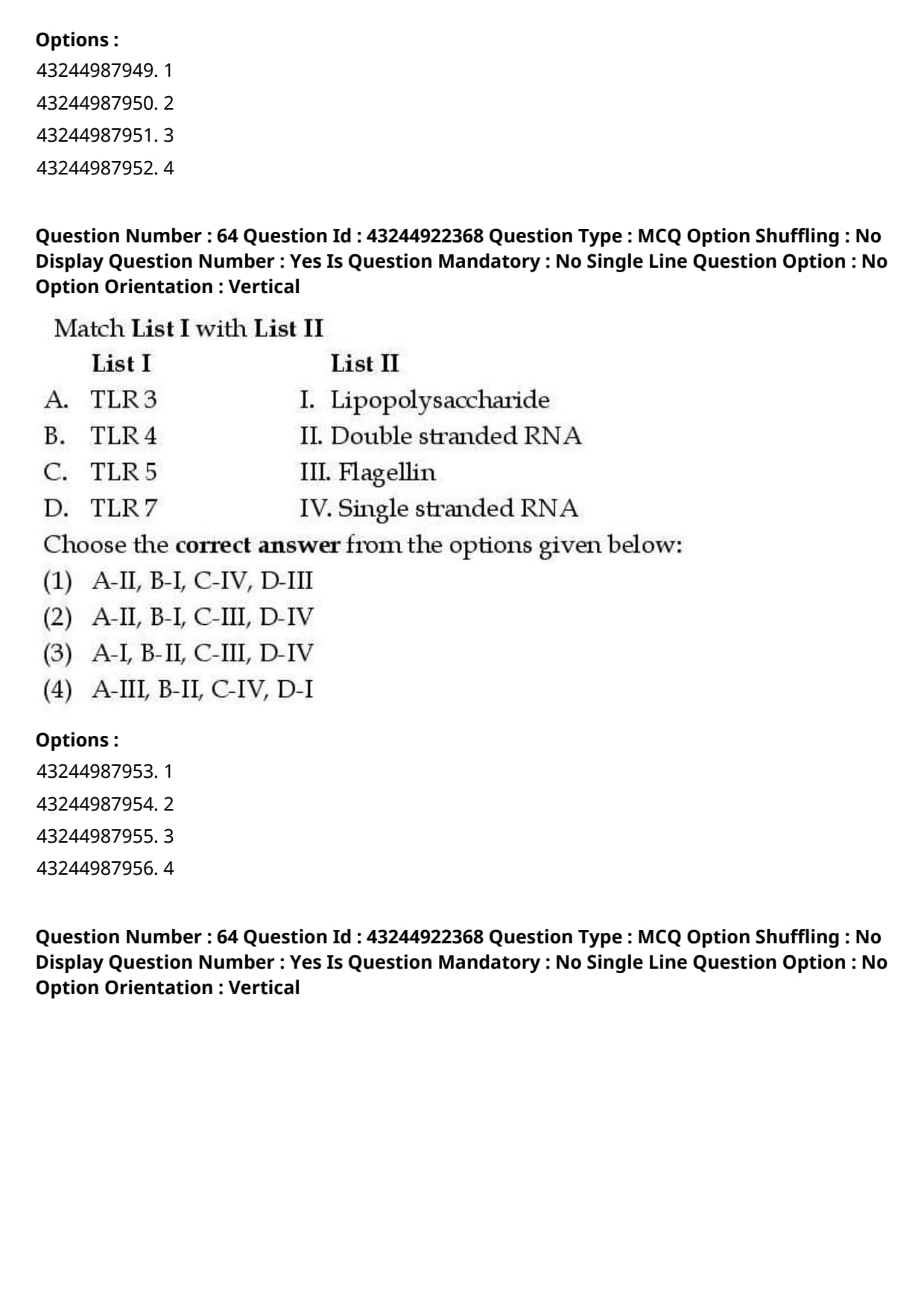
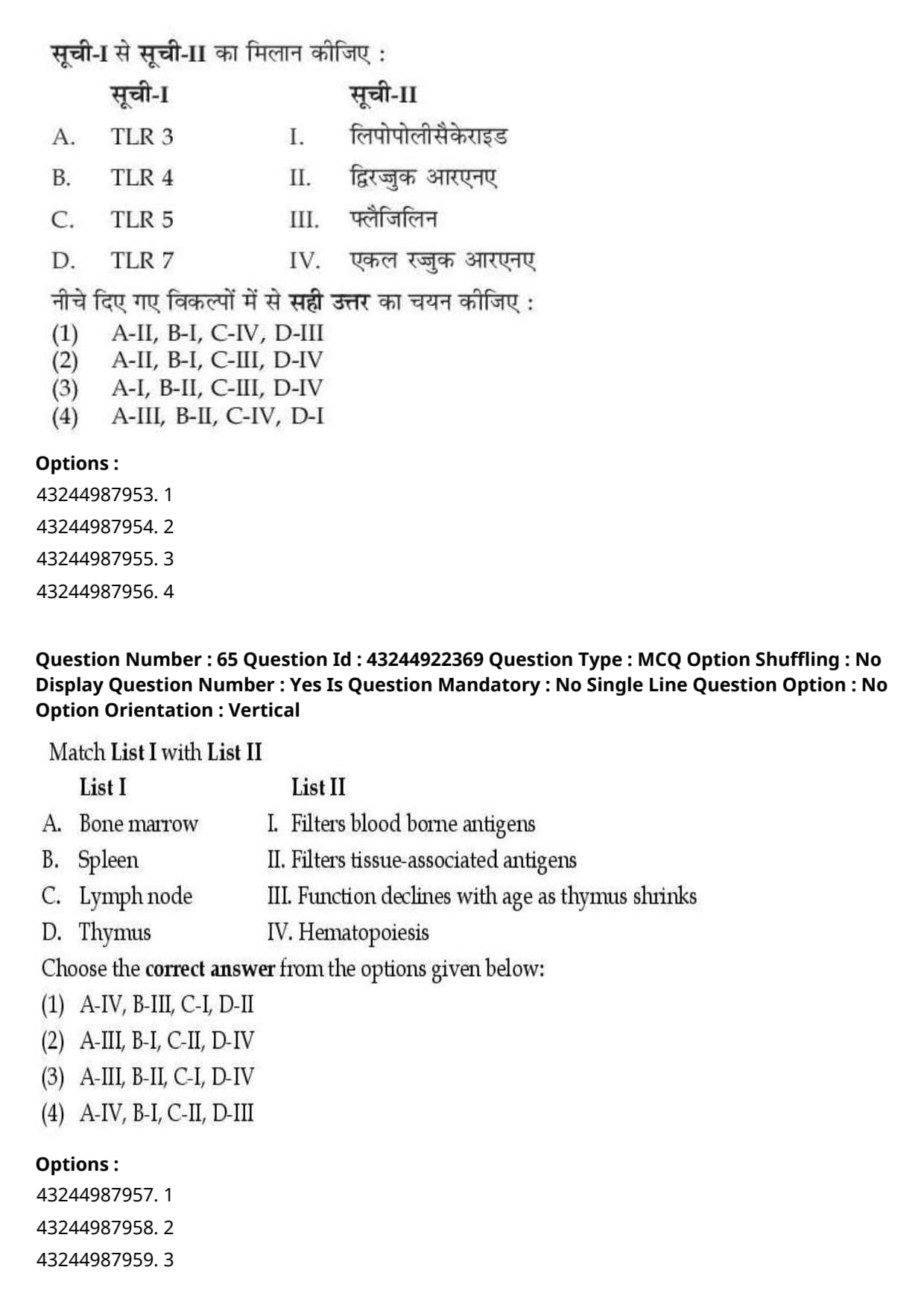
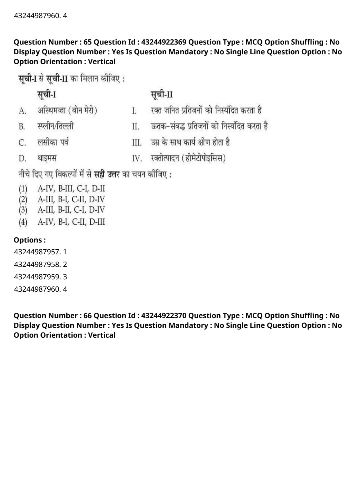
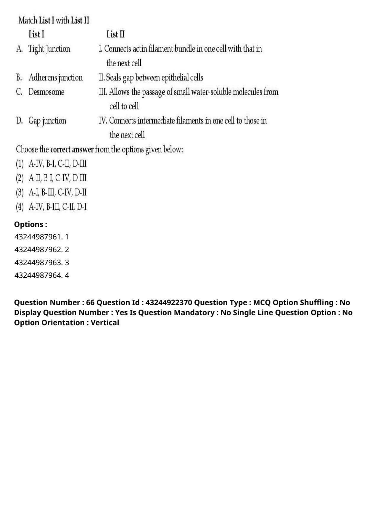
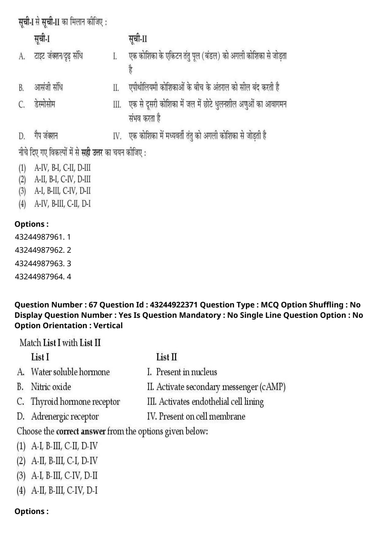
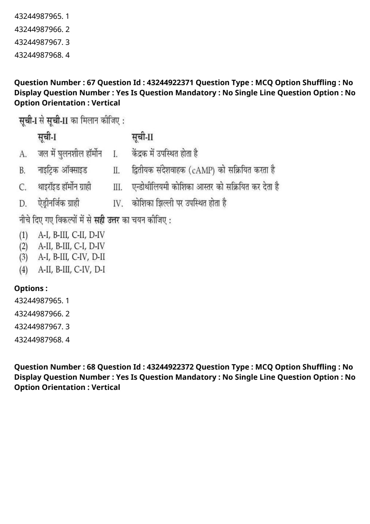
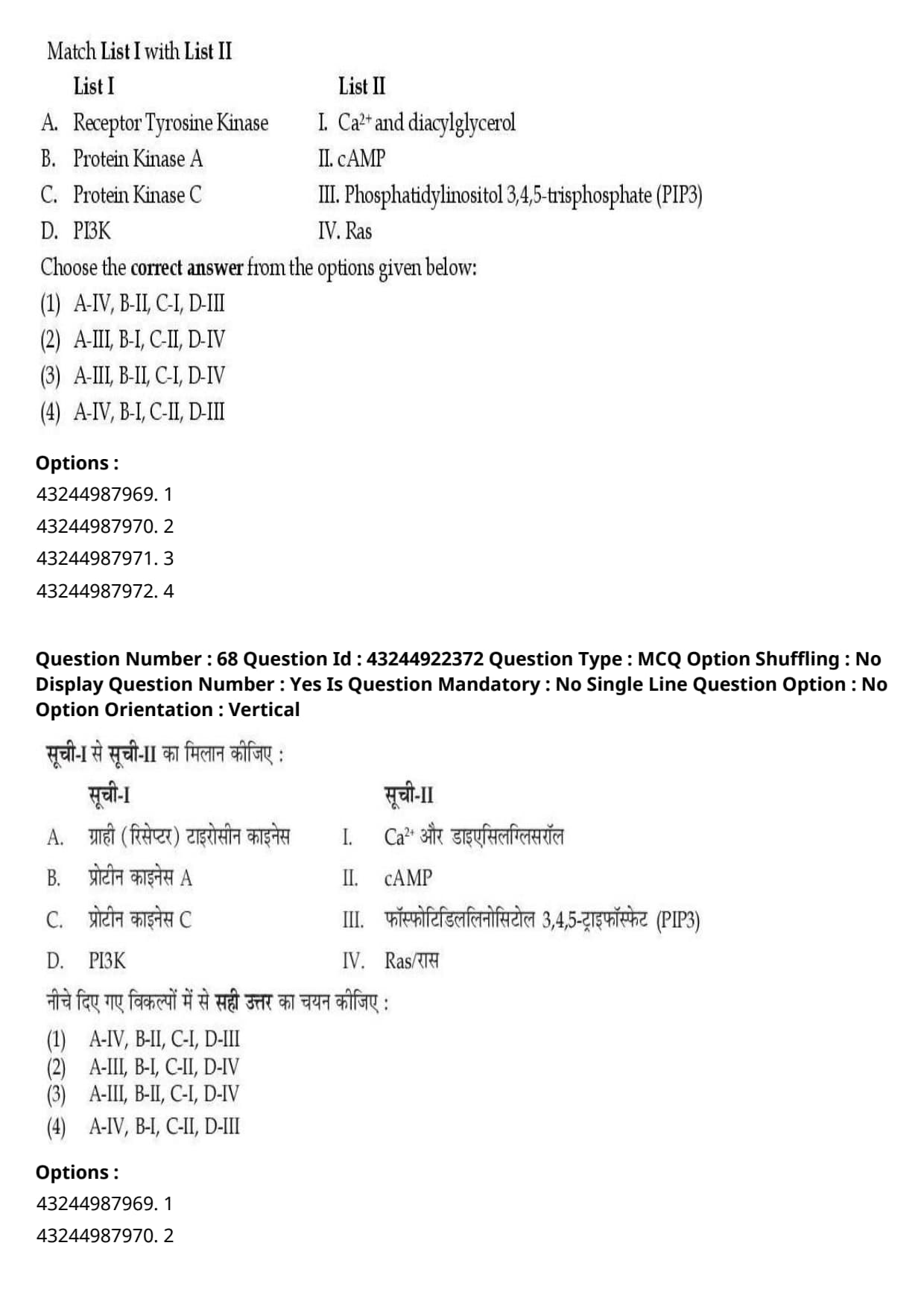
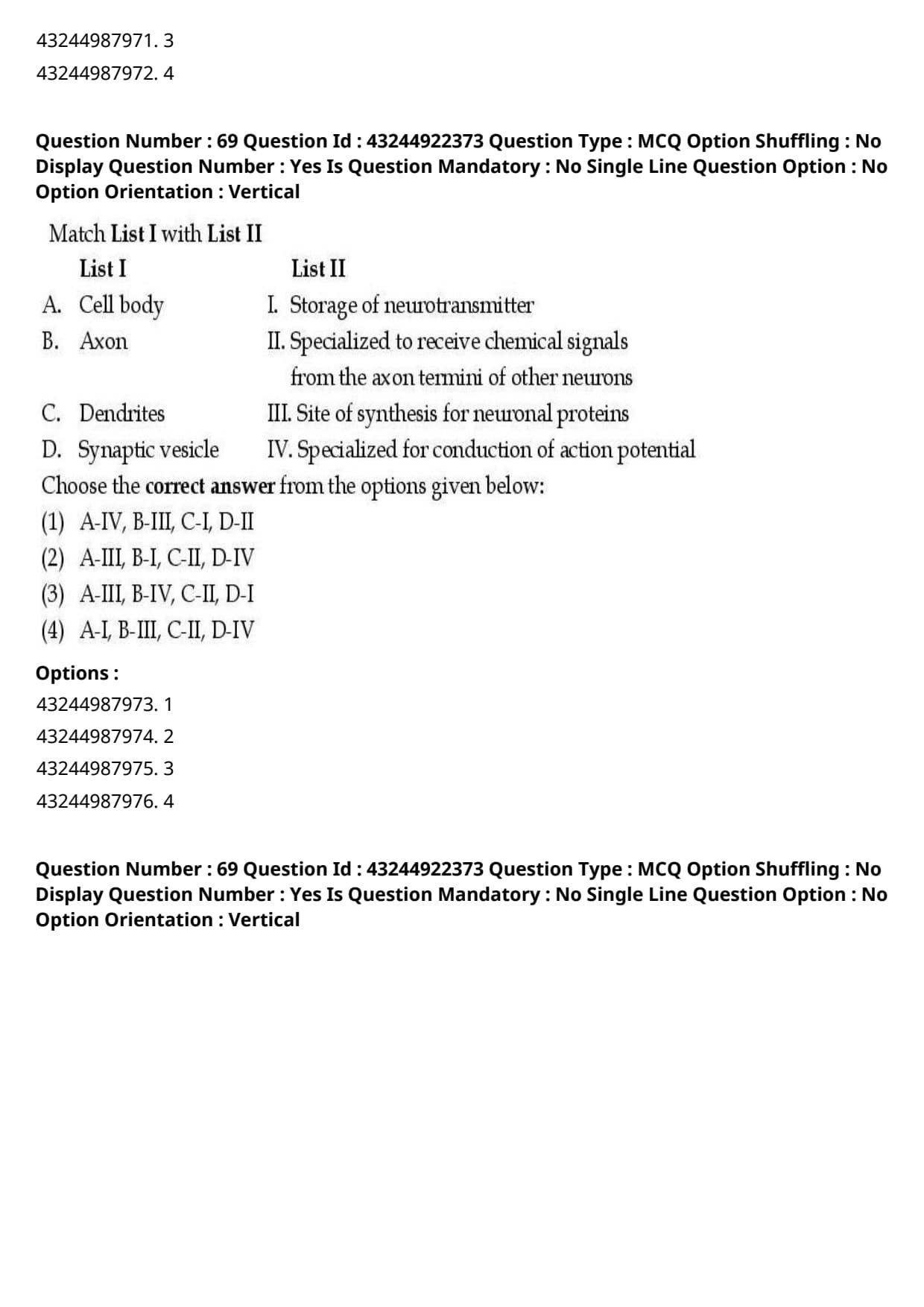
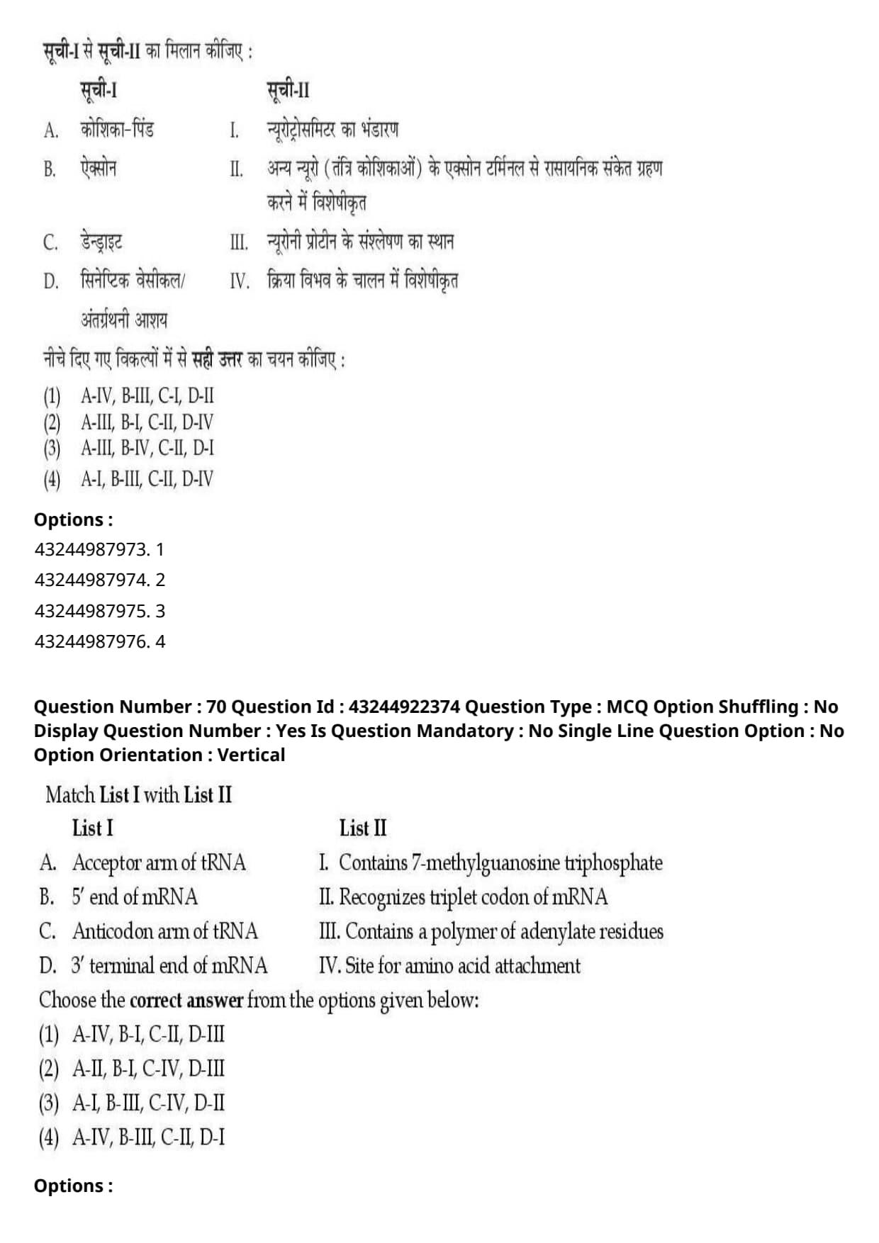
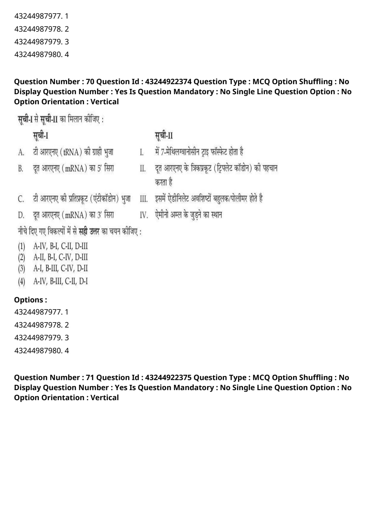
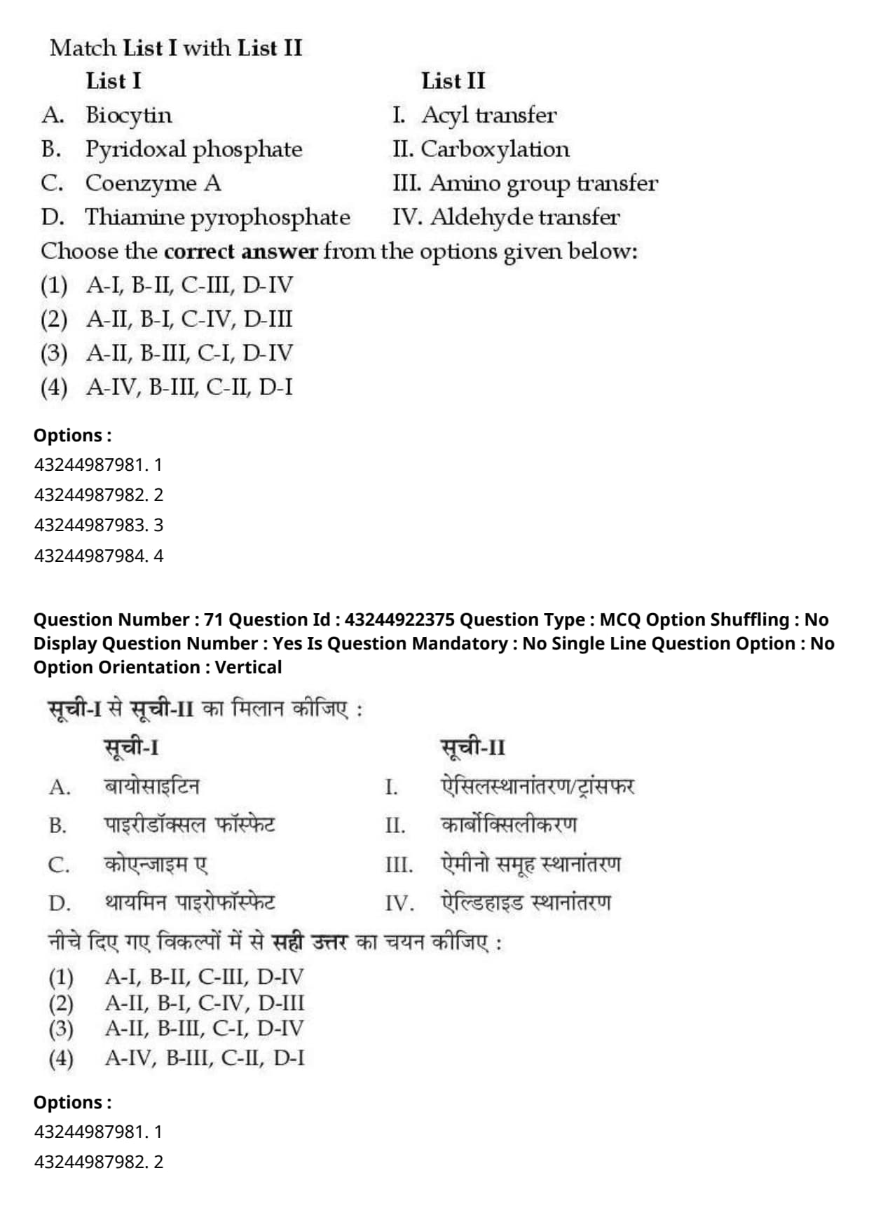
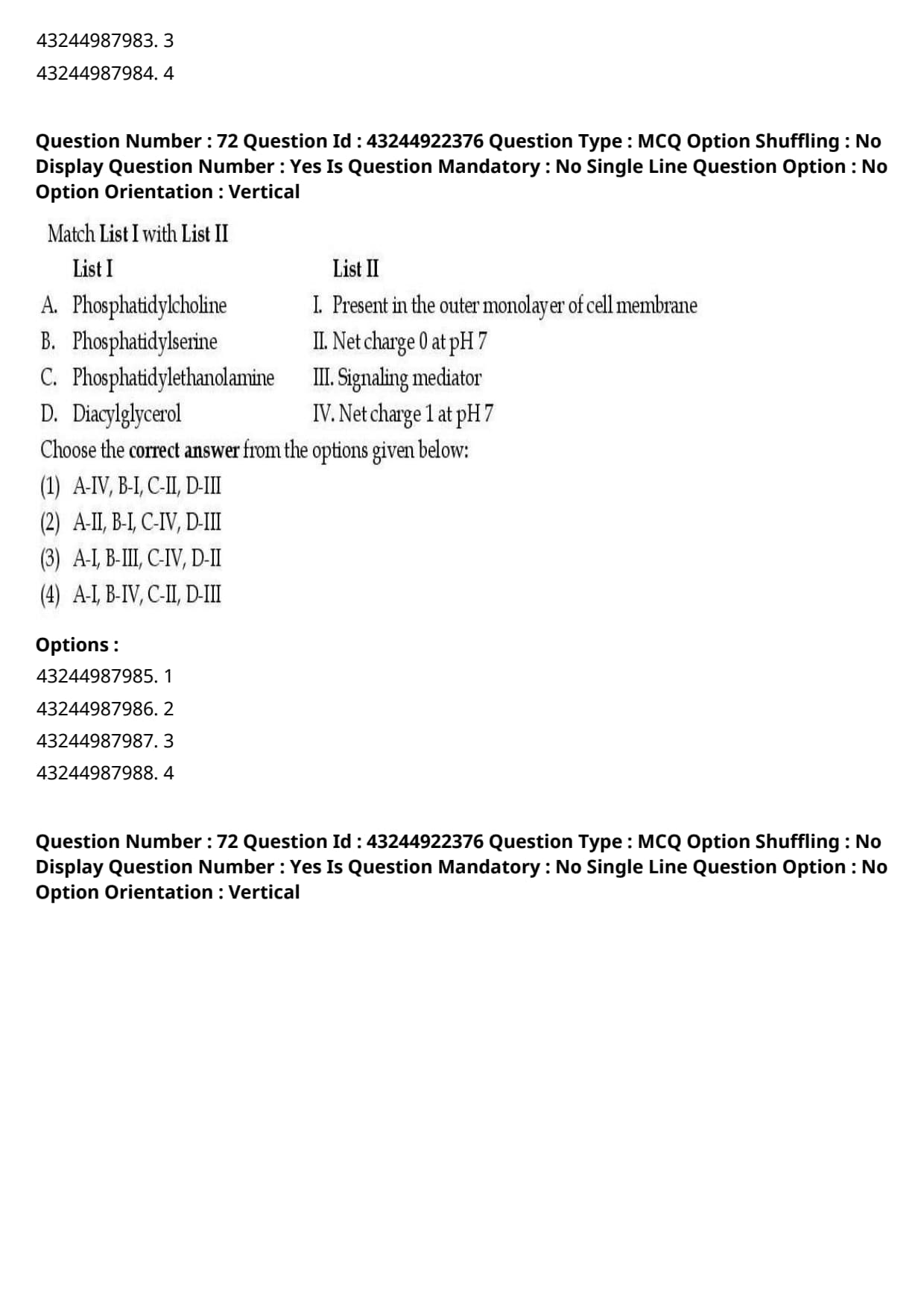
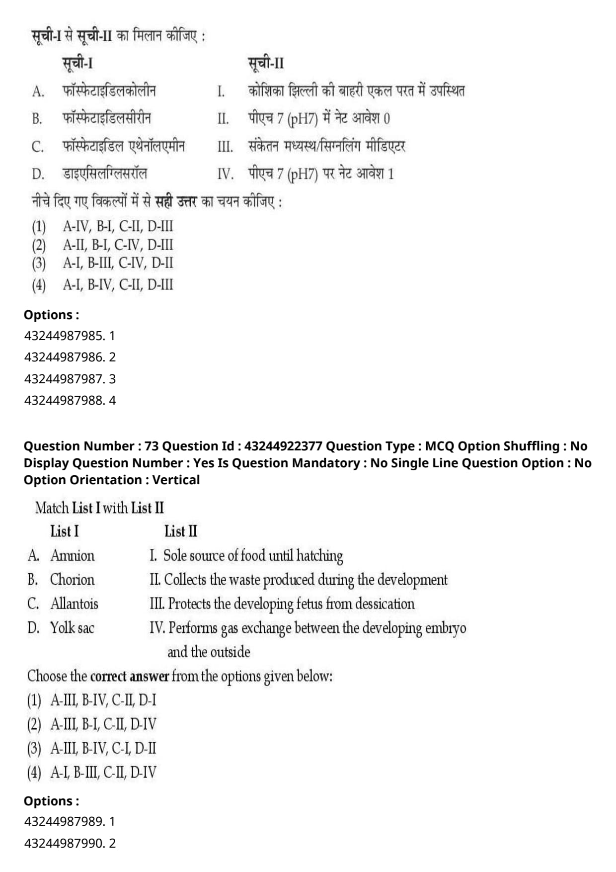
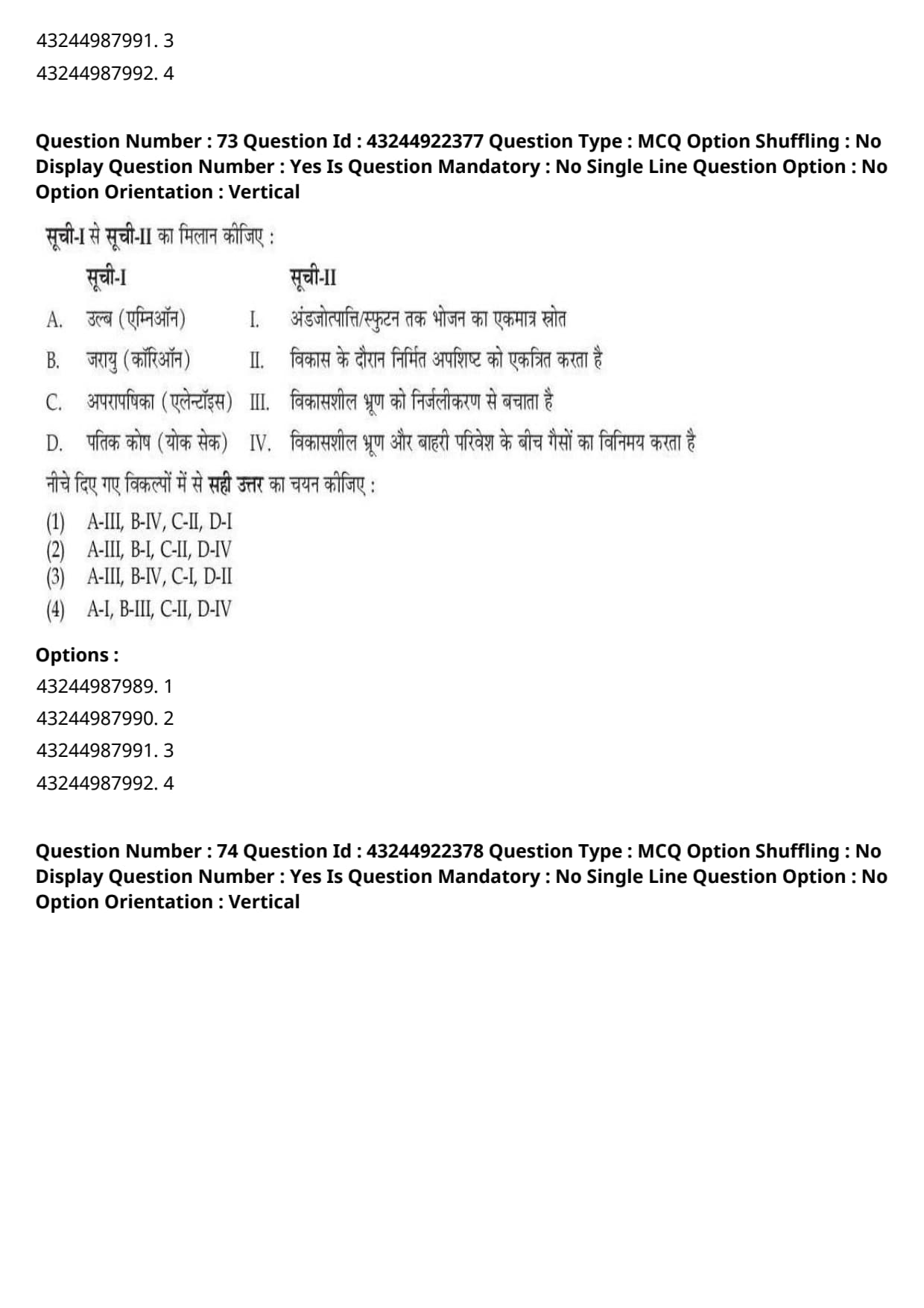
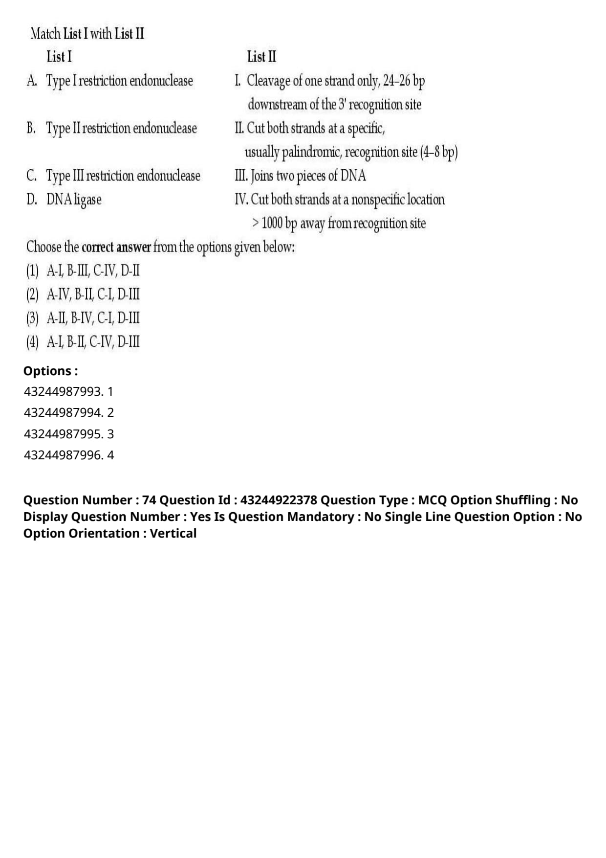
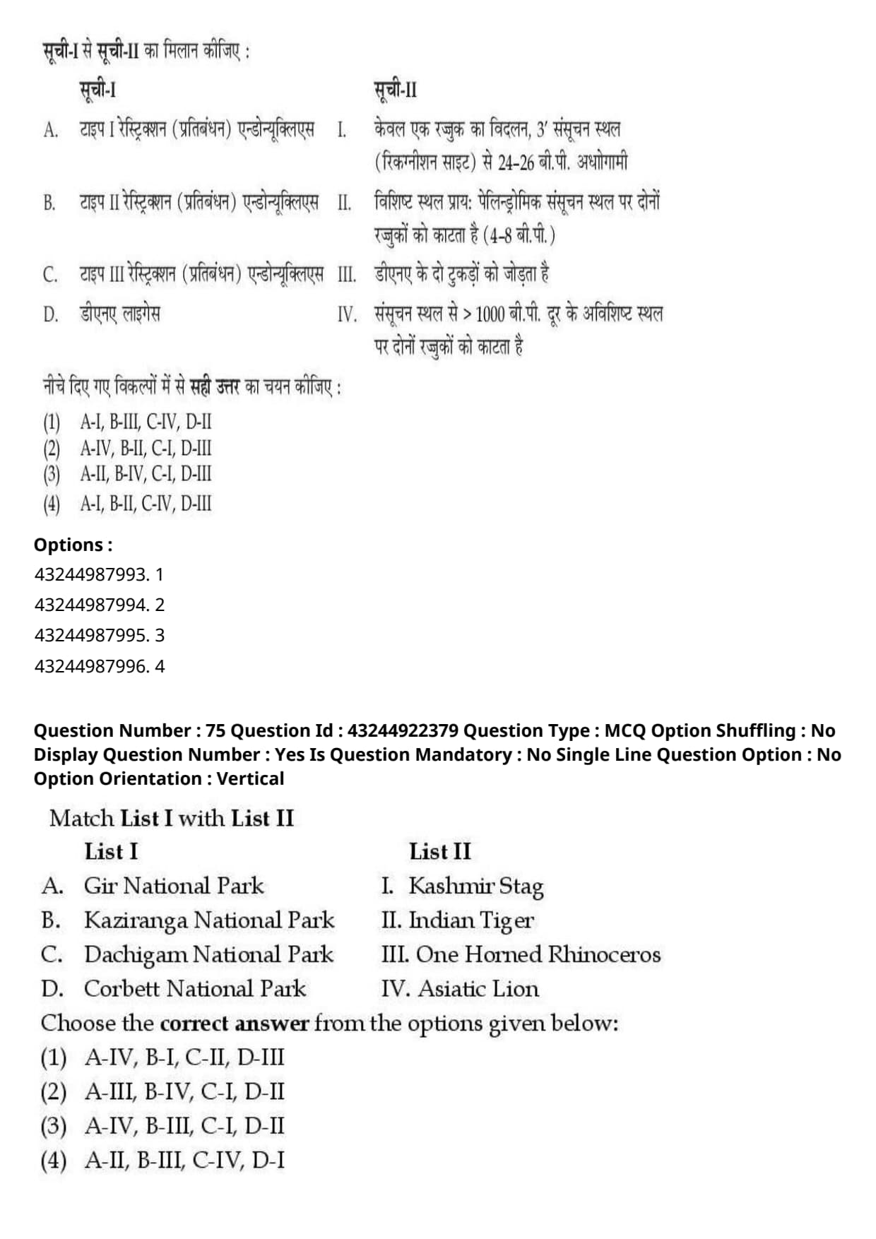
The ecological niches are characterized into different axes except one:
View Solution
Step 1: Understanding the Question:
This question asks us to identify which of the given options is not traditionally classified as a separate, primary axis in the multidimensional characterization of an organism's ecological niche.
Step 2: Detailed Explanation:
An ecological niche describes the total relational position and functional role of a species in its ecosystem.
According to classical ecological theories, particularly the niche partitioning models proposed by G.E. Hutchinson and Eric Pianka, the multidimensional niche space can be simplified into three primary, independent axes:
1. Habitat Axis (or Spatial Niche): This axis represents the physical space, microhabitat, or physical environment where the organism lives (such as height in a forest canopy or depth in a lake).
2. Trophic Axis (or Food Niche): This axis represents the functional role of the species in terms of its food resources, size of prey consumed, and position in the food web.
3. Temporal Axis (or Time Niche): This axis represents the time of daily or seasonal activity, such as being active during the day versus night, or seasonal variations in breeding.
- Because the "Habitat axis" and the "Spatial axis" fundamentally refer to the exact same physical space dimension of the niche, listing both introduces redundancy.
- In standard academic curricula, the three distinct axes of niche characterization are explicitly termed the Habitat, Trophic, and Temporal axes.
- Therefore, the "Spatial axis" is the redundant term and is considered the correct exception in this classification.
Step 3: Final Answer:
Therefore, the spatial axis is the correct exception, corresponding to option (C).
Quick Tip: Remember that the three pillars of ecological niche partitioning are Where (Habitat), What (Trophic), and When (Temporal).
Since Habitat and Spatial refer to the same physical dimension, look for the option that introduces redundancy in the nomenclature.
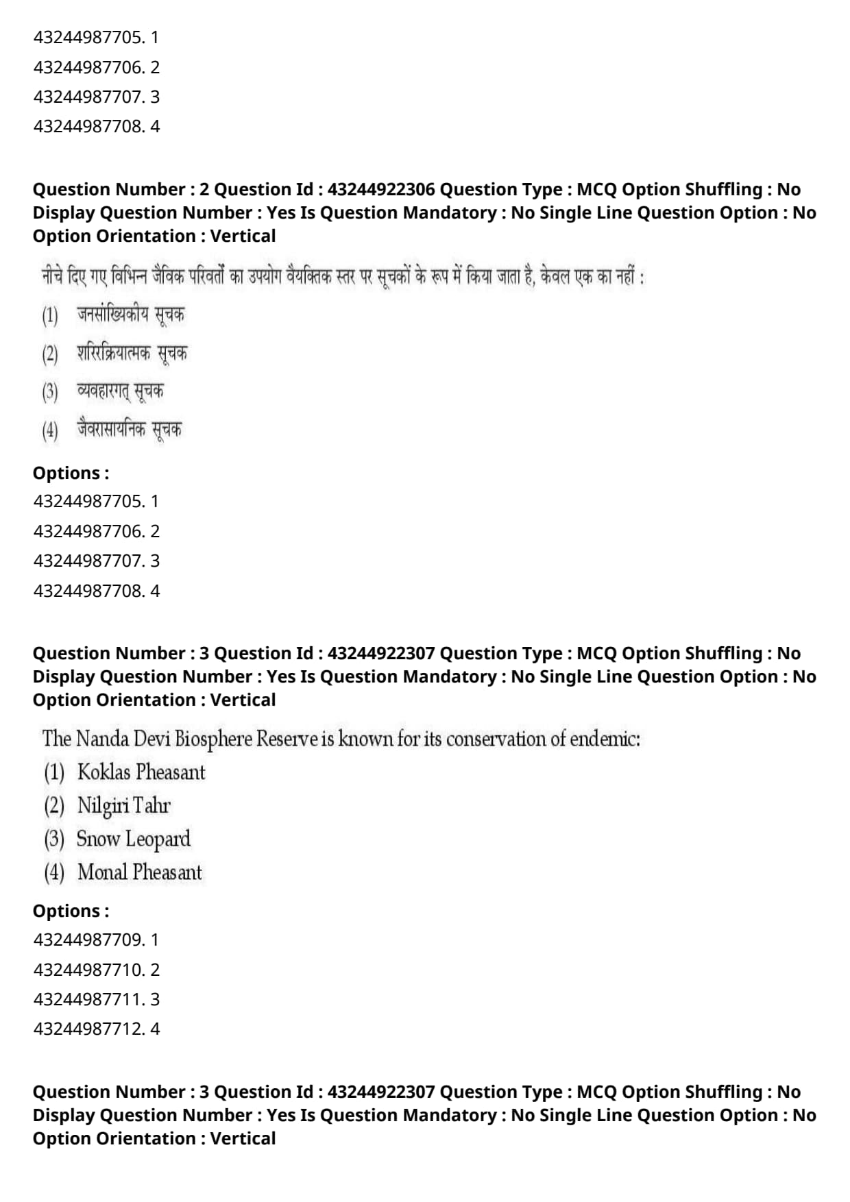
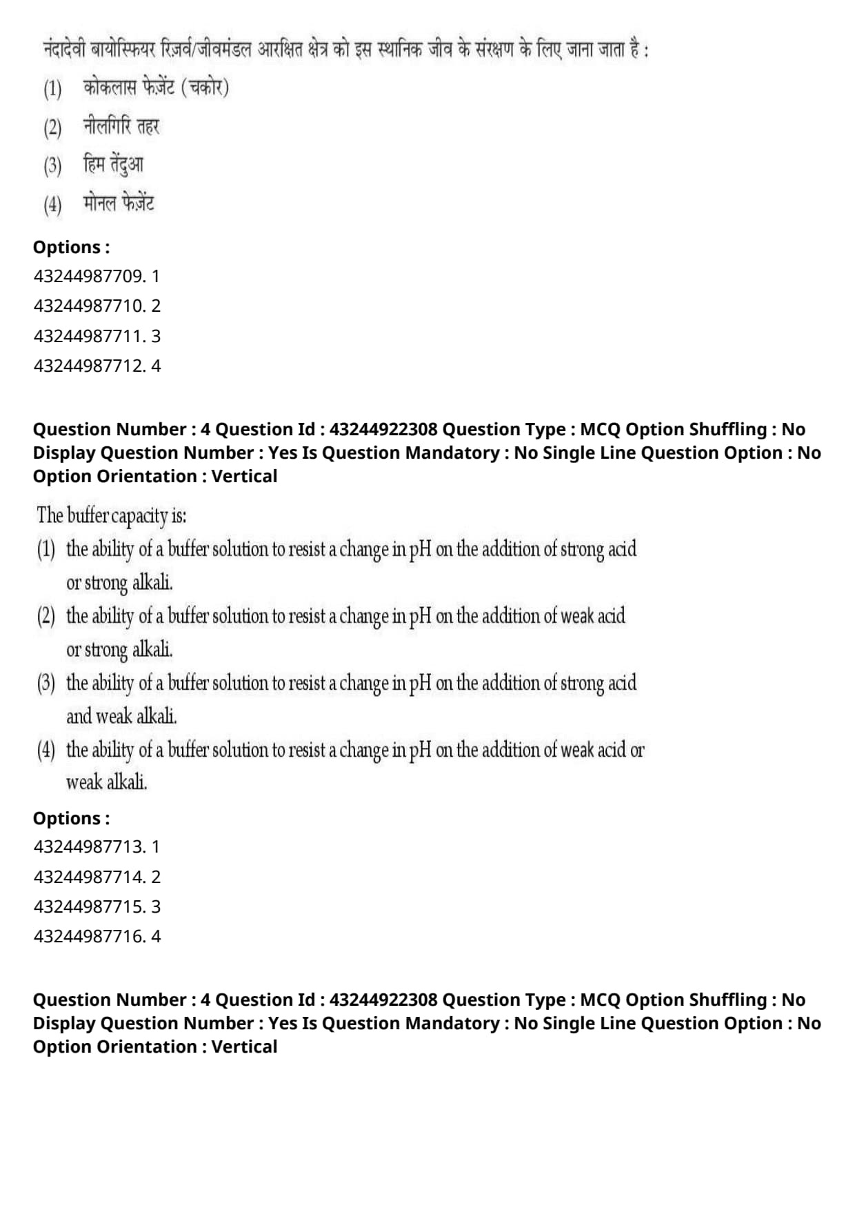
Different biological variables at individual level used as indicators, except one is given below:
View Solution
Step 1: Understanding the Question:
The question requires us to identify which of the listed biological indicators is not measured or applied at the individual level of biological organization.
Step 2: Detailed Explanation:
Biological indicators are used to assess environmental health and ecological stress at various levels of biological hierarchy.
These levels include molecular, cellular, organ, individual, population, and community levels.
1. Individual-Level Indicators: These are biological responses measured directly in a single organism:
- Physiological Indicators: Measurements such as heart rate, oxygen consumption, respiration rate, and osmotic balance reflect the functional health of a single individual.
- Behavioral Indicators: Avoidance behavior, feeding rate, locomotion, and learning capacity are direct actions observed in an individual.
- Biochemical Indicators: The levels of specific enzymes (such as acetylcholinesterase), stress proteins (like heat shock proteins), and molecular damage are measured within an individual's tissues.
2. Population-Level Indicators:
- Demographic Indicators: These deal with statistical characteristics of groups of individuals belonging to the same species in a given area.
- Examples of demographic indicators include birth rate (natality), death rate (mortality), population growth rate, age structure, and sex ratio.
- Because a single individual cannot have a "birth rate" or "age structure," demographic variables are strictly population-level indicators and cannot be measured at the individual level.
Step 3: Final Answer:
Thus, demographic indicators represent the correct population-level exception, corresponding to option (A).
Quick Tip: To quickly solve levels-of-organization questions, ask yourself: "Can I measure this variable on a single isolated organism?"
You can measure heart rate (physiological) or enzyme levels (biochemical) of one individual animal, but you cannot measure a birth rate (demographic) on a single animal.
The Nanda Devi Biosphere Reserve is known for its conservation of endemic:
View Solution
Step 1: Understanding the Question:
This question asks us to identify the primary high-altitude animal of major conservation importance associated with the Nanda Devi Biosphere Reserve.
Step 2: Detailed Explanation:
The Nanda Devi Biosphere Reserve is located in the high-altitude Himalayan region of Uttarakhand, India.
It is a UNESCO World Heritage Site recognized globally for its unique, fragile alpine biodiversity and majestic high-altitude wildlife.
- Snow Leopard (\textit{Panthera uncia) is the key flagship predator species of this high-altitude ecosystem. Nanda Devi is globally renowned for its active conservation programs protecting this highly endangered and endemic cat of the cold deserts.
- Let us analyze the other options to understand why they are incorrect:
- Nilgiri Tahr (\textit{Nilgiritragus hylocrius) is an ungulate endemic strictly to the Western Ghats of Southern India, and is completely absent in the Himalayas.
- Koklas Pheasant (\textit{Pucrasia macrolopha) and Monal Pheasant (\textit{Lophophorus impejanus) are beautifully colored high-altitude birds native to the Western Himalayas, but they are not the primary, high-profile focal conservation species for which the biosphere reserve is globally famous.
- Therefore, the Snow Leopard stands out as the most significant species protected under the reserve's high-altitude conservation mandate.
Step 3: Final Answer:
Hence, the correct option is (C).
Quick Tip: Use geographical elimination to rule out options quickly.
The Nilgiri Tahr is strictly native to Southern India (Western Ghats), so it can be immediately eliminated for a Himalayan biosphere reserve located in Uttarakhand.
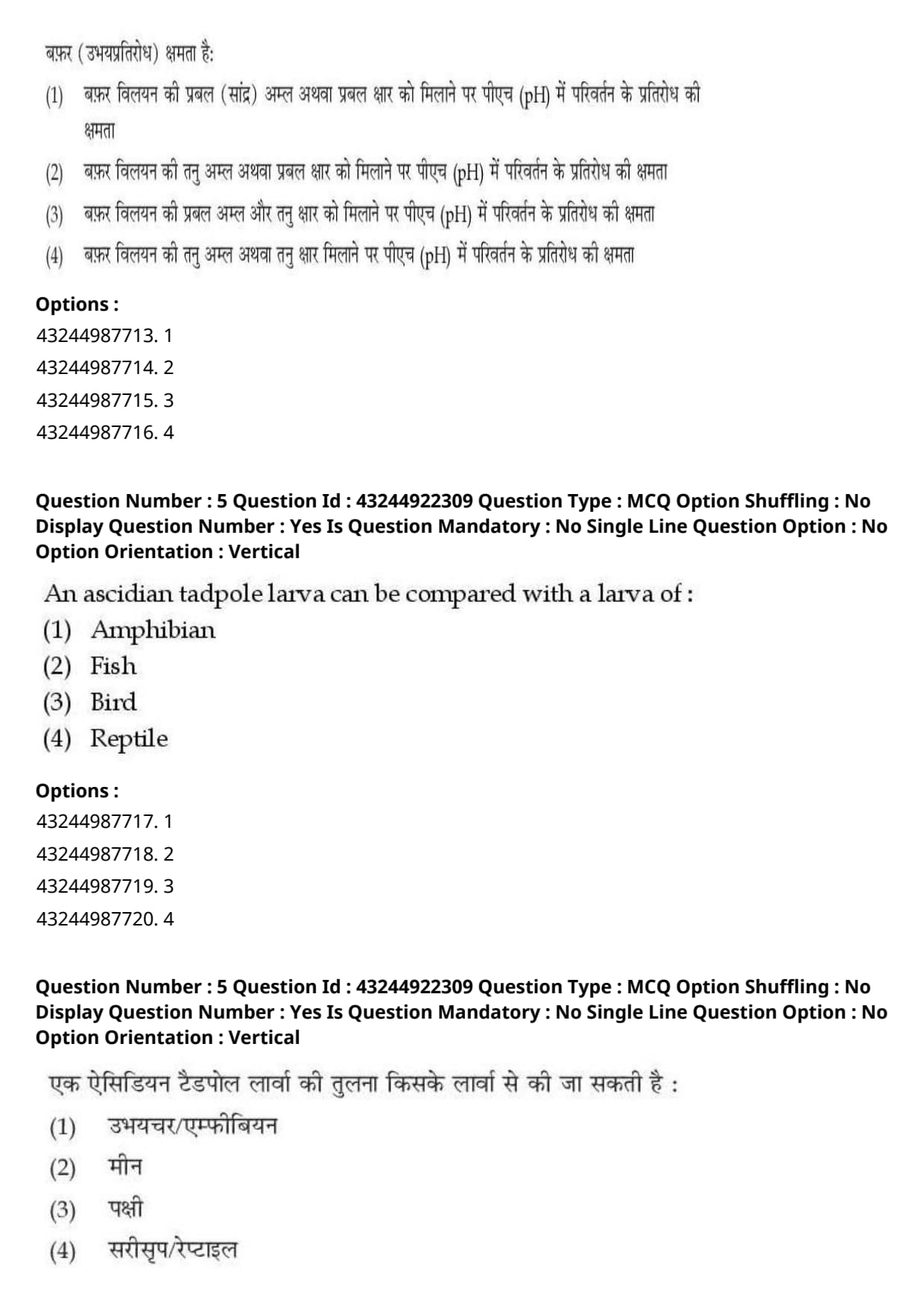
The buffer capacity is:
View Solution
Step 1: Understanding the Question:
The question asks for the standard and precise definition of "buffer capacity" in the context of acid-base chemistry and biochemistry.
Step 2: Key Formula or Approach:
The quantitative measure of buffer capacity (\(\beta\)) is given by the Van Slyke equation:
\[ \beta = \frac{db}{d(pH)} \]
where \(db\) is the amount of strong base (or strong acid) added in moles per liter, and \(d(pH)\) is the resulting change in pH.
Step 3: Detailed Explanation:
A buffer solution is a mixture of a weak acid and its conjugate base, or a weak base and its conjugate acid, which maintains a nearly constant pH when small amounts of acid or base are added.
- Buffer Capacity Defined: Buffer capacity represents the maximum ability of a buffer solution to resist drastic changes in pH when challenged with external acidic or basic substances.
- Why Strong Reagents are Used in the Definition:
- A buffer's true resistance is evaluated by adding strong acids (like \(HCl\)) or strong bases (like \(NaOH\)) because they fully dissociate in water, releasing the maximum concentration of free hydrogen (\(H^+\)) or hydroxide (\(OH^-\)) ions.
- If a buffer can effectively neutralize these fully dissociating strong acids or strong bases, it can easily resist weak acids or bases.
- Therefore, the formal scientific definition of buffer capacity is specifically stated in terms of resisting pH changes upon the addition of a strong acid or strong alkali.
Step 4: Final Answer:
Thus, option (A) provides the most mathematically and scientifically accurate definition.
Quick Tip: To test the maximum capacity of any defensive system, you must challenge it with the strongest possible force.
Therefore, buffer capacity is always defined by adding a strong acid or strong alkali, never weak reagents.
An ascidian tadpole larva can be compared with a larva of:
View Solution
Step 1: Understanding the Question:
This question asks us to identify which vertebrate class possesses a larval stage that is morphologically and developmentally comparable to the ascidian tadpole larva.
Step 2: Detailed Explanation:
Ascidians (commonly known as sea squirts) belong to the subphylum Urochordata (Tunicata).
- Characteristics of the Ascidian Larva:
- Although adult ascidians are sessile, sac-like marine invertebrates that have lost most chordate features, their free-swimming larval stage possesses all classic chordate hallmarks:
1. A supporting notochord restricted to the tail.
2. A dorsal, hollow, tubular nerve cord.
3. Pharyngeal gill slits.
4. A post-anal tail.
- Morphological Resemblance to Amphibian Larva:
- This free-swimming larva is strikingly similar to the aquatic larval stage (tadpole) of an amphibian (such as a frog or toad).
- Both organisms have a distinct head region, a flattened tail with a swimming fin, and undergo a dramatic process of metamorphosis (retrogressive in ascidians, progressive in amphibians) to reach their adult forms.
- Because of this deep structural and developmental homology, the ascidian larva is specifically called a "tadpole" larva, proving a key evolutionary link between invertebrates and vertebrates.
Step 3: Final Answer:
Therefore, the ascidian tadpole larva is compared with the larva of an amphibian, corresponding to option (A).
Quick Tip: The term "tadpole" is a major clue here.
In zoology, "tadpole" is primarily used for the larval stages of only two groups: urochordates (ascidians) and amphibians (frogs), emphasizing their close structural similarity.
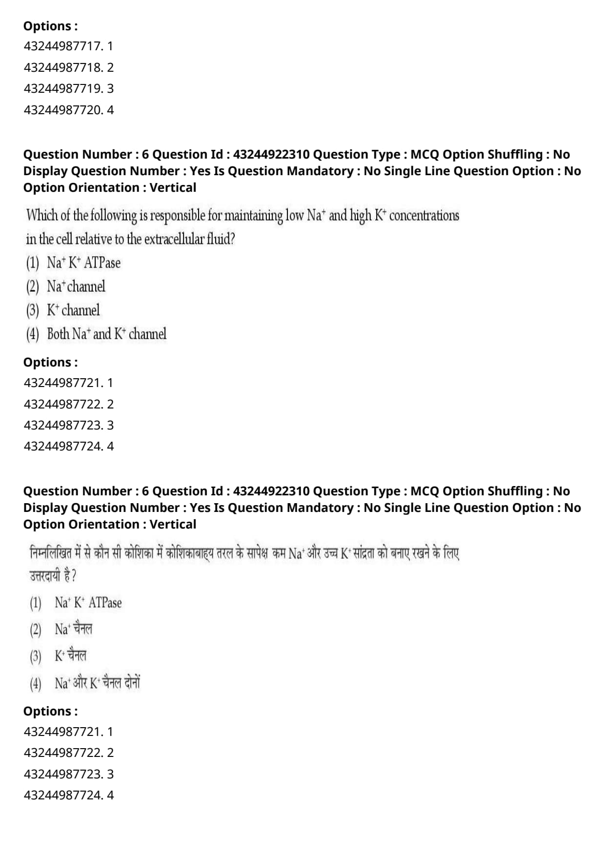
Which of the following is responsible for maintaining low \(Na^+\) and high \(K^+\) concentrations in the cell relative to the extracellular fluid?
View Solution
Step 1: Understanding the Question:
This question asks for the primary membrane transport mechanism responsible for creating and maintaining the steep chemical gradients of sodium (\(Na^+\)) and potassium (\(K^+\)) across the cell membrane of animal cells.
Step 2: Detailed Explanation:
In living cells, a highly asymmetrical distribution of ions is maintained between the cytoplasm and the extracellular environment.
- Specifically, the concentration of potassium (\(K^+\)) is kept very high inside the cell relative to the outside, while the concentration of sodium (\(Na^+\)) is kept extremely low inside the cell relative to the outside.
- Mechanism of Maintenance:
- \(Na^+-K^+\) ATPase (Sodium-Potassium Pump): This is an integral membrane protein that functions as a primary active transport pump.
- Utilizing energy derived from the hydrolysis of one ATP molecule to ADP and inorganic phosphate, the pump undergoes structural changes to transport 3 \(Na^+\) ions out of the cell and 2 \(K^+\) ions into the cell.
- Because both ion species are pushed against their respective concentration gradients, this continuous active pumping is the exclusive mechanism responsible for building and sustaining these concentration gradients.
- Role of Ion Channels:
- Sodium and potassium channels are passive leak or gated channels that allow ions to flow down their gradients (facilitated diffusion).
- If left unchecked, channels would dissipate the gradient. Thus, the active pump must continuously oppose the leak through channels to maintain the concentrations.
Step 3: Final Answer:
Therefore, the \(Na^+\ K^+\) ATPase is the correct option, corresponding to option (A).
Quick Tip: To keep a concentration gradient stable (high on one side, low on the other), the cell must actively pump ions uphill.
Uphill transport always requires active pumps (like ATPases), whereas passive channels only let ions flow downhill, which would destroy the gradient.
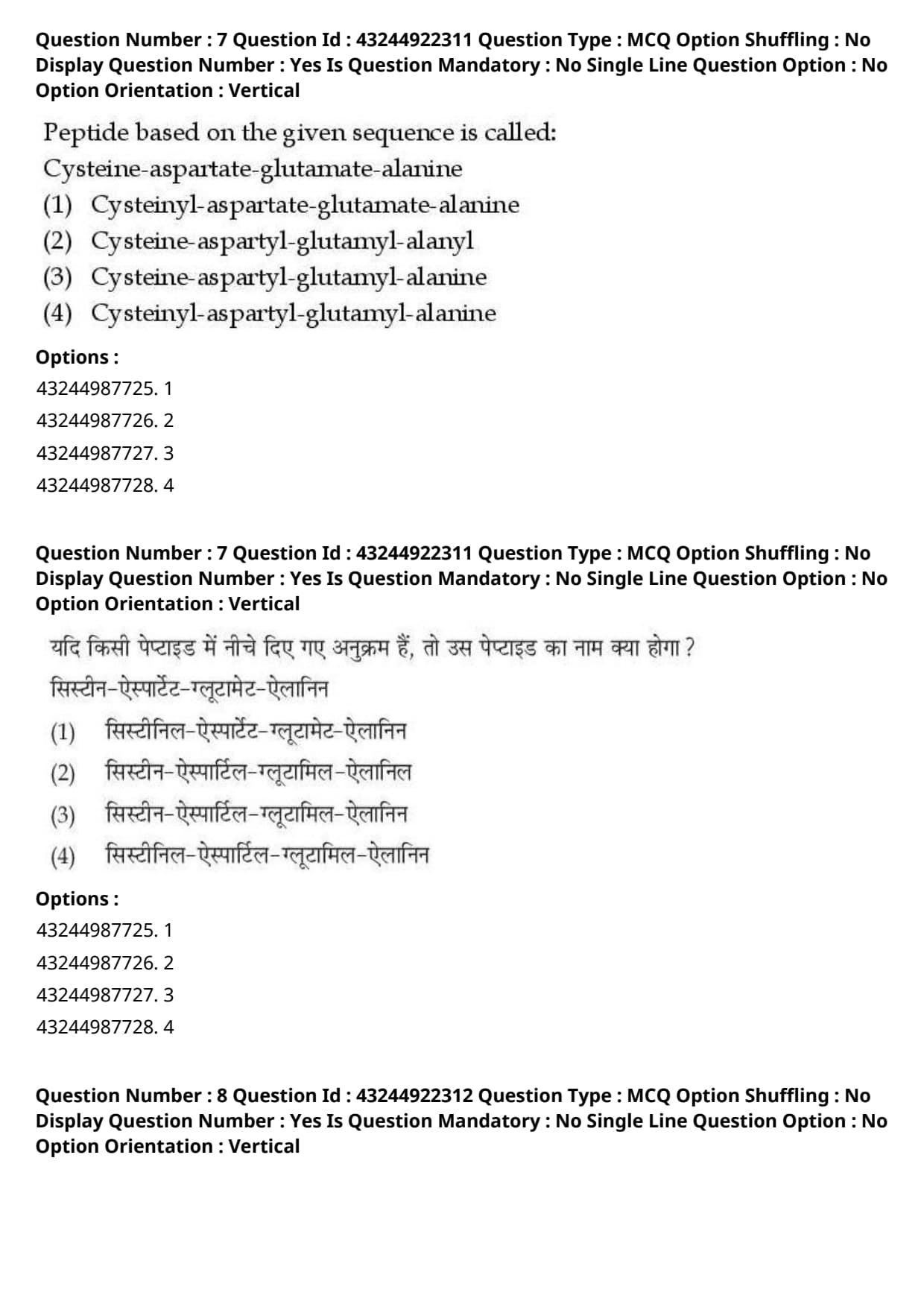
Peptide based on the given sequence is called: Cysteine-aspartate-glutamate-alanine
View Solution
Step 1: Understanding the Question:
The question asks for the formal, systematically correct chemical name of a tetrapeptide sequence made up of Cysteine, Aspartate, Glutamate, and Alanine.
Step 2: Detailed Explanation:
Peptides are formed by linking amino acids through peptide bonds.
According to IUPAC-IUB rules for peptide nomenclature:
1. Direction of Naming: Peptides are named starting from the amino-terminal (N-terminal) amino acid on the left and ending with the carboxyl-terminal (C-terminal) amino acid on the right.
2. Suffix modification: All amino acid residues in the chain except the very last one are named as acyl substituents. Their default endings (such as "-ine" or "-ate") are replaced with the suffix "-yl".
3. The C-terminus: The final, right-most amino acid at the C-terminus retains its standard, unmodified name.
- Let us apply these rules to the given sequence:
- Cysteine (N-terminus) becomes Cysteinyl.
- Aspartate (Middle residue) becomes Aspartyl.
- Glutamate (Middle residue) becomes Glutamyl.
- Alanine (C-terminus) remains alanine.
- Putting these names together yields: Cysteinyl-aspartyl-glutamyl-alanine.
Step 3: Final Answer:
Therefore, the systematically correct name is Cysteinyl-aspartyl-glutamyl-alanine, corresponding to option (D).
Quick Tip: In standard peptide nomenclature, only the very last amino acid at the C-terminus keeps its original name (e.g., "...alanine").
All preceding amino acids must end with "-yl". This simple rule allows you to quickly isolate the correct option in seconds.
Liver exhibits which type of regeneration?
View Solution
Step 1: Understanding the Question:
This question asks us to identify the specific physiological classification of regeneration demonstrated by the mammalian liver during recovery from injury or tissue loss.
Step 2: Detailed Explanation:
Regeneration mechanisms in multicellular animals are broadly categorized into three types:
1. Morphallaxis: Regeneration accomplished by the reorganization or remodeling of remaining tissues with minimal new cell division (classic example is seen in \textit{Hydra).
2. Epimorphosis: Regeneration where a mass of undifferentiated dividing cells (called a blastema) forms at the site of injury to systematically reconstruct the lost limb or organ (classic example is limb regeneration in Salamanders).
3. Compensatory Regeneration: A unique form of tissue restoration where the remaining cells of an organ actively divide to restore the original mass and function of the organ without forming a blastema or regenerating the exact lost physical lobes.
- Regeneration in mammalian Liver:
- When a portion of the liver is surgically excised (partial hepatectomy), the remaining liver lobes do not grow back from the cut margin.
- Instead, the mature, differentiated cells in the remaining lobes (primarily hepatocytes and biliary epithelial cells) exit their resting (\(G_0\)) state and undergo rapid, highly controlled rounds of cell division.
- This hyperplasia continues until the total metabolic mass and volume of the liver are fully restored to baseline.
- Therefore, this is the classic, textbook definition of compensatory regeneration.
Step 3: Final Answer:
Thus, the liver exhibits compensatory regeneration, corresponding to option (D).
Quick Tip: To remember the differences:
- Epimorphosis = New limb grows back from a blastema.
- Compensatory = Remaining cells multiply to compensate for the lost volume (like the liver), but no new lobes/structures are formed at the cut site.
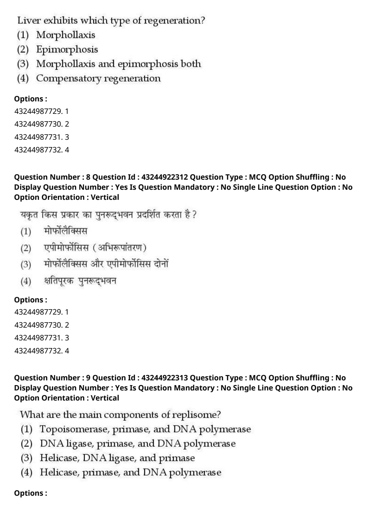
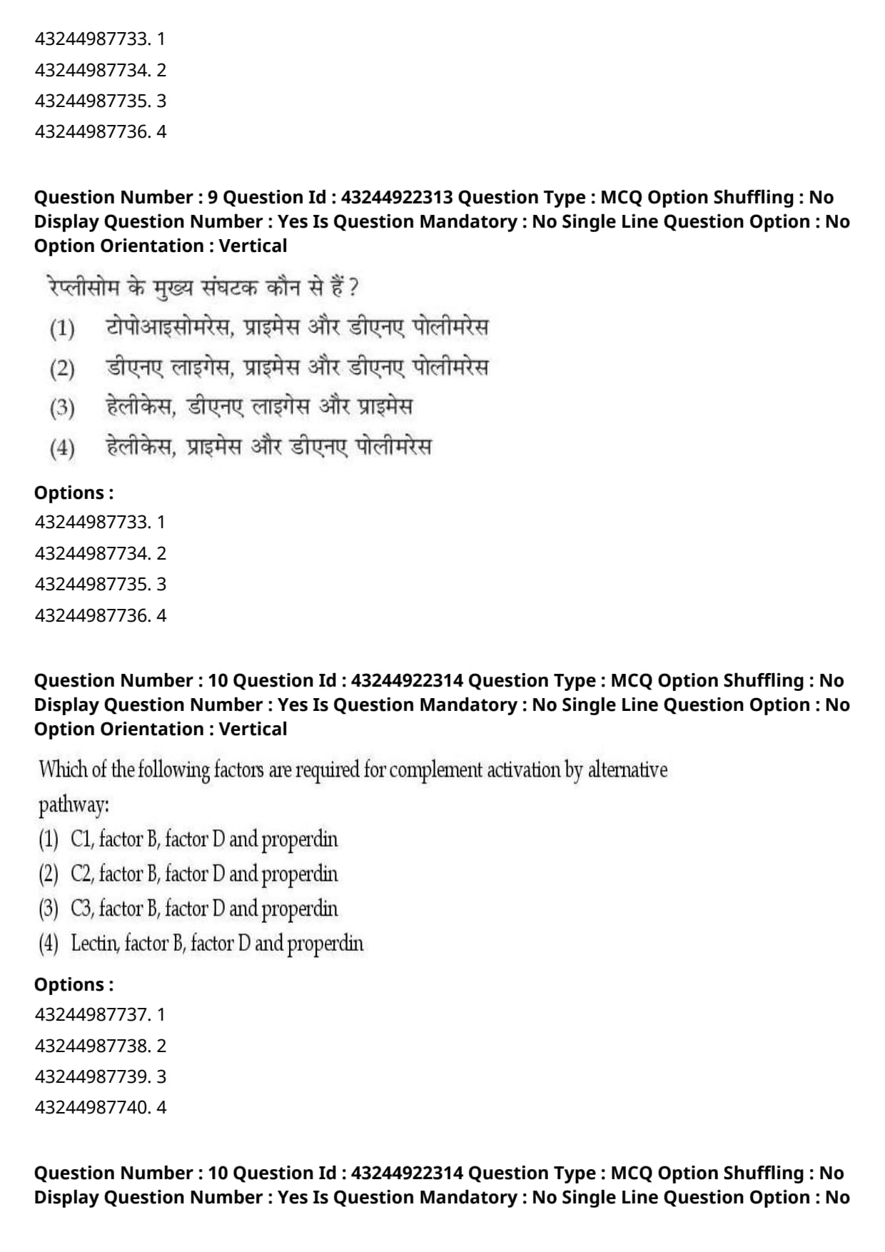
What are the main components of replisome?
View Solution
Step 1: Understanding the Question:
The question asks for the essential, core enzymatic components that form the active molecular machinery (replisome) operating directly at the DNA replication fork.
Step 2: Detailed Explanation:
The replisome is a highly organized, multiprotein complex that carries out DNA replication by coordinating the synthesis of both the leading and lagging strands simultaneously.
- To successfully replicate DNA at a moving replication fork, three primary enzymatic activities must cooperate seamlessly:
1. Helicase: This enzyme acts as the unwinding motor, consuming ATP to break hydrogen bonds between complementary base pairs and separate the double-stranded DNA into single templates.
2. Primase: Because DNA polymerases are absolutely incapable of starting a DNA chain de novo, primase (an RNA polymerase) must synthesize a short RNA primer to provide the free \(3'\)-OH group.
3. DNA Polymerase: This is the main catalytic synthesis engine that adds complementary deoxyribonucleotides to the \(3'\)-OH end of the growing strand.
- Other auxiliary enzymes like Topoisomerase (which relieves supercoiling strain ahead of the fork) and DNA Ligase (which joins Okazaki fragments later) are necessary for the completion of replication, but they do not form the core moving engine of the active replisome complex.
- Hence, the three central, universally accepted components of the replisome are Helicase, Primase, and DNA Polymerase.
Step 3: Final Answer:
Therefore, the correct combination of core enzymes is Helicase, primase, and DNA polymerase, corresponding to option (D).
Quick Tip: To remember the core replisome machinery, think of the absolute minimum tools needed to run a replication fork:
1. Open the DNA zip (Helicase)
2. Mark the starting point (Primase)
3. Build the new sequence (DNA Polymerase)
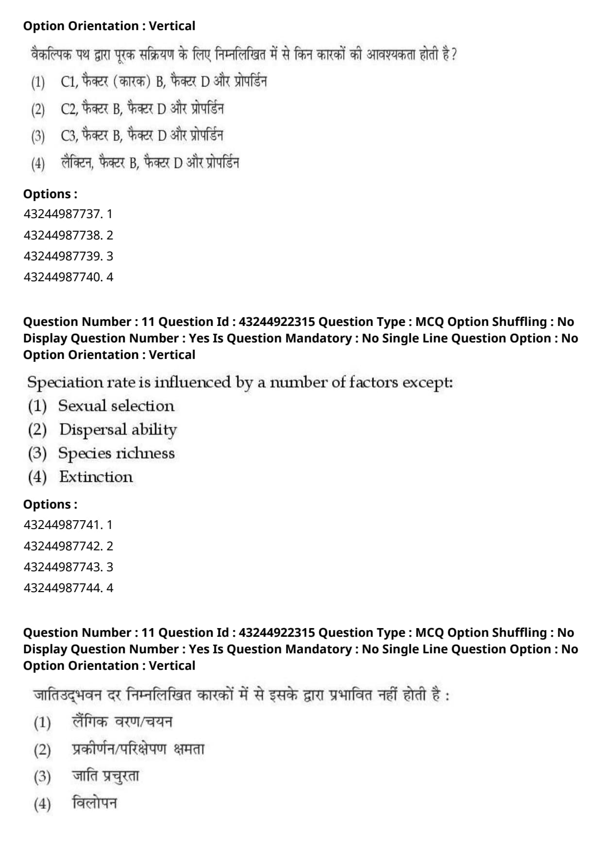
Which of the following factors are required for complement activation by alternative pathway?
View Solution
Step 1: Understanding the Question:
The question asks for the specific set of immune proteins and plasma factors required to initiate and stabilize the alternative pathway of the complement system.
Step 2: Detailed Explanation:
The complement system is a key element of innate immunity consisting of over 30 soluble plasma proteins. It is activated via three distinct biochemical pathways: Classical, Lectin, and Alternative.
- Classical Pathway: Initiated by C1 binding to antibody-antigen complexes. It requires C1, C4, and C2.
- Lectin Pathway: Initiated by mannose-binding lectin binding to microbial surface carbohydrates. It requires MBL, MASP, C4, and C2.
- Alternative Pathway: This pathway is antibody-independent and operates via a continuous low-level activation mechanism. It relies on the following key components:
1. C3: Spontaneously hydrolyzes at a low rate (tick-over) to form \(C3(H_2O)\) or is cleaved to C3b, which deposits directly onto foreign cell surfaces.
2. Factor B: Binds to surface-bound C3b.
3. Factor D: A plasma serine protease that cleaves the bound Factor B into Ba and Bb, leaving the active C3bBb complex (the alternative C3 convertase).
4. Properdin (Factor P): An essential regulatory protein that binds to and stabilizes the highly unstable C3bBb convertase complex on microbial membranes, extending its half-life.
- Since the alternative pathway completely bypasses C1, C2, C4, and Lectins, the key factors required are C3, Factor B, Factor D, and Properdin.
Step 3: Final Answer:
Therefore, the correct option is (C).
Quick Tip: An easy way to distinguish the complement pathways:
- Classical and Lectin pathways rely on C1, C2, and C4.
- The Alternative pathway completely bypasses these and relies on C3, Factor B, Factor D, and Properdin.
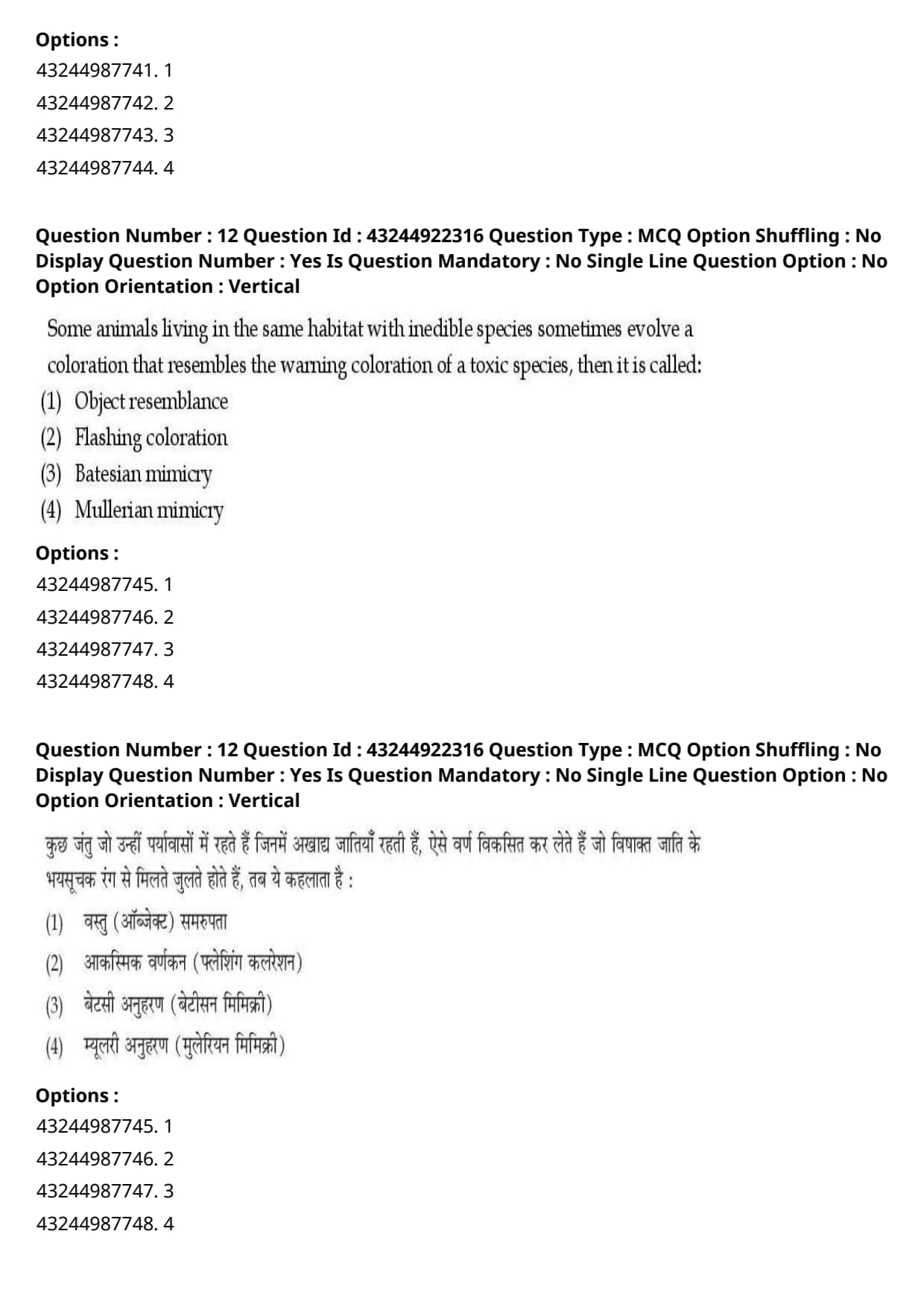
Speciation rate is influenced by a number of factors except:
View Solution
Step 1: Understanding the Question:
The question asks us to identify which of the biological or ecological processes listed does not directly govern or influence the rate at which new species are formed (speciation rate).
Step 2: Detailed Explanation:
Speciation rate is the frequency or velocity with which ancestral biological lineages split to form distinct, reproductively isolated descendant species.
- Let us analyze how the different factors influence this rate:
1. Sexual Selection: Strong sexual selection (such as female preference for specific male ornamental traits or behaviors) drives rapid pre-zygotic reproductive isolation and genetic divergence, which directly increases the rate of speciation.
2. Dispersal Ability: The physical capacity of individuals to travel across distances determines gene flow. High dispersal capability maintains gene flow and prevents divergence (lowering speciation rate), while low dispersal capability promotes rapid geographical isolation and allopatric divergence (increasing speciation rate).
3. Species Richness: High species richness in an ecosystem creates complex ecological competition and niche subdivision, which can drive further adaptive radiation and speciation.
4. Extinction:
- Extinction represents the complete loss or termination of an existing species.
- Although extinction rates and speciation rates together determine the net \textit{diversification rate of a clade (Diversification = Speciation - Extinction), the process of extinction itself does not directly drive or determine the physical and genetic mechanisms that cause new species to arise.
- Therefore, extinction is a separate demographic process and is the correct exception.
Step 3: Final Answer:
Hence, extinction is the correct exception, corresponding to option (D).
Quick Tip: Be careful to distinguish between the rate of species creation (speciation rate) and the net balance of species in existence (diversification rate).
Extinction acts as the loss component of diversity, but it does not directly determine the genetic speed at which new species form.
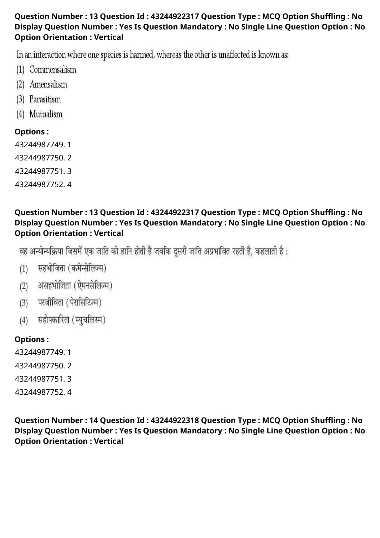
Some animals living in the same habitat with inedible species sometimes evolve a coloration that resembles the warning coloration of a toxic species, then it is called:
View Solution
Step 1: Understanding the Question:
This question describes a scenario where an edible animal living alongside an inedible species evolves to copy the warning coloration of a toxic model, and asks us to identify the name of this ecological phenomenon.
Step 2: Detailed Explanation:
Mimicry is an evolutionary adaptation where one species (the mimic) closely resembles another species (the model) to deceive predators and increase its chances of survival.
- Let us compare the two major types of mimicry:
- Batesian Mimicry:
- Discovered by Henry Walter Bates, this occurs when a harmless, edible (palatable) species evolves warning coloration (aposematism) that resembles a dangerous, inedible (unpalatable/toxic) species.
- Predators that have learned to avoid the toxic model will also avoid the harmless mimic because of their identical visual appearance, offering the mimic strong protection.
- The question states that some animals resemble the warning coloration of a toxic species, which describes a harmless mimic taking advantage of a toxic model's warning signal. This is the classic definition of Batesian mimicry.
- Mullerian Mimicry:
- Discovered by Fritz Müller, this occurs when two or more distinct unpalatable (toxic/inedible) species evolve similar warning coloration. This benefits both species by sharing the cost of predator training.
- Since the mimicking animal in the question is not stated to be toxic itself but is mimicking a toxic species, it represents Batesian mimicry.
Step 3: Final Answer:
Therefore, the correct option is (C).
Quick Tip: To quickly remember the difference between the two main types of mimicry:
- Batesian = One harmless liar (mimic) + One toxic truth-teller (model).
- Mullerian = Two toxic species telling the same honest truth to share the burden of teaching predators.
In an interaction where one species is harmed, whereas the other is unaffected is known as:
View Solution
Step 1: Understanding the Question:
This question asks for the standard ecological term used to describe an interspecific interaction where one species is negatively affected (harmed) while the other species remains completely unaffected.
Step 2: Detailed Explanation:
Interspecific interactions in an ecosystem are classified based on the direct impact they have on the participating species using signs: benefit (\(+\)), harm (\(-\)), or neutral/unaffected (\(0\)).
- Let us analyze each of the provided options:
1. Commensalism (\(+, 0\)): One species benefits, while the other is unaffected (e.g., an orchid growing on a large tree branch).
2. Amensalism (\(-, 0\)): One species is inhibited or harmed, while the other species remains completely unaffected.
- A classic example is the mold \textit{Penicillium secreting penicillin which kills nearby bacteria; the mold is unaffected, but the bacteria are destroyed.
- Another example is a large, tall tree shading out small seedlings growing on the forest floor, where the tall tree is unaffected but the seedlings are starved of light.
3. Parasitism (\(+, -\)): One species (parasite) benefits at the expense of the other (host), which is harmed.
4. Mutualism (\(+, +\)): Both species benefit from the interaction (e.g., mycorrhizal fungi and plant roots).
- Because the question describes a (\(-, 0\)) interaction, it perfectly defines Amensalism.
Step 3: Final Answer:
Therefore, the correct option is (B).
Quick Tip: To easily recall these signs in the exam, write them down:
- Commensalism = \(+\) / \(0\)
- Amensalism = \(-\) / \(0\)
The prefix "a-" in amensalism indicates "asymmetric harm" where one side suffers and the other remains neutral.
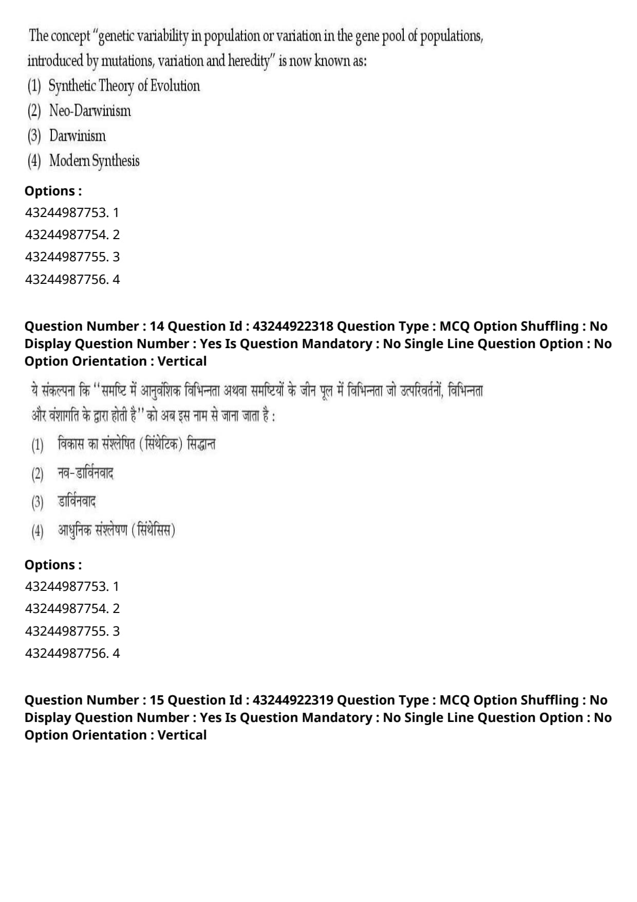
The concept "genetic variability in population or variation in the gene pool of populations, introduced by mutations, variation and heredity" is now known as:
View Solution
Step 1: Understanding the Question:
The question asks for the standard, globally recognized scientific term for the unified theory of evolution that incorporates Mendelian genetics, mutation, and population genetics into Darwin's original theory of natural selection.
Step 2: Detailed Explanation:
Evolutionary biology has evolved through several historical stages:
- Darwinism: Charles Darwin's original 1859 theory focused on natural selection acting on random somatic variations. However, Darwin did not understand the physical mechanism of heredity or how variation arose in the first place.
- Neo-Darwinism: Coined in the late 19th century to denote Darwin's theory as modified by Alfred Russel Wallace and August Weismann, specifically incorporating the complete rejection of Lamarckian inheritance of acquired traits.
- Modern Synthesis: Coined by Julian Huxley in 1942, this represents the grand unification of biology. It successfully integrated Mendelian genetics, Darwin's natural selection, chromosomal theory, and population genetics (formulated by Fisher, Haldane, and Wright).
- It describes how mutations, chromosomal recombination, and heredity continuously introduce genetic variability into a population's gene pool, which is then acted upon by natural selection and genetic drift over generations.
- While "Synthetic Theory of Evolution" is a common synonym, "Modern Synthesis" is the historically accurate and globally accepted term for this paradigm.
Step 3: Final Answer:
Therefore, the most appropriate term is Modern Synthesis, corresponding to option (D).
Quick Tip: "Modern Synthesis" is the official name given to the marriage of genetics (Mendel) and natural selection (Darwin) that occurred in the 1940s.
If both "Modern Synthesis" and "Synthetic Theory" are listed, always select "Modern Synthesis" as it is the primary historical term coined by Julian Huxley.
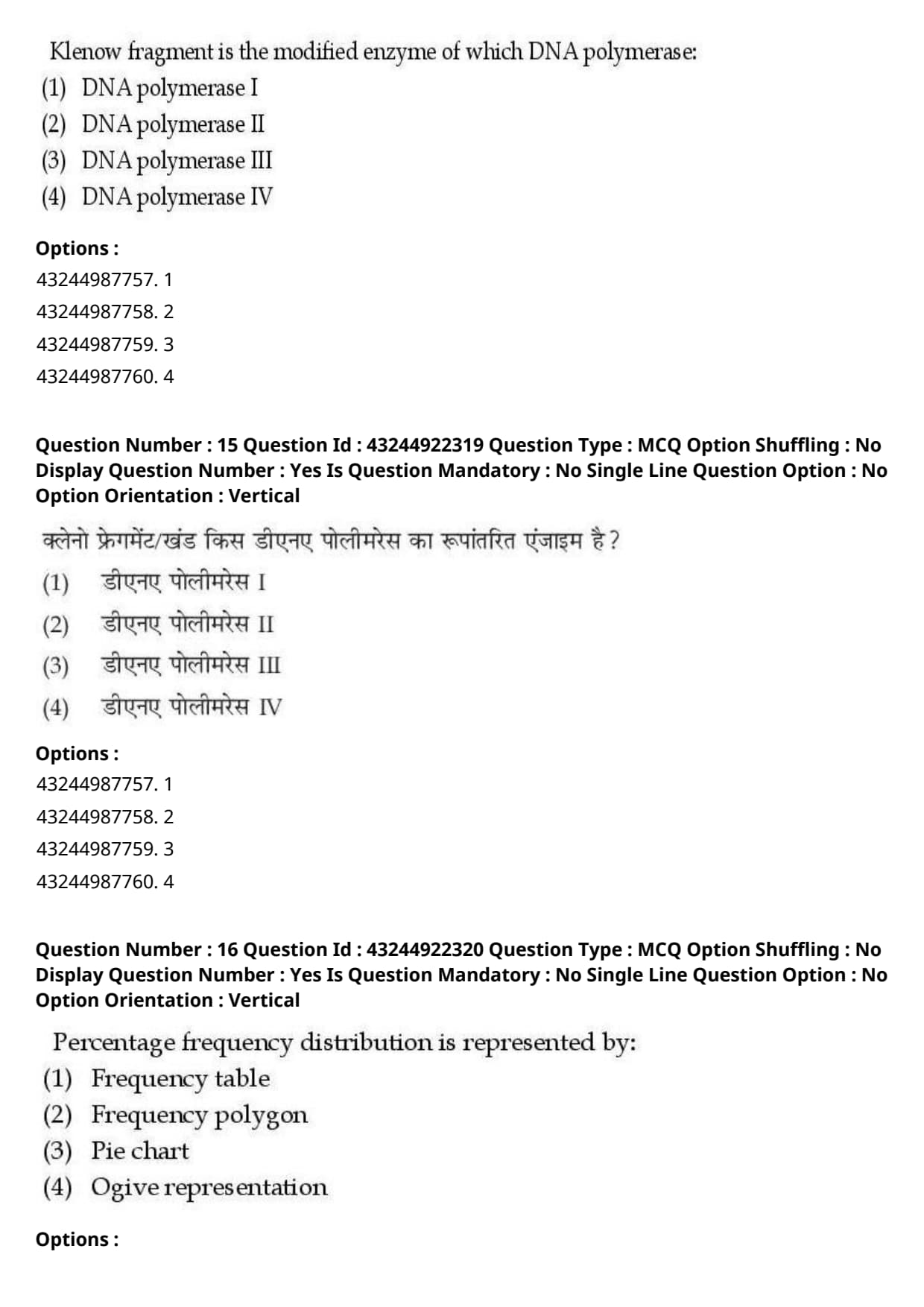
Klenow fragment is the modified enzyme of which DNA polymerase:
View Solution
Step 1: Understanding the Question:
This question asks us to identify the original parental enzyme from which the Klenow fragment is derived via enzymatic cleavage.
Step 2: Detailed Explanation:
The Klenow fragment is a large protein fragment widely used in molecular biology and recombinant DNA technology.
- Origin of the Klenow Fragment:
- \textit{Escherichia coli DNA polymerase I is a single polypeptide chain with three distinct enzymatic activities localized in separate structural domains:
1. \(5' \rightarrow 3'\) polymerase activity.
2. \(3' \rightarrow 5'\) exonuclease (proofreading) activity.
3. \(5' \rightarrow 3'\) exonuclease activity (which removes RNA primers during replication).
- Modification Process:
- When DNA polymerase I is treated with the protease subtilisin, it is cleaved into two fragments:
- A small fragment containing the \(5' \rightarrow 3'\) exonuclease activity.
- A large fragment containing both the \(5' \rightarrow 3'\) polymerase and \(3' \rightarrow 5'\) exonuclease activities, which is known as the Klenow fragment.
- This modified enzyme is highly useful because it can synthesize new DNA strands without degrading existing DNA primers from their \(5'\) end.
Step 3: Final Answer:
Therefore, the Klenow fragment is derived from DNA polymerase I, corresponding to option (A).
Quick Tip: Remember:
- DNA Polymerase I = 3 activities.
- Klenow Fragment = Polymerase I minus \(5' \rightarrow 3'\) exonuclease activity (leaving only 2 activities).
Percentage frequency distribution is represented by:
View Solution
Step 1: Understanding the Question:
The question asks us to identify the most suitable visual or graphical method used to represent a percentage frequency distribution in statistics.
Step 2: Detailed Explanation:
A percentage frequency distribution displays the frequency of each category or class interval as a percentage of the total dataset.
- The sum of all individual percentage frequencies must equal exactly 100%.
- Let us evaluate each of the options:
1. Frequency Table: This is a tabular arrangement of data, not a graphical representation, and typically presents raw values rather than visual proportions.
2. Frequency Polygon: Used primarily to plot continuous, quantitative variables where midpoints of class intervals are plotted against frequencies.
3. Pie Chart (Circular Diagram): A circular diagram divided into sectors (slices). The central angle of each sector is directly proportional to the percentage of the whole it represents.
- Because a pie chart represents a single, complete whole (100%) and clearly communicates how this total is divided among different categories, it is the classic and most effective visual representation of a percentage frequency distribution for discrete or categorical data.
4. Ogive: A cumulative frequency polygon used to determine medians and percentiles of continuous data, not percentage frequencies of separate classes.
Step 3: Final Answer:
Hence, the pie chart is the correct option, corresponding to option (C).
Quick Tip: Think of the phrase "percentage of a whole".
Whenever you need to visualize how 100% of any quantity is divided among various categories, a Pie Chart is always the best and most standard choice in biostatistics.
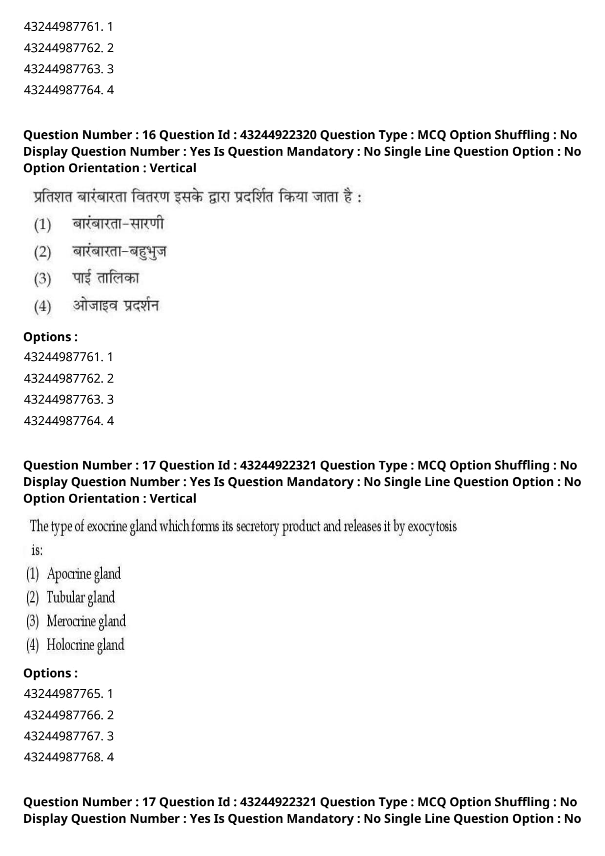
The type of exocrine gland which forms its secretory product and releases it by exocytosis is:
View Solution
Step 1: Understanding the Question:
This question asks us to classify exocrine glands categorized by their mechanism of secretion, specifically selecting the one that releases its secretory product via simple exocytosis.
Step 2: Detailed Explanation:
Exocrine glands are functionally classified into three major groups based on how their secretory cells release their products:
1. Merocrine (or Eccrine) Glands:
- The secretory products are synthesized in ribosomes, processed in the Golgi apparatus, packaged into secretory vesicles, and released from the apical surface of the cell via classic exocytosis.
- No part of the secretory cell is damaged, lost, or pinched off in this process.
- Examples include salivary glands and the exocrine pancreas.
2. Apocrine Glands:
- The secretory product accumulates at the apical tip of the cell. This entire apical portion of the cytoplasm is then pinched off to release the secretion, resulting in minor cell loss.
- Examples include mammary glands.
3. Holocrine Glands:
- The secretory product accumulates within the cell until the entire cell ruptures and dies, becoming the secretion itself. Continuous cell division is required to replace the lost cells.
- Examples include sebaceous (oil) glands of the skin.
- "Tubular gland" is an anatomical classification based on shape, not on the physiological mechanism of secretion.
Step 3: Final Answer:
Therefore, the correct mode of secretion by exocytosis is merocrine, corresponding to option (C).
Quick Tip: To easily remember the secretory modes:
- Merocrine = "Merely" exocytosis (no cell damage/loss).
- Apocrine = "A piece" of the apex pinches off.
- Holocrine = "Whole" cell lyses/destroys.
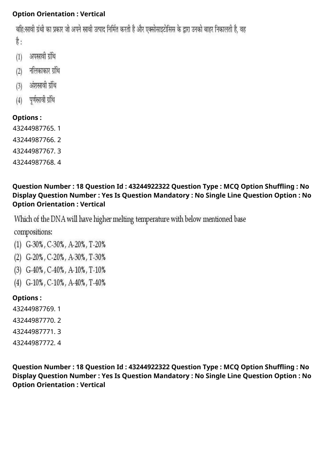
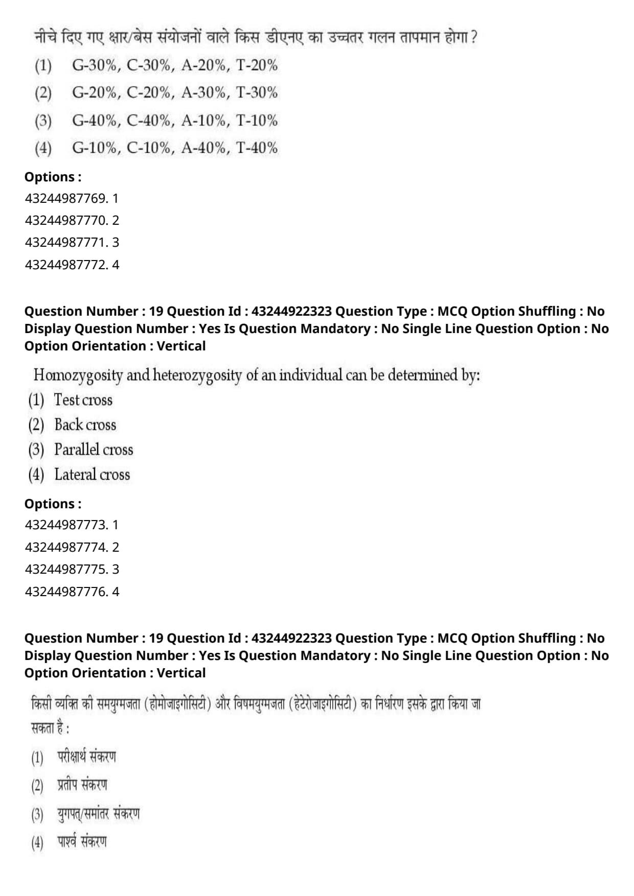
Which of the DNA will have higher melting temperature with below mentioned base compositions:
View Solution
Step 1: Understanding the Question:
This question asks us to determine which of the given DNA base compositions will result in the highest melting temperature (\(T_m\)).
Step 2: Key Formula or Approach:
The melting temperature (\(T_m\)) of a DNA double helix is directly proportional to its Guanine-Cytosine (GC) content:
\[ T_m \propto %(G + C) \]
This relationship exists because G-C base pairs are held together by three hydrogen bonds, whereas A-T base pairs are held together by only two. Additionally, G-C base stacking interactions provide greater thermodynamic stability than A-T stacking.
Step 3: Detailed Explanation:
Denaturation (melting) of DNA involves separating the double helix into two single strands by breaking hydrogen bonds and base-stacking forces.
- A higher percentage of G-C base pairs in a DNA molecule means more thermal energy (a higher temperature) is required to fully dissociate the two strands.
- Let us calculate the total GC content for each of the options:
- Option (A): \(G = 30%\), \(C = 30%\) \(\rightarrow\) Total \(GC = 60%\).
- Option (B): \(G = 20%\), \(C = 20%\) \(\rightarrow\) Total \(GC = 40%\).
- Option (C): \(G = 40%\), \(C = 40%\) \(\rightarrow\) Total \(GC = 80%\).
- Option (D): \(G = 10%\), \(C = 10%\) \(\rightarrow\) Total \(GC = 20%\).
- Option (C) has the highest GC content (\(80%\)), giving it the greatest structural stability and the highest melting temperature.
Step 4: Final Answer:
Therefore, the DNA with the base composition in option (C) will have the highest melting temperature.
Quick Tip: To find the DNA with the highest melting temperature (\(T_m\)), simply add the percentage of G and C.
The option with the highest sum of G + C will always have the highest melting temperature.
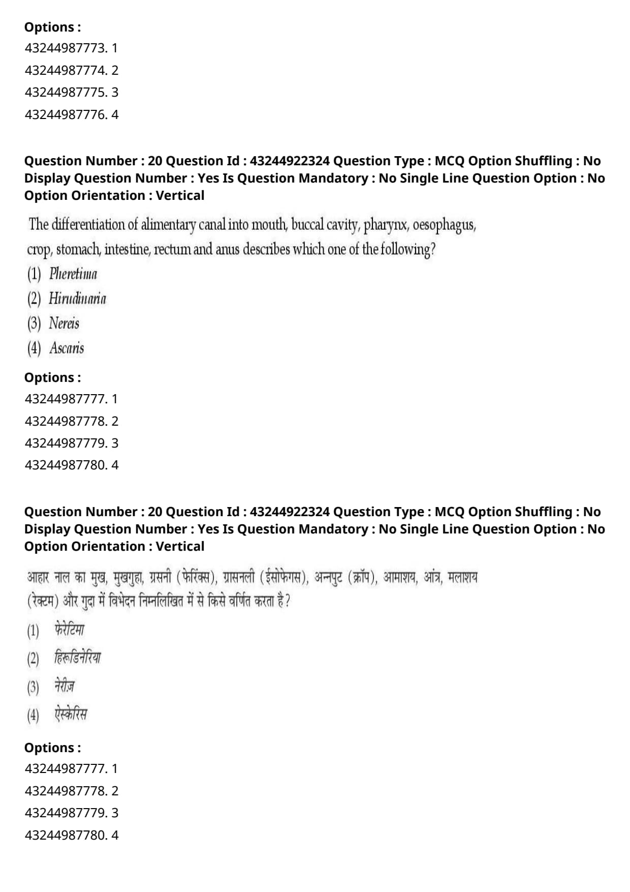
Homozygosity and heterozygosity of an individual can be determined by:
View Solution
Step 1: Understanding the Question:
The question asks us to identify the specific type of genetic cross used to determine whether an organism expressing a dominant phenotype is homozygous dominant or heterozygous.
Step 2: Detailed Explanation:
An individual displaying a dominant phenotype could possess either of two genotypes: homozygous dominant (e.g., \(TT\)) or heterozygous (e.g., \(Tt\)). Phenotypically, these individuals look identical.
- To determine the exact genotype, the individual of unknown genotype is crossed with an organism that is homozygous recessive for the same trait (e.g., \(tt\)). This genetic cross is known as a test cross.
- Let us look at the two possible genetic outcomes of this cross:
- Case 1: If the test individual is Homozygous Dominant (\(TT\)):
- Cross: \(TT \times tt\)
- Offspring: All (\(100%\)) offspring will be heterozygous (\(Tt\)) and display the dominant phenotype.
- Case 2: If the test individual is Heterozygous (\(Tt\)):
- Cross: \(Tt \times tt\)
- Offspring: \(50%\) will be heterozygous (\(Tt\), dominant) and \(50%\) will be homozygous recessive (\(tt\), recessive). The resulting phenotypic ratio is \(1:1\).
- By observing the phenotypes of the offspring, the zygosity of the dominant parent is clearly determined.
- A back cross involves crossing an \(F_1\) hybrid back with either of its parental genotypes (dominant or recessive). A test cross is a specific, diagnostic type of back cross performed with the recessive parent.
Step 3: Final Answer:
Therefore, the correct option is (A).
Quick Tip: Remember Mendel's classical genetic rules:
To test the hidden genotype of an individual showing a dominant phenotype, cross it with a homozygous recessive partner. This diagnostic cross is always called a Test Cross.
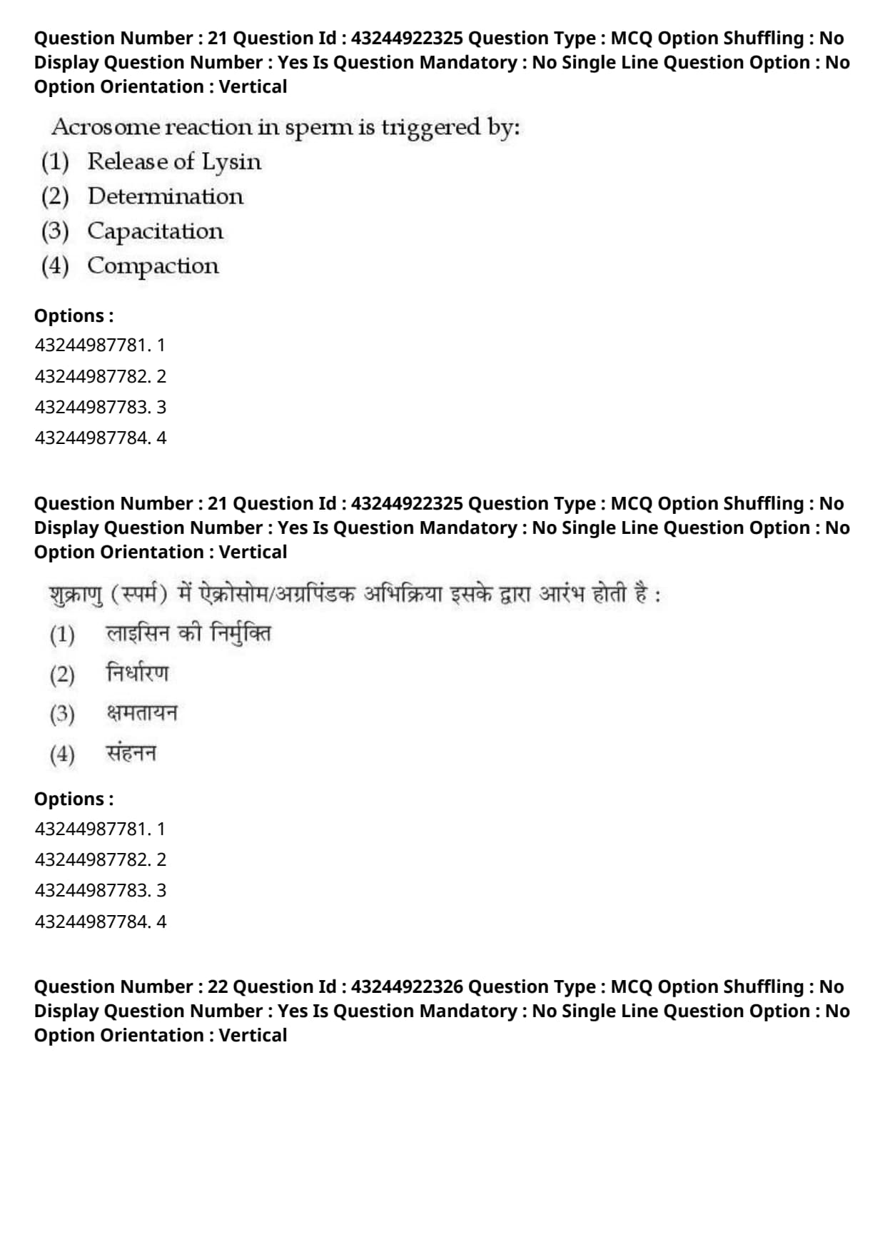
The differentiation of alimentary canal into mouth, buccal cavity, pharynx, oesophagus, crop, stomach, intestine, rectum and anus describes which one of the following?
View Solution
Step 1: Understanding the Question:
The question asks us to identify which of the given invertebrate genera has an alimentary canal anatomically differentiated into the exact structural sequence: mouth, buccal cavity, pharynx, oesophagus, crop, stomach, intestine, rectum, and anus.
Step 2: Detailed Explanation:
Let us review the digestive tract anatomy of each of the options provided:
1. Pheretima (Earthworm):
- The digestive tract of the earthworm consists of a mouth, buccal cavity, pharynx, oesophagus, gizzard (a highly muscular grinding organ), stomach, intestine, and anus. It does not have a "crop" in this sequence.
2. Ascaris (Roundworm):
- Possesses a very simple, straight, non-muscular digestive tube consisting of a mouth, muscular pharynx, a straight, unspecialized intestine, and a rectum ending in the anus. It lacks any division into crop or stomach.
3. Hirudinaria (Indian Cattle Leech):
- The digestive tract of the leech is highly specialized for a blood-feeding (sanguivorous) diet. It is a straight tube running from the mouth to the anus, divided into:
- Mouth and Buccal cavity (equipped with three muscular jaws and salivary glands secreting the anticoagulant hirudin).
- Pharynx (a muscular pumping organ).
- Oesophagus (a short, narrow connecting tube).
- Crop: The largest part of the digestive tract, divided into 10 chambers (from segments 9 to 18), each with a pair of lateral caeca to store large volumes of blood.
- Stomach: A small, heart-shaped chamber in segment 19 where slow digestion takes place.
- Intestine: A narrow, straight tube extending from segment 20 to 22.
- Rectum: A short, thin-walled chamber (segments 23 to 26).
- Anus: A small aperture opening dorsally on the 26th segment.
- This matches the exact sequence of structures described in the question.
Step 3: Final Answer:
Therefore, the described digestive tract belongs to \textit{Hirudinaria, corresponding to option (B).
Quick Tip: To differentiate between these common annelids:
- Leech (\textit{Hirudinaria) = Highly famous for its massive, multi-chambered blood-storing \textbf{Crop}, followed by a stomach.
- Earthworm (Pheretima) = Famous for its highly muscular food-grinding \textbf{Gizzard}.
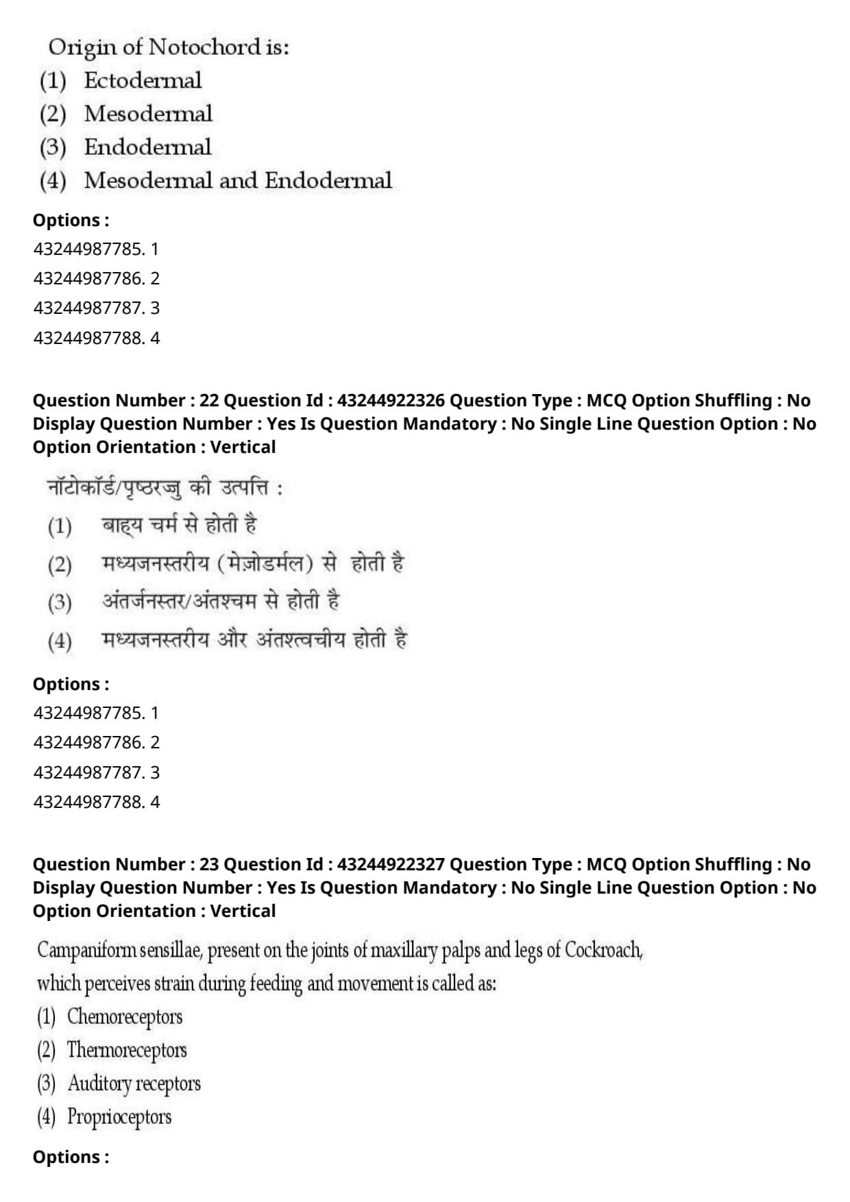
Acrosome reaction in sperm is triggered by:
View Solution
Step 1: Understanding the Question:
The question asks for the biological process that primes and triggers the sperm to undergo the acrosome reaction required for fertilizing an egg.
Step 2: Detailed Explanation:
The acrosome reaction is a specialized exocytotic event in which the acrosomal vesicle of the sperm fuses with its plasma membrane to release hydrolytic enzymes.
- Role of Capacitation:
- Freshly ejaculated mammalian sperm are physiologically incapable of undergoing the acrosome reaction or fertilizing an ovum.
- During transit through the female reproductive tract, sperm undergo a series of biochemical and physiological changes collectively termed capacitation.
- Capacitation involves the removal of inhibitory glycoproteins and cholesterol from the sperm plasma membrane, hyperpolarization of the membrane potential, and an influx of extracellular calcium (\(Ca^{2+}\)) ions.
- This priming process enables hyperactivated motility and triggers the sperm's readiness to undergo the acrosome reaction upon binding to the zona pellucida protein (specifically ZP3 in mammals).
- Evaluation of Other Options:
- Release of Lysin: Acrosomal lysins (such as hyaluronidase and acrosin) are released \textit{as a result of the acrosome reaction, not the trigger for it.
- Determination and Compaction are early developmental processes occurring in the embryonic blastomeres, unrelated to sperm physiology.
Step 3: Final Answer:
Therefore, capacitation is the physiological trigger and prerequisite for the acrosome reaction, corresponding to option (C).
Quick Tip: Remember the sequence of events during fertilization:
Ejaculation \(\rightarrow\) Capacitation (priming) \(\rightarrow\) Zona Pellucida Binding \(\rightarrow\) Acrosome Reaction \(\rightarrow\) Lysin Release \(\rightarrow\) Sperm-Egg Fusion.
Origin of Notochord is:
View Solution
Step 1: Understanding the Question:
This question asks us to identify the primary germ layer from which the notochord originates during embryonic development.
Step 2: Detailed Explanation:
The notochord is a defining chordate feature consisting of a flexible, rod-like structure that defines the primary longitudinal axis of the embryo.
- Embryonic Origin and Gastrulation:
- During gastrulation, cells invaginate through the dorsal lip of the blastopore (in amphibians) or the primitive node/Hensen's node (in birds and mammals).
- A specific population of progenitor cells, termed the chordamesoderm, migrates anteriorly along the midline beneath the overlying ectoderm.
- The chordamesoderm explicitly represents a specialized region of the mesoderm.
- This mesodermal rod condenses to form the definitive notochord, which subsequently signals the overlying ectoderm to induce the formation of the neural tube (neurulation).
- Neither the ectoderm nor the endoderm forms the structural core of the notochord.
Step 3: Final Answer:
Hence, the origin of the notochord is strictly mesodermal, corresponding to option (B).
Quick Tip: Keep germ layer fates clear in mind:
- Ectoderm \(\rightarrow\) Epidermis, Nervous System.
- Mesoderm \(\rightarrow\) Notochord, Muscle, Skeleton, Blood, Kidneys.
- Endoderm \(\rightarrow\) Gut lining, Respiratory tract lining, Liver, Pancreas.
Campaniform sensillae, present on the joints of maxillary palps and legs of Cockroach, which perceives strain during feeding and movement is called as:
View Solution
Step 1: Understanding the Question:
The question asks for the functional classification of campaniform sensilla located on the appendages of a cockroach based on their role in sensing mechanical strain.
Step 2: Detailed Explanation:
Insects possess a wide variety of specialized sensory receptors (sensilla) embedded in their exoskeleton to monitor internal and external environmental changes.
- Campaniform Sensilla Anatomy and Function:
- Campaniform sensilla are dome-shaped mechanoreceptors embedded in the flexible cuticle at areas subject to high mechanical stress, such as the joints of legs, wings, and maxillary palps.
- When the insect moves, feeds, or experiences body weight changes, bending forces cause micro-deformations in the cuticular dome.
- This mechanical stress deforms the underlying sensory neurite, generating nerve impulses that inform the central nervous system about cuticle strain, body position, and load.
- Receptors that monitor self-movement, internal muscle tension, and joint position are functionally defined as proprioceptors.
- Evaluation of Other Options:
- Chemoreceptors sense chemical molecules (taste/smell).
- Thermoreceptors sense temperature variations.
- Auditory receptors perceive sound vibrations.
Step 3: Final Answer:
Therefore, campaniform sensilla act as mechanical proprioceptors, corresponding to option (D).
Quick Tip: Whenever a question mentions sensing "strain," "flexion," "body position," or "joint movement," the receptor type is always a Proprioceptor (mechanoreceptor for self-body awareness).
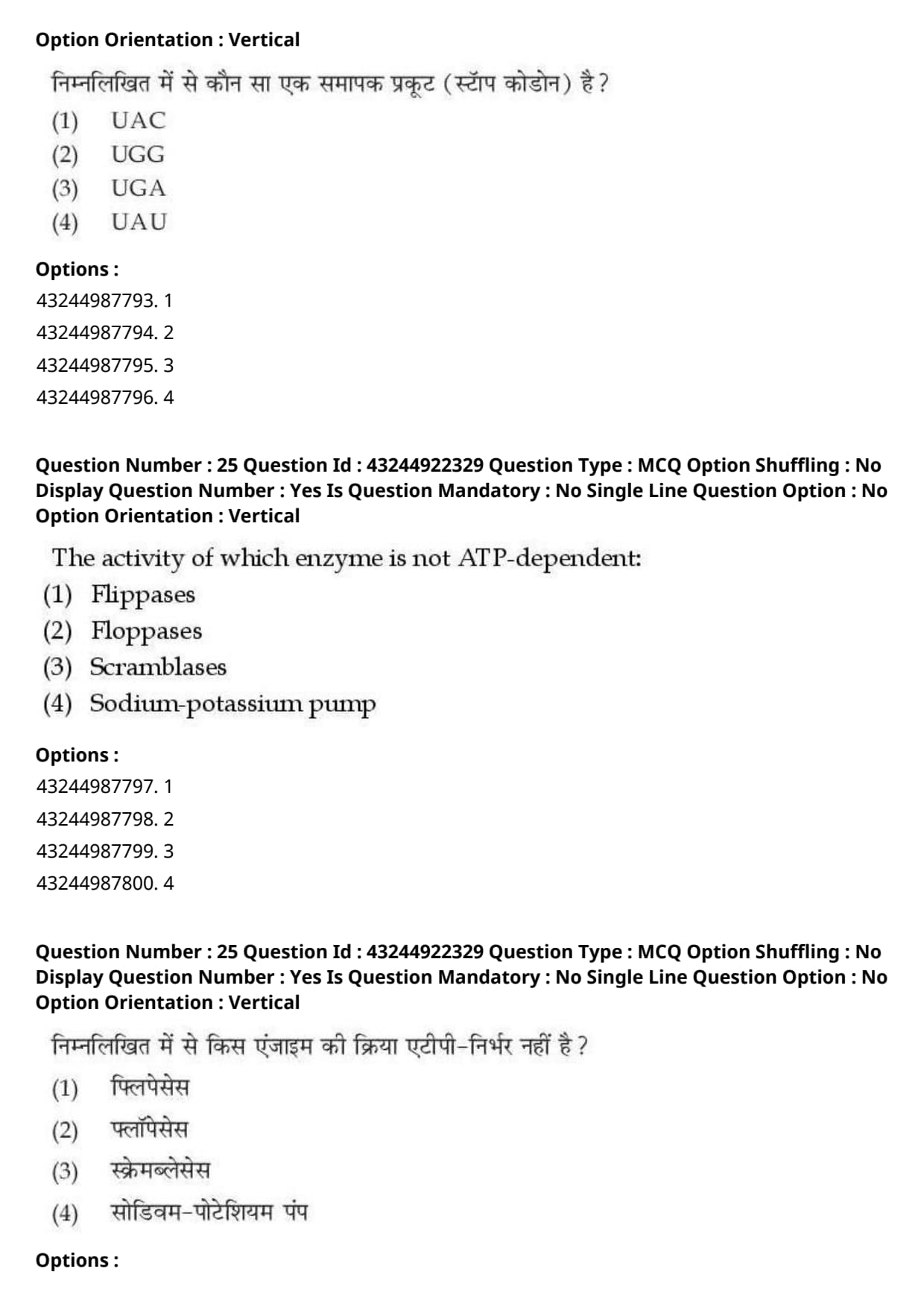
Which of the following is a stop codon:
View Solution
Step 1: Understanding the Question:
This question asks us to identify which of the given trinucleotide mRNA sequences serves as a translation termination signal (stop codon) during protein synthesis.
Step 2: Detailed Explanation:
The genetic code consists of 64 triplet codons written in mRNA.
- Out of these 64 codons, 61 code for specific amino acids, while 3 codons do not code for any amino acid and instead signal the termination of polypeptide chain translation.
- The Three Stop Codons (Nonsense Codons):
1. UAA (traditionally named Ochre)
2. UAG (traditionally named Amber)
3. UGA (traditionally named Opal)
- When a ribosome encounters any of these three codons in the A site during translation, no aminoacyl-tRNA binds. Instead, release factors (RFs) bind to hydrolyze the ester bond between the polypeptide chain and the tRNA, terminating protein synthesis.
- Evaluating the Given Options:
- UAC codes for Tyrosine.
- UGG codes for Tryptophan.
- UGA is a stop codon.
- UAU codes for Tyrosine.
Step 3: Final Answer:
Therefore, UGA is the correct stop codon, corresponding to option (C).
Quick Tip: Easily memorize the three stop codons using a simple mnemonic:
- \textbf{U} \textbf{A}re \textbf{A}way (\textbf{UAA})
- \textbf{U} \textbf{A}re \textbf{G}one (\textbf{UAG})
- \textbf{U} \textbf{G}o \textbf{A}way (\textbf{UGA})
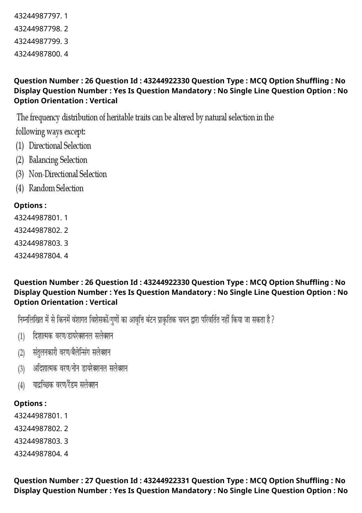
The activity of which enzyme is not ATP-dependent:
View Solution
Step 1: Understanding the Question:
This question asks us to identify which lipid translocator enzyme moves phospholipids across the plasma membrane without consuming energy in the form of ATP.
Step 2: Detailed Explanation:
The biological membrane exhibits phospholipid asymmetry, which is actively established and maintained by specialized membrane transport proteins:
1. Flippases: These are P-type ATPases that specifically move aminophospholipids (phosphatidylserine and phosphatidylethanolamine) from the outer exoplasmic leaflet to the inner cytosolic leaflet. This process requires ATP hydrolysis.
2. Floppases: These are ABC transporters that transport phospholipids in the opposite direction, from the inner cytosolic leaflet to the outer exoplasmic leaflet, driven by ATP hydrolysis.
3. Scramblases: These are calcium-activated (\(Ca^{2+}\)-dependent) membrane proteins that collapse phospholipid asymmetry by facilitating rapid, non-specific, bi-directional movement of phospholipids down their concentration gradients between both leaflets.
- Because scramblases operate via facilitated diffusion rather than active transport, their activity is strictly ATP-independent.
4. Sodium-potassium pump: A primary active transporter (\(Na^+/K^+\) ATPase) that directly hydrolyzes ATP to move ions against their gradients.
Step 3: Final Answer:
Thus, scramblases are ATP-independent enzymes, corresponding to option (C).
Quick Tip: To easily differentiate lipid translocators:
- Flippase = Inward (Requires ATP)
- Floppase = Outward (Requires ATP)
- Scramblase = Scrambles both ways (Requires \(Ca^{2+}\), NO ATP needed)
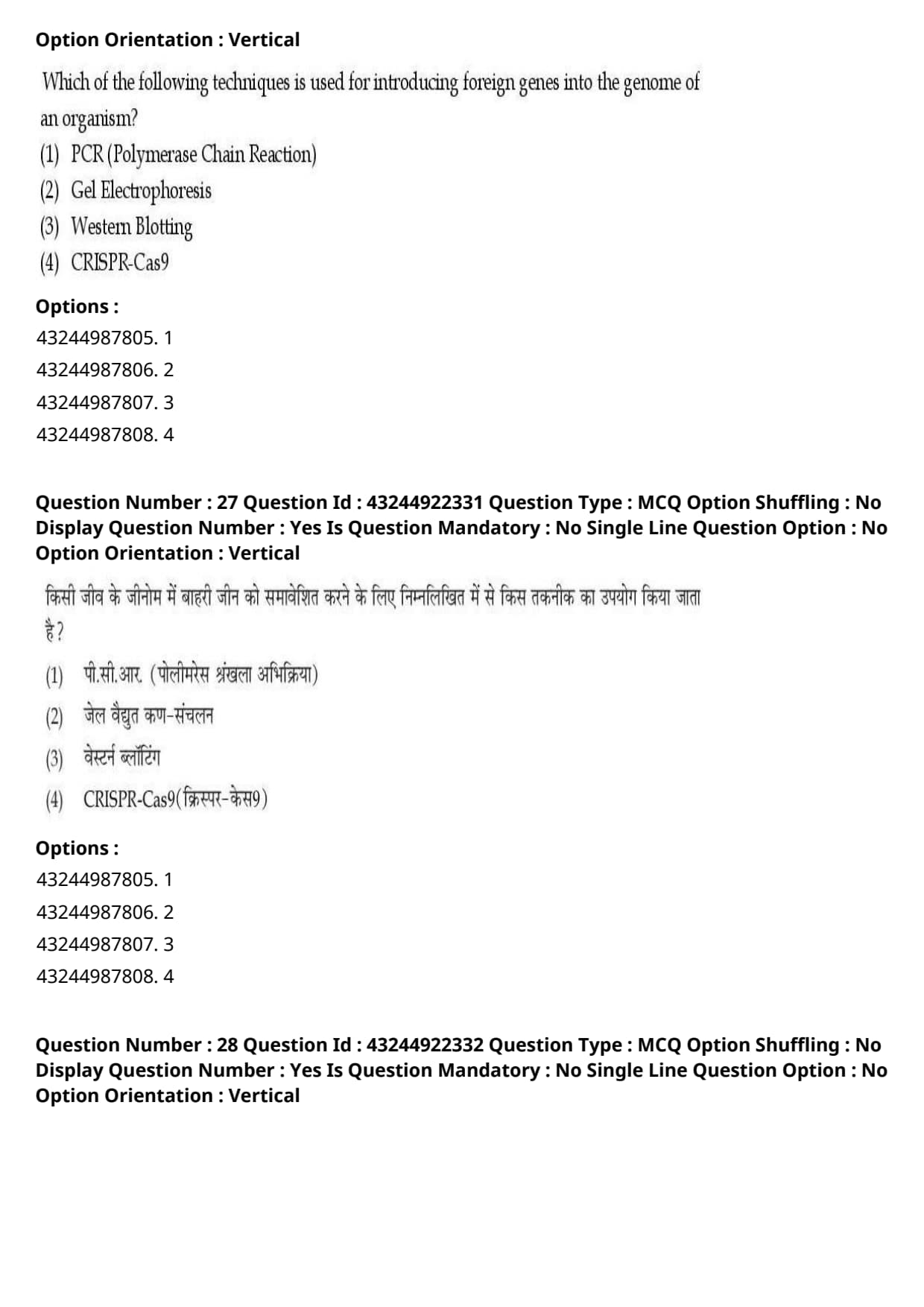
The frequency distribution of heritable traits can be altered by natural selection in the following ways except:
View Solution
Step 1: Understanding the Question:
The question asks us to identify which option does not represent a recognized mode of natural selection that alters phenotypic frequency distributions in populations.
Step 2: Detailed Explanation:
Natural selection acts on phenotypic variation within a population, altering allele and trait frequencies over generations in predictable, non-random ways.
- Modes of Natural Selection:
1. Directional Selection: Favors individuals at one extreme of the phenotypic spectrum, shifting the mean population phenotype toward that direction over time.
2. Balancing Selection: Maintains multiple alleles or phenotypic morphs in a population through mechanisms like heterozygote advantage or frequency-dependent selection.
3. Disruptive / Non-Directional Selection: Favors individuals at both extreme ends of the phenotypic spectrum over intermediate phenotypes, creating a bimodal frequency distribution.
- Why "Random Selection" is Incorrect:
- By definition, natural selection is a non-random process where differential survival and reproductive success are driven by phenotypic fitness differences.
- Random changes in allele or trait frequencies occurring purely by chance represent Genetic Drift, not natural selection.
- Therefore, "Random Selection" is an oxymoron and does not exist as a mode of natural selection.
Step 3: Final Answer:
Consequently, random selection is the correct exception, corresponding to option (D).
Quick Tip: Natural selection is NEVER random! It always favors traits that confer higher relative evolutionary fitness.
Random fluctuations in gene pools are driven by Genetic Drift, not Natural Selection.
Which of the following techniques is used for introducing foreign genes into the genome of an organism?
View Solution
Step 1: Understanding the Question:
The question asks us to identify which biotechnology tool can be used to insert or integrate foreign genetic sequences into the host organism's genome.
Step 2: Detailed Explanation:
Let us analyze the primary function of each listed molecular biology technique:
1. CRISPR-Cas9:
- An advanced, highly targeted genome editing technology derived from bacterial adaptive immune systems.
- It uses a synthetic guide RNA (gRNA) to direct the Cas9 endonuclease enzyme to make a double-strand break at a precise site in the target genome.
- When combined with a donor DNA template via homology-directed repair (HDR), foreign genes or specific modified sequences can be seamlessly integrated directly into the host genome.
2. PCR (Polymerase Chain Reaction): An \textit{in vitro enzymatic technique used to exponentially amplify specific target DNA sequences, but it cannot edit or insert genes into a living organism's genome.
3. Gel Electrophoresis: A physical separation technique used to resolve DNA, RNA, or protein fragments based on their size and charge under an electric field.
4. Western Blotting: An analytical immunoassay used to detect specific protein molecules in a complex tissue extract using antibodies.
Step 3: Final Answer:
Therefore, CRISPR-Cas9 is the technique used for genome insertion and modification, corresponding to option (D).
Quick Tip: Differentiate the core molecular biology techniques:
- PCR = Amplifies DNA
- Electrophoresis = Separates fragments
- Western Blot = Detects proteins
- CRISPR-Cas9 = Edits/Inserts genes into genomes
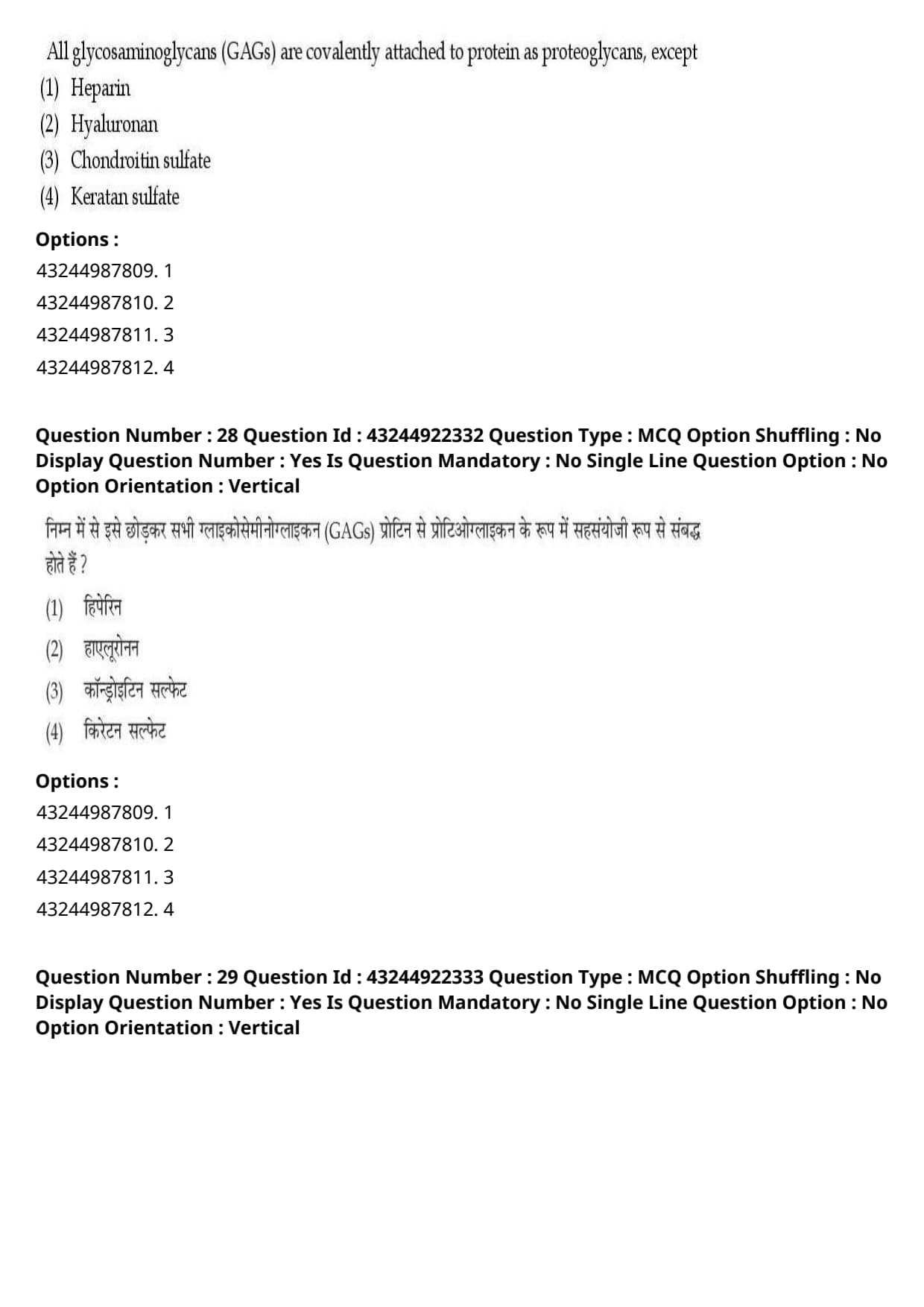
All glycosaminoglycans (GAGs) are covalently attached to protein as proteoglycans, except
View Solution
Step 1: Understanding the Question:
This question asks us to identify which glycosaminoglycan (GAG) exists as a free carbohydrate polymer and is not covalently linked to core proteins to form proteoglycans.
Step 2: Detailed Explanation:
Glycosaminoglycans (GAGs) are long, unbranched heteropolysaccharides consisting of repeating disaccharide units (typically an amino sugar and a uronic acid).
- Characteristics of Proteoglycan GAGs:
- GAGs such as chondroitin sulfate, dermatan sulfate, keratan sulfate, heparan sulfate, and heparin are synthesized in the Golgi apparatus.
- During synthesis, they are covalently linked via a trisaccharide linker to specific serine residues of core proteins to form complex proteoglycan aggregates.
- Additionally, all of these GAGs undergo enzymatic sulfation.
- Unique Features of Hyaluronan (Hyaluronic Acid):
- Hyaluronan is an exceptionally large GAG consisting of up to 25,000 repeating disaccharide units of D-glucuronic acid and N-acetylglucosamine.
- Unlike all other GAGs, hyaluronan is completely unsulfated.
- It is synthesized directly at the plasma membrane by hyaluronan synthases and extruded into the extracellular space.
- Crucially, hyaluronan is never covalently attached to a core protein during its synthesis. Instead, it interacts non-covalently with proteoglycans via link proteins in the extracellular matrix.
Step 3: Final Answer:
Therefore, hyaluronan is the correct exception, corresponding to option (B).
Quick Tip: Remember the two unique rules for Hyaluronan:
1. It is the ONLY GAG that contains NO sulfate groups.
2. It is the ONLY GAG that NEVER covalently binds to core proteins to form proteoglycans.
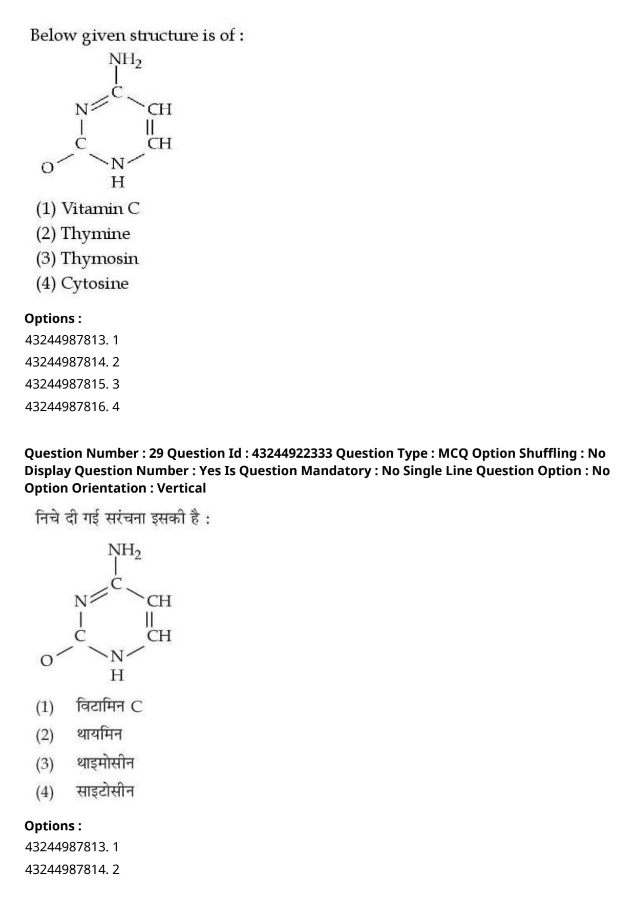
Below given structure is of:

View Solution
Step 1: Understanding the Question:
The question asks us to identify the nitrogenous base represented by the chemical structure provided in the image.
Step 2: Detailed Explanation:
Let us analyze the chemical structure shown:
- The image displays a six-membered pyrimidine ring containing two nitrogen atoms (at positions 1 and 3).
- Functional Groups Present:
- A carbonyl group (\(=O\)) attached at the C2 position.
- An amino group (\(-NH_2\)) attached at the C4 position.
- A double bond between C5 and C6 with no methyl group attached at C5.
- Differentiating Pyrimidine Bases:
1. Cytosine: 2-oxy-4-aminopyrimidine (\(=O\) at C2, \(-NH_2\) at C4).
2. Uracil: 2,4-dioxypyrimidine (\(=O\) at C2, \(=O\) at C4).
3. Thymine: 5-methyluracil (\(=O\) at C2, \(=O\) at C4, plus a \(-CH_3\) methyl group at C5).
- Since the molecule possesses an amino group (\(-NH_2\)) at C4 and a keto group (\(=O\)) at C2 without a C5 methyl group, it is unequivocally Cytosine.
Step 3: Final Answer:
Therefore, the chemical structure represents Cytosine, corresponding to option (D).
Quick Tip: Quick rule to identify pyrimidine chemical structures:
- Cytosine has an Amino group (\(-NH_2\)).
- Uracil has two Keto groups (\(=O\)).
- Thymine is Uracil with a Methyl group (\(-CH_3\)).
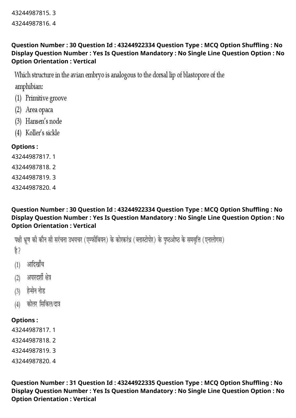
Which structure in the avian embryo is analogous to the dorsal lip of blastopore of the amphibian:
View Solution
Step 1: Understanding the Question:
This question asks us to identify the avian embryonic structure that serves as the primary organizer, equivalent to the amphibian dorsal lip of the blastopore.
Step 2: Detailed Explanation:
During embryonic development, the "primary organizer" is a region of embryonic tissue that induces surrounding uncommitted ectodermal cells to form the neural tube and establishes the primary body axes.
- The Amphibian Organizer:
- In amphibian embryos, Hans Spemann and Hilde Mangold discovered that the dorsal lip of the blastopore acts as the primary organizer.
- The Avian Homolog/Analog:
- In avian (bird) embryos, gastrulation occurs along the primitive streak formed on the blastoderm.
- At the anterior tip of the primitive streak, a regional thickening of cells forms, known as Hensen's node (primitive knot).
- Grafting experiments demonstrate that Hensen's node secretes neural inducers (such as Chordin and Noggin) and can induce a secondary body axis when transplanted into another blastoderm, exactly like the dorsal lip of the amphibian blastopore.
- Thus, Hensen's node in birds (and mammals) is the functional analog and structural homolog of the amphibian dorsal lip of the blastopore.
Step 3: Final Answer:
Therefore, the correct option is (C).
Quick Tip: Remember the primary organizer across animal groups:
- Amphibians \(\rightarrow\) Dorsal lip of the blastopore
- Birds and Mammals \(\rightarrow\) Hensen's node
- Teleost Fish \(\rightarrow\) Embryonic shield
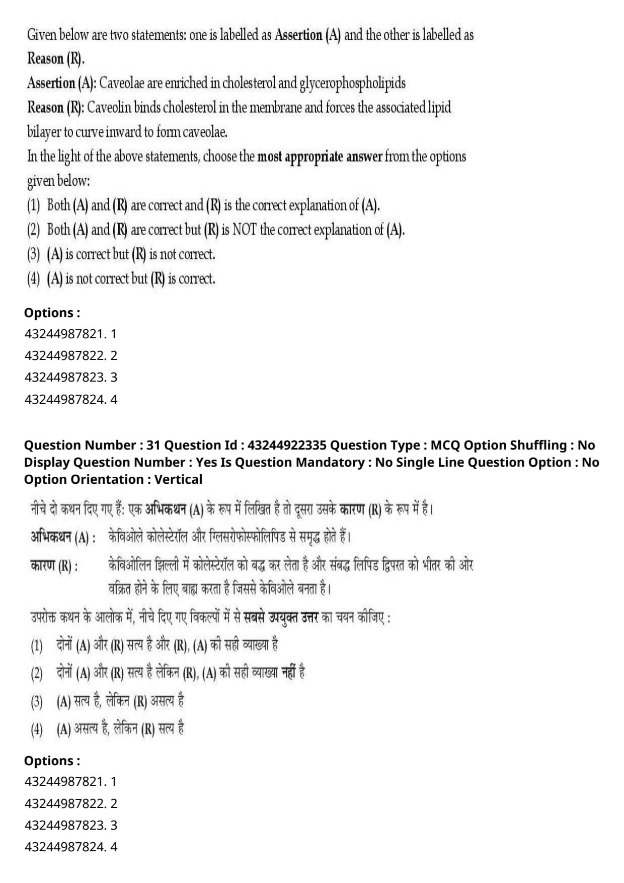
Given below are two statements: one is labelled as Assertion (A) and the other is labelled as Reason (R).
Assertion (A): Caveolae are enriched in cholesterol and glycerophospholipids
Reason (R): Caveolin binds cholesterol in the membrane and forces the associated lipid bilayer to curve inward to form caveolae.
In the light of the above statements, choose the most appropriate answer from the options given below:
View Solution
Step 1: Understanding the Question:
This question evaluates two statements regarding the molecular composition and mechanism of formation of caveolae in cell membranes.
Step 2: Detailed Explanation:
- Evaluating Assertion (A):
- Caveolae are specialized flask-shaped invaginations of the plasma membrane that function in endocytosis, signal transduction, and mechanosensing.
- Biochemically, caveolae are special microdomains of lipid rafts that are enriched specifically in cholesterol and sphingolipids (such as sphingomyelin and glycosphingolipids), not glycerophospholipids.
- Glycerophospholipids are the standard bulk components of the surrounding membrane bilayer, whereas lipid rafts and caveolae exclude glycerophospholipids to maintain a tightly packed liquid-ordered phase.
- Therefore, Assertion (A) is incorrect due to the misstatement about glycerophospholipids.
- Evaluating Reason (R):
- Caveolin is an integral dimeric protein that directly binds cholesterol molecules in the inner leaflet of the plasma membrane.
- Insertion of caveolin oligomers into the cytosolic leaflet displaces lipid headgroups and forces the membrane bilayer to curve inward, generating the characteristic invaginated flask structure of caveolae.
- Thus, Reason (R) is scientifically correct.
Step 3: Final Answer:
Therefore, Assertion (A) is not correct but Reason (R) is correct, corresponding to option (D).
Quick Tip: Lipid rafts and caveolae are ALWAYS composed of Cholesterol + Sphingolipids.
Glycerophospholipids make up the fluid non-raft bilayer, so any statement claiming rafts are enriched in glycerophospholipids is false.
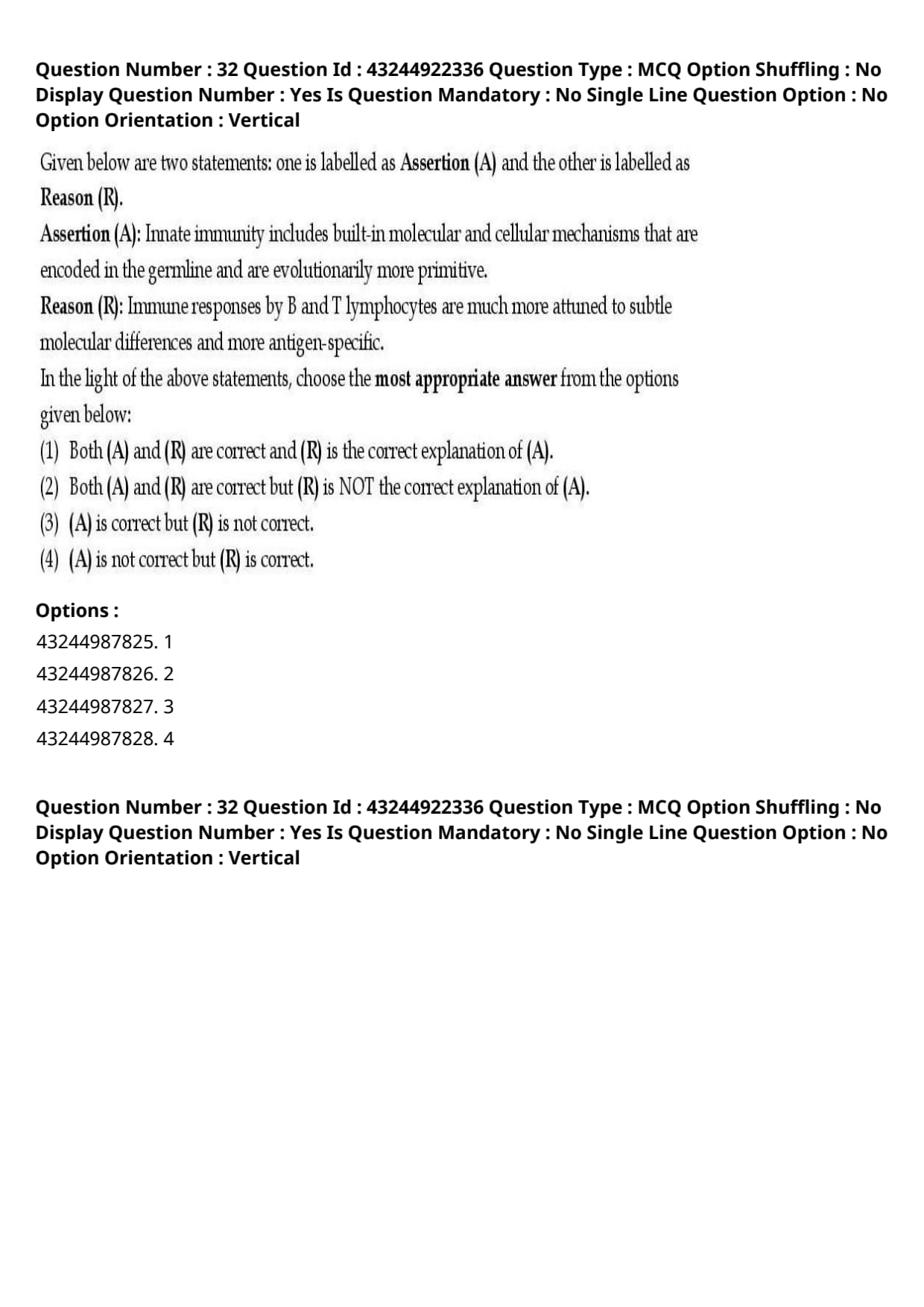
Given below are two statements: one is labelled as Assertion (A) and the other is labelled as Reason (R).
Assertion (A): Innate immunity includes built-in molecular and cellular mechanisms that are encoded in the germline and are evolutionarily more primitive.
Reason (R): Immune responses by B and T lymphocytes are much more attuned to subtle molecular differences and more antigen-specific.
In the light of the above statements, choose the most appropriate answer from the options given below:
View Solution
Step 1: Understanding the Question:
This question requires us to evaluate the truth of two statements comparing innate and adaptive immune mechanisms and determine if the reason provides a direct cause-and-effect explanation for the assertion.
Step 2: Detailed Explanation:
- Evaluating Assertion (A):
- Innate immunity is the first line of defense found in virtually all multicellular organisms.
- Its components (such as pattern recognition receptors like Toll-like receptors, phagocytes, and antimicrobial peptides) are encoded directly in the germline DNA without requiring somatic gene rearrangement, making it evolutionarily ancient and primitive.
- Thus, Assertion (A) is completely correct.
- Evaluating Reason (R):
- Adaptive immunity is mediated by B and T lymphocytes, which generate highly diverse antigen receptors (antibodies and T-cell receptors) via somatic V(D)J recombination.
- This enables adaptive immunity to recognize subtle molecular differences between specific antigens with exquisite specificity.
- Thus, Reason (R) is also completely correct as a standalone fact.
- Evaluating Explanation Link:
- Reason (R) describes the highly specific nature of adaptive immunity, whereas Assertion (A) describes the germline-encoded, primitive nature of innate immunity.
- The characteristics of B and T cells (Reason) do not explain \textit{why innate immunity is germline-encoded and primitive (Assertion). They are simply independent truths describing the two distinct arms of the immune system.
Step 3: Final Answer:
Therefore, both statements are correct, but (R) is NOT the correct explanation of (A), corresponding to option (B).
Quick Tip: When solving Assertion-Reason questions:
If both statements are true facts, check if placing the word "BECAUSE" between Assertion and Reason makes a logical sentence.
Innate immunity is primitive NOT BECAUSE B and T cells are specific. Thus, the reason is not a direct explanation.
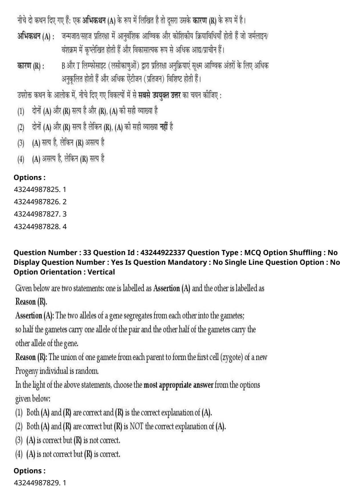
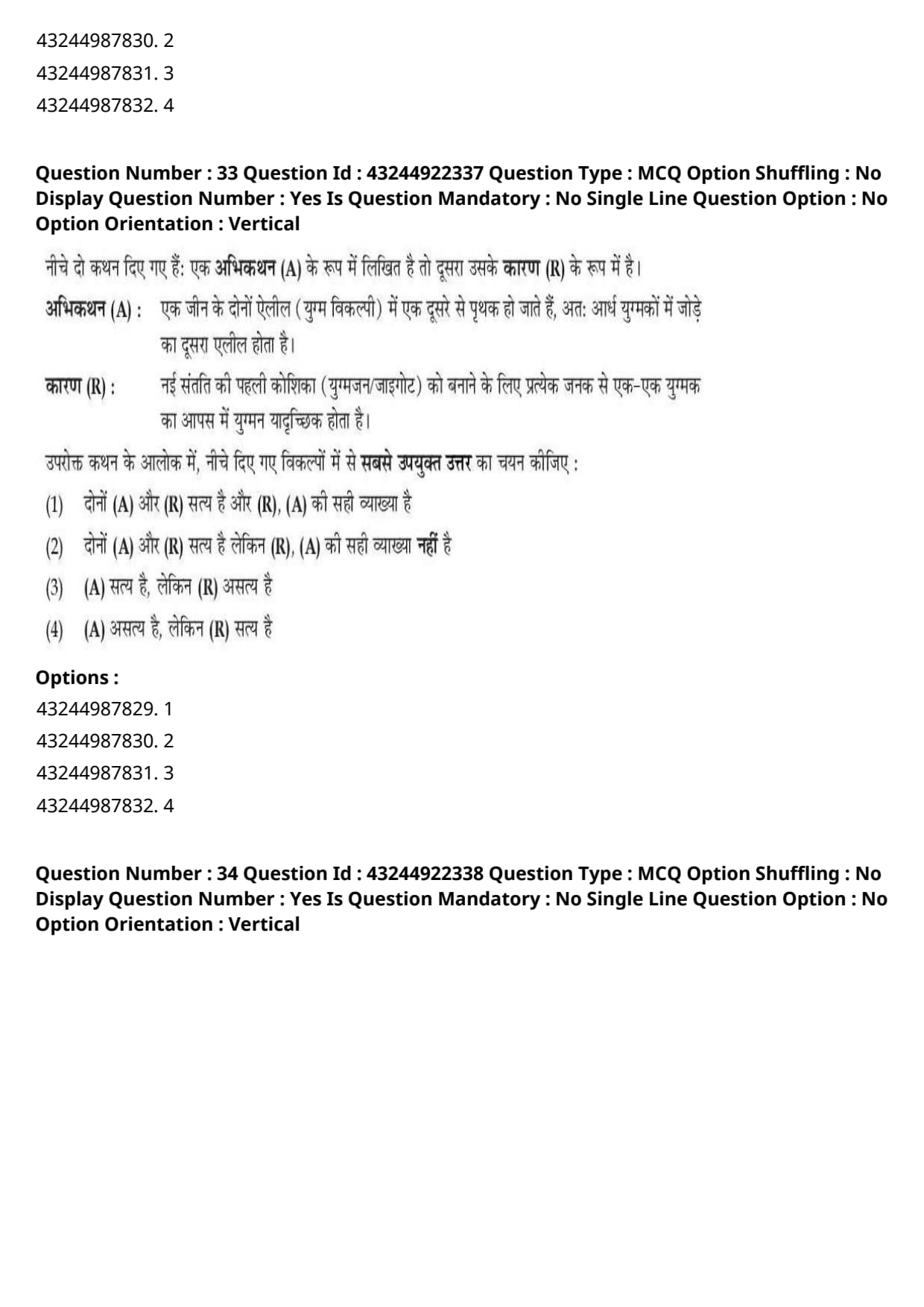
Given below are two statements: one is labelled as Assertion (A) and the other is labelled as Reason (R).
Assertion (A): The two alleles of a gene segregates from each other into the gametes; so half the gametes carry one allele of the pair and the other half of the gametes carry the other allele of the gene.
Reason (R): The union of one gamete from each parent to form the first cell (zygote) of a new Progeny individual is random.
In the light of the above statements, choose the most appropriate answer from the options given below:
View Solution
Step 1: Understanding the Question:
This question tests Mendelian genetics, specifically evaluating the Law of Segregation and the randomness of fertilization.
Step 2: Detailed Explanation:
- Evaluating Assertion (A):
- Assertion (A) is a direct statement of Mendel's First Law (Law of Segregation).
- During gametogenesis (specifically at Anaphase I of meiosis), homologous chromosomes carrying different alleles separate so that each gamete receives only one allele of the pair, resulting in a 50% (1:1) distribution among gametes.
- Thus, Assertion (A) is correct.
- Evaluating Reason (R):
- Reason (R) states that fertilization (the fusion of a male gamete with a female gamete to form a zygote) occurs at random with respect to the alleles carried by the gametes.
- Thus, Reason (R) is also correct as a fundamental law of genetics.
- Evaluating Explanation Link:
- Allelic segregation (Assertion) happens during \textit{meiosis in a single parent during gamete formation.
- Random fertilization (Reason) happens during \textit{conception when two separate parental gametes fuse.
- Random fertilization explains why offspring genotypes appear in predictable Punnett square ratios (such as 1:2:1), but it does not cause or explain the physical separation of homologous chromosomes during meiosis.
Step 3: Final Answer:
Therefore, both (A) and (R) are correct, but (R) is NOT the correct explanation of (A), corresponding to option (B).
Quick Tip: Keep genetic events in chronological order:
1. Segregation of alleles happens during Meiosis (Anaphase I).
2. Random union of gametes happens later during Fertilization.
Fertilization cannot be the explanation for why meiosis separates alleles!
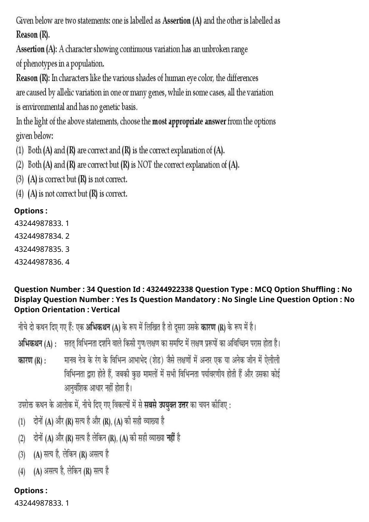
Given below are two statements: one is labelled as Assertion (A) and the other is labelled as Reason (R).
Assertion (A): A character showing continuous variation has an unbroken range of phenotypes in a population.
Reason (R): In characters like the various shades of human eye color, the differences are caused by allelic variation in one or many genes, while in some cases, all the variation is environmental and has no genetic basis.
In the light of the above statements, choose the most appropriate answer from the options given below:
View Solution
Step 1: Understanding the Question:
This question evaluates concepts related to continuous variation, quantitative traits, polygenic inheritance, and environmental influences on phenotypes.
Step 2: Detailed Explanation:
- Evaluating Assertion (A):
- Continuous variation describes traits that do not fall into discrete, clear-cut phenotypic classes (unlike Mendelian traits such as smooth vs wrinkled seeds).
- Instead, traits showing continuous variation display a smooth, unbroken spectrum or continuum of phenotypes across a population (such as human height, skin color, and eye color).
- Thus, Assertion (A) is correct.
- Evaluating Reason (R):
- Continuous phenotypic variation arises because the phenotypic expression is controlled by multiple additive genes (polygenic inheritance) and/or influenced by non-genetic environmental factors that smooth out discrete genotypic boundaries into a continuous distribution.
- In traits like eye color, subtle shades are generated by variations in multiple genes (such as \textit{OCA2 and \textit{HERC2) interacting together, alongside environmental factors during development.
- Because polygenic variation and environmental influences smooth out phenotypic classes into a spectrum, Reason (R) directly explains why continuous variation produces an unbroken range of phenotypes.
Step 3: Final Answer:
Therefore, both (A) and (R) are correct, and (R) is the correct explanation of (A), corresponding to option (A).
Quick Tip: Continuous variation = Unbroken spectrum of traits (bell curve).
This continuum occurs BECAUSE multiple genes (polygenes) and environmental influences blur discrete Mendelian classes together.
Given below are two statements: one is labelled as Assertion (A) and the other is labelled as Reason (R).
Assertion (A): Scleroderma is a systemic autoimmune disease.
Reason (R): In Scleroderma, immune response is directed toward nuclei, heart, lungs, Kidney and gastrointestinal tract.
In the light of the above statements, choose the most appropriate answer from the options given below:
View Solution
Step 1: Understanding the Question:
The question asks us to evaluate the pathology of Scleroderma and determine whether the systemic organ involvement explains why it is classified as a systemic autoimmune disease.
Step 2: Detailed Explanation:
- Evaluating Assertion (A):
- Scleroderma (also known as Systemic Sclerosis) is an autoimmune connective tissue disorder characterized by abnormal collagen deposition, vascular dysfunction, and tissue fibrosis.
- Because it is not restricted to a single organ (unlike organ-specific autoimmune diseases like Hashimoto's thyroiditis), it is classified as a systemic autoimmune disease.
- Thus, Assertion (A) is correct.
- Evaluating Reason (R):
- In scleroderma, autoantibodies (such as anti-topoisomerase I / Scl-70 and anti-centromere antibodies) target nuclear antigens across diverse cell types.
- This aberrant immune response causes systemic inflammation and microvascular damage affecting multiple internal visceral organs, including the skin, heart, lungs, kidneys, and gastrointestinal tract.
- Evaluating Explanation Link:
- The reason an autoimmune disease is termed "systemic" is precisely because its immune reactivity and pathological damage affect multiple organ systems and ubiquitous cellular components (like nuclei and connective tissue) throughout the entire body.
- Therefore, Reason (R) provides the exact medical explanation for Assertion (A).
Step 3: Final Answer:
Hence, both (A) and (R) are correct, and (R) is the correct explanation of (A), corresponding to option (A).
Quick Tip: Organ-Specific vs Systemic Autoimmune Diseases:
- Organ-Specific = Targets 1 organ (e.g., Insulin-dependent Diabetes targets pancreas).
- Systemic = Targets widespread nuclear/connective tissue affecting multiple organs (e.g., Scleroderma, Lupus).
Given below are two statements: one is labelled as Assertion (A) and the other is labelled as Reason (R).
Assertion (A): The transcripts of RNA polymerases I and III are not processed.
Reason (R): RNA polymerases I and III lack a C-terminal domain (CTD).
In the light of the above statements, choose the most appropriate answer from the options given below:
View Solution
Step 1: Understanding the Question:
This question evaluates statements regarding eukaryotic RNA polymerases, their structural domains, and the post-transcriptional processing of their RNA transcripts.
Step 2: Detailed Explanation:
- Evaluating Assertion (A):
- Transcripts synthesized by RNA Polymerase I (pre-rRNA) and RNA Polymerase III (pre-tRNA, 5S rRNA) undergo extensive post-transcriptional processing.
- Pre-rRNA undergoes endonuclease cleavage, exonucleolytic trimming, pseudouridylation, and ribose methylation guided by snoRNAs.
- Pre-tRNA undergoes \(5'\) cleavage by RNase P, \(3'\) processing, addition of the \(CCA\) tail, extensive base modifications, and intron splicing.
- Therefore, claiming that transcripts of RNA polymerases I and III are "not processed" is completely false.
- Evaluating Reason (R):
- RNA Polymerase II uniquely possesses a carboxyl-terminal domain (CTD) consisting of multiple repeats of the heptapeptide consensus sequence \(Tyr-Ser-Pro-Thr-Ser-Pro-Ser\).
- This CTD gets phosphorylated to orchestrate mRNA-specific processing events (capping, splicing, polyadenylation).
- Neither RNA Polymerase I nor RNA Polymerase III possesses this CTD structure.
- Thus, Reason (R) is a true molecular fact.
Step 3: Final Answer:
Therefore, Assertion (A) is not correct, but Reason (R) is correct, corresponding to option (D).
Quick Tip: ALL major eukaryotic RNAs (mRNA, rRNA, tRNA) undergo post-transcriptional processing!
RNA Polymerase II is unique in possessing the CTD tail, but lack of CTD in Pol I and Pol III does NOT mean their RNA is unprocessed.
Given below are two statements: one is labelled as Assertion (A) and the other is labelled as Reason (R).
Assertion (A): Isoelectric focusing (IEF) is a highly sensitive analytical technique and is particularly useful for studying micro-heterogeneity in a protein.
Reason (R): A protein may show a single band on SDS gel, but may show three bands on an IEF gel. The method is particularly useful for separating isoenzymes, which are same forms of the different enzyme.
In the light of the above statements, choose the most appropriate answer from the options given below:
View Solution
Step 1: Understanding the Question:
This question evaluates the analytical technique of Isoelectric Focusing (IEF) and its ability to resolve protein micro-heterogeneity compared to SDS-PAGE.
Step 2: Detailed Explanation:
- Evaluating Assertion (A):
- Isoelectric Focusing (IEF) separates proteins based on their unique isoelectric point (\(pI\)) along a stable pH gradient established in an electric field.
- It is an exceptionally high-resolution technique capable of detecting subtle charge variations (micro-heterogeneity) caused by post-translational modifications such as phosphorylation, deamidation, or sialylation.
- Thus, Assertion (A) is correct.
- Evaluating Reason (R):
- SDS-PAGE separates protein subunits strictly based on molecular weight, denaturing them and imparting a uniform negative charge.
- Proteins with identical molecular weights but slightly different net charges (such as isoenzymes or post-translationally modified isoforms) will resolve into a single band on SDS-PAGE.
- However, when subjected to IEF, these subtle charge variations cause the proteins to migrate to different \(pI\) positions, resolving into multiple distinct bands.
- This ability to separate same-mass, charge-variant isoforms directly explains why IEF is such a powerful tool for analyzing protein micro-heterogeneity.
Step 3: Final Answer:
Therefore, both (A) and (R) are correct, and (R) is the correct explanation of (A), corresponding to option (A).
Quick Tip: Understand the separation principles:
- SDS-PAGE = Separates strictly by Molecular Weight (Mass).
- IEF = Separates strictly by Isoelectric Point (\(pI\)/Charge).
Combining both gives 2D Gel Electrophoresis!
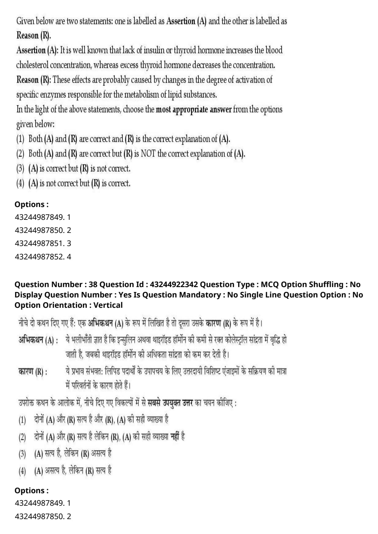
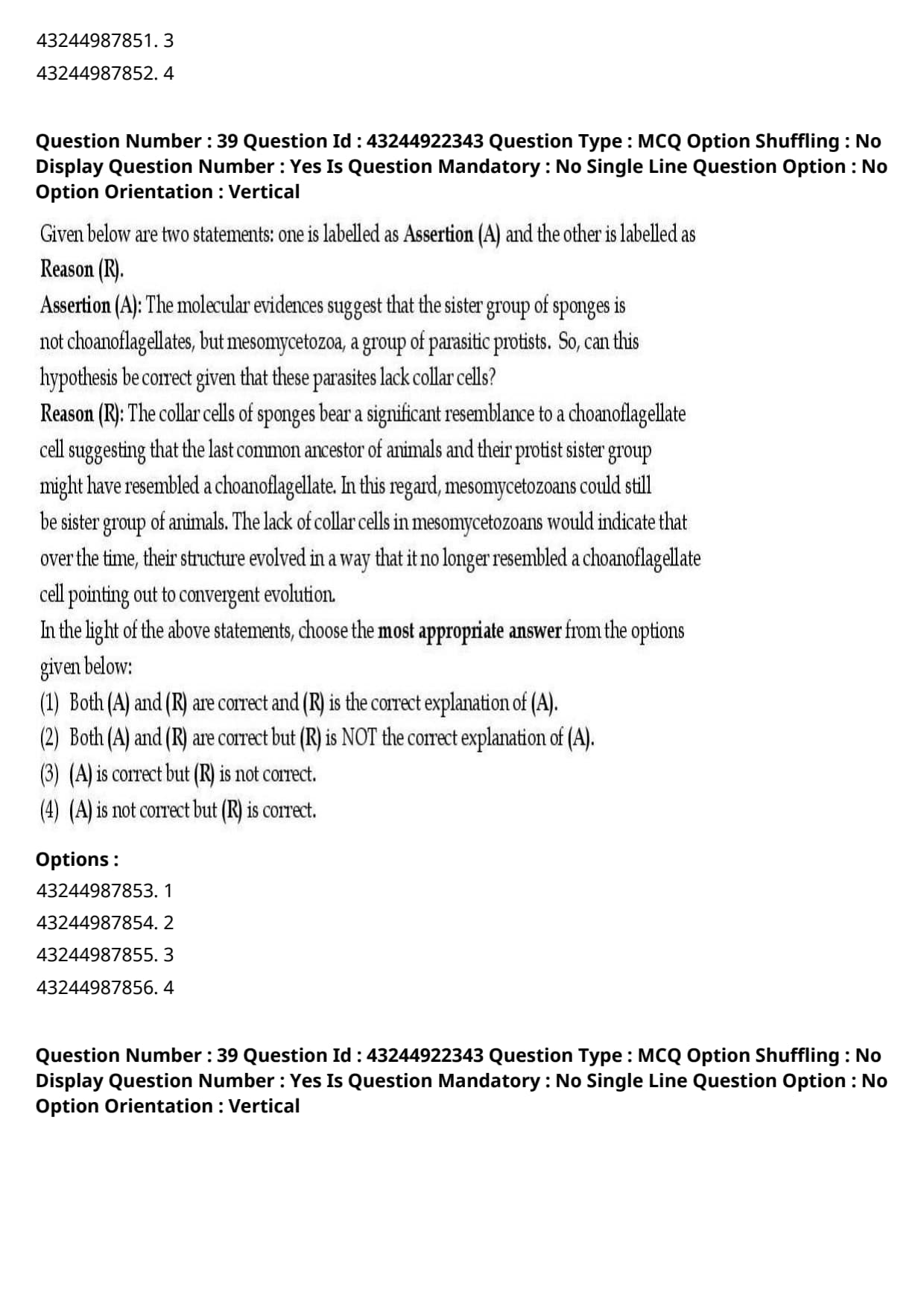
Given below are two statements: one is labelled as Assertion (A) and the other is labelled as Reason (R).
Assertion (A): It is well known that lack of insulin or thyroid hormone increases the blood cholesterol concentration, whereas excess thyroid hormone decreases the concentration.
Reason (R): These effects are probably caused by changes in the degree of activation of specific enzymes responsible for the metabolism of lipid substances.
In the light of the above statements, choose the most appropriate answer from the options given below:
View Solution
Step 1: Understanding the Question:
This question evaluates endocrine regulation of lipid metabolism, specifically how thyroid hormones and insulin influence blood cholesterol concentration.
Step 2: Detailed Explanation:
- Evaluating Assertion (A):
- Thyroid hormones (\(T_3\) and \(T_4\)) and insulin are key endocrine regulators of metabolic homeostasis.
- Hypothyroidism and insulin deficiency (Diabetes mellitus) lead to hypercholesterolemia (elevated blood cholesterol levels).
- Conversely, hyperthyroidism increases hepatic clearance and catabolism of cholesterol, resulting in significantly decreased plasma cholesterol levels.
- Thus, Assertion (A) is correct.
- Evaluating Reason (R):
- Thyroid hormones increase the transcription of hepatic LDL receptors, stimulate cholesterol 7\(\alpha\)-hydroxylase (converting cholesterol to bile acids), and activate lipoprotein lipase.
- Insulin regulates HMG-CoA reductase and lipoprotein lipase activity.
- Therefore, alterations in hormone levels change the expression and activation state of key metabolic enzymes and receptors governing lipid breakdown and clearance.
- This enzymatic and receptor regulation directly provides the physiological explanation for the clinical observations stated in Assertion (A).
Step 3: Final Answer:
Therefore, both (A) and (R) are correct, and (R) is the correct explanation of (A), corresponding to option (A).
Quick Tip: Clinical Hormone-Lipid Connection:
- Hypothyroidism \(\rightarrow\) Reduced LDL receptors \(\rightarrow\) High Blood Cholesterol.
- Hyperthyroidism \(\rightarrow\) Increased LDL receptors \(\rightarrow\) Low Blood Cholesterol.
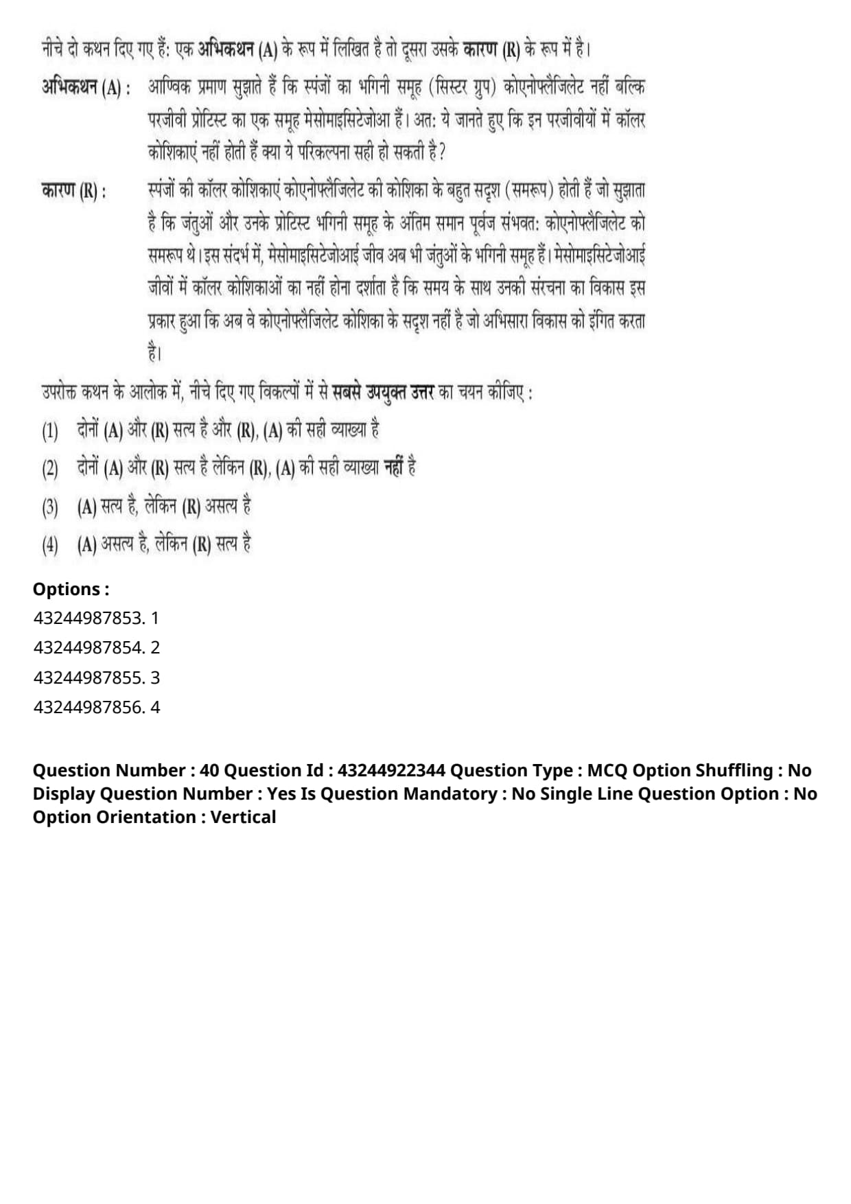
Given below are two statements: one is labelled as Assertion (A) and the other is labelled as Reason (R).
Assertion (A): The molecular evidences suggest that the sister group of sponges is not choanoflagellates, but mesomycetozoa, a group of parasitic protists. So, can this hypothesis be correct given that these parasites lack collar cells?
Reason (R): The collar cells of sponges bear a significant resemblance to a choanoflagellate cell suggesting that the last common ancestor of animals and their protist sister group might have resembled a choanoflagellate. In this regard, mesomycetozoans could still be sister group of animals. The lack of collar cells in mesomycetozoans would indicate that over time, their structure evolved in a way that it no longer resembled a choanoflagellate cell pointing out to convergent evolution.
In the light of the above statements, choose the most appropriate answer from the options given below:
View Solution
Step 1: Understanding the Question:
This question evaluates evolutionary theories regarding the origin of Metazoa (animals/sponges) and their relationship with unicellular protistan sister groups.
Step 2: Detailed Explanation:
- Evaluating Assertion (A):
- Robust phylogenomic studies consistently establish that Choanoflagellates (and Filasterea) represent the true living protistan sister group to Metazoa (animals/sponges).
- Mesomycetozoans (Teretosporea) are a clade of unicellular Opisthokonts, but they branch earlier and are more distant from animals than choanoflagellates.
- The premise in Assertion (A) claiming that molecular evidence identifies mesomycetozoa as the sister group of sponges is scientifically incorrect.
- Thus, Assertion (A) is not correct.
- Evaluating Reason (R):
- The remarkable morphological and molecular homology between sponge choanocytes (collar cells) and free-living choanoflagellates provides classic evolutionary evidence that the last common ancestor of metazoans possessed a choanoflagellate-like cellular morphology.
- Reason (R) correctly outlines the evolutionary reasoning regarding collar cell resemblance and theoretical loss/divergence scenarios in protistan clades.
- Thus, Reason (R) is correct.
Step 3: Final Answer:
Therefore, Assertion (A) is not correct, but Reason (R) is correct, corresponding to option (D).
Quick Tip: Consistently remember the animal sister group in evolution:
Choanoflagellata is unequivocally established as the immediate sister group to Metazoa (animals).
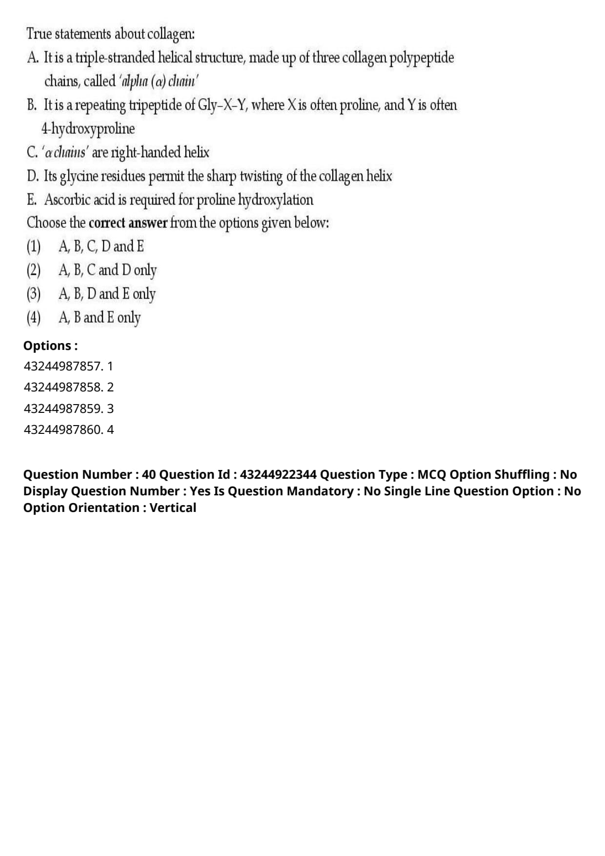
True statements about collagen:
A. It is a triple-stranded helical structure, made up of three collagen polypeptide chains, called 'alpha (\(\alpha\)) chain'
B. It is a repeating tripeptide of Gly-X-Y, where X is often proline, and Y is often 4-hydroxyproline
C. '\(\alpha\) chains' are right-handed helix
D. Its glycine residues permit the sharp twisting of the collagen helix
E. Ascorbic acid is required for proline hydroxylation
Choose the correct answer from the options given below:
View Solution
Step 1: Understanding the Question:
This question asks us to identify the correct structural and biochemical statements regarding collagen protein.
Step 2: Detailed Explanation:
Let us analyze each statement individually:
- Statement A: Correct. Collagen is a triple-stranded fibrous protein composed of three polypeptide chains referred to as \(\alpha\)-chains.
- Statement B: Correct. Collagen has a strict repeating tripeptide sequence pattern of \(Gly-X-Y\), where \(X\) is frequently proline and \(Y\) is frequently 4-hydroxyproline (or hydroxylysine).
- Statement C: Incorrect. The individual \(\alpha\)-chains are left-handed polyproline II-like helices. These three left-handed helices then wrap around each other to form a right-handed superhelix. Therefore, describing the individual \(\alpha\)-chains as right-handed is incorrect.
- Statement D: Correct. Glycine is the smallest amino acid with only a single hydrogen atom as its side chain. It occurs at every third position (\(Gly-X-Y\)) because only glycine is small enough to fit inside the crowded central core of the triple helix, allowing tight packing and sharp twisting.
- Statement E: Correct. Ascorbic acid (Vitamin C) is an essential cofactor for prolyl 4-hydroxylase, maintaining the active \(Fe^{2+}\) state of the enzyme required for proline hydroxylation.
Step 3: Final Answer:
Therefore, the correct statements are A, B, D, and E only, corresponding to option (C).
Quick Tip: Collagen Helix Handedness Rule:
- Individual \(\alpha\)-chains = Left-handed helix.
- Overall Triple Superhelix = Right-handed helix.
Keep this distinction clear to avoid tricky options!
Choose the correct sequence of events involved in receptor-mediated endocytosis of transferrin:
A. The high pH in the endosome induces transferrin to release its bound iron
B. The low pH in the endosome induces transferrin to completely release its bound iron
C. Iron free transferrin is degraded in lysosome
D. Cell-surface transferrin receptors deliver transferrin with its bound iron to early endosomes by receptor-mediated endocytosis
E. Transferrin receptor along with iron free transferrin is recycled back to the plasma membrane
Choose the correct answer from the options given below:
View Solution
Step 1: Understanding the Question:
This question asks for the correct chronological sequence of biological events that occur during the receptor-mediated endocytosis and recycling of transferrin.
Step 2: Detailed Explanation:
Introduction to the Topic:
Transferrin is an extracellular glycoprotein that transports iron (\(Fe^{3+}\)) through blood.
Cells internalize this complex via receptor-mediated endocytosis, separating the iron and recycling the carrier protein.
Analysis of Individual Steps:
- Step D: Cell-surface transferrin receptors bind iron-bound transferrin (ferrotransferrin) and are internalized into early endosomes via clathrin-mediated endocytosis. This is the first step.
- Step B: Within the endosome, V-type ATPases pump protons into the lumen to lower the pH (\(\sim 5.5\)). This acidic environment induces a conformational change causing the release of iron (\(Fe^{3+}\)).
- Step E: The apotransferrin (iron-free) remains bound to the transferrin receptor at this low pH. The entire complex is recycled back to the neutral-pH environment of the plasma membrane, where apotransferrin dissociates.
Analysis of Incorrect Statements:
- Step A: High pH does not release iron; acidity is required. Thus, A is incorrect.
- Step C: Apotransferrin is spared from lysosomal degradation to be reused. Thus, C is incorrect.
Step 3: Final Answer:
The valid, sequential events are: D \(\rightarrow\) B \(\rightarrow\) E.
Therefore, the correct option is (A).
Quick Tip: The transferrin pathway is a classic exception to lysosomal degradation.
Both the receptor and the ligand (apotransferrin) are recycled back to the cell surface, ensuring highly efficient iron delivery.
Choose the correct sequence of events involved in the synthesis of a secretory protein:
A. Signal peptide binds to the interior of the translocon
B. As the signal sequence emerges from the ribosome, it binds to the SRP
C. The SRP-ribosome complex then binds to an SRP receptor situated within the ER membrane
D. Synthesis of the polypeptide begins on a free ribosome
E. After the nascent polypeptide passes into the lumen of the ER, the signal peptide is cleaved by a membrane protein
Choose the correct answer from the options given below:
View Solution
Step 1: Understanding the Question:
This question asks for the correct chronological order of molecular events during the co-translational translocation of secretory proteins across the Endoplasmic Reticulum (ER) membrane.
Step 2: Detailed Explanation:
Introduction to the Topic:
Secretory proteins are targeted to the ER via their N-terminal signal sequences.
This process is governed by the signal recognition particle (SRP) and its receptor on the ER membrane.
Chronological Sequence of Events:
- Step D: Translation of the mRNA begins on free ribosomes in the cytosol, and the N-terminal signal peptide is synthesized.
- Step B: As the signal sequence emerges from the ribosome, it is recognized and bound by the SRP, temporarily halting translation.
- Step C: The SRP-ribosome-nascent chain complex is targeted to the ER membrane, where SRP binds to its receptor.
- Step A: The ribosome is transferred to the translocon (Sec61 complex), and the signal peptide binds to the interior of the translocon channel.
- Step E: Translation resumes, translocating the growing polypeptide into the ER lumen. Once inside, signal peptidase (a membrane protein) cleaves the signal peptide.
Step 3: Final Answer:
The correct order of events is: D \(\rightarrow\) B \(\rightarrow\) C \(\rightarrow\) A \(\rightarrow\) E.
Therefore, the correct option is (B).
Quick Tip: Synthesis must start on a free ribosome (D) before any targeting can happen, and ends with the final cleavage of the signal peptide inside the ER lumen (E).
This starting-and-ending pattern uniquely directs you to Option (B).
Correctly arrange the below given Transcription Factors (TFs) as per their involvement in initiation of eukaryotic transcription:
A. TFIIB
B. TFIID
C. TFIIE
D. TFIIF
E. TFIIH
Choose the correct answer from the options given below:
View Solution
Step 1: Understanding the Question:
This question asks for the step-by-step assembly sequence of general transcription factors (GTFs) during the formation of the eukaryotic preinitiation complex (PIC).
Step 2: Detailed Explanation:
Introduction to the Topic:
Initiation of eukaryotic gene transcription by RNA Polymerase II requires the highly coordinated sequential binding of GTFs at the promoter.
Sequential Assembly Pathway:
- Step B (TFIID): First, TFIID binds to the TATA box via its TATA-binding protein (TBP) subunit.
- Step A (TFIIB): TFIIB binds to TFIID and acts as a bridge to align RNA Polymerase II.
- Step D (TFIIF): TFIIF, associated with RNA Polymerase II, is recruited to the complex next.
- Step C (TFIIE): TFIIE binds next and creates a docking platform for the final factor.
- Step E (TFIIH): TFIIH binds last, completing the preinitiation complex. It functions as a helicase to open the promoter and a kinase to initiate transcription.
Step 3: Final Answer:
The correct order is: B \(\rightarrow\) A \(\rightarrow\) D \(\rightarrow\) C \(\rightarrow\) E.
Therefore, the correct option is (C).
Quick Tip: A popular mnemonic to remember the binding order of eukaryotic transcription factors is:
\textbf{D}ogs \textbf{A}nd \textbf{B}irds \textbf{F}ly \textbf{E}verywhere \textbf{H}appily
This translates to: TFIID \(\rightarrow\) TFIIA \(\rightarrow\) TFIIB \(\rightarrow\) TFIIF \(\rightarrow\) TFIIE \(\rightarrow\) TFIIH.
Correct sequence of events in cytosolic pathway of antigen presentation:
A. Calnexin dissociates, thereafter class I molecule associates with the chaperone calreticulin and with tapasin
B. Peptides are transported to RER (Rough Endoplasmic Reticulum) via TAP (Transport Associated Protein)
C. Endogenous antigen is degraded by proteasome in cytosol
D. Class I MHC \(\alpha\) chain binds calnexin, then \(\beta_2\) microglobulin
E. MHC captures peptide and chaperones dissociate
Choose the correct answer from the options given below:
View Solution
Step 1: Understanding the Question:
This question asks for the correct order of events involved in the processing and loading of endogenous antigens onto MHC Class I molecules for cell-surface presentation.
Step 2: Detailed Explanation:
Introduction to the Topic:
MHC Class I molecules present endogenous proteins to cytotoxic T cells (\(CD8^+\)).
The sequence involves cytosolic degradation, MHC chaperone assembly, peptide transport into the ER, and peptide loading.
Analysis of Sequential Steps:
- Step C: First, the endogenous cellular proteins are degraded in the cytosol by the proteasome.
- Step D: In the RER, the newly synthesized MHC Class I heavy (\(\alpha\)) chain binds to calnexin and then associates with \(\beta_2\)-microglobulin.
- Step A: Upon \(\beta_2\)-microglobulin binding, calnexin is released, and the heterodimer associates with calreticulin and tapasin.
- Step E: The MHC Class I complex is assembled in a peptide-receptive state.
- Step B: Proteasome-derived peptides are actively transported across the ER membrane into the lumen via the TAP transporter, where they are loaded onto the waiting MHC molecules.
*(Note: While peptide transport (B) biochemically occurs prior to the final peptide-capture stability (E), the steps in option D outline the sequence of structural assembly).*
Step 3: Final Answer:
The correct option as per the official answer key is C, D, A, E and B.
Therefore, the correct option is (D).
Quick Tip: The cytosolic pathway of antigen presentation always begins with proteasomal degradation (C) of foreign or abnormal cellular proteins in the cytosol.
Identifying C as the first step eliminates several incorrect option paths.
Correct sequence of embryonic developmental stages:
A. Blastula
B. Neurula
C. Morula
D. Fertilized egg
E. Gastrula
Choose the correct answer from the options given below:
View Solution
Step 1: Understanding the Question:
This question asks for the chronological developmental order of animal embryonic stages, beginning from fertilization.
Step 2: Detailed Explanation:
Detailed Chronology of Embryogenesis:
- Step D (Fertilized egg): Development begins when fertilization forms a single-celled diploid zygote.
- Step C (Morula): The zygote undergoes rapid mitotic cleavages to produce a solid, mulberry-like sphere of cells called a morula.
- Step A (Blastula): Continued cell divisions lead to a hollow blastula surrounding a fluid-filled blastocoel cavity.
- Step E (Gastrula): Gastrulation involves morphogenetic movements that invaginate cells, forming the three primary germ layers (ectoderm, mesoderm, endoderm).
- Step B (Neurula): In chordate embryos, neurulation occurs next, folding ectoderm to form the neural tube.
Step 3: Final Answer:
The correct order of stages is: D \(\rightarrow\) C \(\rightarrow\) A \(\rightarrow\) E \(\rightarrow\) B.
Therefore, the correct option is (D).
Quick Tip: Remember this simple and logical alphabetical rule:
\textbf{Z}ygote (Fertilized egg) \(\rightarrow\) \textbf{M}orula \(\rightarrow\) \textbf{B}lastula \(\rightarrow\) \textbf{G}astrula \(\rightarrow\) \textbf{N}eurula.
Correct sequence of events in mammalian fertilization:
A. Lysis of zona pellucida by sperm
B. Sperm activated by female reproductive tract
C. Acrosomal reaction
D. Fusion of sperm and egg
E. Sperm binds zona pellucida
Choose the correct answer from the options given below:
View Solution
Step 1: Understanding the Question:
This question asks for the correct physiological sequence of events leading to successful sperm-egg fusion in mammals.
Step 2: Detailed Explanation:
Detailed Analysis of Fertilization Steps:
- Step B: First, sperm must undergo capacitation (activation) within the female reproductive tract to gain hypermotility.
- Step E: Next, the activated sperm travels to the oocyte and binds to specific glycoproteins of the egg's extracellular coat, the zona pellucida.
- Step C: Binding triggers the acrosomal reaction, releasing hydrolytic enzymes from the sperm head.
- Step A: These enzymes digest a pathway through the protective zona pellucida.
- Step D: The sperm traverses this path and fuses with the oocyte plasma membrane.
Step 3: Final Answer:
The correct order of events is: B \(\rightarrow\) E \(\rightarrow\) C \(\rightarrow\) A \(\rightarrow\) D.
Therefore, the correct option is (B).
Quick Tip: Capacitation (B) is the mandatory physiological priming step that must occur in the female reproductive tract before sperm can interact with the egg envelope (E).
Correct sequence of events on larval development of Herdmania:
A. The blastopore of embryo closes and rudiment of tail formed
B. The chorion ruptured due to an enzyme secreted by inner follicle cells
C. The archenteron produces presumptive mesoderm as solid band
D. The presumptive notochordal occupy the central core of larva
E. The fully formed free swimming larva is produced
Choose the correct answer from the options given below:
View Solution
Step 1: Understanding the Question:
This question asks for the correct developmental timeline of embryonic and larval structures in the marine urochordate *Herdmania*.
Step 2: Detailed Explanation:
Sequential Events in Herdmania Development:
- Step A: Gastrulation occurs first, resulting in the closure of the blastopore and the formation of a tail bud.
- Step C: The archenteron invaginates and differentiates to form a solid band of presumptive mesoderm.
- Step D: Presumptive notochordal cells organize and form the central structural core of the developing tail.
- Step B: Once fully developed, hatching enzymes secreted by follicle cells digest and rupture the chorion.
- Step E: The free-swimming tunicate tadpole larva escapes into the water column.
Step 3: Final Answer:
The correct developmental sequence is: A \(\rightarrow\) C \(\rightarrow\) D \(\rightarrow\) B \(\rightarrow\) E.
Therefore, the correct option is (A).
Quick Tip: Larval release (E) must always be the final step, preceded immediately by the enzymatic hatching process (B) of chorion rupture.
Choose the correct sequence of enzymes involved in the preparatory phase of glycolysis:
A. Hexokinase
B. Phospho-fructo kinase I
C. Phospho-hexose isomerase
D. Aldolase
E. Triose-phosphate Isomerase
Choose the correct answer from the options given below:
View Solution
Step 1: Understanding the Question:
This question asks for the correct order of enzymes that catalyze steps 1 through 5 of the glycolytic pathway (the preparatory phase).
Step 2: Detailed Explanation:
Enzymatic Steps of the Preparatory Phase:
- Step A (Hexokinase): Step 1 converts glucose to glucose-6-phosphate.
- Step C (Phospho-hexose isomerase): Step 2 converts glucose-6-phosphate to fructose-6-phosphate.
- Step B (PFK-1): Step 3 converts fructose-6-phosphate to fructose-1,6-bisphosphate.
- Step D (Aldolase): Step 4 splits the hexose bisphosphate into GAP and DHAP.
- Step E (Triose-phosphate Isomerase): Step 5 interconverts DHAP into a second molecule of GAP.
Step 3: Final Answer:
The correct enzymatic sequence is: A \(\rightarrow\) C \(\rightarrow\) B \(\rightarrow\) D \(\rightarrow\) E.
Therefore, the correct option is (B).
Quick Tip: Glycolysis always starts with the phosphorylation of glucose by Hexokinase (A) and concludes its investment phase with the isomerization of triose phosphates by Triose-phosphate Isomerase (E).
Choose the correct sequence of events on short term endocrine response to stress:
A. Hypothalamus
B. Stressor
C. Sympathetic centers
D. Sympathetic nerve & adrenal medulla
E. Adrenaline & nor-adrenaline response
Choose the correct answer from the options given below:
View Solution
Step 1: Understanding the Question:
This question asks for the physiological signal transduction sequence in the body's acute (fight-or-flight) stress response.
Step 2: Detailed Explanation:
Acute Sympathomedullary Pathway Sequence:
- Step B (Stressor): The stress response is initiated by a perceived external or internal stressor.
- Step A (Hypothalamus): The sensory signals are received and integrated by the hypothalamus in the brain.
- Step C (Sympathetic centers): The hypothalamus activates sympathetic outflow centers in the CNS.
- Step D (Sympathetic nerve \& adrenal medulla): Neural impulses travel via sympathetic nerves to stimulate the chromaffin cells of the adrenal medulla.
- Step E (Adrenaline release): The adrenal medulla rapidly releases adrenaline and noradrenaline into the blood to activate systemic tissues.
Step 3: Final Answer:
The correct physiological sequence is: B \(\rightarrow\) A \(\rightarrow\) C \(\rightarrow\) D \(\rightarrow\) E.
Therefore, the correct option is (D).
Quick Tip: The response must start with the Stressor (B) and end with the systemic Adrenaline response (E). This quickly limits options to (C) and (D), with the hypothalamus (A) always acting as the primary brain coordinator.
Choose the correct stages in the development of Gastropods to indicate occurrence of torsion:
A. Pre-torsional stage
B. 90° lateral anti-clockwise torsion
C. Movement of mantle cavity \& anus to the right
D. Larva with ventral flexure in exo-gastric shell
E. 90° torsion clockwise - Adult stage
View Solution
Step 1: Understanding the Question:
This question asks for the correct chronological sequence of morphological stages and physiological changes that occur during the developmental process of gastropod torsion.
Step 2: Detailed Explanation:
Anatomical Concept of Torsion:
Torsion is a highly specific, defining developmental process in gastropod molluscs where the visceral mass, mantle, and shell rotate by \(180^\circ\) relative to the head and foot.
This rotation shifts the mantle cavity and anus from their ancestral posterior position to an anterior position located directly above the head.
Analysis of Individual Developmental Milestones:
- Stage A (Pre-torsional stage): The development begins with a symmetric veliger larva. In this early pre-torsional stage, the body is bilaterally symmetrical, and the mantle cavity is located at the posterior end.
- Stage B (\(90^\circ\) lateral anti-clockwise torsion): The first phase of torsion is rapid and is driven by the asymmetrical contraction of the larval retractor muscle. This muscle rotates the visceral mass by \(90^\circ\) in a counter-clockwise direction.
- Stage D (Larva with ventral flexure in exogastric shell): Following the initial rotation, the dorsal and ventral developmental axes undergo a differential shift. This produces a ventral flexure of the visceral mass, causing the shell to become exogastric (coiling anteriorly over the head).
- Stage C (Movement of mantle cavity \& anus to the right): Due to the physical changes induced by the initial rotation and body flexure, the mantle cavity and the anus are relocated to the right side of the larval body.
- Stage E (\(90^\circ\) torsion clockwise - Adult stage): The second phase of torsion is much slower and is driven by differential tissue growth rather than muscle contraction. This final \(90^\circ\) rotation completes the full \(180^\circ\) rearrangement, bringing the mantle cavity and anus to their permanent anterior-dorsal adult positions.
Step 3: Final Answer:
Combining these morphological milestones in chronological order yields the sequence: A \(\rightarrow\) B \(\rightarrow\) D \(\rightarrow\) C \(\rightarrow\) E.
Therefore, the correct option is (D).
Quick Tip: In gastropod development, the first \(90^\circ\) of torsion is rapid and muscle-driven, while the remaining \(90^\circ\) is slow and driven by differential growth.
Recognizing that the symmetric pre-torsional stage (A) is always the starting point, and the adult stage (E) is the final destination, helps narrow down the choices.
Choose the correct sequence of events during a Cardiac cycle:
A. SA (Sino-Atrial) node
B. Atrial depolarization
C. Conduction through AV (Atrio-Ventricular) node
D. Ventricular repolarization
E. Ventricular depolarization
Choose the correct answer from the options given below:
View Solution
Step 1: Understanding the Question:
This question asks for the correct sequence of electrical events during a single cardiac cycle that drive coordinate pumping.
Step 2: Detailed Explanation:
Electrical Conduction in the Cardiac Cycle:
- Step A: The action potential is initiated spontaneously at the pacemaker SA node in the right atrium.
- Step B: The impulse travels across the atria, causing atrial depolarization (P wave) and triggering atrial contraction.
- Step C: The signal reaches the AV node, where it is delayed to allow the ventricles to fill with blood.
- Step E: The impulse enters the Purkinje system, causing rapid ventricular depolarization (QRS complex) and contraction.
- Step D: Ventricular cells repolarize (T wave) during relaxation, preparing for the next heartbeat.
Step 3: Final Answer:
The correct order of events is: A \(\rightarrow\) B \(\rightarrow\) C \(\rightarrow\) E \(\rightarrow\) D.
Therefore, the correct option is (D).
Quick Tip: Recall the sequence of waves on a standard ECG trace:
Pacemaker (SA node) \(\rightarrow\) P-wave (Atria) \(\rightarrow\) PR interval (AV delay) \(\rightarrow\) QRS complex (Ventricles depol) \(\rightarrow\) T-wave (Ventricles repol).
Which of the following statements are correct:
A. Phosphorylation of mannose residues is a critical step in sorting lysosomal proteins
B. Phosphorylated mannose residues are recognized by a mannose-6-phosphate receptor in the cis Golgi network
C. N-linked glycosylation takes place in both endoplasmic reticulum and Golgi body
D. O-linked glycosylation occurs entirely in Golgi body
Choose the correct answer from the options given below:
View Solution
Step 1: Understanding the Question:
This question asks us to identify the correct statements regarding protein sorting pathways and eukaryotic glycosylation sites.
Step 2: Detailed Explanation:
Analysis of Statement A:
Phosphorylation of mannose on soluble lysosomal hydrolases creates a Mannose-6-Phosphate (M6P) tag, which is the key signal for sorting them to lysosomes. Thus, Statement A is correct.
Analysis of Statement B:
While mannose is phosphorylated in the *cis*-Golgi, the Mannose-6-phosphate receptor (MPR) recognizes and binds this tag in the trans-Golgi network (TGN) to package them into clathrin-coated vesicles. Thus, Statement B is incorrect.
Analysis of Statement C:
N-linked glycosylation begins in the rough ER (with transfer of a core 14-sugar oligosaccharide) and is further trimmed and modified in the Golgi apparatus. Thus, Statement C is correct.
Analysis of Statement D:
Unlike N-linked, O-linked glycosylation (addition of sugars to Ser/Thr residues) occurs almost exclusively within the Golgi body. Thus, Statement D is correct.
Step 3: Final Answer:
Statements A, C, and D are correct, while B is incorrect.
Therefore, the correct option is (B).
Quick Tip: Lysosomal sorting receptors (MPRs) always reside at the exit face of the Golgi, the \textbf{trans-Golgi network (TGN)}, and not the entering *cis*-Golgi network.
This key detail invalidates Statement B.
Which of the following statements are correct:
A. In bacteria, RNA primers are removed by the action of polymerase II.
B. In bacteria, polymerase III synthesizes both leading and lagging strands of DNA.
C. In eukaryotes, Polymerases \(\delta\) is responsible for synthesis of the lagging strand.
D. In eukaryotes, Polymerases \(\epsilon\) is responsible for synthesis of leading strands.
Choose the correct answer from the options given below:
View Solution
Step 1: Understanding the Question:
This question asks us to identify the correct replication roles of DNA Polymerases in prokaryotes and eukaryotes.
Step 2: Detailed Explanation:
Analysis of Statement A:
In bacteria (*E. coli*), RNA primers are removed by DNA Polymerase I because it is the only polymerase with \(5' \rightarrow 3'\) exonuclease activity. DNA Pol II is involved in SOS DNA repair. Thus, Statement A is incorrect.
Analysis of Statement B:
DNA Polymerase III is the main replicative enzyme in bacteria, synthesizing both leading and lagging strands. Thus, Statement B is correct.
Analysis of Statement C:
In eukaryotes, DNA Polymerase \(\delta\) is the major enzyme responsible for lagging strand synthesis. Thus, Statement C is correct.
Analysis of Statement D:
In eukaryotes, DNA Polymerase \(\epsilon\) is highly processive and is the primary enzyme responsible for leading strand synthesis. Thus, Statement D is correct.
Step 3: Final Answer:
Statements B, C, and D are correct, while Statement A is incorrect.
Therefore, the correct option is (C).
Quick Tip: Prokaryotic \textbf{Pol I} is unique for its \(5' \rightarrow 3'\) exonuclease activity, enabling it to clean up RNA primers.
Recognizing Pol I's unique role makes Statement A false, narrowing down the choices.
Which of the following amino acids are encoded by only one codon:
A. Methionine
B. Tyrosine
C. Tryptophan
D. Arginine
Choose the correct answer from the options given below:
View Solution
Step 1: Understanding the Question:
This question tests our knowledge of standard codon degeneracy and amino acid assignment in the genetic code.
Step 2: Detailed Explanation:
Codon Assignment Analysis:
- A. Methionine (Met): Encoded only by the start codon, AUG. Thus, Statement A is correct.
- B. Tyrosine (Tyr): Encoded by two codons: UAU, UAC. Thus, Statement B is incorrect.
- C. Tryptophan (Trp): Encoded only by one codon, UGG. Thus, Statement C is correct.
- D. Arginine (Arg): Highly degenerate, encoded by six codons: CGU, CGC, CGA, CGG, AGA, AGG. Thus, Statement D is incorrect.
Step 3: Final Answer:
Only Methionine (A) and Tryptophan (C) are encoded by a single codon.
Therefore, the correct option is (B).
Quick Tip: Out of the 20 standard amino acids, only two have a single unique codon: \textbf{Methionine (AUG)} and \textbf{Tryptophan (UGG)}. All others have 2, 3, 4, or 6 codons.
Which of the following statements are correct:
A. RNA polymerase I transcribes all ribosomal rRNAs genes
B. RNA polymerase I and III transcribes all ribosomal rRNAs genes
C. RNA polymerase III transcribes genes for tRNAs
D. RNA polymerase III also transcribes microRNAs (miRNAs) and long noncoding RNAs (lncRNAs)
Choose the correct answer from the options given below:
View Solution
Step 1: Understanding the Question:
This question asks us to identify the correct functional statements regarding transcription by eukaryotic RNA Polymerases I, II, and III.
Step 2: Detailed Explanation:
Analysis of Statement A:
RNA Polymerase I transcribes 28S, 18S, and 5.8S rRNAs, but not the 5S rRNA (which is transcribed by RNA Polymerase III). Thus, Statement A is incorrect.
Analysis of Statement B:
Together, RNA Polymerase I (for 28S, 18S, 5.8S) and RNA Polymerase III (for 5S) transcribe all ribosomal RNA genes. Thus, Statement B is correct.
Analysis of Statement C:
RNA Polymerase III is specifically responsible for transcribing tRNA genes. Thus, Statement C is correct.
Analysis of Statement D:
The vast majority of miRNAs and lncRNAs are transcribed by RNA Polymerase II, as they feature typical eukaryotic \(5'\) capping and poly-A tail modifications. Thus, Statement D is incorrect.
Step 3: Final Answer:
Statements B and C are correct, while A and D are incorrect.
Therefore, the correct option is (B).
Quick Tip: Remember the primary eukaryotic polymerase target division:
- Pol I \(\rightarrow\) Large rRNAs (28S, 18S, 5.8S)
- Pol II \(\rightarrow\) mRNAs, most miRNAs, and lncRNAs
- Pol III \(\rightarrow\) tRNAs and the small 5S rRNA
Which of the following statements are correct about poly-A-tail addition to a pre-mRNA:
A. Polyadenylation signals in mammalian cells consist of the hexanucleotide AAUAAA in addition to upstream and downstream (U- or GU-rich) elements
B. Polyadenylation signals in mammalian cells consist of the hexanucleotide GGUAAC in addition to upstream and downstream (U- or GU-rich) elements
C. Poly-A polymerase adds a poly-A tail consisting of about 50 Adenines (A) to the 3' end of the RNA
D. Poly-A polymerase adds a poly-A tail consisting of about 200 Adenines (A) to the 3' end of the RNA
Choose the correct answer from the options given below:
View Solution
Step 1: Understanding the Question:
The question requires identifying the correct features of polyadenylation of pre-mRNA in mammalian cells, specifically focusing on the signaling consensus sequence and the length of the poly-A tail added by poly-A polymerase.
Step 2: Key Concepts or Approach:
Eukaryotic post-transcriptional processing involves 5' capping, splicing, and 3' polyadenylation.
The polyadenylation process is directed by specific signal sequences in the pre-mRNA transcript and is catalyzed by the enzyme poly-A polymerase.
Step 3: Detailed Explanation:
\(\bullet\) Statement A is correct because the consensus polyadenylation signal in mammalian cells is the hexanucleotide sequence AAUAAA, which is located 10 to 30 nucleotides upstream of the cleavage site. This works in conjunction with downstream U-rich or GU-rich elements that help recruit the cleavage and polyadenylation machinery.
\(\bullet\) Statement B is incorrect because GGUAAC is not the consensus polyadenylation signal sequence in mammalian systems.
\(\bullet\) Statement C is incorrect because poly-A polymerase synthesizes a longer tail than 50 adenines in order to properly protect the mRNA from degradation and assist in nuclear export.
\(\bullet\) Statement D is correct because, in mammalian cells, the poly-A polymerase adds a tail consisting of approximately 200 to 250 adenylate residues to the 3' end of the nascent pre-mRNA transcript.
\(\bullet\) Combining these, only statements A and D are correct, which corresponds to option (4).
Step 4: Final Answer:
The correct options are A and D only, which corresponds to option (4).
Quick Tip: Remember that the highly conserved hexanucleotide AAUAAA is the signature mammalian polyadenylation signal.
The average poly-A tail length added initially in mammals is around 200 residues, which is significantly longer than that in lower eukaryotes like yeast (~80 residues).
Which of the following complement components are larger fragments:
A. C3a
B. C3b
C. C2a
D. C2b
Choose the correct answer from the options given below:
View Solution
Step 1: Understanding the Question:
The question asks to identify which of the listed cleavage products of the complement proteins (specifically C2 and C3) represent the larger fragments.
Step 2: Key Concepts or Approach:
The complement system cascade relies on proteolytic cleavage of inactive glycoproteins.
As a general rule, cleavage of a complement protein yields a smaller fragment (designated with the suffix 'a') and a larger fragment (designated with the suffix 'b'), with the historical exception of C2.
Step 3: Detailed Explanation:
\(\bullet\) Cleavage of C3 by C3 convertase generates C3a and C3b. The C3a fragment is small (~9 kDa) and acts as an anaphylatoxin. The C3b fragment is much larger (~180 kDa) and binds covalently to foreign surfaces, serving as an opsonin. Thus, C3b is the larger fragment.
\(\bullet\) For the C2 protein, historical nomenclature designated C2a as the larger cleavage fragment (~70 kDa) which binds to C4b to form the C3 convertase (C4b2a). The smaller fragment released is designated C2b (~30 kDa). Therefore, C2a is the larger fragment.
\(\bullet\) Hence, the larger fragments among the choices are B (C3b) and C (C2a).
\(\bullet\) This corresponds to "B and C Only" which is option (2).
Step 4: Final Answer:
The larger complement fragments are C3b and C2a, which matches option (2).
Quick Tip: While 'b' typically denotes the larger fragment in complement proteins (e.g., C3b, C4b, C5b), C2 is the classic exception in standard textbooks where 'C2a' is designated as the larger enzymatically active fragment.
'Sea urchin' avoids polyspermy by a:
A. Fast reaction, accomplished by an electric change in the egg plasma membrane
B. Slower reaction, caused by the exocytosis of the cortical granules
C. Fast reaction, caused by the exocytosis of the cortical granules
D. Slower reaction, accomplished by an electric change in the egg plasma membrane
Choose the correct answer from the options given below:
View Solution
Step 1: Understanding the Question:
The question asks about the biological mechanisms that sea urchin eggs utilize to block polyspermy (fertilization by multiple sperm).
Step 2: Key Concepts or Approach:
Fertilization requires strict prevention of polyspermy to avoid abnormal polyploid development.
In sea urchins, this is accomplished via two mechanisms: a transient fast block and a permanent slow block.
Step 3: Detailed Explanation:
\(\bullet\) Fast Block (Statement A): Immediately upon the binding of the first sperm, there is a rapid influx of sodium ions (\( Na^+ \)) into the egg. This causes an electrical depolarization of the egg plasma membrane, shifting the membrane potential from -70 mV to approximately +20 mV. Sperm cannot fuse with a positively charged egg membrane, providing a rapid, temporary block.
\(\bullet\) Slow Block (Statement B): The electrical change is followed by a release of intracellular calcium (\( Ca^{2+} \)) ions. This wave of calcium triggers the exocytosis of cortical granules lying immediately beneath the plasma membrane. The contents of these granules alter the vitelline envelope, lifting it and forming a tough, permanent fertilization membrane that acts as a slow physical block.
\(\bullet\) Statements C and D are incorrect because they mix up the fast and slow reactions with the wrong biological processes.
\(\bullet\) Thus, statements A and B are correct, which corresponds to option (1).
Step 4: Final Answer:
The fast block is electrical (A), and the slow block is chemical/mechanical via cortical granules (B). This corresponds to option (1).
Quick Tip: Remember: "Fast block is electrical (ions shift voltage)" and "Slow block is physical/chemical (cortical granules form the fertilization envelope)".
This simple association helps solve developmental biology questions quickly.
Which of the following statements are correct:
A. In glycolysis, glyceraldehyde 3-phosphate dehydrogenase converts 1,3-Bisphosphoglycerate into 3-Phosphoglycerate
B. In glycolysis, hexokinase converts glucose into glucose-6-phosphate
C. In glycolysis, Phosphoglycerate kinase converts 1,3-Bisphosphoglycerate into 3-Phosphoglycerate
D. In glycolysis, Pyruvate isomerase converts Phosphoenolpyruvate into pyruvate
Choose the correct answer from the options given below:
View Solution
Step 1: Understanding the Question:
The question asks to identify the correct enzymatic steps occurring within the glycolysis pathway.
Step 2: Key Concepts or Approach:
Glycolysis is a universal ten-step cytosolic pathway that breaks down one molecule of glucose into two molecules of pyruvate, generating a net of two ATP and two NADH.
To solve this, each statement must be verified by matching the specified enzyme with its correct substrates and products.
Step 3: Detailed Explanation:
\(\bullet\) Statement A is incorrect because glyceraldehyde 3-phosphate dehydrogenase converts glyceraldehyde 3-phosphate into 1,3-bisphosphoglycerate, producing NADH. It does not catalyze the conversion of 1,3-bisphosphoglycerate to 3-phosphoglycerate.
\(\bullet\) Statement B is correct because hexokinase catalyzes the first step of glycolysis, transferring a phosphate group from ATP to glucose to produce glucose-6-phosphate.
\(\bullet\) Statement C is correct because phosphoglycerate kinase converts 1,3-bisphosphoglycerate into 3-phosphoglycerate. This reaction transfers a high-energy phosphate group to ADP, representing the first substrate-level phosphorylation step of glycolysis.
\(\bullet\) Statement D is incorrect because the final step converting phosphoenolpyruvate into pyruvate is catalyzed by pyruvate kinase, not a pyruvate isomerase.
\(\bullet\) Thus, statements B and C are correct, which corresponds to option (4).
Step 4: Final Answer:
The correct statements are B and C, which matches option (4).
Quick Tip: Memorizing the key regulated steps in glycolysis can save time.
The three irreversible enzymes of glycolysis are Hexokinase (Step 1), Phosphofructokinase-1 (Step 3), and Pyruvate Kinase (Step 10).
Which of the statements regarding the sequence of events from DNA to RNA to protein to enzyme (hexokinase) is correct?
A. The linear sequence of deoxyribonucleotides in the DNA that encodes the protein hexokinase is transcribed into a ribonucleic acid molecule with the complementary ribonucleotide sequence.
B. The RNA sequence is translated into the linear protein chain of hexokinase.
C. The linear sequence of deoxyribonucleotides in the DNA encodes for protein hexokinase.
D. The linear protein chain of hexokinase, which folds into its native three-dimensional shape, aided by molecular chaperones to catalyze the phosphorylation of glucose, using ATP as the phosphoryl group donor.
Choose the correct answer from the options given below:
View Solution
Step 1: Understanding the Question:
The question asks to identify the correct chronological sequence of biological events that transition genetic information from DNA to RNA, then to a linear protein, and finally to a functional, folded enzyme (hexokinase).
Step 2: Key Concepts or Approach:
The central dogma of molecular biology dictates that genetic information flows from DNA to RNA through transcription, and then from RNA to protein through translation.
The newly synthesized polypeptide must then fold into its native conformation to achieve catalytic activity.
Step 3: Detailed Explanation:
\(\bullet\) Statement A describes transcription. The deoxyribonucleotide sequence in the DNA template strand is transcribed into a complementary ribonucleotide sequence of messenger RNA (mRNA). This is correct.
\(\bullet\) Statement B describes translation. The linear sequence of codons in the mRNA is read by ribosomes to assemble the linear polypeptide chain of hexokinase. This is correct.
\(\bullet\) Statement C is a general static statement stating that DNA encodes the protein, but does not depict the sequential event-driven pathway from gene to functional enzyme as requested by the premise of the question.
\(\bullet\) Statement D describes post-translational folding and enzyme function. The linear polypeptide chain folds into its native three-dimensional tertiary conformation, a process often assisted by molecular chaperones, allowing it to perform its biological function (catalyzing the phosphorylation of glucose using ATP). This is correct.
\(\bullet\) Thus, the sequential progression is correctly represented by statements A, B, and D.
\(\bullet\) This corresponds to option (1).
Step 4: Final Answer:
Statements A, B, and D correctly describe the sequence of physical events from gene to active enzyme. This corresponds to option (1).
Quick Tip: Central dogma questions typically follow a specific flow: DNA sequence (transcription) \(\rightarrow\) mRNA sequence (translation) \(\rightarrow\) Polypeptide chain (folding) \(\rightarrow\) Active functional enzyme.
Polar and non-polar biomolecules differ in their solubility in water because:
A. Polar biomolecules dissolve readily in water because they interfere with water-water interactions hence unable to form water-solute interactions.
B. Nonpolar biomolecules are insoluble in water because they can replace water-water interactions with more energetically favorable water-solute interactions.
C. Polar biomolecules dissolve readily in water because they can replace water-water interactions with more energetically favorable water-solute interactions.
D. Nonpolar biomolecules are poorly soluble in water because they interfere with water-water interactions hence unable to form water-solute interactions.
Choose the correct answer from the options given below:
View Solution
Step 1: Understanding the Question:
The question asks to identify the molecular and thermodynamic reasons why polar biomolecules readily dissolve in water while nonpolar biomolecules remain poorly soluble.
Step 2: Key Concepts or Approach:
Solubility depends on the balance of intermolecular forces.
Water is a highly polar solvent capable of forming strong hydrogen bonds.
A solute dissolves if the newly formed water-solute interactions are thermodynamically favorable compared to the original water-water and solute-solute interactions.
Step 3: Detailed Explanation:
\(\bullet\) Statement C is correct because polar biomolecules contain functional groups (like hydroxyl, carboxyl, or amino groups) that form favorable hydrogen bonds or electrostatic interactions with water molecules. These new water-solute interactions are energetically favorable enough to replace the pre-existing water-water interactions, making the substance soluble.
\(\bullet\) Statement D is correct because nonpolar biomolecules lack charge or dipole moments and cannot form hydrogen bonds. When introduced into water, they disrupt the local water-water hydrogen-bonding network without forming compensating water-solute interactions. This forces water molecules to organize into rigid, cage-like structures (clathrates) around the nonpolar solute, which is entropically highly unfavorable (the hydrophobic effect), leading to poor solubility.
\(\bullet\) Statement A is incorrect because it falsely states that polar molecules are unable to form water-solute interactions.
\(\bullet\) Statement B is incorrect because it falsely claims that nonpolar molecules can form favorable water-solute interactions.
\(\bullet\) Thus, statements C and D are correct, matching option (3).
Step 4: Final Answer:
The differences in solubility are explained by statements C and D, which matches option (3).
Quick Tip: "Like dissolves like" is governed by thermodynamics.
Dissolution of polar molecules is enthalpically favored by hydrogen bonding, whereas nonpolar molecules remain insoluble primarily due to the entropic penalty associated with the hydrophobic effect.
Which of the following statements are correct:
A. 70% of carbon dioxide is carried in the solution as bicarbonate ions in plasma
B. 7% of carbon dioxide is carried bound to hemoglobin
C. 1.5% of oxygen is carried in the solution in plasma
D. 98.5% of oxygen is carried bound to hemoglobin
Choose the correct answer from the options given below:
View Solution
Step 1: Understanding the Question:
The question tests knowledge regarding the quantitative mechanisms of carbon dioxide (\( CO_2 \)) and oxygen (\( O_2 \)) transport in human blood.
Step 2: Key Concepts or Approach:
Gases are transported in the blood in two ways: dissolved in plasma or bound to chemical components (like hemoglobin or converted to bicarbonate).
Understanding the specific physiological percentages for each pathway is necessary to solve this question.
Step 3: Detailed Explanation:
\(\bullet\) Statement A is correct. Approximately 70% of physiological carbon dioxide is transported in blood as bicarbonate (\( HCO_3^- \)) ions dissolved in the plasma, after conversion inside red blood cells by carbonic anhydrase.
\(\bullet\) Statement B is incorrect. Around 23% of carbon dioxide is carried bound to hemoglobin as carbaminohemoglobin. Only about 7% is carried directly dissolved in physical solution in plasma.
\(\bullet\) Statement C is correct. Because oxygen has low solubility in aqueous solutions, only about 1.5% of the total oxygen in blood is carried physically dissolved in plasma.
\(\bullet\) Statement D is correct. The remaining 98.5% of oxygen is transported chemically bound to hemoglobin inside red blood cells as oxyhemoglobin (\( HbO_2 \)).
\(\bullet\) Consequently, statements A, C, and D are correct, which corresponds to option (2).
Step 4: Final Answer:
The correct statements are A, C, and D, which is represented by option (2).
Quick Tip: Remember the main proportions:
Oxygen: 98.5% bound to Hemoglobin, 1.5% dissolved in Plasma.
Carbon dioxide: 70% as Bicarbonate, 23% bound to Hemoglobin, 7% dissolved in Plasma.
Which of the following statements are correct:
A. The Bengal bustard has dwindled due to habitat loss and considered as critically endangered
B. The Forest Owlet is endemic to India and considered as critically endangered species
C. The Jerdon's Courser comes under critical endangered due to clearing of scrub jungle and growing of dry land crops
D. The Greater Flamingo feeds on shrimps and comes under critically endangered due to habitat loss
Choose the correct answer from the options given below:
View Solution
Step 1: Understanding the Question:
The question requires identifying the correct conservation status (according to the IUCN Red List) and ecological threats for several bird species found in India.
Step 2: Key Concepts or Approach:
The IUCN Red List categorizes species based on their risk of extinction.
"Critically Endangered" represents the highest risk category for wild species.
Step 3: Detailed Explanation:
\(\bullet\) Statement A is correct. The Bengal Florican (historically referred to as the Bengal bustard) has suffered extensive habitat loss due to grassland conversion and is classified as Critically Endangered.
\(\bullet\) Statement B is correct. The Forest Owlet (\textit{Heteroglaux blewitti) is endemic to the deciduous forests of central India and is classified as Critically Endangered due to logging and habitat fragmentation.
\(\bullet\) Statement C is correct. Jerdon's Courser (\textit{Rhinoptilus bitorquatus) is an extremely rare, critically endangered nocturnal bird endemic to Andhra Pradesh, threatened by the clearing of scrub jungles.
\(\bullet\) Statement D is incorrect. Although Greater Flamingos (\textit{Phoenicopterus roseus) feed on shrimps and other small organisms, they are highly adaptable, widely distributed, and classified as "Least Concern" by the IUCN, not critically endangered.
\(\bullet\) Thus, statements A, B, and C are correct, which matches option (A).
Step 4: Final Answer:
The correct combination is A, B, and C, which corresponds to option (A).
Quick Tip: Greater Flamingos are abundant globally and in India (e.g., Chilika Lake, Rann of Kutch) and are not threatened.
Eliminating option D immediately leaves option (1) as the correct choice.
Match List I with List II:

Choose the correct answer from the options given below:
View Solution
Step 1: Understanding the Question:
The question is a matching exercise between Toll-Like Receptors (TLRs) and their corresponding pathogen-associated molecular pattern (PAMP) ligands.
Step 2: Key Concepts or Approach:
TLRs are pattern recognition receptors (PRRs) of the innate immune system.
They recognize conserved molecular structures produced by pathogens. Each TLR class has a highly specific set of ligands.
Step 3: Detailed Explanation:
\(\bullet\) A. TLR 3 recognizes double-stranded RNA (dsRNA), which is typical of replicating viruses. This matches with II.
\(\bullet\) B. TLR 4 recognizes lipopolysaccharide (LPS), a major endotoxin component found in the outer membrane of Gram-negative bacteria. This matches with I.
\(\bullet\) C. TLR 5 recognizes flagellin, the main structural protein component of bacterial flagella. This matches with III.
\(\bullet\) D. TLR 7 recognizes single-stranded RNA (ssRNA) of viral origin within endosomal compartments. This matches with IV.
\(\bullet\) This gives the matching pattern: A-II, B-I, C-III, D-IV.
\(\bullet\) This corresponds to option (B).
Step 4: Final Answer:
The correct matching is A-II, B-I, C-III, D-IV, which matches option (B).
Quick Tip: To quickly solve TLR matching questions, remember:
TLR 4 is the universal sensor for Gram-negative bacterial LPS.
TLR 5 is the sensor for flagellin (associated with motility).
Endosomal TLRs (3, 7, 8, 9) detect nucleic acids (TLR3 \(\rightarrow\) dsRNA, TLR7 \(\rightarrow\) ssRNA).
Match List I with List II:

Choose the correct answer from the options given below:
View Solution
Step 1: Understanding the Question:
The question asks to match primary and secondary lymphoid organs with their corresponding primary physiological functions or anatomical features.
Step 2: Key Concepts or Approach:
Lymphoid organs are divided into primary (where lymphocytes are produced and mature, e.g., bone marrow, thymus) and secondary (where immune responses are initiated, e.g., spleen, lymph nodes).
Step 3: Detailed Explanation:
\(\bullet\) A. Bone marrow is the primary site of hematopoiesis (the production of cellular components of blood, including leukocytes). This matches with IV.
\(\bullet\) B. Spleen is a secondary lymphoid organ designed to filter blood-borne antigens, removing foreign materials and old erythrocytes from circulation. This matches with I.
\(\bullet\) C. Lymph nodes are secondary lymphoid organs strategically positioned along lymphatic vessels to filter lymph and tissue-associated antigens. This matches with II.
\(\bullet\) D. Thymus is a primary lymphoid organ where T cells mature. Its size and functional activity undergo a progressive decline with age (thymic involution). This matches with III.
\(\bullet\) Combining these matches: A-IV, B-I, C-II, D-III.
\(\bullet\) This corresponds to option (D).
Step 4: Final Answer:
The correct matching is A-IV, B-I, C-II, D-III, which corresponds to option (D).
Quick Tip: Keep in mind:
Spleen \(\rightarrow\) Blood filter.
Lymph Nodes \(\rightarrow\) Tissue/Lymph filter.
Bone marrow \(\rightarrow\) Hematopoiesis.
Thymus \(\rightarrow\) Involution/Shrinking with age.
Match List I with List II:

Choose the correct answer from the options given below:
View Solution
Step 1: Understanding the Question:
The question requires matching different types of cell junctions with their respective structures, cellular partners, or functions in animal tissues.
Step 2: Key Concepts or Approach:
Cell junctions are multiprotein complexes that provide contact or communication between neighboring cells.
They are classified as occluding (tight), anchoring (adherens, desmosomes), and communicating (gap) junctions.
Step 3: Detailed Explanation:
\(\bullet\) A. Tight Junctions (zonula occludens) are occluding junctions that seal the intercellular space between adjacent epithelial cells, preventing paracellular transport of water and solutes. This matches with II.
\(\bullet\) B. Adherens junctions are anchoring junctions that link the actin cytoskeleton of one cell to that of an adjacent cell via cadherin proteins. This matches with I.
\(\bullet\) C. Desmosomes (macula adherens) are anchoring junctions that connect intermediate filaments (like keratin) of one cell to those of the neighboring cell, providing mechanical strength to tissues. This matches with IV.
\(\bullet\) D. Gap junctions are communicating junctions composed of connexin proteins that form pores, allowing direct passage of ions and small water-soluble molecules between the cytoplasm of adjacent cells. This matches with III.
\(\bullet\) Combining these gives the match: A-II, B-I, C-IV, D-III.
\(\bullet\) This matches option (2).
Step 4: Final Answer:
The correct matching sequence is A-II, B-I, C-IV, D-III, which corresponds to option (2).
Quick Tip: A quick way to distinguish anchoring junctions:
Adherens junctions bind to \textbf{actin} filaments.
Desmosomes bind to \textbf{intermediate} filaments.
Gap junctions act as channels for communication.
Match List I with List II:

Choose the correct answer from the options given below:
View Solution
Step 1: Understanding the Question:
The question is a matching test pairing hormones, receptors, and signaling molecules with their specific cellular localization or immediate physiological signaling pathways.
Step 2: Key Concepts or Approach:
Hormones are categorized by chemical nature. Hydrophilic/water-soluble hormones bind membrane receptors, triggering second messengers like cAMP.
Lipophilic/gas molecules cross membranes and act on intracellular receptors, or are produced by specific cells (like nitric oxide).
Step 3: Detailed Explanation:
\(\bullet\) A. Water-soluble hormones (like epinephrine or glucagon) are hydrophilic and cannot cross the cell membrane. They bind to extracellular cell-surface receptors, which often activate cyclic AMP (cAMP) as a intracellular second messenger. This matches with II.
\(\bullet\) B. Nitric oxide (NO) is a gas signaling molecule synthesized in endothelial cells. It diffuses rapidly to target smooth muscle cells, regulating vascular tone. This matches with III.
\(\bullet\) C. Thyroid hormone receptor is a nuclear receptor. Thyroid hormones are lipophilic and act directly on receptors already bound to DNA in the cell nucleus. This matches with I.
\(\bullet\) D. Adrenergic receptors (alpha and beta) are G-protein coupled receptors that are integral proteins present on the target cell membrane. This matches with IV.
\(\bullet\) Combining these matches: A-II, B-III, C-I, D-IV.
\(\bullet\) This corresponds to option (B).
Step 4: Final Answer:
The matching combination is A-II, B-III, C-I, D-IV, which matches option (B).
Quick Tip: Thyroid hormone receptors are nuclear receptors (C-I), which is highly characteristic.
Once you match C to I, you can narrow down the choices easily to option (2).
Match List I with List II:

Choose the correct answer from the options given below:
View Solution
Step 1: Understanding the Question:
The question is a matching exercise linking key signal transduction enzymes/receptors to their downstream activators or immediate lipid/second messenger products.
Step 2: Key Concepts or Approach:
Cell signaling pathways utilize cascades of kinases and second messengers.
To solve this, trace the downstream signaling components and regulatory steps for RTKs, PKA, PKC, and PI3K.
Step 3: Detailed Explanation:
\(\bullet\) A. Receptor Tyrosine Kinase (RTK) activation leads to the recruitment of adaptor proteins (like Grb2 and Sos), which act as guanine nucleotide exchange factors to activate the monomeric G-protein Ras. This matches with IV.
\(\bullet\) B. Protein Kinase A (PKA) is a serine/threonine kinase that is directly activated by the second messenger cyclic AMP (cAMP). This matches with II.
\(\bullet\) C. Protein Kinase C (PKC) is classically activated by calcium ions (\( Ca^{2+} \)) and the membrane-bound lipid messenger diacylglycerol (DAG). This matches with I.
\(\bullet\) D. Phosphoinositide 3-kinase (PI3K) phosphorylates phosphatidylinositol 4,5-bisphosphate (\( PIP_2 \)) to generate the lipid second messenger phosphatidylinositol 3,4,5-trisphosphate (\( PIP_3 \)). This matches with III.
\(\bullet\) The correct match is: A-IV, B-II, C-I, D-III.
\(\bullet\) This corresponds to option (A).
Step 4: Final Answer:
The matching combination is A-IV, B-II, C-I, D-III, which matches option (A).
Quick Tip: Recall these classic pairings:
PKA \(\rightarrow\) cAMP.
PKC \(\rightarrow\) DAG + Calcium (\( Ca^{2+} \)).
PI3K \(\rightarrow\) \( PIP_3 \).
RTK \(\rightarrow\) Ras.
Match List I with List II:

Choose the correct answer from the options given below:
View Solution
Step 1: Understanding the Question:
The question is a matching exercise between different anatomical regions of a neuron and their specific physiological functions.
Step 2: Key Concepts or Approach:
A neuron is a highly polarized cell containing specialized zones for signal reception, protein synthesis, action potential propagation, and chemical transmission.
Step 3: Detailed Explanation:
\(\bullet\) A. Cell body (or soma) contains the nucleus, rough endoplasmic reticulum, and ribosomes, serving as the primary site of synthesis for neuronal proteins. This matches with III.
\(\bullet\) B. Axon is a long cytoplasmic extension specialized for the rapid conduction of action potentials from the axon hillock to the nerve terminals. This matches with IV.
\(\bullet\) C. Dendrites are highly branched projections that increase surface area, specialized to receive chemical signals (via neurotransmitter receptors) from the pre-synaptic terminals of other neurons. This matches with II.
\(\bullet\) D. Synaptic vesicles are membrane-bound organelles located in the axon terminal specialized for the storage of neurotransmitters. This matches with I.
\(\bullet\) Combining these matches: A-III, B-IV, C-II, D-I.
\(\bullet\) This corresponds to option (C).
Step 4: Final Answer:
The correct matching sequence is A-III, B-IV, C-II, D-I, which matches option (C).
Quick Tip: To quickly solve neuronal structure matches:
Cell body \(\rightarrow\) Protein synthesis (contains Nissl substance/ribosomes).
Axon \(\rightarrow\) Action potential conduction.
Dendrite \(\rightarrow\) Receiver.
Vesicle \(\rightarrow\) Storage.
Match List I with List II:

Choose the correct answer from the options given below:
View Solution
Step 1: Understanding the Question:
The question is a matching exercise involving specific functional domains and modifications on transfer RNA (tRNA) and messenger RNA (mRNA).
Step 2: Key Concepts or Approach:
Both mRNA and tRNA possess highly distinct structural motifs that allow translation to occur with high fidelity.
Verify the terminal modifications of eukaryotic mRNA and the structural arms of tRNA.
Step 3: Detailed Explanation:
\(\bullet\) A. Acceptor arm of tRNA is the region of the tRNA molecule ending in the conserved sequence 5'-CCA-3'. This serves as the covalent site for amino acid attachment catalyzed by aminoacyl-tRNA synthetases. This matches with IV.
\(\bullet\) B. 5' end of mRNA is modified post-transcriptionally in eukaryotes to contain a 5'-to-5' triphosphate-linked 7-methylguanosine cap. This matches with I.
\(\bullet\) C. Anticodon arm of tRNA contains the anticodon loop, which possesses three nucleotides that base-pair specifically with the complementary triplet codon of the mRNA. This matches with II.
\(\bullet\) D. 3' terminal end of mRNA is modified post-transcriptionally to contain a poly-A tail composed of a polymer of adenylate residues. This matches with III.
\(\bullet\) Combining these matches: A-IV, B-I, C-II, D-III.
\(\bullet\) This corresponds to option (A).
Step 4: Final Answer:
The correct matching sequence is A-IV, B-I, C-II, D-III, which matches option (A).
Quick Tip: Remember:
mRNA 5' end \(\rightarrow\) Cap (7-methylguanosine).
mRNA 3' end \(\rightarrow\) Tail (poly-A).
tRNA Acceptor arm \(\rightarrow\) Amino acid attachment.
tRNA Anticodon arm \(\rightarrow\) Codon recognition.
Match List I with List II:

Choose the correct answer from the options given below:
View Solution
Step 1: Understanding the Question:
The question is a biochemical matching exercise pairing coenzymes with the specific types of functional groups they transiently carry and transfer during enzymatic catalysis.
Step 2: Key Concepts or Approach:
Coenzymes are organic non-protein cofactor molecules essential for enzyme action, acting as transient intermediate carriers of electrons or specific chemical groups.
Step 3: Detailed Explanation:
\(\bullet\) A. Biocytin is the covalently bound form of biotin (linked to a lysine residue of the enzyme). It functions as a specialized carrier of activated carbon dioxide (\( CO_2 \)) in carboxylation reactions. This matches with II.
\(\bullet\) B. Pyridoxal phosphate (PLP) is derived from Vitamin B6 and acts as a coenzyme in amino acid metabolism, participating directly in transamination reactions (amino group transfer). This matches with III.
\(\bullet\) C. Coenzyme A (CoA-SH) is a central coenzyme that forms high-energy thioester bonds with organic acids, functioning primarily in acyl group transfer. This matches with I.
\(\bullet\) D. Thiamine pyrophosphate (TPP) is derived from Vitamin B1 and acts as a coenzyme in decarboxylation and transketolase reactions, carrying activated aldehyde groups. This matches with IV.
\(\bullet\) Thus, the correct matching is: A-II, B-III, C-I, D-IV.
\(\bullet\) This corresponds to option (C).
Step 4: Final Answer:
The correct matching sequence is A-II, B-III, C-I, D-IV, which matches option (C).
Quick Tip: To easily recall these functional coenzyme pairs:
Biotin/Biocytin \(\rightarrow\) \( CO_2 \) (Carboxylation).
PLP \(\rightarrow\) Amino group (Transamination).
Coenzyme A \(\rightarrow\) Acyl transfer (Acetyl-CoA).
TPP \(\rightarrow\) Aldehyde transfer.
Match List I with List II:

Choose the correct answer from the options given below:
View Solution
Step 1: Understanding the Question:
The question requires matching major membrane lipids and derivatives with their physiological distribution, head-group charges at physiological pH, or functional roles.
Step 2: Key Concepts or Approach:
Phospholipids have different polar head groups that determine their physical properties, charge, and asymmetric distribution between the outer and inner leaflets of the lipid bilayer.
Step 3: Detailed Explanation:
\(\bullet\) A. Phosphatidylcholine (PC) is the most abundant phospholipid in eukaryotic membranes and is predominantly localized in the outer monolayer of the plasma membrane. This matches with I.
\(\bullet\) B. Phosphatidylserine (PS) is an acidic phospholipid containing a serine head group. At physiological pH 7, it carries a net negative charge of -1. This matches with IV (interpreted as net charge of magnitude 1).
\(\bullet\) C. Phosphatidylethanolamine (PE) has an ethanolamine head group, containing a positive amino group and a negative phosphate group, giving it a net charge of 0 at pH 7 (zwitterionic). This matches with II.
\(\bullet\) D. Diacylglycerol (DAG) lacks a polar head group and functions as a lipid-derived second messenger (signaling mediator) that activates protein kinase C (PKC). This matches with III.
\(\bullet\) Combining these matches: A-I, B-IV, C-II, D-III.
\(\bullet\) This matches option (D).
Step 4: Final Answer:
The correct matching sequence is A-I, B-IV, C-II, D-III, which corresponds to option (D).
Quick Tip: Remember that eukaryotic membranes are asymmetric:
Phosphatidylcholine (PC) and Sphingomyelin (SM) are enriched in the outer leaflet.
Phosphatidylserine (PS) carries a negative charge and is restricted to the inner leaflet.
Match List I with List II:

Choose the correct answer from the options given below:
View Solution
Step 1: Understanding the Question:
The question requires matching different extraembryonic membranes of amniotic embryos with their respective physiological functions during development.
Step 2: Key Concepts or Approach:
Amniotes (reptiles, birds, and mammals) develop extraembryonic membranes to support life outside of an aquatic environment, allowing respiration, waste storage, nutrition, and protection.
Step 3: Detailed Explanation:
\(\bullet\) A. Amnion forms a fluid-filled cavity (amniotic cavity) enclosing the embryo, which serves to absorb physical shocks and protect the developing fetus from desiccation. This matches with III.
\(\bullet\) B. Chorion is the outermost extraembryonic membrane. It lines the eggshell and, in conjunction with the vascularized allantois, functions to perform gas exchange between the embryo and the exterior environment. This matches with IV.
\(\bullet\) C. Allantois is a sac-like structure that serves to store nitrogenous waste products (such as uric acid in birds) produced by the developing embryo. This matches with II.
\(\bullet\) D. Yolk sac is a highly vascularized membrane that surrounds the yolk, serving as the primary or sole source of nutrients (food) for the embryo until hatching. This matches with I.
\(\bullet\) Thus, the correct matching is: A-III, B-IV, C-II, D-I.
\(\bullet\) This corresponds to option (A).
Step 4: Final Answer:
The correct matching sequence is A-III, B-IV, C-II, D-I, which matches option (A).
Quick Tip: Remember:
Amnion \(\rightarrow\) Aquafit protection (Prevents drying).
Yolk sac \(\rightarrow\) Nutrition (Food).
Allantois \(\rightarrow\) Waste storage.
Chorion \(\rightarrow\) Gas exchange (Outermost).
Match List I with List II:

Choose the correct answer from the options given below:
View Solution
Step 1: Understanding the Question:
The question is a matching exercise between types of restriction endonucleases and DNA ligase, and their specific enzymatic activities and cleavage sites.
Step 2: Key Concepts or Approach:
Restriction enzymes (endonucleases) are bacterial enzymes that cleave DNA. They are classified into different types based on composition, cofactor requirements, and the position of the cleavage site relative to the recognition sequence.
Step 3: Detailed Explanation:
\(\bullet\) A. Type I restriction endonucleases cut both strands at a non-specific location that is located at a distance of greater than 1000 base pairs (\(>\) 1000 bp) away from the recognition site. They require ATP, S-adenosylmethionine, and magnesium ions. This matches with IV.
\(\bullet\) B. Type II restriction endonucleases are the standard enzymes used in recombinant DNA technology. They cut both strands at specific, usually palindromic, recognition sequences of 4 to 8 base pairs. This matches with II.
\(\bullet\) C. Type III restriction endonucleases cleave DNA at a short distance downstream (usually 24-26 bp) of the 3' recognition site. This matches with I.
\(\bullet\) D. DNA ligase is an enzyme that catalyzes the formation of phosphodiester bonds between adjacent 3'-hydroxyl and 5'-phosphate ends, thus joining two DNA fragments together. This matches with III.
\(\bullet\) The matching is: A-IV, B-II, C-I, D-III.
\(\bullet\) This corresponds to option (B).
Step 4: Final Answer:
The correct matching sequence is A-IV, B-II, C-I, D-III, which matches option (B).
Quick Tip: To quickly solve restriction enzyme questions, recall that:
Type II enzymes are the only ones that cleave specifically within or very close to their recognition sites, making them highly valuable tools in genetic engineering.
Match List I with List II:

Choose the correct answer from the options given below:
View Solution
Step 1: Understanding the Question:
The question is a geography-based matching test pairing major Indian National Parks with their iconic or protected flagship animal species.
Step 2: Key Concepts or Approach:
National Parks in India often serve as sanctuary zones established specifically to protect threatened flagship species.
Identifying the primary protected species for each of the listed parks will provide the correct matching.
Step 3: Detailed Explanation:
\(\bullet\) A. Gir National Park, located in Gujarat, is internationally famous as the sole remaining natural habitat of the endangered Asiatic Lion (\textit{Panthera leo persica). This matches with IV.
\(\bullet\) B. Kaziranga National Park, located in Assam, is a UNESCO World Heritage site known for harboring the world's largest population of the Great Indian One-Horned Rhinoceros (\textit{Rhinoceros unicornis). This matches with III.
\(\bullet\) C. Dachigam National Park, located near Srinagar in Jammu \& Kashmir, is the last remaining home of the critically endangered Hangul, also known as the Kashmir Stag (\textit{Cervus hanglu hanglu). This matches with I.
\(\bullet\) D. Jim Corbett National Park, located in Uttarakhand, was the first national park established in India and is a famous tiger reserve, protecting the Bengal/Indian Tiger (\textit{Panthera tigris). This matches with II.
\(\bullet\) The correct match is: A-IV, B-III, C-I, D-II.
\(\bullet\) This corresponds to option (C).
Step 4: Final Answer:
The correct matching sequence is A-IV, B-III, C-I, D-II, which corresponds to option (C).
Quick Tip: Remember these iconic flagship pairings:
Gir \(\rightarrow\) Asiatic Lion (Gujarat).
Kaziranga \(\rightarrow\) Rhinoceros (Assam).
Dachigam \(\rightarrow\) Hangul/Kashmir Stag (J \& K).



Comments