




NEET SS 2024 exam for Paediatric Group was conducted by NBEMS on 30th March 2025. NEET SS 2024 was conducted as part of the national-level super speciality exam for admissions into DM and DrNB programmes. Candidates are required to attempt 150 questions in 150 minutes, with a total of 600 marks. Each correct response carries 4 marks, while 1 mark is deducted for every incorrect answer. The NEET SS 2024 exam for Paediatric Group covers 40% questions from general paediatrics and 60% super-specialty questions focused on child health, clinical case scenarios, and systems-based subspecialties like neonatology, neurology, cardiology, and inborn errors of metabolism.
NEET SS 2024 Paediatric Group Question Paper with Solutions
| NEET SS 2024 Paediatric Group Question Paper | Download PDF | Check Solutions |
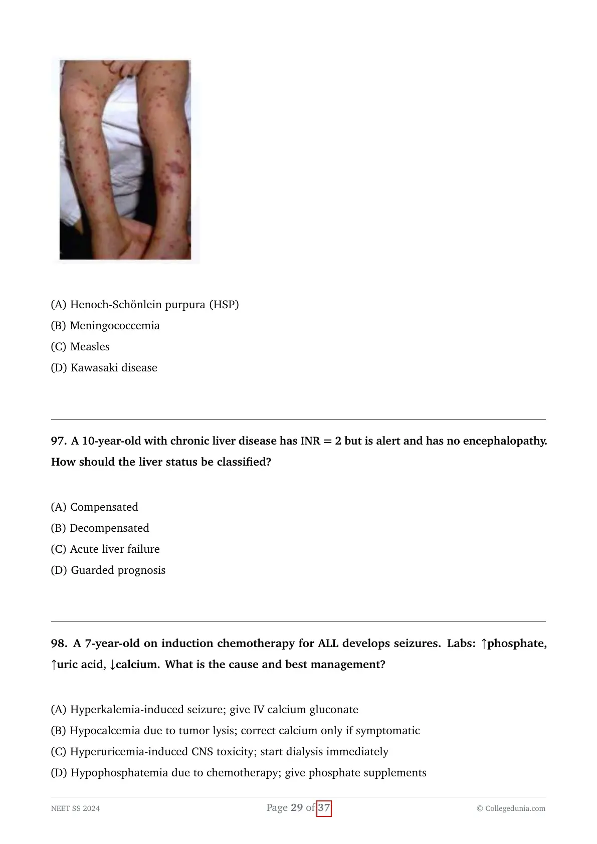
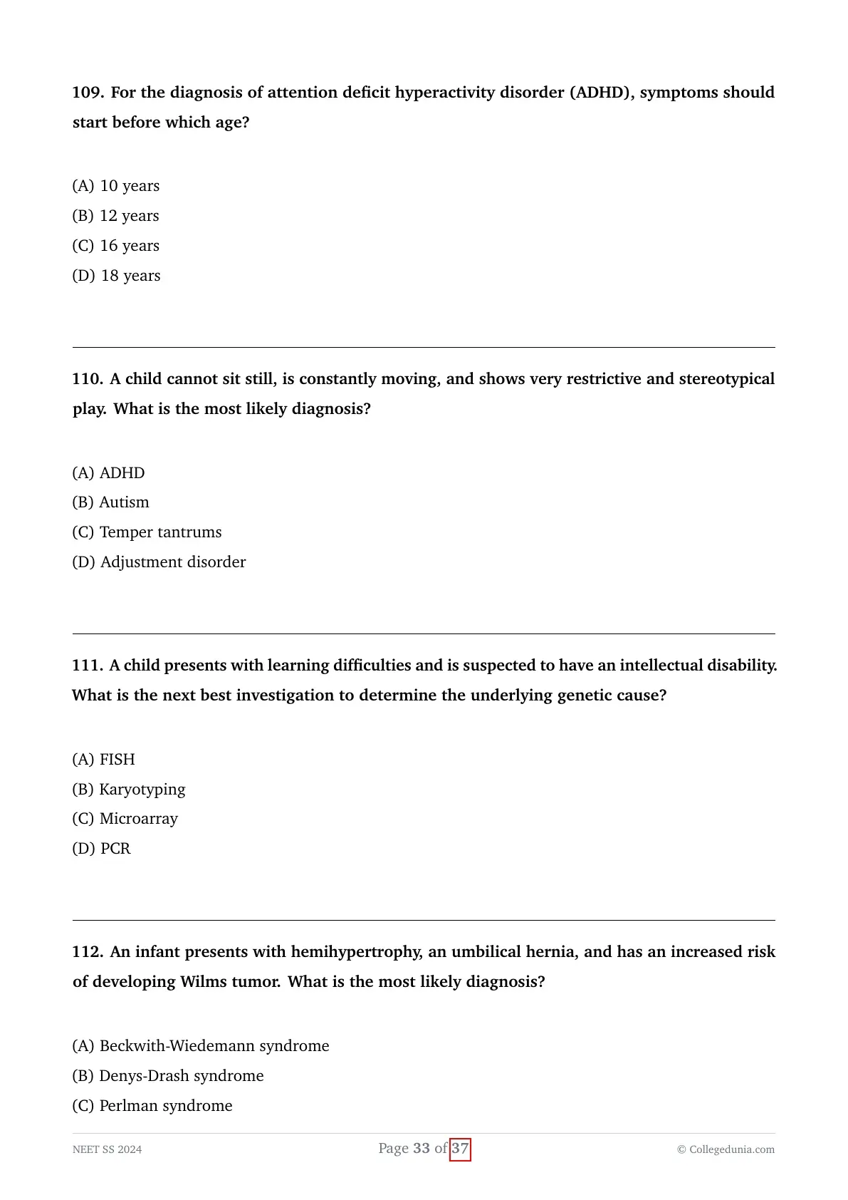
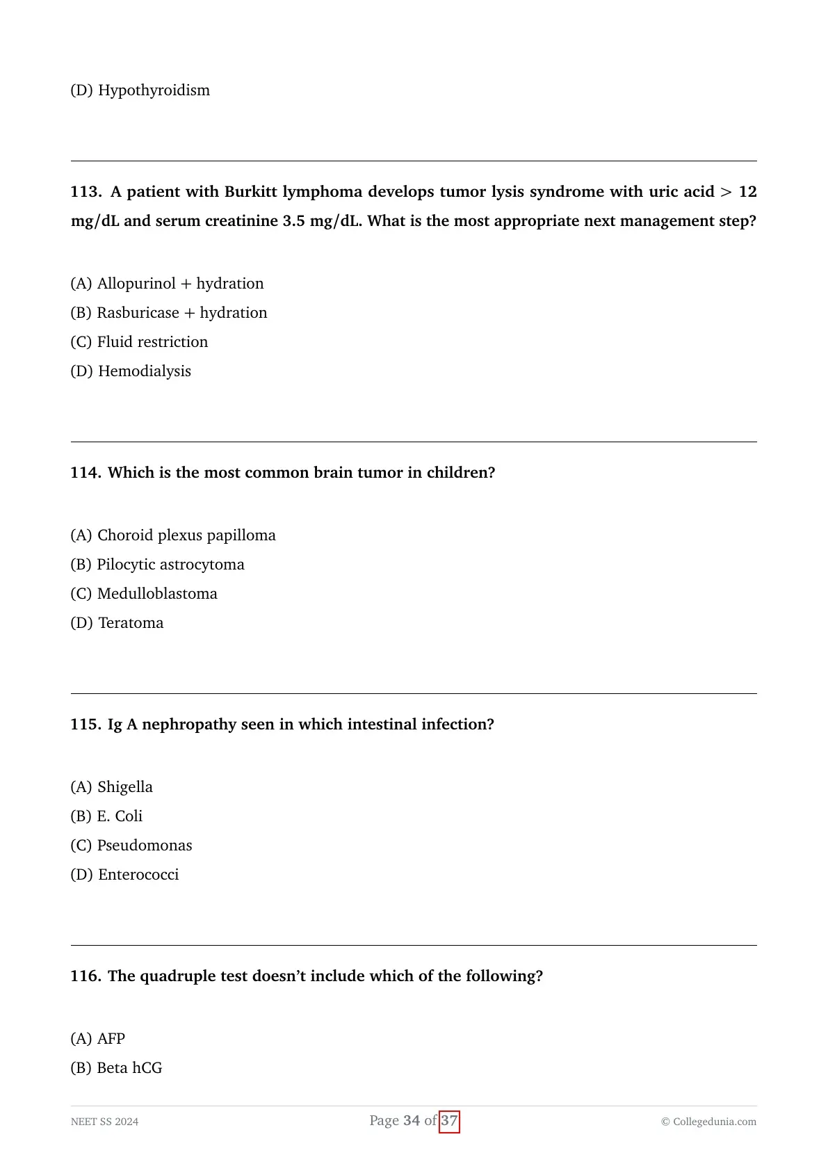
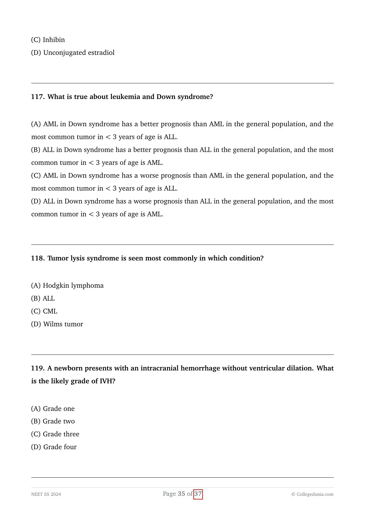
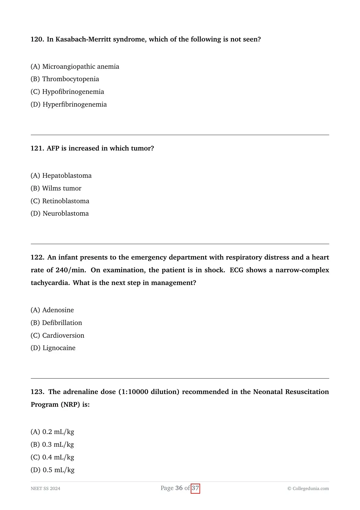
A 2-year-old child presents with persistent fever for 4 days. Examination shows conjunctival injection and periungual desquamation, but criteria for complete Kawasaki disease are not met. You plan to evaluate for incomplete Kawasaki disease as per AHA guidelines. Which of the following investigations is NOT routinely recommended in this evaluation?
View Solution
Concept:
Incomplete (atypical) Kawasaki disease (KD) is suspected in infants and children who have prolonged unexplained fever with fewer than four of the classic diagnostic criteria.
The American Heart Association (AHA) provides a well-defined diagnostic algorithm comprising specific laboratory parameters and echocardiography to guide evaluation and treatment in these suspected cases.
Explanation:
- Kawasaki disease is an acute, self-limiting systemic vasculitis primarily affecting medium-sized arteries, especially the coronary arteries, in children younger than 5 years of age.
- When a child presents with fever for $\ge 4$--$5$ days and fewer than four principal clinical features, an algorithm based on laboratory findings is utilized to confirm incomplete Kawasaki disease.
- The initial workup recommended by the AHA guidelines includes inflammatory markers, specifically C-reactive protein (CRP $\ge 3 mg/dL$) and erythrocyte sedimentation rate (ESR $\ge 40 mm/hr$).
- Supplemental laboratory criteria include:
1. Serum albumin $\le 3.0 g/dL$
2. Anemia for age (evaluated on complete blood count)
3. Platelet count $>450,000/mm^3$ after day 7
4. White blood cell count $\ge 15,000/mm^3$
5. Serum alanine aminotransferase (ALT) elevation
6. Urine $WBC \ge 10/HPF$ (sterile pyuria) - Serum ferritin is not a component of the standard AHA algorithm for incomplete KD; it is predominantly measured when evaluating for secondary Hemophagocytic Lymphohistiocytosis (HLH) or Macrophage Activation Syndrome (MAS).
Final Answer:
Serum ferritin is not routinely recommended as part of the supplemental laboratory algorithm for evaluating incomplete Kawasaki disease according to the AHA guidelines.
A 27-year-old pregnant woman with systemic lupus erythematosus (SLE) is concerned about the risk of congenital heart block in her fetus due to anti-Ro/SSA antibodies. Which of the following medications is not recommended antenatally to prevent cardiac manifestations of neonatal lupus?
View Solution
Concept:
Neonatal lupus erythematosus (NLE) is an acquired autoimmune disorder caused by the transplacental transfer of maternal anti-SSA/Ro and/or anti-SSB/La autoantibodies.
The most severe complication is congenital heart block (CHB), which carries high fetal and neonatal morbidity, necessitating strict antenatal monitoring and safe pharmacotherapy while strictly avoiding teratogens.
Explanation:
- Congenital complete heart block develops predominantly between 16 and 28 weeks of gestation due to antibody-mediated autoimmune injury and subsequent fibrosis of the fetal atrioventricular (AV) node.
- Hydroxychloroquine (HCQ) is strongly recommended during pregnancy in anti-Ro/SSA-positive mothers because it significantly reduces the recurrence and incidence of fetal cardiac lupus by downregulating TLR signaling.
- Fluorinated corticosteroids such as dexamethasone or betamethasone cross the placenta without being inactivated by $11\beta$-hydroxysteroid dehydrogenase type 2 and are utilized to treat emerging incomplete heart block (first- or second-degree) or associated fetal myocarditis.
- Beta-sympathomimetic agents such as terbutaline are often combined with dexamethasone when significant fetal bradycardia (heart rate $<55 bpm$) is present to improve fetal cardiac output.
- Leflunomide is an inhibitor of pyrimidine synthesis that is categorized as Category X (strictly contraindicated in pregnancy) due to potent embryotoxic and teratogenic effects; it must never be used antenatally.
Final Answer:
Leflunomide is strictly contraindicated and not recommended antenatally because of its severe teratogenicity and lack of efficacy for fetal cardiac lupus.
A 2-week-old infant presents with a photosensitive annular erythematous rash on the face. ECG reveals complete heart block. Which is the most likely diagnosis?
View Solution
Concept:
Neonatal lupus erythematosus (NLE) is a passively acquired autoimmune syndrome occurring in infants born to mothers with autoantibodies against Ro/SSA, La/SSB, or rarely U1-RNP.
The hallmark clinical triad consists of cutaneous lesions, congenital conduction abnormalities (most characteristically third-degree AV block), and transient hepatobiliary or hematologic disturbances.
Explanation:
- Cutaneous manifestations typically appear in the first few weeks of life and classically present as annular, polycyclic, erythematous, scaly plaques predominantly located in periorbital (giving a "raccoon-eye" or "owl-eye" appearance) and facial regions following sun exposure.
- The cutaneous rash is transient and gradually resolves within 6 to 9 months as maternal IgG antibodies are cleared from the infant's circulation, leaving minimal or no scarring.
- Cardiac involvement, unlike cutaneous manifestations, is permanent and irreversible because antibody-mediated inflammation triggers fibrosis and calcification of the fetal conduction system.
- Congenital cytomegalovirus (CMV) infection presents with sensorineural hearing loss, periventricular calcifications, and microcephaly, but not heart block.
- Atopic dermatitis typically appears after 2--3 months of age, involves flexural or extensor surfaces, and is not associated with complete heart block.
Final Answer:
The constellation of a photosensitive annular facial rash combined with complete heart block in a neonate is diagnostic of neonatal lupus erythematosus.
A 10-year-old child presents with arthritis involving the wrist and first MCP joint. Examination shows nail pitting. Which of the following is the most likely diagnosis?
View Solution
Concept:
Juvenile psoriatic arthritis (PsA) is a distinct subtype of Juvenile Idiopathic Arthritis (JIA) characterized by the co-occurrence of arthritis and psoriasis or characteristic psoriasis-related clinical features.
According to the International League of Associations for Rheumatology (ILAR) classification criteria, definitive skin psoriasis is not strictly required if other diagnostic features such as nail pitting or dactylitis are present alongside arthritis.
Explanation:
- Juvenile psoriatic arthritis typically presents in a bimodal age distribution: an early peak in preschool-aged females (often presenting as oligoarthritis) and a late childhood peak in males and females.
- The ILAR criteria define juvenile psoriatic arthritis as arthritis with either:
1. Definite psoriasis, OR
2. At least two of the following: dactylitis ("sausage digit"), nail pitting ($>2$ pits) or onycholysis, and a family history of psoriasis in a first-degree relative. - Nail changes, particularly punctate nail pitting, are strongly associated with distal interphalangeal (DIP) and MCP joint arthritis in psoriatic disease.
- Systemic sclerosis causes sclerodactyly and Raynaud phenomenon rather than isolated pitting with oligoarthritis/polyarthritis.
- Systemic lupus erythematosus causes non-deforming, non-erosive (Jaccoud-type) arthritis accompanied by multiorgan involvement and malar rash, not isolated nail pitting.
Final Answer:
The combination of arthritis in peripheral joints along with characteristic nail pitting is diagnostic of juvenile psoriatic arthritis.
A 14-year-old girl with SLE presents with edema and nephrotic-range proteinuria. Renal biopsy shows diffuse thickening of the glomerular basement membrane without proliferative lesions. This corresponds to which class of lupus nephritis?
View Solution
Concept:
Lupus nephritis is categorized according to the International Society of Nephrology/Renal Pathology Society (ISN/RPS) histological classification into six distinct classes based on the pattern and extent of glomerular involvement.
Class V lupus nephritis represents membranous nephropathy, which typically presents clinically with marked proteinuria and full-blown nephrotic syndrome.
Explanation:
- Class I (Minimal mesangial lupus nephritis): Normal glomeruli on light microscopy, with mesangial immune deposits on immunofluorescence.
- Class II (Mesangial proliferative lupus nephritis): Purely mesangial hypercellularity with mesangial immune deposits, without subendothelial deposits.
- Class III (Focal lupus nephritis): Active or inactive endocapillary or extracapillary glomerulonephritis involving $<50\%$ of all glomeruli, usually presenting with nephritic features and active sediment.
- Class IV (Diffuse lupus nephritis): Involves $\ge 50\%$ of glomeruli with marked proliferative and necrotizing lesions, often showing wire-loop lesions and the worst renal prognosis.
- Class V (Membranous lupus nephritis): Characterized by diffuse thickening of the glomerular basement membrane (GBM) due to continuous global or segmental subepithelial immune deposits (demonstrating "spikes" on silver stain) without significant endocapillary proliferation.
- Class VI (Advanced sclerosing lupus nephritis): Global sclerosis of $\ge 90\%$ of glomeruli, representing end-stage scarring.
Final Answer:
Diffuse thickening of the glomerular basement membrane without proliferative lesions in a patient presenting with nephrotic-range proteinuria is the histological hallmark of Class V (Membranous) Lupus Nephritis.
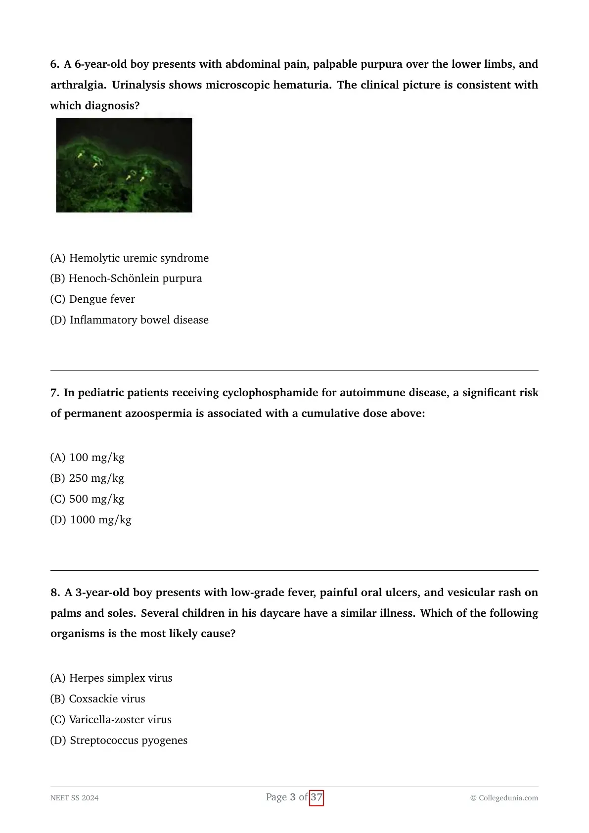
A 6-year-old boy presents with abdominal pain, palpable purpura over the lower limbs, and arthralgia. Urinalysis shows microscopic hematuria. The clinical picture is consistent with which diagnosis?

View Solution
Concept:
Henoch-Schönlein purpura (HSP), also recognized as IgA vasculitis, is the most common systemic small-vessel vasculitis in childhood.
It is characterized pathologically by leukocytoclastic vasculitis with predominant IgA-dominant immune complex deposition in small vessels of the skin, gastrointestinal tract, joints, and kidneys.
Explanation:
- The EULAR/PRINTO/PRES consensus criteria require the presence of palpable purpura (mandatory criterion, predominantly on dependent areas such as buttocks and lower extremities) without thrombocytopenia or coagulopathy, PLUS at least one of the following:
1. Diffuse colicky abdominal pain (often complicated by intussusception)
2. Arthritis or arthralgia (acute onset, non-migratory, non-deforming)
3. Renal involvement (hematuria and/or proteinuria)
4. Biopsy showing predominant IgA deposition - The provided immunofluorescence image typically demonstrates prominent mesangial or capillary wall IgA deposition within the renal glomerulus or dermal venules.
- Hemolytic Uremic Syndrome (HUS) is characterized by the classic triad of microangiopathic hemolytic anemia, thrombocytopenia, and acute kidney injury, usually following bloody diarrhea without palpable purpura.
- Dengue fever presents with acute febrile illness, retro-orbital pain, severe myalgia, thrombocytopenia, and petechial (non-palpable) rash.
Final Answer:
The classic tetrad of palpable purpura, colicky abdominal pain, arthralgia, and renal involvement (microscopic hematuria) is diagnostic of Henoch-Schönlein Purpura (IgA Vasculitis).
In pediatric patients receiving cyclophosphamide for autoimmune disease, a significant risk of permanent azoospermia is associated with a cumulative dose above:
View Solution
Concept:
Cyclophosphamide is a potent alkylating agent used in the induction therapy of severe, life-threatening pediatric autoimmune conditions, such as proliferative lupus nephritis and systemic vasculitis.
Gonadotoxicity is one of its most critical long-term dose-limiting adverse effects, causing destruction of rapidly dividing germ cells (spermatogonia) in males and depletion of primordial ovarian follicles in females.
Explanation:
- Alkylating agents cause cross-linking of DNA, which selectively damages actively proliferating spermatogonia in the testicular seminiferous tubules, leading to oligospermia or irreversible azoospermia.
- In pediatric and adolescent males, the risk of permanent gonadal damage is directly correlated with the cumulative lifetime dose administered.
- A cumulative dose exceeding $250 mg/kg$ (or approximately $>7.5$--$10 g/m^2$) carries a substantially increased and significant risk of permanent germ cell failure and permanent azoospermia.
- Doses below $150$--$200 mg/kg$ generally have a lower risk of permanent infertility, with potential for recovery of spermatogenesis years after cessation of therapy.
- To minimize this risk in pediatric rheumatology, intravenous pulse cyclophosphamide regimens or alternative immunosuppressive agents like mycophenolate mofetil (MMF) or rituximab are preferred over prolonged daily oral cyclophosphamide.
Final Answer:
A cumulative cyclophosphamide dose above $250\text{ mg/kg}$ is associated with a significant risk of permanent azoospermia in male pediatric patients.
A 3-year-old boy presents with low-grade fever, painful oral ulcers, and vesicular rash on palms and soles. Several children in his daycare have a similar illness. Which of the following organisms is the most likely cause?
View Solution
Concept:
Hand-Foot-and-Mouth Disease (HFMD) is a highly contagious, common viral exanthem of early childhood, frequently occurring in institutional settings like daycares and preschools.
It is caused by viruses of the Enterovirus genus within the Picornaviridae family, most commonly Coxsackievirus A16 and Enterovirus A71.
Explanation:
- HFMD presents characteristically with a mild prodrome of low-grade fever, malaise, and poor oral intake followed by the eruption of distinct mucocutaneous lesions.
- The oral enanthem consists of painful erythematous macules that rapidly evolve into small, shallow vesicles and ulcers located on the buccal mucosa, tongue, hard palate, and gingiva.
- The exanthem features non-pruritic, tender or painless oval, elliptical, or football-shaped vesicles on an erythematous base distributed symmetrically over the palms, soles, fingers, toes, and occasionally buttocks.
- Coxsackievirus A16 is the most frequent global cause of typical, mild HFMD, whereas Enterovirus 71 is associated with more severe neurological complications (e.g., aseptic meningitis, encephalitis, and acute flaccid paralysis).
- Herpes simplex virus (HSV-1) primarily causes herpetic gingivostomatitis involving anterior oral cavity and perioral skin without the characteristic palm-and-sole exanthem.
- Varicella-zoster virus produces generalized, highly pruritic polymorphic lesions in crops with a central/centripetal distribution.
Final Answer:
The clinical triad of fever, painful oral ulcers, and vesicular lesions on the palms and soles is typical of Hand-Foot-and-Mouth Disease caused by Coxsackievirus.
A neutropenic child with acute myeloid leukemia presents with fever and cough. CT chest shows a halo sign, and serum galactomannan is positive. Which antifungal is the drug of choice?
View Solution
Concept:
Invasive Pulmonary Aspergillosis (IPA) is a major life-threatening opportunistic fungal infection in severely immunocompromised and prolonged neutropenic pediatric oncology patients.
Prompt recognition via imaging findings (CT chest halo sign) and biomarkers (serum galactomannan assay) followed by immediate targeted antifungal therapy is crucial for survival.
Explanation:
- Aspergillus species (most commonly Aspergillus fumigatus) are angioinvasive molds that cause pulmonary tissue infarction and necrosis.
- The classic CT chest finding is the "halo sign", which represents an area of ground-glass attenuation surrounding a pulmonary nodule or consolidation, corresponding to alveolar hemorrhage surrounding ischemic fungal necrosis.
- Galactomannan is a polysaccharide cell wall component released during active hyphal growth of Aspergillus, serving as a validated diagnostic serological biomarker.
- Voriconazole, an extended-spectrum triazole, is recommended by the Infectious Diseases Society of America (IDSA) and European guidelines as the first-line drug of choice for targeted treatment of invasive aspergillosis.
- Fluconazole is entirely inactive against Aspergillus species (it has activity limited mainly to Candida and Cryptococcus).
- Caspofungin and other echinocandins are considered second-line or salvage agents, not the primary treatment of choice for invasive pulmonary aspergillosis.
Final Answer:
The first-line drug of choice for the treatment of invasive pulmonary aspergillosis is voriconazole.
A 16-year-old boy develops sore throat followed by high fever and rigors. After a week, he presents with septicemia and neck pain. Imaging reveals thrombosis of the internal jugular vein. This clinical scenario is most consistent with:
View Solution
Concept:
Lemierre's syndrome (post-anginal septicemia) is a severe, potentially fatal complication of acute bacterial pharyngitis/tonsillitis seen predominantly in healthy adolescents and young adults.
It is classically characterized by an antecedent oropharyngeal infection leading to secondary septic thrombophlebitis of the internal jugular vein (IJV) and disseminated septic embolization.
Explanation:
- The primary causative organism in the vast majority of cases is Fusobacterium necrophorum, an obligate anaerobic, gram-negative bacillus that is part of the normal oral flora.
- The disease begins as a primary oropharyngeal infection (pharyngitis, peritonsillar abscess, or tonsillitis) that invades the lateral pharyngeal (parapharyngeal) space through contiguous spread or regional lymphatics.
- This invasion triggers severe localized inflammation and septic thrombophlebitis within the adjacent internal jugular vein.
- Patients present with unilateral neck swelling, tenderness along the sternocleidomastoid muscle, spiking fevers with rigors, and signs of systemic sepsis.
- Metastatic septic emboli frequently shower the lungs (causing cavitary nodular infiltrates, empyema, and hemoptysis) and large joints (septic arthritis).
- Contrast-enhanced CT of the neck is the imaging modality of choice, demonstrating intraluminal filling defects in the IJV with surrounding soft tissue inflammation.
Final Answer:
Lemierre's syndrome refers to internal jugular vein septic thrombophlebitis and bacteremia following a primary oropharyngeal infection.
A 10-year-old boy presents with focal seizures. MRI brain shows multiple small ring-enhancing lesions suggestive of neurocysticercosis. What is the appropriate management?
View Solution
Concept:
Neurocysticercosis (NCC) is the most common parasitic infection of the central nervous system, caused by the larval stage of the tapeworm Taenia solium.
The management of active parenchymal neurocysticercosis with multiple lesions requires a multimodal strategy addressing seizure control, inflammation, and antiparasitic eradication.
Explanation:
- Seizures are the most common clinical manifestation of parenchymal NCC in pediatric patients; thus, antiepileptic drug (AED) monotherapy is mandatory as the initial symptomatic step to achieve complete seizure control.
- Cysticidal therapy (albendazole at $15 mg/kg/day$ in two divided doses for 10--14 days) hastens the resolution of active parenchymal cysts and decreases long-term seizure recurrence.
- For patients with multiple non-encephalitic viable parenchymal cysticerci ($>2$ lesions), dual cysticidal therapy combining albendazole and praziquantel ($50 mg/kg/day$) has demonstrated superior cyst clearance compared to albendazole monotherapy.
- Destruction of the parasite releases antigenic material that triggers severe local host inflammatory response and perilesional brain edema; therefore, concomitant corticosteroid therapy (dexamethasone or prednisolone) is essential and must be initiated prior to or simultaneously with cysticidal agents.
Final Answer:
The appropriate comprehensive management of multiple parenchymal lesions in neurocysticercosis consists of Albendazole + Antiepileptics + Corticosteroids $\pm$ Praziquantel.
A 5-day-old newborn presents with mild conjunctivitis and cough. The baby subsequently develops interstitial pneumonia. Gram stain of conjunctival discharge shows no organisms. What is the most likely etiological agent?
View Solution
Concept:
Chlamydia trachomatis (serovars D--K) is a major cause of neonatal conjunctivitis (ophthalmia neonatorum) and afebrile interstitial pneumonitis in early infancy.
Infection is acquired vertically during passage through an infected maternal birth canal during vaginal delivery.
Explanation:
- Neonatal chlamydial conjunctivitis typically presents between 5 and 14 days of life with watery to mucopurulent discharge, chemosis, and pseudomembrane formation.
- Chlamydia trachomatis is an obligate intracellular bacterium and therefore does not show organisms on standard Gram stain, but diagnostic Giemsa staining reveals characteristic basophilic intracytoplasmic inclusion bodies.
- Approximately 10--20\% of infants born to infected mothers develop chlamydial pneumonia, typically presenting at 2 to 12 weeks of age with a classic "staccato" cough, tachypnea, lack of fever, and bilateral diffuse interstitial/hyperinflation patterns on chest radiography.
- Peripheral eosinophilia ($>400 cells/\muL$) and elevated serum immunoglobulins (IgM and IgG) are characteristically observed.
- Neisseria gonorrhoeae presents earlier (days 2--5) with hyperacute, copious, purulent eye discharge and gram-negative intracellular diplococci on Gram stain, but does not cause interstitial pneumonia.
Final Answer:
The clinical course of neonatal conjunctivitis evolving into interstitial pneumonia with negative Gram staining is classic for Chlamydia trachomatis infection.
A 7-year-old boy presents with right lower quadrant abdominal pain and fever, mimicking acute appendicitis. Stool culture grows the likely organism. Which infection best explains this clinical scenario?
View Solution
Concept:
"Pseudoappendicitis" is a recognized clinical syndrome in children and adolescents presenting with acute right lower quadrant abdominal pain, fever, and leukocytosis that closely mimics acute appendicitis.
The classic bacterial pathogen responsible for mesenteric adenitis and terminal ileitis leading to this presentation is Yersinia enterocolitica.
Explanation:
- Yersinia enterocolitica is a gram-negative, non-lactose-fermenting, zoonotic coccobacillus that exhibits psychrotrophic properties (ability to grow at refrigeration temperatures, $4^\circC$).
- Transmission occurs primarily via ingestion of contaminated, undercooked pork products, unpasteurized milk, or contaminated water.
- In older children and adolescents, the organism invades the intestinal mucosa through M cells overlying Peyer's patches, producing marked acute terminal ileitis and mesenteric lymphadenitis.
- This pronounced inflammation localized to the ileocecal region produces tenderness and guarding in the right iliac fossa, leading to surgical consultations for suspected appendicitis where normal appendices are found on laparoscopy/laparotomy.
- Stool culture on selective media, such as Cefsulodin-Irgasan-Novobiocin (CIN) agar, readily confirms the growth of Yersinia.
Final Answer:
The infectious etiology most commonly responsible for right lower quadrant abdominal pain mimicking acute appendicitis (pseudoappendicitis) is Yersinia enterocolitica.
A 6-year-old child with culture-proven Salmonella septicemia presents in cardiogenic shock. Ceftriaxone therapy has already been initiated. What is the next best step in management?
View Solution
Concept:
Severe enteric (typhoid) fever can be complicated by severe septic/cardiogenic shock, altered mental status, stupor, or delirium, carrying substantial mortality.
High-dose adjunctive corticosteroid therapy alongside effective bactericidal antibiotics is proven to reduce mortality in severe, complicated typhoid state shock.
Explanation:
- Severe typhoid fever is mediated by a hyperinflammatory cytokine storm triggered by endotoxin and macrophage activation during systemic bacteremia.
- Landmark clinical trials have demonstrated that adding high-dose intravenous dexamethasone to standard antimicrobials significantly reduces mortality in severe typhoid fever associated with shock, coma, delirium, or stupor.
- The recommended regimen consists of intravenous dexamethasone: an initial loading dose of $3 mg/kg$ infused over 30 minutes, followed by $1 mg/kg$ every 6 hours for an additional 48 hours (total of 8 doses).
- Third-generation cephalosporins (such as ceftriaxone) remain the antimicrobial backbone; switching or substituting antibiotics is unnecessary when the organism is responsive and already on effective coverage.
- Dexamethasone acts by blunting the massive systemic release of pro-inflammatory cytokines, including TNF-$\alpha$, IL-1, and IL-6, which drive systemic vasodilation and myocardial depression.
Final Answer:
In severe culture-proven Salmonella septicemia complicated by shock or encephalopathy, adding high-dose intravenous dexamethasone is the next best life-saving step.
A 5-year-old boy is diagnosed with Plasmodium vivax malaria and presents with danger signs (drowsiness, persistent vomiting). What is the most appropriate initial treatment?
View Solution
Concept:
Severe malaria can be caused by both Plasmodium falciparum and Plasmodium vivax.
The presence of danger signs (e.g., impaired consciousness/drowsiness, persistent vomiting, severe anemia, jaundice, shock, or respiratory distress) defines complicated/severe malaria and warrants immediate parenteral antimalarial therapy regardless of the species.
Explanation:
- According to World Health Organization (WHO) and National Malaria Guidelines, all cases of severe malaria must be treated urgently with intravenous or intramuscular artesunate as the drug of choice.
- The recommended pediatric dose of IV artesunate is $2.4 mg/kg$ body weight administered intravenously at 0, 12, and 24 hours, and then once daily until the child can tolerate oral medications.
- Intravenous artesunate rapidly reduces parasitemia, is well-tolerated, and has a significantly superior survival benefit compared to intravenous quinine or chloroquine.
- Oral antimalarials (such as oral ACT or oral chloroquine) are strictly contraindicated as initial therapy in the presence of persistent vomiting and impaired consciousness due to the risk of poor absorption and aspiration.
- Once the patient is clinically stabilized and able to tolerate oral medication, a complete course of oral Artemisinin-based Combination Therapy (ACT) is completed, followed by primaquine (after testing for G6PD deficiency) for radical cure of hypnozoites in P. vivax.
Final Answer:
The drug of choice for the immediate management of severe malaria with danger signs is intravenous (IV) artesunate.
In a case of extra-thoracic airway obstruction, where does the airway collapse occur and during which phase of respiration?
View Solution
Concept:
The mechanics of airway collapse during the respiratory cycle depend heavily on whether the obstruction is extrathoracic (e.g., larynx, upper cervical trachea) or intrathoracic (e.g., intrathoracic trachea, bronchi).
Dynamic pressure differentials between the intraluminal airway pressure and surrounding atmospheric or pleural pressure dictate the phase and site of dynamic airway narrowing.
Explanation:
- In an extrathoracic airway segment (above the thoracic inlet), the outer surface of the airway is exposed to atmospheric pressure ($P_{atm}$).
- During inspiration, the generation of negative intrathoracic pressure by the diaphragm creates subatmospheric intraluminal pressure within the extrathoracic airway ($P_{luminal} < P_{atm}$).
- When the intraluminal pressure drops below the surrounding atmospheric pressure, a positive transmural pressure gradient acts inward across the compliant airway walls, leading to dynamic narrowing/collapse during inspiration.
- By contrast, during expiration, the airway pressure inside the extrathoracic lumen increases above atmospheric pressure ($P_{luminal} > P_{atm}$), which forces the extrathoracic airway open.
- Consequently, extrathoracic lesions characteristically manifest with dynamic collapse and inspiratory stridor, whereas intrathoracic airway lesions collapse during expiration to produce expiratory wheeze.
Final Answer:
In extrathoracic airway obstruction, dynamic collapse occurs prominently during inspiration.
A 3-year-old child accidentally swallowed a coin. X-ray shows the coin is now in the stomach, and the child is asymptomatic. What is the correct next step?
View Solution
Concept:
Ingested foreign bodies in children are a common pediatric emergency.
Management algorithms depend strictly on the anatomical location of the foreign object, its characteristics (e.g., blunt coin vs. button battery vs. sharp object vs. multiple magnets), and the presence or absence of symptoms.
Explanation:
- A smooth, blunt foreign body such as a standard coin that has successfully negotiated the lower esophageal sphincter into the stomach will pass through the remainder of the gastrointestinal tract uneventfully in $>95\%$ of cases.
- For an asymptomatic child with a coin confirmed in the stomach on radiograph, the gold-standard recommendation is expectant management (observation) on a normal diet while monitoring stools for passage.
- Endoscopic removal is only indicated if the coin remains lodged in the stomach for longer than $2$--$4$ weeks without progressing, or if the child develops symptoms such as abdominal pain, vomiting, or gastrointestinal bleeding.
- Urgent endoscopic intervention is reserved for coins lodged in the esophagus, button batteries in the esophagus/stomach, sharp-pointed objects, or multiple magnets.
- Cathartics, emetics, and laxatives are completely unnecessary, unproven, and potentially harmful in pediatric foreign body ingestion.
Final Answer:
The correct and safe next step for an asymptomatic child with a coin in the stomach is observation to allow spontaneous passage through the gastrointestinal tract.
What is the first line management of raised ICP in children?
View Solution
Concept:
Increased intracranial pressure (ICP) is a life-threatening pediatric emergency that can lead to brain herniation and secondary ischemic brain injury.
Management is structured in a stepwise, tiered approach, beginning with foundational baseline neuroprotective and physiological stabilization measures.
Explanation:
- Tier 0 / Baseline First-Line Measures:
1. Airway stabilization, adequate oxygenation, and normocapnia ($PaCO_2$ $35$--$40 mmHg$)
2. Maintaining cerebral perfusion pressure by securing hemodynamic stability
3. Elevating the head of the bed to $30^\circ$ with the neck kept in a neutral midline position to maximize cerebral venous drainage without compromising arterial perfusion
4. Normothermia maintenance and adequate analgesia/sedation to minimize metabolic demand - Tier 1 Measures: Initiated if ICP remains elevated despite baseline optimization; includes osmotherapy using Hypertonic ($3\%$) Saline or Mannitol.
- Tier 2 Measures: Neuromuscular blockade, mild hyperventilation ($PaCO_2$ $30$--$35 mmHg$) as a temporary bridge.
- Tier 3 / Refractory Measures: High-dose barbiturate coma, moderate therapeutic hypothermia, or emergency decompressive craniectomy.
- Aggressive hyperventilation to $PaCO_2 < 30 mmHg$ causes severe cerebral vasoconstriction and is contraindicated as it triggers profound cerebral ischemia.
Final Answer:
The primary first-line baseline management of raised ICP begins with airway stabilization and head-of-bed elevation to $30^\circ$ in the midline.
A child was involved in an RTA. Below the CT image is given., What is the diagnosis?
View Solution
Concept:
Traumatic brain injury (TBI) from road traffic accidents (RTA) in children commonly results in extra-axial intracranial hemorrhages.
Non-contrast head CT is the gold standard imaging modality for distinguishing epidural hematoma (EDH) from subdural hematoma (SDH) and intraparenchymal contusions.
Explanation:
- Epidural Hematoma (EDH):
- Results from arterial bleeding, most classically rupture of the middle meningeal artery associated with a temporal/parietal bone fracture.
- Appears on CT as a classic well-defined, hyperdense, biconvex (lenticular) collection.
- The hematoma is confined by the tight adherence of the dura mater to the cranial sutures, so it does not cross cranial suture lines (but can cross the falx/tentorium). - Subdural Hematoma (SDH):
- Results from tearing of bridging cortical veins.
- Appears as a crescent-shaped (concavo-convex) hyperdense collection that crosses cranial sutures along the hemisphere, limited only by dural reflections (falx cerebri). - Intraparenchymal Hemorrhage:
- Appears as hyperdense hemorrhagic foci within the brain parenchyma itself, typically associated with surrounding edema.
Final Answer:
The classic biconvex, lenticular hyperdense lesion limited by cranial sutures on head CT following trauma is diagnostic of an Epidural Hematoma (EDH).
$\bullet$ EDH = Biconvex / Lenticular shape | Middle meningeal artery | Does NOT cross suture lines.
$\bullet$ SDH = Crescent shape | Bridging cortical veins | CROSSES suture lines.
A child presents with tachypnea, vomiting, and altered sensorium after accidentally ingesting a drug used by his father. Labs show respiratory alkalosis and hyperglycemia. Which of the following is most likely the ingested drug?
View Solution
Concept:
Salicylate (aspirin) toxicity is a classic, complex poisoning presenting with a hallmark biphasic or mixed acid-base disturbance and characteristic metabolic derangements.
It directly stimulates the central respiratory center while simultaneously uncoupling oxidative phosphorylation in cellular mitochondria.
Explanation:
- Direct stimulation of the medullary respiratory center by salicylates leads to hyperventilation, causing marked tachypnea, hyperpnea, and an early primary respiratory alkalosis.
- Concurrently, salicylates uncouple mitochondrial oxidative phosphorylation and inhibit Krebs cycle dehydrogenases, resulting in cellular anaerobic metabolism, accumulation of lactic acid, and ketoacids, which drives a high anion gap metabolic acidosis.
- In older children and adults, the classic hallmark is a mixed primary respiratory alkalosis and primary metabolic acidosis. In toddlers, metabolic acidosis may dominate early.
- Metabolic derangements include hyperglycemia (early, due to catecholamine-induced glycogenolysis) or profound neuroglycopenia/hypoglycemia.
- Iron toxicity presents with severe hemorrhagic gastroenteritis, shock, and high anion gap metabolic acidosis, but not primary respiratory alkalosis.
- Tricyclic antidepressants (TCAs) cause anticholinergic toxidrome, sedation, respiratory depression (respiratory acidosis), and QRS widening on ECG.
Final Answer:
The classic clinical picture of tachypnea, vomiting, altered sensorium, primary respiratory alkalosis, and hyperglycemia following ingestion is indicative of Aspirin (salicylate) toxicity.
Venturi mask delivers oxygen and flow?
View Solution
Concept:
The Venturi (air-entrainment) mask is a high-flow oxygen delivery device designed to deliver a precise, fixed, and predictable fraction of inspired oxygen ($\text{FiO}_2$) regardless of the patient's respiratory rate or tidal volume.
It operates based on the Bernoulli principle and jet mixing, entraining room air through calibrated side ports.
Explanation:
- Venturi masks use color-coded interchangeable adapters or adjustable barrel valves that entrain specific proportions of ambient room air with pure oxygen.
- The system delivers a precise $FiO_2$ ranging from $24\%$ to $50\%$ (or up to $60\%$), utilizing oxygen flow rates between $2$ and $15 L/min$.
- Standard color codes and settings include:
- Blue: $24\% FiO_2$ at $2--4 L/min$
- White: $28\% FiO_2$ at $4--6 L/min$
- Yellow / Orange: $31\%--35\% FiO_2$ at $6--8 L/min$
- Red / Green: $40\%--50\% FiO_2$ at $8--15 L/min$ - Because the total flow delivered (oxygen plus entrained air) exceeds the patient's peak inspiratory flow rate, the delivered $FiO_2$ remains constant and accurate, making it ideal for patients with chronic hypercapnic respiratory failure.
Final Answer:
The Venturi mask delivers a controlled $\text{FiO}_2$ of $24\%\text{--}50\%$ at flow rates of $2\text{--}15\text{ L/min}$.
$\bullet$ Nasal Cannula: $24\text{--}44\%$ ($1\text{--}6\text{ L/min}$)
$\bullet$ Simple Face Mask: $35\text{--}55\%$ ($5\text{--}10\text{ L/min}$)
$\bullet$ Venturi Mask: $24\text{--}50\%$ ($2\text{--}15\text{ L/min}$, high-flow, fixed $\text{FiO}_2$)
$\bullet$ Non-Rebreather Mask: $60\text{--}90\%$ ($10\text{--}15\text{ L/min}$)
Capillary Wedge pressure is decreased in what type of shock?
View Solution
Concept:
Pulmonary Capillary Wedge Pressure (PCWP) provides an accurate indirect estimate of left ventricular end-diastolic pressure (LVEDP) and left atrial filling pressure (preload).
Hemodynamic profiling using PCWP, Cardiac Output/Index, and Systemic Vascular Resistance (SVR) allows precise classification of the underlying type of circulatory shock.
Explanation:
- Distributive Shock (e.g., Early/Warm Septic Shock):
- Widespread arterial and venous vasodilation due to inflammatory mediators leads to pooling of blood in capacitance vessels and increased capillary permeability.
- This causes a substantial reduction in effective circulating volume and venous return, leading to a decreased PCWP ($<8--12 mmHg$) and decreased SVR, typically with a compensatory elevated cardiac output. - Cardiogenic Shock:
- Primary pump failure causes blood to back up into the pulmonary circulation, leading to an elevated PCWP ($>15--18 mmHg$) and decreased cardiac output with elevated SVR. - Obstructive Shock (e.g., Cardiac Tamponade, Tension Pneumothorax):
- Extrinsic compression impairs diastolic filling; intracardiac/wedge pressures are characteristically elevated or equalized. - Late/Cold Septic Shock:
- Secondary myocardial depression and profound hypovolemia occur; if fluid resuscitation has taken place, PCWP may normalize or elevate due to myocardial dysfunction.
Final Answer:
Capillary wedge pressure is decreased in distributive shock states, such as Early Septic Shock, due to systemic vasodilation and reduced venous return.
$\bullet$ Hypovolemic: PCWP $\downarrow$, CO $\downarrow$, SVR $\uparrow$
$\bullet$ Cardiogenic: PCWP $\uparrow$, CO $\downarrow$, SVR $\uparrow$
$\bullet$ Early Septic (Distributive): PCWP $\downarrow$ (or normal), CO $\uparrow$, SVR $\downarrow$
$\bullet$ Obstructive: PCWP $\uparrow$ (or equalized), CO $\downarrow$, SVR $\uparrow$
A child was operated on for a Posterior urethral Valve abnormality. What will be the electrolyte abnormality?
View Solution
Concept:
Posterior urethral valves (PUV) cause severe congenital lower urinary tract obstruction in male infants, leading to secondary obstructive uropathy and renal dysplasia.
Surgical valve ablation relieves the anatomical obstruction but frequently triggers a profound, life-threatening post-obstructive diuresis (POD) accompanied by severe fluid and electrolyte disturbances.
Explanation:
- Relief of severe bilateral urinary tract obstruction precipitates post-obstructive diuresis due to accumulated urea excretion, fluid retention, and transient or permanent tubular dysfunction.
- Chronic elevated backpressure disrupts the renal medullary architecture and induces medullary insensitivity of the collecting ducts to antidiuretic hormone (acquired nephrogenic diabetes insipidus).
- Impaired collecting duct responsiveness to vasopressin leads to massive excretion of hyposthenuric, dilute urine containing excessive free water relative to solutes.
- If free water losses are not aggressively and accurately replaced milliliter-for-milliliter, the rapid unreplaced excretion of hypotonic urine results in severe dehydration and hypovolemic hypernatremia.
- In addition, impaired distal tubular hydrogen and potassium secretion during obstructive uropathy may cause type 4 renal tubular acidosis, but marked free water wasting following surgical relief primarily manifests as hypernatremia.
Final Answer:
Following relief of posterior urethral valves, acquired nephrogenic diabetes insipidus and profound post-obstructive free water loss characteristically lead to hypernatremia.
Which of the following statements about PDA is not true?
View Solution
Concept:
Patent ductus arteriosus (PDA) is a persistent vascular structure derived embryologically from the distal portion of the left sixth aortic arch.
In normal fetal circulation, the ductus arteriosus shunts deoxygenated blood away from high-resistance fetal pulmonary vasculature into the descending aorta.
Explanation:
- Anatomically, the ductus arteriosus originates from the bifurcation of the main pulmonary trunk (or origin of the left pulmonary artery) and inserts into the aortic isthmus distal to the origin of the left subclavian artery.
- Stating that the PDA is located proximal to the left subclavian artery is anatomically incorrect; preductal coarctation occurs proximal to it, but the PDA itself inserts distally.
- The PDA provides a direct connection between the pulmonary arterial tree and the descending thoracic aorta.
- Small ("restrictive") PDAs generate minimal left-to-right shunting, preserve normal pulmonary pressures, and are typically asymptomatic, often discovered incidentally during routine auscultation.
- The classic auscultatory finding of a hemodynamically significant PDA is a continuous "machinery" (Gibson) murmur, heard loudest in the left upper sternal border/infraclavicular area, spilling past the second heart sound ($S_2$) into diastole.
Final Answer:
The false statement is that the PDA is located proximal to the left subclavian artery; it anatomically inserts distal to the left subclavian artery.
Which is the correct statement regarding fetal circulation?
View Solution
Concept:
The fetal circulation is uniquely adapted to receive oxygenated, nutrient-rich blood from the placenta and direct it preferentially toward developing vital organs (the brain and myocardium) through specialized fetal shunts.
These key anatomical shunts include the ductus venosus, foramen ovale, and ductus arteriosus.
Explanation:
- Well-oxygenated blood ($80$--$85\%$ oxygen saturation) enters the fetus from the placenta via the single umbilical vein.
- Approximately $50\%$ of umbilical venous blood enters the hepatic microcirculation to perfuse the liver, while the remaining $50\%$ bypasses the liver parenchyma via the ductus venosus directly into the inferior vena cava (IVC).
- Oxygenated IVC blood reaches the right atrium (RA) where the Eustachian valve and the crista dividens preferentially streamline and shunt the majority of this high-velocity flow across the foramen ovale into the left atrium (LA).
- From the LA, oxygen-rich blood passes to the left ventricle (LV) and is ejected into the ascending aorta to preferentially supply the cerebral and coronary circulations.
- Superior vena cava (SVC) blood is deoxygenated ($30$--$40\%$ saturation) and drains directly through the tricuspid valve into the right ventricle (RV), not the left atrium, and is subsequently pumped into the pulmonary artery and across the ductus arteriosus.
Final Answer:
The correct statement is that blood from the IVC is preferentially directed from the right atrium across the foramen ovale into the left atrium.
Which of the following is true about VSD?
View Solution
Concept:
Ventricular septal defect (VSD) is the most common congenital heart malformation, characterized by an abnormal communication between the left and right ventricles.
The hemodynamic consequences of a VSD depend directly on the anatomical size of the defect and the ratio of pulmonary to systemic vascular resistance ($Q_p/Q_s$).
Explanation:
- Small (restrictive) VSDs: High resistance to flow generates a large left-to-right pressure gradient, producing a loud, harsh, pansystolic murmur (maladie de Roger) with a systolic thrill, without chamber dilation or left ventricular hypertrophy (LVH). Most small defects close spontaneously ($>75\%$) and do not require surgery.
- Large (non-restrictive) VSDs: Equalize left and right ventricular pressures, resulting in massive left-to-right shunting and pulmonary overcirculation.
- The increased pulmonary venous return overloads the left atrium and left ventricle, leading to left ventricular volume overload, LVH (and biventricular hypertrophy), and congestive heart failure within the first $2$--$8$ weeks of life as pulmonary vascular resistance drops.
- Because there is minimal pressure gradient across a large non-restrictive VSD, its murmur is softer and less harsh than that of a small restrictive defect.
Final Answer:
Large ventricular septal defects cause significant left-to-right shunting, leading to left ventricular volume overload, LVH, and congestive heart failure.
Which of the following is a known complication of subpulmonic VSD
View Solution
Concept:
Subpulmonic (also called doubly committed subarterial, supracristal, or infundibular) VSD is a defect located immediately beneath both the aortic and pulmonary semilunar valve rings.
This defect is particularly prevalent in Asian populations (accounting for up to $30\%$ of all VSDs in these groups) and carries a well-known risk of progressive valve distortion.
Explanation:
- In subpulmonic VSD, there is a lack of superior muscular and fibrous support beneath the right coronary cusp (and occasionally the non-coronary cusp) of the aortic valve.
- During systole, high-velocity jet flow through the defect creates a localized low-pressure field (the Venturi effect) that sucks the unsupported right coronary aortic cusp down into the defect.
- This repetitive mechanical pulling leads to progressive cusp elongation, cusp prolapse, and progressive aortic regurgitation (AR).
- Once aortic regurgitation develops, it tends to progress rapidly and rarely resolves spontaneously; therefore, early surgical closure is indicated whenever cusp prolapse or AR is detected, regardless of shunt magnitude.
Final Answer:
Aortic regurgitation resulting from aortic valve cusp prolapse via the Venturi effect is a well-established classic complication of subpulmonic (supracristal) VSD.
A 2-year-old child with cyanotic congenital heart disease presents with sudden onset of deep cyanosis and irritability while crying. On evaluation, the child is diagnosed with a hypercyanotic spell. Regarding management and timing of surgery in Tetralogy of Fallot, which of the following is most accurate?
View Solution
Concept:
Hypercyanotic ("tet") spells are acute, life-threatening paroxysmal episodes of severe hypoxemia in children with Tetralogy of Fallot (TOF).
They occur due to acute infundibular spasm or a decrease in systemic vascular resistance (SVR), which dramatically increases right-to-left shunting across the non-restrictive VSD.
Explanation:
- Acute medical management of a hypercyanotic spell focuses on increasing SVR, decreasing infundibular spasm, and calming the child (knee-chest position, high-flow oxygen, morphine, IV fluid bolus, $\beta$-blockers like propranolol/esmolol, and systemic vasoconstrictors like phenylephrine).
- The occurrence of even a single hypercyanotic spell indicates severe, unstable dynamic infundibular obstruction and serves as an absolute indication for urgent/early surgical repair or palliative shunt surgery.
- Elective complete surgical repair of TOF is routinely performed in infancy (typically between $3$ and $6$ months of age), not delayed until 10 years of age.
- Chronic hypoxemia triggers secondary erythrocytosis, which substantially increases blood viscosity and elevates the risk of cerebral thromboembolism and brain abscess (especially in the setting of relative iron deficiency).
Final Answer:
The development of hypercyanotic spells represents a major surgical indication for expedited intervention in children with Tetralogy of Fallot.
A 2-day-old neonate presents with pallor and cyanosis. Chest X-ray shows decreased pulmonary blood flow, and ECG reveals left axis deviation. Which is the most likely diagnosis?
View Solution
Concept:
Tricuspid atresia is a cyanotic congenital heart lesion characterized by complete agenesis of the tricuspid valve, leading to absent direct communication between the right atrium and right ventricle.
It produces a classic electrocardiographic signature that uniquely separates it from nearly all other cyanotic heart defects in neonates.
Explanation:
- In normal neonates, right ventricular dominance produces a physiological right axis deviation (RAD) and right ventricular hypertrophy (RVH) on ECG.
- In tricuspid atresia, systemic venous blood in the right atrium must shunt right-to-left through an obligatory ASD/PFO into the left atrium, where it mixes with pulmonary venous blood and enters the dominant left ventricle.
- Because the right ventricle is hypoplastic and the left ventricle handles total systemic and pulmonary cardiac output, the neonatal ECG characteristically demonstrates Left Axis Deviation (LAD, typically $-30^\circ to -90^\circ$) and Left Ventricular Hypertrophy (LVH).
- Chest radiography shows decreased pulmonary vascular markings (oligemic lung fields) because of severe subpulmonary stenosis or pulmonary atresia.
- By comparison, Tetralogy of Fallot, TGA, and Truncus arteriosus exhibit right axis deviation and right ventricular dominance on neonatal ECG.
Final Answer:
A cyanotic neonate with decreased pulmonary blood flow and an ECG demonstrating left axis deviation with LVH has the hallmark findings of Tricuspid Atresia.
A 10-year-old boy presents with exertional dyspnea and cyanosed lips. On examination, the precordium is silent, a systolic click is heard, but no cardiac murmur is present. Which is the most likely diagnosis?
View Solution
Concept:
Eisenmenger syndrome is the end-stage complication of an uncorrected large systemic-to-pulmonary (left-to-right) shunt, such as a large VSD, ASD, or PDA.
Severe, irreversible pulmonary arterial hypertension develops, leading to equalization of pressures and eventual shunt reversal (right-to-left) with central cyanosis.
Explanation:
- Chronic pulmonary overcirculation induces irreversible vascular remodeling with plexiform lesions, muscular hypertrophy of the media, and intimal proliferation in the pulmonary arterial bed.
- When pulmonary vascular resistance equals or exceeds systemic vascular resistance, the original left-to-right shunt diminishes and then reverses to a right-to-left shunt, causing central cyanosis, polycythemia, and digital clubbing.
- With equalization of ventricular or arterial pressures across the defect, high-velocity turbulent flow ceases; hence, the original systolic murmur disappears (the precordium becomes "silent").
- Auscultation classically reveals physical signs of severe pulmonary hypertension: a single or narrowly split, palpable and loudly accentuated pulmonic second sound ($P_2$), a pulmonary ejection systolic click due to sudden dilation of the main pulmonary artery, and possibly a high-pitched early diastolic Graham Steell murmur of functional pulmonary regurgitation.
Final Answer:
A child with cyanosis, disappearance of a previously present shunt murmur (silent precordium), and an audible pulmonary ejection click is exhibiting classic Eisenmenger syndrome.
A 14-year-old adolescent is noted to have a mid-systolic click on cardiac auscultation. Regarding mitral valve prolapse in children, which of the following is true?
View Solution
Concept:
Mitral valve prolapse (MVP), or Barlow syndrome, is characterized by superior displacement of abnormally thickened, redundant mitral valve leaflets into the left atrium during ventricular systole.
It is the most common valvular abnormality in adolescents and young adults and is frequently associated with heritable connective tissue disorders such as Marfan syndrome and Ehlers-Danlos syndrome.
Explanation:
- MVP is rare in infants and young children; its prevalence steadily increases with age throughout adolescence and into adulthood as myxomatous degenerative changes in the valve leaflets progress over time.
- Pathophysiologically, MVP is characterized by myxomatous expansion of the spongiosa layer with accumulation of glycosaminoglycans and disruption of collagen bundles, predominantly involving the posterior mitral leaflet (or both leaflets), rather than isolated septal/anterior elongation.
- Most pediatric and adolescent patients with MVP are entirely asymptomatic and have mild or no mitral regurgitation; significant MR is uncommon in childhood.
- The classic auscultatory finding is a non-ejection mid-to-late systolic click, often followed by a high-pitched late systolic murmur if mitral regurgitation is present. Maneuvers that decrease LV volume (such as standing or Valsalva) cause the click and murmur to occur earlier in systole.
Final Answer:
Mitral valve prolapse exhibits an age-dependent increase in prevalence, becoming significantly more evident during late childhood and adolescence.
An infant presents with tachypnea, diaphoresis during feeds, and poor weight gain. ECG reveals deep Q waves in leads I and aVL with ST depression in lateral leads. Echocardiography shows dilated LV with reduced ejection fraction. Which is the most likely diagnosis?
View Solution
Concept:
Anomalous Origin of the Left Coronary Artery from the Pulmonary Artery (ALCAPA), also known as Bland-White-Garland syndrome, is a rare but critical congenital coronary anomaly.
It leads to progressive myocardial ischemia, anterolateral left ventricular infarction, and severe heart failure in early infancy.
Explanation:
- In the early neonatal period, high pulmonary vascular resistance (PVR) maintains antegrade perfusion of the anomalous LCA from the pulmonary artery with relatively desaturated blood.
- As PVR physiological drops during the first $4$--$8$ weeks of life, perfusion pressure in the pulmonary artery falls below systemic levels. This causes a coronary steal phenomenon, where blood flows retrogradely from right coronary collaterals through the LCA into the low-pressure pulmonary artery.
- This produces severe myocardial ischemia and infarction of the anterolateral left ventricle, manifesting clinically during feeds (infantile angina) with diaphoresis, irritability, pallor, tachypnea, and failure to thrive.
- The pathognomonic ECG findings are deep, wide Q waves ($>0.04 s$ duration, $>3 mm$ depth) with inverted T waves in lateral leads (I, aVL, $V_5$--$V_6$), representing anterolateral myocardial infarction.
- Echocardiography demonstrates a dilated left ventricle with impaired systolic function, fibroelastosis, and retrograde flow from the LCA into the main pulmonary trunk on color Doppler.
Final Answer:
The presentation of infantile heart failure with anterolateral infarction pattern (deep Q waves in I and aVL) and dilated LV is diagnostic of ALCAPA.
Which of the following statements regarding aortic stenosis in children is correct?
View Solution
Concept:
Congenital aortic stenosis (AS) encompasses a spectrum of left ventricular outflow tract (LVOT) obstructions, anatomically categorized into valvular ($70\%$), subvalvular ($15$--$20\%$), and supravalvular ($5$--$10\%$) levels.
Valvular AS is the most common form in children, predominantly caused by a congenital bicuspid or dysplastic aortic valve.
Explanation:
- Congenital valvular aortic stenosis is frequently associated with other left-sided obstructive cardiac lesions, most notably Coarctation of the Aorta (found in up to $20$--$30\%$ of cases, as part of the Shone complex).
- Valvular aortic stenosis is the most common anatomical subtype, making the statement that subvalvular is more common incorrect.
- Severe aortic stenosis delays left ventricular ejection time, causing delayed closure of the aortic valve component ($A_2$) relative to the pulmonic component ($P_2$), leading to paradoxical (reversed) splitting of $S_2$ (splits during expiration and closes during inspiration).
- Bicuspid aortic valve (BAV) is the most common congenital cardiac anomaly overall (prevalence of $1$--$2\%$) and exhibits high familial heritability (autosomal dominant pattern with variable penetrance, linked to NOTCH1 mutations).
Final Answer:
Congenital aortic stenosis is well known to be frequently associated with coarctation of the aorta.
A 15-year-old adolescent presents with exertional syncope. Echocardiogram shows asymmetric septal hypertrophy with LVOT obstruction. Which of the following is the treatment of choice?
View Solution
Concept:
Hypertrophic Obstructive Cardiomyopathy (HOCM) is a genetic disorder of cardiac sarcomere proteins characterized by unexplained left ventricular hypertrophy, predominantly affecting the interventricular septum.
Dynamic left ventricular outflow tract (LVOT) obstruction results from the combination of septal hypertrophy and systolic anterior motion (SAM) of the anterior mitral valve leaflet.
Explanation:
- First-line medical therapy for symptomatic HOCM with LVOT obstruction consists of Beta-blockers (e.g., propranolol, atenolol, metoprolol).
- Beta-blockers exert negative inotropic effects (reducing contractility and lessening SAM) and negative chronotropic effects (prolonging ventricular diastole, allowing better LV filling, and increasing end-diastolic volume), thereby expanding the LVOT and reducing dynamic gradient and exertional symptoms.
- Vasodilators (such as nitroprusside or ACE inhibitors) are strictly contraindicated because decreasing afterload exacerbates dynamic LVOT obstruction.
- Pure diuretics worsen LVOT obstruction by reducing preload and decreasing LV cavity size.
- Positive inotropes like digoxin and dobutamine are strictly contraindicated because increasing contractility worsens the LVOT gradient.
Final Answer:
Beta-blockers are the first-line medical treatment of choice for reducing dynamic LVOT obstruction and symptoms in HOCM.
$\bullet$ DO GIVE: Beta-blockers (first-line), Non-dihydropyridine CCBs (verapamil).
$\bullet$ DO NOT GIVE: Digoxin (increases inotropy), Diuretics (decreases preload), Vasodilators (decreases afterload) $\rightarrow$ ALL worsen LVOT gradient!
A 7-year-old boy presents with palpitations, hypotension, and altered mental status. ECG confirms supraventricular tachycardia (SVT). What is the next best step?
View Solution
Concept:
Supraventricular tachycardia (SVT) is the most common symptomatic pathological tachyarrhythmia in the pediatric population.
The management algorithm depends strictly on hemodynamic stability: stable SVT is treated medically, whereas hemodynamically unstable SVT requires immediate electrical cardioversion.
Explanation:
- Hemodynamic instability is defined by signs of poor end-organ perfusion, such as hypotension, altered mental status, diaphoresis, delayed capillary refill, weak peripheral pulses, or acute pulmonary edema.
- According to Pediatric Advanced Life Support (PALS) resuscitation guidelines, an unstable patient with SVT requires immediate synchronized electrical cardioversion.
- The initial recommended energy dose is $0.5 to 1.0 J/kg$; if ineffective, the energy dose is increased to $2 J/kg$.
- Synchronizing the shock with the R wave on the ECG is critical to avoid delivering energy during the vulnerable ventricular repolarization period (the T wave), which could induce ventricular fibrillation.
- Intravenous adenosine ($0.1 mg/kg$ rapid push) or vagal maneuvers are first-line for hemodynamically stable SVT, or can be attempted while preparing the defibrillator in unstable SVT only if vascular access is immediately available without delaying cardioversion.
Final Answer:
In a child presenting with hemodynamically unstable SVT (hypotension and altered mental status), immediate synchronized cardioversion is the mandatory next step.
$\bullet$ Stable $\rightarrow$ Vagal maneuvers $\rightarrow$ IV Adenosine ($0.1\text{ mg/kg} \rightarrow 0.2\text{ mg/kg}$).
$\bullet$ Unstable (Shock/Altered mental state) $\rightarrow$ Immediate Synchronized Cardioversion ($0.5\text{--}1\text{ J/kg} \rightarrow 2\text{ J/kg}$).
During embryonic development of the heart, looping occurs to align cardiac chambers. Which of the following is true?
View Solution
Concept:
Cardiac looping (d-looping) is a fundamental morphogenetic process during early embryonic heart development that breaks bilateral symmetry to establish the normal left-right spatial orientation (situs solitus) of the cardiac chambers and great vessels.
Explanation:
- The primitive straight cardiac tube begins to loop during the 4th week of human development (approximately day 22--23, completing by day 28), not the 5th week.
- In normal development (dextro-looping), the straight heart tube bends to the right side (d-loop) and anteriorly, bringing the primitive ventricle to the left and shifting the primitive atrium dorsally and cranially.
- The direction of cardiac looping is tightly orchestrated by an evolutionary conserved cascade of asymmetric left-right signaling pathways, mediated by nodal cilia, left-sided expression of NODAL, LEFTY-2, and the homeobox transcription factor PITX2c.
- Disruption of this asymmetric molecular signaling leads to abnormal looping (l-looping), resulting in L-transposition (congenitally corrected TGA), heterotaxy syndromes, or dextrocardia.
- During looping, the primitive right ventricle (bulbus cordis) aligns with the truncus arteriosus, while the primitive ventricle forms the left ventricle and aligns with the atrioventricular canal.
Final Answer:
The accurate statement regarding cardiac looping is that asymmetric molecular signaling regulates the direction of looping.
Which best describes the embryological basis of the foramen ovale?
View Solution
Concept:
Interatrial septation in the embryonic heart involves the sequential development and interaction of two distinct partitions: the septum primum and the septum secundum.
The foramen ovale is the physiological flap-valve orifice that allows right-to-left shunting of blood in the fetal heart.
Explanation:
- The septum primum grows downward from the atrial roof toward the endocardial cushions, leaving an initial opening (ostium primum). Before the ostium primum completely closes, apoptosis in the superior portion creates the ostium secundum.
- Subsequently, a thick, muscular septum secundum grows downward immediately to the right of the septum primum.
- The septum secundum does not form a complete wall; its free lower crescentic margin leaves an oval aperture called the foramen ovale.
- The thin, flexible upper remnant of the septum primum remains on the left atrial side and acts as the flap valve of the foramen ovale.
- Thus, the foramen ovale exists as an oblique, tunnel-like passage formed between the overlapping margins of the rigid septum secundum and the mobile flap of the septum primum.
- At birth, left atrial pressure rises above right atrial pressure, pressing the valve of the septum primum firmly against the septum secundum and functionally sealing the foramen.
Final Answer:
The foramen ovale is embryologically formed as an overlapping valvular passage between the margins of the septum primum and septum secundum.
$\bullet$ Valve of foramen ovale = Septum Primum (thin, on left atrial side).
$\bullet$ Limbus of foramen ovale = Free edge of Septum Secundum (thick, on right atrial side).
A newborn presents with stridor and severe respiratory distress. Imaging reveals compression of trachea and right main bronchus by vascular structures. Which anomaly is most likely?
View Solution
Concept:
Vascular rings are congenital malformations of the aortic arch complex that encircle and compress the trachea and esophagus, causing stridor, persistent wheezing, barky cough, and dysphagia.
A double aortic arch is the most common type of complete, symptomatic vascular ring in neonates and early infancy.
Explanation:
- Double Aortic Arch: Arises from persistence of both the right and left embryonic fourth branchial arches. The ascending aorta bifurcates into a right/posterior arch and a left/anterior arch that encircle both the trachea and esophagus before reuniting into a single descending aorta.
- Because it forms a tight, rigid, non-compliant ring, it produces severe, early-onset biphasic stridor and dynamic compression of the trachea and adjacent mainstem bronchi (frequently right main bronchus compression by the larger, dominant right arch).
- Pulmonary artery sling (anomalous left pulmonary artery arising from right PA) compresses between the trachea and esophagus, specifically narrowing the distal trachea and right main bronchus ("ring-sling complex"), but double aortic arch is the most common cause of complete circumferential tracheal compression.
- Isolated right-sided aortic arch without an aberrant left subclavian artery and ductus arteriosus does not form a complete ring and rarely causes compression.
Final Answer:
The most likely vascular ring anomaly causing severe respiratory distress and tracheobronchial compression in a neonate is a Double Aortic Arch.
$\bullet$ Double Aortic Arch: Most common symptomatic vascular ring (anterior and posterior compression of trachea + esophagus).
$\bullet$ Pulmonary Artery Sling: Arises from RPA, passes BETWEEN trachea and esophagus $\rightarrow$ right main bronchus compression.
Nasal nitric oxide measurement is primarily used as a screening test for which of the following?
View Solution
Concept:
Primary Ciliary Dyskinesia (PCD), including Kartagener syndrome, is an inherited autosomal recessive disorder causing defects in the structural and functional machinery of motile cilia (such as inner/outer dynein arms).
This leads to impaired mucociliary clearance, recurrent sinopulmonary infections, bronchiectasis, and situs inversus.
Explanation:
- Paranasal sinus epithelium normally produces extremely high concentrations of nitric oxide (NO) via inducible nitric oxide synthase (iNOS), which maintains sterile, host-defended upper airway passages.
- In patients with primary ciliary dyskinesia, nasal nitric oxide (nNO) levels are characteristically extremely low or nearly absent (typically $<77 nL/min$ or $<100 ppb$).
- Measurement of nasal nitric oxide using chemiluminescence during a breath-hold (or tidal breathing in infants) serves as the primary, non-invasive, highly sensitive ($>95\%$) and specific first-line screening test recommended by international guidelines (ATS/ERS) for PCD.
- In bronchial asthma, fractional exhaled nitric oxide in the lower airways ($FeNO$) is typically elevated, reflecting eosinophilic airway inflammation.
- In cystic fibrosis, the gold standard screening/diagnostic tool is the quantitative pilocarpine iontophoresis sweat chloride test.
Final Answer:
Nasal nitric oxide (nNO) measurement is primarily utilized as a sensitive first-line screening test for Primary Ciliary Dyskinesia (PCD).
$\bullet$ Low Nasal NO (nNO) = Primary Ciliary Dyskinesia / Kartagener Syndrome.
$\bullet$ High Exhaled NO (FeNO) = Eosinophilic / Atopic Bronchial Asthma.
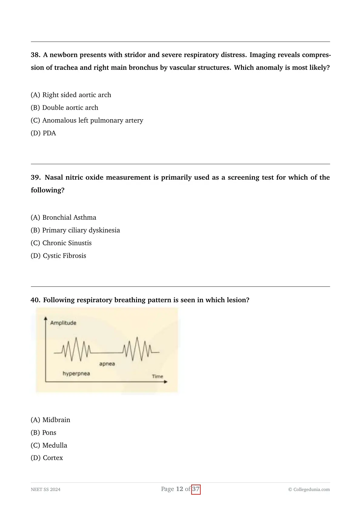
Following respiratory breathing pattern is seen in which lesion?

View Solution
Concept:
Abnormal respiratory patterns provide essential localizing neuroanatomical value in comatose or brain-injured pediatric patients.
The pattern illustrating a crescendo-decrescendo hyperpneic phase alternating with periods of apnea is known as Cheyne-Stokes respiration.
Explanation:
- Cheyne-Stokes Respiration: Characterized by a cyclic pattern of smoothly waxing and waning tidal volumes (crescendo-decrescendo hyperpnea) followed by a period of central apnea.
- This breathing pattern reflects an increased ventilatory response to carbon dioxide (hypersensitivity to $PaCO_2$) combined with delayed circulatory feedback.
- It classically localizes to bilateral deep cerebral hemisphere (cortex/subcortex) or diencephalic lesions, or severe diffuse metabolic encephalopathy.
- Central Neurogenic Hyperventilation: Sustained, rapid, deep breathing seen in lesions of the midbrain (upper tegmentum).
- Apneustic Breathing: Prolonged inspiratory pauses followed by expiratory pauses, localizing to lesions of the middle/lower pons.
- Ataxic (Biot) Breathing: Completely irregular, chaotic breaths with unpredictable apneas, localizing to the respiratory centers in the dorsomedial medulla and signifying impending respiratory arrest.
Final Answer:
Cheyne-Stokes breathing with alternating hyperpnea and apnea is seen in lesions of the bilateral cerebral cortex and diencephalon.
$\bullet$ Cheyne-Stokes $\rightarrow$ Bilateral Cerebral Cortex / Diencephalon
$\bullet$ Central Neurogenic Hyperventilation $\rightarrow$ Midbrain
$\bullet$ Apneustic Breathing $\rightarrow$ Pons
$\bullet$ Ataxic / Biot Breathing $\rightarrow$ Medulla (pre-terminal)
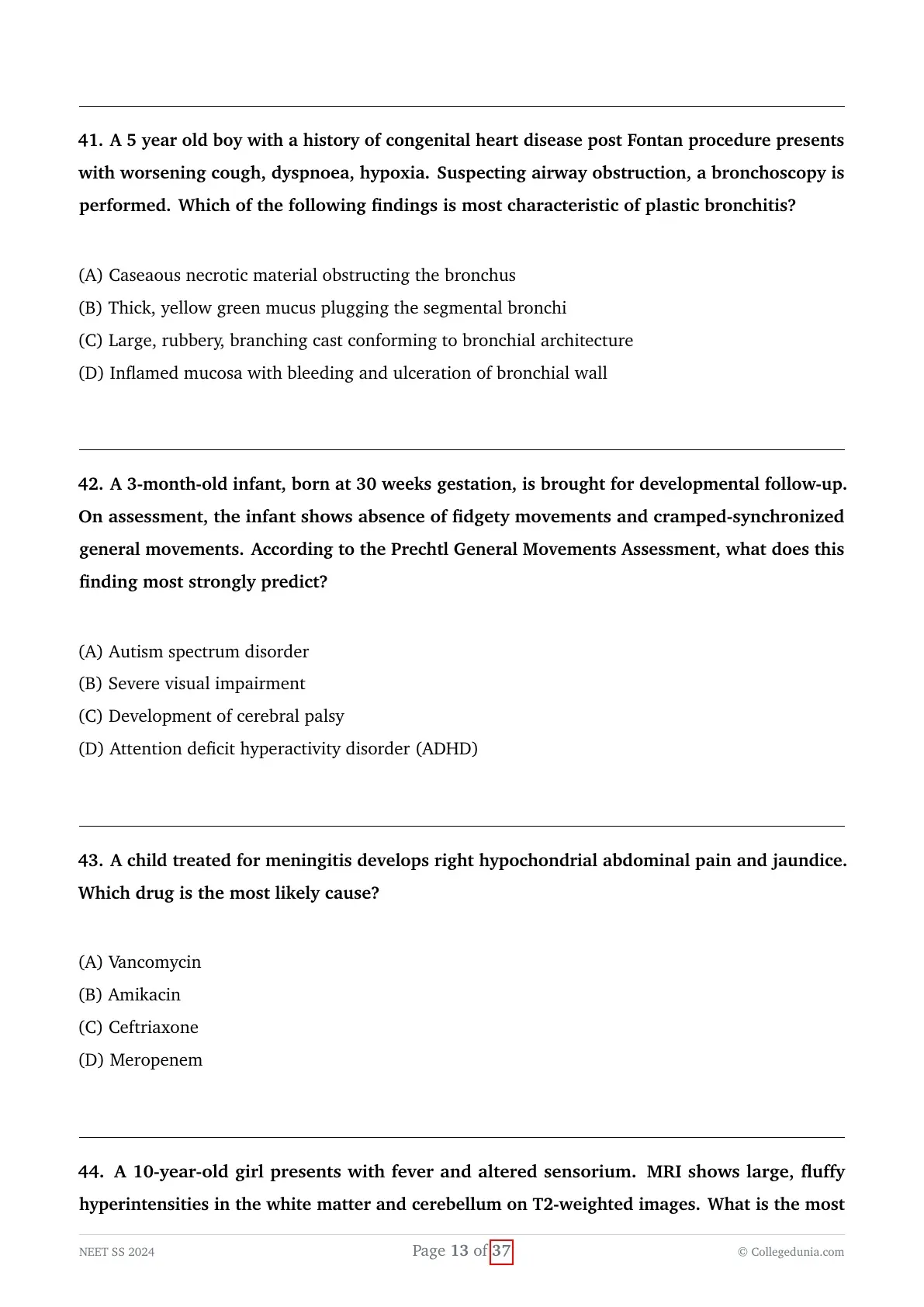
A 5 year old boy with a history of congenital heart disease post Fontan procedure presents with worsening cough, dyspnoea, hypoxia. Suspecting airway obstruction, a bronchoscopy is performed. Which of the following findings is most characteristic of plastic bronchitis?
View Solution
Concept:
Plastic bronchitis (PB) is a rare and potentially fatal disorder characterized by the formation and expectoration of large, cohesive, cohesive, branching casts that conform to the exact branching architecture of the tracheobronchial tree.
It is a well-recognized, severe complication following single-ventricle palliation surgeries, particularly the Fontan procedure.
Explanation:
- Following a Fontan procedure, elevated systemic venous pressures and altered thoracic lymphatic dynamics lead to lymphatic hypertension and abnormal lymphatic leakage directly into the airway lumen.
- In the presence of high pulmonary venous pressures and lymphatic leakage, proteinaceous, chylous, and fibrin-rich lymph fluid coagulates inside the airways.
- These solidified secretions form large, thick, rubbery, branching casts that precisely mirror the shapes of the segmental and subsegmental bronchial branches ("casts of the bronchial tree").
- These cohesive casts cause critical mechanical obstruction of major airways, precipitating acute respiratory failure, atelectasis, severe dyspnea, and life-threatening asphyxiation.
- Rigid bronchoscopy provides immediate life-saving therapeutic cast removal, while definitive diagnosis and management involve dynamic contrast-enhanced magnetic resonance lymphangiography (DCMRL) followed by lymphatic intervention/embolization.
Final Answer:
The pathognomonic bronchoscopic finding of plastic bronchitis is the presence of large, rubbery, cohesive branching casts that replicate the bronchial architecture.
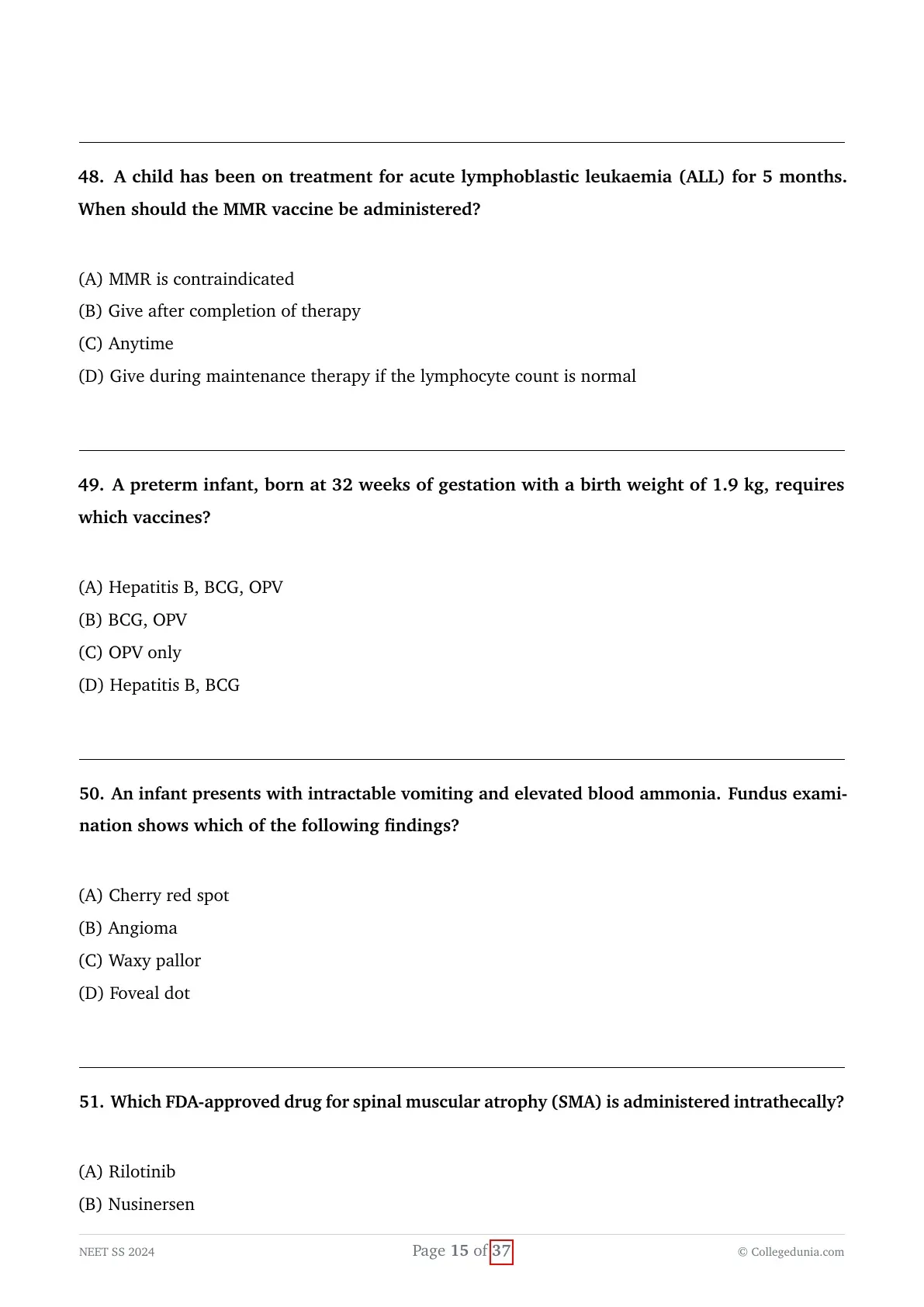
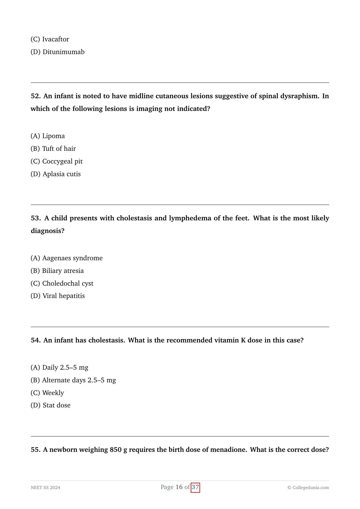
A 3-month-old infant, born at 30 weeks gestation, is brought for developmental follow-up. On assessment, the infant shows absence of fidgety movements and cramped-synchronized general movements. According to the Prechtl General Movements Assessment, what does this finding most strongly predict?
View Solution
Concept:
Prechtl's Qualitative Assessment of General Movements (GMA) is a highly sensitive, standardized, non-invasive neuromotor evaluation tool used in high-risk preterm and term infants.
It analyzes spontaneous, endogenous movement patterns generated by the brain to detect early signs of neurological dysfunction.
Explanation:
- Spontaneous general movements are observed from early fetal life until approximately 20 weeks post-term age and occur in two age-specific stages:
1. Writhing movements: From preterm period until $6$--$9$ weeks post-term.
2. Fidgety movements: Tiny, continuous movements of moderate speed and variable acceleration of the neck, trunk, and limbs present from $9$ to $20$ weeks post-term age. - Normal fidgety movements indicate intact neural integrity.
- Pathological patterns include cramped-synchronized movements (rigid, simultaneous contraction and relaxation of limb and trunk muscles) during the writhing stage, and the absence of fidgety movements during the 3- to 5-month post-term window.
- The combination of absent fidgety movements and cramped-synchronized patterns has a sensitivity and specificity exceeding $95\%$ for predicting the subsequent development of Cerebral Palsy (particularly bilateral spastic CP).
Final Answer:
According to Prechtl's General Movements Assessment, the absence of fidgety movements combined with cramped-synchronized movements strongly predicts the development of cerebral palsy.
A child treated for meningitis develops right hypochondrial abdominal pain and jaundice. Which drug is the most likely cause?
View Solution
Concept:
Ceftriaxone is a broad-spectrum, third-generation cephalosporin commonly utilized at high doses ($100\text{ mg/kg/day}$) in pediatric bacterial meningitis due to its excellent cerebrospinal fluid penetration.
A well-documented adverse effect associated with high-dose or prolonged ceftriaxone therapy is the formation of biliary sludge and pseudolithiasis.
Explanation:
- Ceftriaxone has a dual elimination pathway, with approximately $40\%$ excreted unchanged into the bile where it reaches high concentrations.
- In the gallbladder, ceftriaxone anions bind with divalent calcium ions ($Ca^{2+}$) to form an insoluble, radio-opaque precipitate of calcium-ceftriaxone salt.
- This precipitates clinically as biliary pseudolithiasis (biliary sludging or reversible gallstones), occurring in up to $15$--$40\%$ of children receiving high-dose regimens.
- Affected children can present with symptoms mimicking acute cholecystitis, including right upper quadrant (hypochondrial) pain, vomiting, nausea, and obstructive jaundice.
- Unlike true cholesterol or pigment stones, ceftriaxone-induced pseudolithiasis is completely reversible and spontaneously resolves within several days to weeks after discontinuing the drug.
- Vancomycin and amikacin are nephrotoxic and ototoxic, while meropenem rarely induces biliary sludging.
Final Answer:
Ceftriaxone is the drug responsible for calcium-ceftriaxone biliary precipitation, causing biliary pseudolithiasis, right hypochondrial pain, and jaundice.
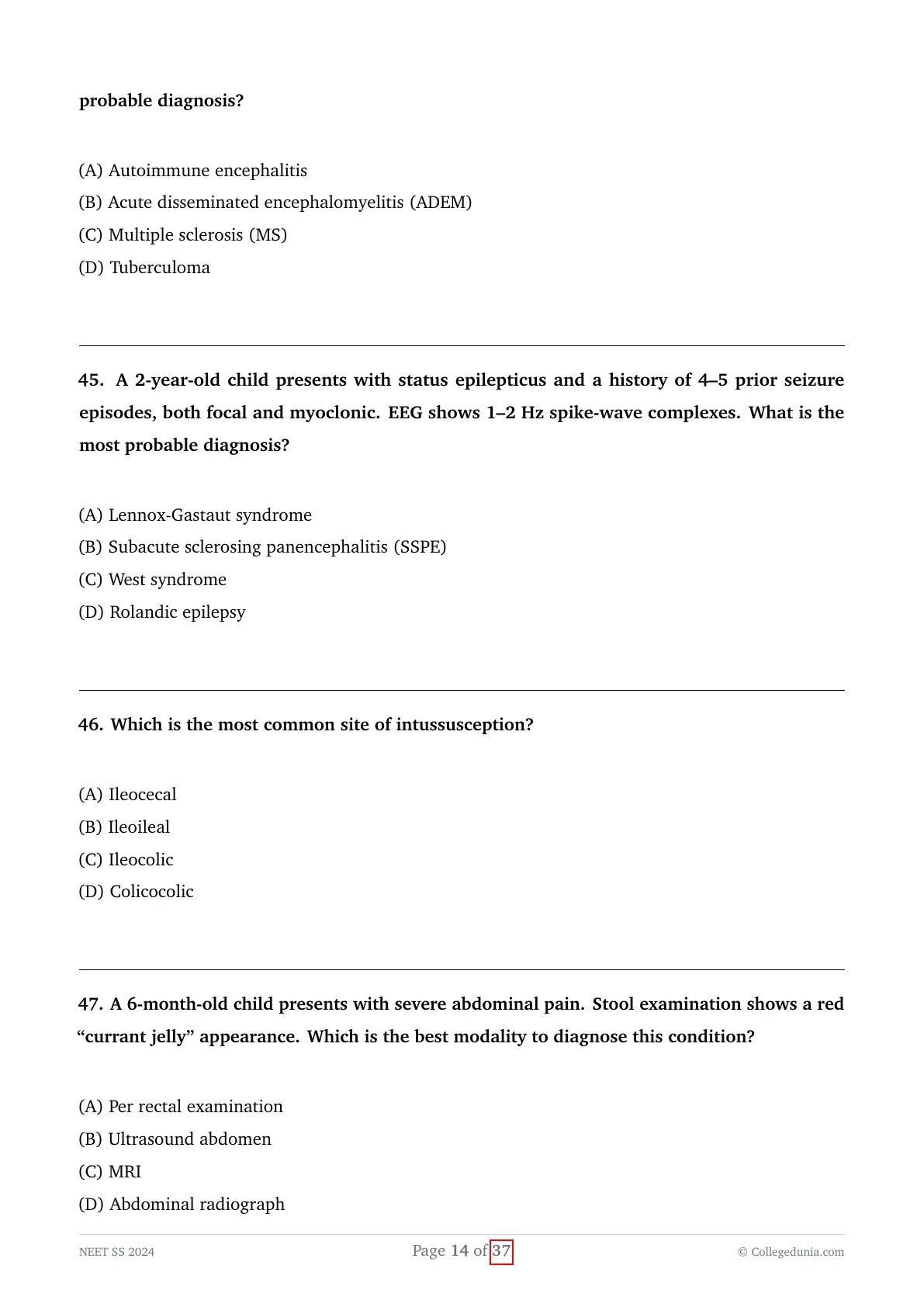
A 10-year-old girl presents with fever and altered sensorium. MRI shows large, fluffy hyperintensities in the white matter and cerebellum on T2-weighted images. What is the most probable diagnosis?
View Solution
Concept:
Acute Disseminated Encephalomyelitis (ADEM) is an acute, immune-mediated, post-infectious or post-vaccinal inflammatory demyelinating condition of the central nervous system.
It predominantly affects children and is characterized by acute multifocal neurological deficits combined with encephalopathy (altered mental status).
Explanation:
- Diagnostic criteria for ADEM require an acute or subacute onset of multifocal neurological symptoms accompanied by encephalopathy (lethargy, confusion, altered consciousness) not explained by fever alone.
- Brain MRI (T2-weighted and FLAIR sequences) characteristically reveals large ($>1--2 cm$), asymmetric, poorly marginated, "fluffy" or cloud-like hyperintense lesions involving the subcortical and deep cerebral white matter, basal ganglia, brainstem, and cerebellum.
- These lesions are typically in the same stage of evolution (monophasic demyelinating event).
- Multiple Sclerosis (MS): Lesions are typically small, well-demarcated, ovoid, located perpendicularly along the corpus callosum (Dawson's fingers), and clinical presentation rarely presents with prominent encephalopathy in pediatric onset.
- Autoimmune Encephalitis: Typically causes hyperintensities restricted to the limbic structures (medial temporal lobes) on MRI, presenting with severe behavioral changes, psychosis, and refractory seizures.
- First-line treatment for ADEM is high-dose intravenous pulse methylprednisolone ($30 mg/kg/day$ for $3$--$5$ days), leading to rapid clinical recovery.
Final Answer:
The clinical presentation of acute encephalopathy following a febrile episode with MRI demonstrating large, fluffy T2 hyperintensities in cerebral white matter and cerebellum is characteristic of Acute Disseminated Encephalomyelitis (ADEM).
$\bullet$ ADEM = Post-infectious + Encephalopathy mandatory + Large, fluffy, poorly marginated white matter/cerebellar lesions (Monophasic).
$\bullet$ MS = No encephalopathy + Dawson's fingers / periventricular discrete lesions (Dissemination in time and space).
A 2-year-old child presents with status epilepticus and a history of 4–5 prior seizure episodes, both focal and myoclonic. EEG shows 1–2 Hz spike-wave complexes. What is the most probable diagnosis?
View Solution
Concept:
Lennox-Gastaut Syndrome (LGS) is a severe, refractory developmental and epileptic encephalopathy of early childhood.
It is defined clinically by a classic diagnostic triad consisting of polymorphic intractable seizures, intellectual impairment/cognitive dysfunction, and a characteristic slow spike-and-wave pattern on electroencephalography (EEG).
Explanation:
- Lennox-Gastaut syndrome typically manifests between 2 and 8 years of age, often evolving from infantile spasms (West syndrome).
- The syndrome features multiple seizure types occurring in the same patient, including tonic seizures (especially during non-REM sleep), atonic/drop attacks, atypical absences, myoclonic jerks, and episodes of non-convulsive status epilepticus.
- The pathognomonic interictal EEG finding is generalized slow spike-and-wave complexes at a frequency of $<2.5 Hz$ (typically $1.5--2.0 Hz$), often accompanied by paroxysmal fast activity ($10--20 Hz$) during sleep.
- West syndrome presents earlier in infancy ($3--12 months$) with epileptic spasms, developmental arrest, and an EEG pattern of hypsarrhythmia (chaotic, high-voltage, disorganized slow waves).
- Subacute sclerosing panencephalitis (SSPE) occurs years after measles infection in older children and presents with periodic, high-amplitude, stereotyped slow-wave complexes (Radermecker complexes).
- Self-limited (benign) Rolandic epilepsy presents with nocturnal focal orofacial motor seizures and centrotemporal spikes on a normal background EEG without status epilepticus or cognitive decline.
Final Answer:
The clinical presentation of intractable polymorphic seizures, status epilepticus, and an EEG demonstrating slow ($1\text{--}2\text{ Hz}$) spike-wave complexes confirms Lennox-Gastaut syndrome.
1. Multiple intractable seizure types (Tonic, Atonic, Atypical Absence, Myoclonic)
2. Cognitive impairment / neurodevelopmental regression
3. Slow spike-and-wave discharges ($<2.5\text{ Hz}$, usually $1.5\text{--}2\text{ Hz}$) on EEG.
Which is the most common site of intussusception?
View Solution
Concept:
Intussusception is the telescoping or invagination of a proximal segment of the bowel (the intussuscipiens) into an adjacent, immediately distal bowel segment (the intussuscipiens).
It represents the most common cause of acute intestinal obstruction in infants and young children between 3 months and 3 years of age.
Explanation:
- In over $85--90\%$ of pediatric cases, intussusception is idiopathic and occurs in the ileocolic region, where the terminal ileum invaginates through the ileocecal valve into the colon.
- This anatomical site is particularly vulnerable because the terminal ileum contains abundant lymphoid tissue (Peyer patches) that readily undergoes hypertrophy following common viral infections (such as adenovirus, rotavirus, or enterovirus), serving as a physiological lead point.
- Ileoileal intussusception (small-bowel intussusception) is less frequent and is often transient, non-obstructive, or associated with a pathological lead point like a Meckel diverticulum or Henoch-Schönlein purpura hematoma.
- Colicocolic intussusception accounts for less than $5\%$ of cases and typically occurs in older children or adults in association with discrete colonic polyps or neoplasms.
- Ileocecal intussusception refers specifically to invagination of the ileocecal valve itself, which is less precise than the classic ileocolic progression.
Final Answer:
The most frequent anatomical subtype of pediatric intussusception is ileocolic intussusception.
A 6-month-old child presents with severe abdominal pain. Stool examination shows a red “currant jelly” appearance. Which is the best modality to diagnose this condition?
View Solution
Concept:
The classic clinical triad of acute intussusception consists of intermittent colicky abdominal pain (screaming episodes with legs pulled up to the abdomen), vomiting, and passage of blood and mucus per rectum ("currant jelly" stools).
Establishing a prompt, non-invasive, radiation-free, and definitive anatomical diagnosis is essential to prevent bowel ischemia, necrosis, and perforation.
Explanation:
- Abdominal Ultrasonography is the gold-standard and primary diagnostic imaging modality of choice for pediatric intussusception, boasting a sensitivity and specificity approaching $100\%$ in experienced hands.
- On transverse sonographic cross-sections, it reveals the pathognomonic "target", "doughnut", or "bull's eye" sign, representing concentric alternating hyperechoic and hypoechoic rings of bowel wall edema and invaginated mesenteric fat.
- On longitudinal views, it demonstrates the "pseudokidney" or "sandwich" sign, where layers of the intussusceptum are seen telescoping within the intussuscipiens.
- Abdominal radiographs (X-rays) have low sensitivity and are mainly used to rule out bowel perforation (pneumoperitoneum) prior to reduction.
- MRI is impractical in acute emergencies due to long acquisition times and the requirement for sedation.
- Digital rectal examination may confirm the presence of blood or a palpable "cervix-like" mass (Dance's sign), but it cannot definitively confirm the diagnosis or evaluate bowel viability.
Final Answer:
Abdominal ultrasonography is the most accurate and best imaging modality to diagnose intussusception in children.
$\bullet$ Transverse USG: Target / Doughnut / Bull's eye sign
$\bullet$ Longitudinal USG: Pseudokidney / Sandwich sign
Initial treatment of choice (if no perforation): Ultrasound-guided Hydrostatic or Air Enema reduction.
A child has been on treatment for acute lymphoblastic leukaemia (ALL) for 5 months. When should the MMR vaccine be administered?
View Solution
Concept:
The MMR (Measles, Mumps, Rubella) vaccine is a live-attenuated viral vaccine.
In patients undergoing active chemotherapy for hematologic malignancies like Acute Lymphoblastic Leukemia (ALL), profound cell-mediated and humoral immunosuppression contraindicates the administration of all live vaccines due to the risk of severe vaccine-strain viral replication and dissemination.
Explanation:
- Immunosuppressive chemotherapy severely depletes T-cell and B-cell populations, impairing the host's ability to clear attenuated viral strains, which can lead to life-threatening complications such as measles inclusion body encephalitis or giant cell pneumonia.
- Current international guidelines (CDC, AAP, and IDSA) state that live-attenuated vaccines, including MMR and Varicella, are strictly contraindicated during active chemotherapy (including induction, consolidation, and maintenance phases).
- Following the completion of chemotherapy, immune reconstitution gradually occurs over several months.
- Live-attenuated vaccines such as MMR should be administered only after the completion of all immunosuppressive therapy, with guidelines recommending a minimum waiting period of $3 to 6 months$ post-chemotherapy (provided the disease is in remission and total lymphocyte counts have normalized).
- Inactivated vaccines can typically be administered starting 3 months after chemotherapy completion, with revaccination strategies to ensure protective titers.
Final Answer:
The MMR vaccine must be withheld during active chemotherapy and should be administered after the completion of therapy once immune recovery has occurred.
$\bullet$ Live vaccines (MMR, Varicella): Withhold during therapy; administer $\ge 6\text{ months}$ AFTER completion of chemotherapy.
$\bullet$ Inactivated vaccines: Can be given $\ge 3\text{ months}$ post-chemotherapy.
A preterm infant, born at 32 weeks of gestation with a birth weight of 1.9 kg, requires which vaccines?
View Solution
Concept:
Preterm and low-birth-weight infants are at heightened risk for vaccine-preventable infections due to lower baseline maternal transplacental antibody transfer and an immature immune system.
Standard national and international immunization guidelines dictate that medically stable preterm infants should receive routine birth vaccines according to their chronological age.
Explanation:
- In standard national immunization programs (such as the Universal Immunization Programme / WHO guidelines), the scheduled birth vaccines include:
1. BCG (Bacille Calmette-Guérin)
2. Oral Polio Vaccine (OPV zero dose)
3. Hepatitis B birth dose - For preterm infants with a birth weight $\ge 2.0 kg$, the birth dose of Hepatitis B is counted as part of the primary series. For infants $<2.0 kg$ (like this $1.9 kg$ infant), the birth dose of Hepatitis B is still administered at birth for post-exposure prophylaxis / early protection, though it is not counted as part of the 3-dose primary series (a 4-dose schedule is completed).
- BCG and OPV zero dose are safely administered at birth or upon clinical stabilization prior to hospital discharge.
- Therefore, a clinically stable $32$-week preterm infant weighing $1.9 kg$ is eligible to receive all three standard birth vaccines: Hepatitis B, BCG, and OPV.
Final Answer:
The newborn is eligible to receive Hepatitis B, BCG, and OPV at birth.
An infant presents with intractable vomiting and elevated blood ammonia. Fundus examination shows which of the following findings?
View Solution
Concept:
Inborn errors of metabolism presenting in infancy with metabolic decompensation (vomiting, lethargy, encephalopathy, hyperammonemia, and metabolic acidosis) encompass organic acidemias, urea cycle defects, and lysosomal storage disorders.
The presence of distinct ophthalmologic abnormalities, particularly a macular "cherry-red spot", narrows the diagnostic differentials among neurometabolic storage disorders.
Explanation:
- A cherry-red spot at the macula occurs when lipid, ganglioside, or complex carbohydrate storage materials accumulate pathologically within the multi-layered retinal ganglion cells surrounding the fovea.
- Because the central foveola is anatomically devoid of ganglion cell layers, the underlying highly vascularized choroid shines through as a striking bright red circle surrounded by a pale, opacified, lipid-laden retina.
- Conditions classically presenting with a macular cherry-red spot include:
1. Sphingolipidoses (Tay-Sachs disease, Sandhoff disease, Niemann-Pick disease type A)
2. Generalized GM1 gangliosidosis (which can present with severe neurovisceral storage, liver dysfunction, vomiting, and secondary hyperammonemia)
3. Sialidosis and Farber disease - While primary urea cycle disorders do not show a cherry-red spot, multiorgan storage diseases and organic acidemias associated with secondary hyperammonemia and metabolic crisis feature this distinctive fundoscopic hallmark.
Final Answer:
The characteristic fundus examination finding associated with metabolic neurometabolic storage disease is a cherry red spot.
$\bullet$ Central retinal artery occlusion
$\bullet$ Tay-Sachs disease
$\bullet$ Niemann-Pick disease
$\bullet$ GM1 gangliosidosis
$\bullet$ Sandhoff disease / Sialidosis
Which FDA-approved drug for spinal muscular atrophy (SMA) is administered intrathecally?
View Solution
Concept:
Spinal Muscular Atrophy (SMA) is an autosomal recessive neuromuscular disease caused by homozygous deletions or loss-of-function mutations in the SMN1 (Survival Motor Neuron 1) gene on chromosome 5q.
This leads to progressive degeneration of anterior horn alpha motor neurons in the spinal cord, causing proximal muscle weakness, hypotonia, and motor disability.
Explanation:
- Humans possess a nearly identical centromeric paralog gene, SMN2, which contains a single nucleotide transition ($C\rightarrow T$) in exon 7 that causes alternative splicing, producing primarily an unstable, truncated protein ($SMN\Delta 7$) with only $10$--$20\%$ fully functional SMN protein.
- Nusinersen (Spinraza) is an antisense oligonucleotide (ASO) specifically designed to bind to the intronic splice-silencing site (ISS-N1) of the SMN2 pre-mRNA, promoting exon 7 inclusion and drastically increasing the production of full-length, functional SMN protein.
- Because antisense oligonucleotides do not cross the blood-brain barrier, nusinersen must be administered directly into the cerebrospinal fluid via intrathecal injection (administered in 4 loading doses over 2 months, followed by maintenance injections every 4 months).
- Onasemnogene abeparvovec (Zolgensma) is an AAV9 vector-mediated gene replacement therapy administered as a single intravenous infusion.
- Risdiplam (Evrysdi) is a small molecule SMN2 splicing modifier administered as a daily oral liquid.
- Ivacaftor is a CFTR potentiator used in cystic fibrosis, not SMA.
Final Answer:
Nusinersen is the FDA-approved antisense oligonucleotide therapy for SMA that is administered intrathecally.
$\bullet$ Nusinersen (Spinraza): Intrathecal ASO modifier of SMN2 splicing.
$\bullet$ Risdiplam (Evrysdi): Oral small-molecule SMN2 splicing modifier.
$\bullet$ Onasemnogene abeparvovec (Zolgensma): Single IV dose AAV9 gene therapy.
An infant is noted to have midline cutaneous lesions suggestive of spinal dysraphism. In which of the following lesions is imaging not indicated?
View Solution
Concept:
Occult spinal dysraphism (OSD) refers to closed neural tube defects where the underlying spinal cord or filum terminale malformation is covered by intact skin.
Cutaneous markers along the midline back serve as crucial surface indicators of underlying dysraphism or tethered cord syndrome, requiring radiological screening (spinal ultrasound in infants $<6$ months or MRI in older infants).
Explanation:
- High-risk cutaneous stigmata that strictly require neuroimaging (spinal ultrasound or MRI) include:
1. Subcutaneous lipoma or fibrofatty mass
2. Hypertrichosis / localized hairy patch (faun tail)
3. Atypical dimples (located $>2.5 cm$ above the anal verge, diameter $>5 mm$, or multiple dimples)
4. Aplasia cutis congenita or dermal sinus tract
5. Hemangiomas ($>2.5 cm$), caudal appendages / pseudotails - A simple coccygeal pit / simple sacral dimple is an extremely common, benign variant found in up to $4$--$5\%$ of normal healthy newborns.
- A simple dimple is defined as a small ($<5 mm$ in diameter) pit situated within the gluteal cleft within $2.5 cm$ of the anal verge, where the base of the pit can be clearly visualized without associated cutaneous markers.
- These low-risk simple coccygeal pits do not communicate with the subarachnoid space and do not represent occult spinal dysraphism; hence, diagnostic neuroimaging is not indicated.
Final Answer:
A simple, solitary coccygeal pit located within the gluteal cleft is a benign finding for which spinal neuroimaging is not indicated.
$\bullet$ High Risk (Image!): $>2.5\text{ cm}$ above anus, $>5\text{ mm}$ size, base not visible, hairy tuft, lipoma, or hemangioma.
$\bullet$ Low Risk (Do NOT image): Single simple dimple $<5\text{ mm}$ within gluteal cleft $\le 2.5\text{ cm}$ from anus with visible base.
A child presents with cholestasis and lymphedema of the feet. What is the most likely diagnosis?
View Solution
Concept:
Aagenaes syndrome, also termed Lymphedema-Cholestasis Syndrome (LCS1), is an extremely rare autosomal recessive disorder characterized by the unique co-occurrence of neonatal intrahepatic cholestasis and generalized lymphatic vessel hypoplasia.
Explanation:
- Aagenaes syndrome is caused by homozygous mutations in the LCS1 gene locus on chromosome 15q.
- It is characterized by abnormal development and generalized hypoplasia of lymphatic vessels throughout the body, including both peripheral tissues and the liver.
- Clinically, affected patients present in the neonatal period with severe conjugated hyperbilirubinemia, pruritus, pale stools, and jaundice mimicking other causes of neonatal cholestasis.
- The cholestasis typically exhibits an episodic or fluctuating course that tends to improve spontaneously during childhood, only to recur later in life (e.g., during intercurrent illnesses or pregnancy).
- The pathognomonic hallmark is the development of chronic, progressive lymphedema of the lower extremities (feet and legs), which typically becomes clinically prominent around school age or early childhood.
- Biliary atresia, choledochal cysts, and viral hepatitis present with neonatal cholestasis, but none of these conditions are associated with congenital peripheral lymphedema.
Final Answer:
The distinctive association of idiopathic neonatal cholestasis with lower-limb lymphedema is the hallmark of Aagenaes syndrome.
$\bullet$ Cholestasis + Lower limb lymphedema = Aagenaes syndrome (LCS1)
$\bullet$ Cholestasis + Butterfly vertebrae + Peripheral pulmonary stenosis = Alagille syndrome
$\bullet$ Cholestasis + Normal GGT = PFIC-1 / PFIC-2 / Inborn errors of bile acid synthesis
An infant has cholestasis. What is the recommended vitamin K dose in this case?
View Solution
Concept:
Neonatal cholestasis impairs the normal delivery of bile salts into the duodenal lumen, disrupting micellar solubilization and absorption of fat-soluble vitamins (A, D, E, and K).
Vitamin K deficiency rapidly impairs hepatic gamma-carboxylation of coagulation factors II, VII, IX, and X, predisposing the infant to coagulopathy and life-threatening intracranial hemorrhage.
Explanation:
- In chronic cholestatic liver disease, intestinal absorption of dietary fat-soluble vitamins is profoundly reduced.
- Standard pediatric gastroenterology and hepatology consensus guidelines (ESPGHAN/NASPGHAN) mandate regular oral supplementation with fat-soluble vitamins for all cholestatic infants.
- Because of poor intestinal absorption, standard physiological replacement doses are inadequate; high-dose oral water-soluble / micellar preparations are required.
- The recommended routine maintenance dosage for Vitamin K in pediatric cholestasis is $2.5 to 5 mg administered orally daily$, or $5 mg$ twice weekly, or periodic parenteral doses ($2.5--5 mg$ IM/SC every $1--4 weeks$) if oral absorption is severely compromised.
- Daily oral supplementation ensures consistent maintenance of prothrombin complex activity and prevents coagulopathy during periods of poor dietary intake.
Final Answer:
The recommended oral vitamin K maintenance supplementation in an infant with cholestasis is 2.5–5 mg daily.
$\bullet$ Vitamin A: $5,000\text{--}10,000\text{ IU/day}$
$\bullet$ Vitamin D: $1,200\text{--}5,000\text{ IU/day}$ (or calcidiol $3\text{--}5\text{ }\mu\text{g/kg/day}$)
$\bullet$ Vitamin E (TPGS): $15\text{--}25\text{ IU/kg/day}$
$\bullet$ Vitamin K: $2.5\text{--}5\text{ mg/day}$ orally (or $2.5\text{--}5\text{ mg}$ IM every $2\text{--}4$ weeks).
A newborn weighing 850 g requires the birth dose of menadione. What is the correct dose?
View Solution
Concept:
Vitamin K prophylaxis is administered to all newborns at birth to prevent Vitamin K Deficiency Bleeding (VKDB), formerly known as Hemorrhagic Disease of the Newborn.
Dosing is weight-stratified to achieve adequate hemostasis while avoiding unnecessary tissue toxicity or overdosing in extremely low birth weight (ELBW) infants.
Explanation:
- Vitamin K deficiency bleeding can occur early (first 24 hours), classically (days 2--7), or late (weeks 2--24, presenting with intracranial hemorrhage).
- Standard guidelines (AAP, ESPGHAN, and WHO) recommend weight-adjusted intramuscular administration of vitamin K1 (phytomenadione/phytonadione) at birth:
1. Birth weight $>1000 g$ (or term neonates): $1.0 mg$ intramuscularly.
2. Preterm infants weighing $<1000 g$ (ELBW): Reduced dose of $0.3 mg$ to $0.5 mg$ (specifically $0.3 mg/kg$ or a fixed dose of $0.3 mg$) intramuscularly. - An infant weighing $850 g$ falls into the extremely low birth weight category ($<1000 g$), where a single IM dose of $0.3 mg$ provides optimal plasma vitamin K levels without overloading hepatic metabolic pathways.
Final Answer:
For an ELBW preterm infant weighing 850 g ($<1000\text{ g}$), the recommended birth dose of vitamin K is 0.3 mg IM.
$\bullet$ Weight $\ge 1000\text{ g}$: $1\text{ mg}$ IM
$\bullet$ Weight $<1000\text{ g}$: $0.3\text{ mg}$ (or $0.5\text{ mg}$) IM.
A neonate presents with respiratory distress at birth, and a congenital diaphragmatic hernia (CDH) is suspected. What is the initial management?
View Solution
Concept:
Congenital Diaphragmatic Hernia (CDH) is a developmental defect of the posterolateral diaphragm (most commonly the left-sided Bochdalek hernia), allowing herniation of abdominal viscera into the thoracic cavity.
This causes severe pulmonary hypoplasia, pulmonary arterial hypertension, and acute respiratory distress in the delivery room.
Explanation:
- When CDH is known antenatally or suspected immediately after birth (scaphoid abdomen, shifted heart sounds, respiratory distress), standard bag-mask ventilation (BMV) and non-invasive continuous positive airway pressure (CPAP) are strictly contraindicated.
- Bag-mask ventilation forces air into the stomach and herniated loops of intestine, causing progressive gastrointestinal distension. This acute bowel distension increases mass effect in the hemithorax, further compresses the already hypoplastic lungs, shifts the mediastinum, and severely compromises both ventilation and venous return.
- The mandatory initial management in the delivery room is immediate endotracheal intubation without prior bag-mask ventilation.
- Simultaneously, a large-bore orogastric/nasogastric tube (e.g., $10 Fr$ Replogle tube) must be placed to continuous low suction to decompress the stomach and prevent bowel distension.
- Gentle mechanical ventilation utilizing low peak inspiratory pressures (lung-protective strategy) is employed to prevent barotrauma to the hypoplastic contralateral lung.
Final Answer:
The mandatory initial step in managing a neonate with suspected congenital diaphragmatic hernia is immediate endotracheal intubation while strictly avoiding bag-mask ventilation.
Which gene is associated with congenital nephrotic syndrome?
View Solution
Concept:
Congenital Nephrotic Syndrome (CNS) is defined as nephrotic syndrome manifesting in utero or within the first 3 months of life.
It is overwhelmingly genetic in etiology, caused by autosomal recessive mutations in key structural genes encoding the slit diaphragm and podocyte foot process proteins of the glomerular filtration barrier.
Explanation:
- The podocyte slit diaphragm is the critical size- and charge-selective filtration barrier in the glomerulus.
- NPHS2 (located on chromosome 1q25--q31) encodes podocin, a hairpin-like integral membrane protein localized to the insertion site of the slit diaphragm that interacts directly with nephrin and CD2AP.
- Mutations in NPHS2 are the most common genetic cause of steroid-resistant nephrotic syndrome (SRNS) and familial focal segmental glomerulosclerosis (FSGS), as well as congenital/infantile nephrotic syndrome.
- NPHS1 (on chromosome 19q13.1) encodes nephrin, mutations in which cause Congenital Nephrotic Syndrome of the Finnish type (CNSF).
- TP53 is a classic tumor suppressor gene involved in cell-cycle arrest (mutated in Li-Fraumeni syndrome).
- RET proto-oncogene is associated with Hirschsprung disease and MEN 2.
- NOTCH2 mutations are associated with Alagille syndrome.
Final Answer:
NPHS2 (encoding podocin) is a key gene fundamentally associated with congenital and early-onset steroid-resistant nephrotic syndrome.
$\bullet$ NPHS1 $\rightarrow$ Nephrin (Finnish Congenital Nephrotic Syndrome)
$\bullet$ NPHS2 $\rightarrow$ Podocin (Steroid-Resistant Nephrotic Syndrome / FSGS)
$\bullet$ WT1 $\rightarrow$ Denys-Drash / Frasier Syndrome
$\bullet$ LAMB2 $\rightarrow$ Pierson Syndrome.
A 7-year-old boy presents with cola-colored urine and facial puffiness 2 weeks after an episode of sore throat. He has hypertension and periorbital edema. Which of the following statements regarding his condition is incorrect?
View Solution
Concept:
Post-Streptococcal Glomerulonephritis (PSGN) is the classic prototype of acute post-infectious glomerulonephritis in children.
It occurs following infection with nephritogenic strains of Group A $\beta$-hemolytic Streptococcus (GAS) and is mediated by immune complex deposition activating the alternative complement pathway.
Explanation:
- PSGN develops $1--3$ weeks after streptococcal pharyngitis (or $3--6$ weeks after streptococcal impetigo/pyoderma) and presents with acute nephritic syndrome: hematuria (tea- or cola-colored urine), oliguria, hypertension, and periorbital edema.
- Immune complex deposition (involving streptococcal antigens like SpeB/NAPlr) triggers intense activation of the alternative complement pathway, causing marked consumption of serum complement C3.
- Consequently, serum C3 levels are significantly decreased/depressed in $>90\%$ of patients during the acute phase (typically normalizing within $6--8$ weeks). Therefore, stating that C3 levels are elevated is incorrect.
- Serum Antistreptolysin O (ASO) titers are elevated in approximately $70--80\%$ of cases following streptococcal pharyngitis (anti-DNase B is elevated after skin infections).
- Electron microscopy characteristically demonstrates large, discrete subepithelial electron-dense "humps" ("dome-shaped humps") along the glomerular basement membrane.
- Because the disease is self-limiting with an excellent long-term prognosis ($>95\%$ complete recovery), management is supportive, focusing on salt and water restriction, loop diuretics (furosemide) for hypervolemia/hypertension, and antihypertensives.
Final Answer:
The incorrect statement is that C3 levels are elevated; in acute PSGN, serum C3 levels are characteristically low due to alternative complement pathway activation.
MRI brain of a 6-year-old boy with refractory seizures shows cortical thickening with a “blurred” gray-white matter junction. Which of the following best describes the underlying pathogenesis?

View Solution
Concept:
Malformations of cortical development (MCD) are a major cause of medically refractory epilepsy, neurodevelopmental delay, and intellectual disability in children.
They result from genetic or environmental disruptions occurring during the critical embryonic stages of neurogenesis, neuronal proliferation, neuronal migration, or cortical organization.
Explanation:
- During normal fetal brain development (between 12 and 24 weeks of gestation), immature post-mitotic neuroblasts migrate radially from the periventricular germinal matrix along radial glial fibers outward to form the 6-layered cerebral neocortex.
- Disruptions in this process are termed neuronal migration disorders, which encompass a spectrum including lissencephaly (pachygyria/agyria), band heterotopia (double cortex), periventricular nodular heterotopia, and Focal Cortical Dysplasia (FCD Type II).
- On high-resolution T1/T2 MRI, impaired migration and abnormal cortical lamination characteristically produce localized cortical thickening, broad flat gyri, shallow sulci, and blurring/loss of the sharp boundary at the gray-white matter junction.
- Histologically, these areas contain disorganized cortical layers, dysmorphic ectopic neurons in the subcortical white matter, and abnormal balloon cells.
- These malformed, dysplastic neuronal networks are intrinsically hyper-excitable and represent one of the most common causes of surgically remediable pediatric drug-resistant epilepsy.
Final Answer:
Cortical thickening accompanied by blurring of the gray-white matter junction on neuroimaging is characteristic of a neuronal migration disorder.
$\bullet$ Abnormal Proliferation $\rightarrow$ Microcephaly, Hemimegalencephaly
$\bullet$ Abnormal Migration $\rightarrow$ Lissencephaly, Pachygyria, Heterotopias, Blurred gray-white junction
$\bullet$ Abnormal Post-migrational Organization $\rightarrow$ Polymicrogyria, Schizencephaly.
A child is able to recite short rhymes, copy a square, dress and undress without help, and participates in group play. What is the most likely developmental age of this child?
View Solution
Concept:
Child developmental milestones are systematically assessed across four primary functional domains: gross motor, fine motor/adaptive, language/cognitive, and personal-social.
Specific drawing capabilities and independent activities of daily living provide precise developmental age benchmarks.
Explanation:
- Fine Motor Milestones (Drawing / Copying):
- 2 years: Copies a vertical line
- 3 years: Copies a circle
- 4 years: Copies a cross ($+$) and a square ($\square$)
- 5 years: Copies a triangle ($\triangle$)
- 6 years: Copies a diamond / rectangle - Language / Cognitive: At 4 years, a child recites poems/songs/short nursery rhymes, tells stories, knows 4 colors, and uses sentences of 4--5 words.
- Personal-Social / Self-Help: At 4 years, a child can dress and undress independently (except for difficult back buttons/shoelaces), goes to the toilet alone, and actively engages in associative/group and cooperative play.
- By 5 years, the child can copy a triangle, skip on alternating feet, tie shoelaces, and understand rules in competitive games.
- By 3 years, the child copies a circle, pedals a tricycle, and engages in parallel play, but cannot copy a square.
Final Answer:
The ability to copy a square, recite short rhymes, dress independently, and engage in group play corresponds precisely to a developmental age of 4 years.
$\bullet$ Circle = $3\text{ years}$
$\bullet$ Cross / Square = $4\text{ years}$
$\bullet$ Triangle = $5\text{ years}$
$\bullet$ Diamond = $6\text{ years}$
A 5-year-old child presents with acute gastroenteritis. Antibiotics should be used with caution in which infection as it may worsen hemolytic uremic syndrome (HUS)?
View Solution
Concept:
Typical (diarrhea-associated, $D^+$) Hemolytic Uremic Syndrome (HUS) is the leading cause of acute kidney injury in children, caused predominantly by Shiga toxin-producing Escherichia coli (STEC), most notably serotype O157:H7.
Antimicrobial therapy during the acute diarrheal prodrome is contraindicated due to its association with a significantly increased risk of developing full-blown HUS.
Explanation:
- E. coli O157:H7 produces potent Shiga toxins (Stx1 and Stx2) that bind to Gb3 receptors on endothelial cells, triggering endothelial injury, platelet-fibrin microthrombi formation, microangiopathic hemolytic anemia, thrombocytopenia, and renal failure.
- Treating STEC colitis with certain bactericidal antibiotics (especially fluoroquinolones, co-trimoxazole, and $\beta$-lactams) induces bacterial SOS DNA-repair pathways and bacterial cell lysis.
- This massive bacterial lysis releases vast quantities of preformed intracellular Shiga toxins into the intestinal lumen, enhancing systemic absorption into the circulation.
- Multiple prospective studies and meta-analyses have shown that antibiotic administration during STEC O157:H7 infection increases the relative risk of developing HUS by up to 2- to 17-fold.
- Therefore, in children presenting with acute bloody diarrhea where STEC is suspected, antibiotics and antimotility agents should be strictly avoided, and supportive fluid therapy with isotonic crystalloids is the cornerstone of management.
Final Answer:
Antibiotics are avoided and contraindicated in Escherichia coli O157:H7 gastroenteritis because they induce toxin release and worsen the risk and severity of Hemolytic Uremic Syndrome.
A 15-year-old boy presents with progressive abdominal distension and weight loss. Imaging reveals a cystic lesion in the liver, and following sudden severe abdominal pain, free peritoneal fluid is noted. Which is the most likely diagnosis?
View Solution
Concept:
Cystic echinococcosis (hydatid disease) is a zoonotic parasitic infection caused by the larval metacestode stage of the dog tapeworm, Echinococcus granulosus.
The liver is the most common organ involved ($>70\%$ of cases). Hydatid cysts can remain asymptomatic for years until growth produces mass effect or acute complications like cyst rupture occur.
Explanation:
- In humans (accidental intermediate hosts), ingested oncospheres penetrate the intestinal mucosa, enter the portal circulation, and develop into slow-growing, fluid-filled unilocular or multilocular hydatid cysts with inner germinal layers and surrounding daughter cysts.
- A well-recognized life-threatening emergency in cystic echinococcosis is intraperitoneal rupture of the hydatid cyst, which can occur spontaneously due to high intracystic pressure or secondary to blunt abdominal trauma.
- Rupture presents with sudden-onset acute severe abdominal pain, signs of peritonitis, free fluid (hydatid fluid and daughter cysts) in the peritoneal cavity on ultrasonography/CT, and a high risk of life-threatening anaphylactic shock due to the release of highly antigenic cyst contents.
- Hepatoblastoma and hepatocellular carcinoma present as solid, vascular hepatic masses rather than clear unilocular/multilocular fluid-filled cysts with rupture causing free fluid.
- Liver cirrhosis presents with diffuse parenchymal nodularity, portal hypertension, and ascites without an isolated cystic mass.
Final Answer:
The clinical course of an expanding hepatic cyst suffering acute rupture with severe pain and free peritoneal fluid is indicative of a ruptured Hydatid Cyst.
A 10-day-old neonate presents with jaundice, poor feeding, fever, and hepatomegaly. Urine shows pyuria and culture is pending. Which of the following is the most common causative organism of UTI in this child?
View Solution
Concept:
Urinary Tract Infection (UTI) in the neonatal period is a serious systemic bacterial infection, often occurring in the setting of bacteremia or underlying congenital anomalies of the kidney and urinary tract (CAKUT).
Neonates present with non-specific systemic signs such as unexplained jaundice (conjugated or unconjugated), fever or hypothermia, poor feeding, lethargy, and vomiting.
Explanation:
- Across all pediatric age groups, including the neonatal period, Escherichia coli is overwhelmingly the most common causative uropathogen, responsible for $75 to 80\%$ of all community-acquired and neonatal urinary tract infections.
- Uropathogenic E. coli (UPEC) possesses specialized virulence factors, including P-fimbriae (pyelonephritis-associated pili), type 1 fimbriae, and hemolysin, allowing adhesion to uroepithelial cells and ascending migration against urinary flow.
- Other uropathogens accounting for the remaining minority of neonatal cases include Klebsiella pneumoniae, Enterococcus faecalis, Group B Streptococcus (GBS), Proteus mirabilis, and Pseudomonas aeruginosa.
- Jaundice in neonatal UTI is mediated by bacterial endotoxins and inflammatory cytokines causing microcanalicular cholestasis and hemolysis.
- Due to the high incidence of structural urinary anomalies in neonates with UTI, renal and bladder ultrasonography (RBUS) is mandatory after initiating parenteral antibiotic therapy.
Final Answer:
Escherichia coli is the most frequent pathogen causing urinary tract infection in neonates.
A newborn presents with recurrent tonic seizures, generalized stiffness, and exaggerated response to light (photosensitivity). EEG is normal. There is no improvement with standard anticonvulsants. What is the most likely diagnosis?
View Solution
Concept:
Hyperekplexia (startle disease / stiff-baby syndrome) is a rare, non-epileptic neurogenetic disorder characterized by exaggerated, non-habituating startle responses to tactile, auditory, or visual stimuli, and generalized neonatal hypertonia.
Explanation:
- Hyperekplexia is most commonly inherited in an autosomal dominant or recessive pattern due to mutations in the GLRA1 gene (encoding the alpha-1 subunit of the inhibitory glycine receptor), or less commonly GLRB or SLC6A5.
- Impaired glycinergic inhibitory neurotransmission in the brainstem and spinal cord leads to uninhibited motor startle reflexes and persistent muscular rigidity that diminishes during sleep.
- The hallmark presentation is marked neonatal stiffness, exaggerated startle response to sudden unexpected sensory stimuli (tapping the nose, tactile taps, auditory or visual stimuli), and episodic life-threatening apnea or tonic spasms.
- Because these paroxysmal episodes are non-epileptic in origin, the interictal and ictal EEG is completely normal, and standard antiepileptic drugs (e.g., phenobarbital, phenytoin) are entirely ineffective.
- Tapping the tip of the nose elicits an exaggerated startle reflex with non-habituating head retraction (pathognomonic clinical test).
- The therapeutic drug of choice is Clonazepam (a GABA-A receptor agonist that enhances inhibitory tone), and acute hypertonia-induced apnea is relieved by the Vigevano maneuver (forced head-to-knees flexion).
Final Answer:
The clinical features of neonatal stiffness, exaggerated stimulus-induced startle responses, a normal EEG, and non-responsiveness to anticonvulsants are diagnostic of Hyperekplexia.
A 10-year-old boy presents with recurrent episodes of muscle cramps and fatigue. Investigations reveal hypokalemia, hypomagnesemia, metabolic alkalosis, and low urinary calcium excretion. Blood pressure is normal. What is the most likely diagnosis?
View Solution
Concept:
Hereditary tubulopathies affecting renal electrolyte handling typically present with distinct urinary electrolyte excretion patterns, acid-base imbalances, and blood pressure profiles.
Gitelman syndrome is an autosomal recessive salt-wasting tubulopathy of the distal convoluted tubule (DCT) that mimics the chronic administration of thiazide diuretics.
Explanation:
- Gitelman syndrome is caused by loss-of-function mutations in the SLC12A3 gene, which encodes the thiazide-sensitive Sodium-Chloride Cotransporter (NCCT) in the distal convoluted tubule.
- Impaired NaCl reabsorption in the DCT increases distal sodium and fluid delivery to the collecting duct, stimulating aldosterone-mediated sodium reabsorption in exchange for potassium and hydrogen ion excretion, leading to hypokalemia and metabolic alkalosis.
- The two distinguishing laboratory hallmarks that differentiate Gitelman syndrome from Bartter syndrome are:
1. Severe Hypomagnesemia with renal magnesium wasting
2. Hypocalciuria (low urinary calcium excretion, urine calcium-to-creatinine ratio $<0.2$) due to enhanced passive calcium reabsorption in the DCT. - Bartter syndrome (loop-diuretic phenotype affecting NKCC2/ROMK in thick ascending limb) presents earlier in infancy with polyhydramnios, severe failure to thrive, and hypercalciuria with nephrocalcinosis.
- Liddle syndrome is characterized by constitutive activation of ENaC, causing hypokalemic metabolic alkalosis accompanied by severe hypertension and low renin/aldosterone levels.
Final Answer:
The combination of normal blood pressure, hypokalemia, metabolic alkalosis, hypomagnesemia, and characteristic hypocalciuria is diagnostic of Gitelman syndrome.
$\bullet$ Bartter: Thick ascending limb (Loop diuretic-like) $\rightarrow$ Normotensive, Hypercalciuria, Nephrocalcinosis.
$\bullet$ Gitelman: Distal tubule (Thiazide-like) $\rightarrow$ Normotensive, Hypocalciuria + Hypomagnesemia.
$\bullet$ Liddle: ENaC gain of function $\rightarrow$ Hypertension + Hypokalemic alkalosis.
A 1.4 kg preterm infant presents with anemia and marked reticulocytosis at 6 weeks of life. Which nutrient deficiency is the most likely cause?
View Solution
Concept:
Preterm infants are born with significantly reduced body stores of iron because approximately two-thirds of fetal iron accretion occurs during the third trimester of pregnancy.
Anemia in low-birth-weight preterm infants results from early iron depletion combined with rapid postnatal growth and iatrogenic phlebotomy blood losses.
Explanation:
- Preterm infants weighing $<1.5 kg$ (Very Low Birth Weight, VLBW) exhaust their meager transplacental iron stores by $6 to 8 weeks$ of life due to rapid post-natal body growth and expanded blood volume.
- Iron deficiency is the most prevalent nutritional deficiency leading to early-onset microcytic anemia in preterm infants if prophylactic iron supplementation is not initiated by 2--4 weeks of chronological age.
- In response to declining hemoglobin levels and tissue hypoxia, endogenous erythropoietin secretion increases, stimulating active erythropoiesis in the bone marrow that manifests with marked reticulocytosis.
- Copper deficiency can also produce anemia and neutropenia but typically manifests later ($>3--6 months$) with associated skeletal changes (osteopenia, cupping of metaphyses).
- Vitamin B12 deficiency causes megaloblastic anemia with inappropriately low reticulocyte counts.
- Standard guidelines recommend daily enteral elemental iron supplementation ($2--4 mg/kg/day$) for all VLBW infants starting at 2 to 4 weeks of life through the first year.
Final Answer:
Iron deficiency is the most common nutrient deficiency causing anemia with compensatory reticulocytosis in very low birth weight preterm infants.
$\bullet$ Dose: $2\text{--}4\text{ mg/kg/day}$ of elemental iron.
$\bullet$ Timing: Start at $2\text{--}4\text{ weeks}$ of life in all infants born $<37$ weeks or weighing $<2.5\text{ kg}$ and continue until 12 months.
A 6-year-old child presents with progressive ataxia, loss of deep tendon reflexes, and peripheral neuropathy. Vitamin B12 deficiency has been excluded, and MRI spine is normal. Which vitamin deficiency is most likely responsible?
View Solution
Concept:
Vitamin E ($\alpha$-tocopherol) is a critical lipid-soluble antioxidant that protects long, heavily myelinated axons and neuronal membranes from reactive oxygen species-induced peroxidative damage.
Chronic Vitamin E deficiency leads to a severe, progressive spinocerebellar neurodegenerative syndrome that clinically mirrors Friedreich ataxia.
Explanation:
- Vitamin E deficiency in children occurs secondary to severe fat malabsorption (e.g., cystic fibrosis, biliary atresia, cholestatic syndromes, abetalipoproteinemia) or an autosomal recessive genetic defect in the $\alpha$-tocopherol transfer protein (Ataxia with Vitamin E Deficiency, AVED, due to TTPA gene mutations).
- The pathological hallmarks are degeneration of the large-caliber myelinated sensory fibers in the posterior columns (gracile and cuneate fasciculi), spinocerebellar tracts, and peripheral sensory nerves.
- Clinically, affected children develop progressive sensory and cerebellar ataxia, dysmetria, intentional tremor, loss of deep tendon reflexes (areflexia), and impaired vibration and proprioception sensations with a positive Romberg sign.
- Advanced or prolonged deficiency causes secondary axonal peripheral sensorimotor neuropathy, ophthalmoplegia, and pigmented retinopathy.
- While subacute combined degeneration of the cord from Vitamin B12 deficiency produces similar posterior/lateral column signs, normal B12 levels point directly to Vitamin E deficiency.
Final Answer:
Chronic Vitamin E deficiency is responsible for the Friedreich ataxia-like syndrome of progressive ataxia, areflexia, and peripheral neuropathy.
$\bullet$ Vitamin E deficiency $\rightarrow$ Spinocerebellar ataxia + Areflexia + Loss of proprioception/vibration (mimics Friedreich ataxia).
$\bullet$ Vitamin B1 (Thiamine) $\rightarrow$ Wernicke encephalopathy / Beriberi.
$\bullet$ Vitamin B12 $\rightarrow$ Subacute combined degeneration (posterior + lateral columns).
Which of the following best describes the serologic profile of a patient in the immunotolerant phase of chronic hepatitis B infection?
View Solution
Concept:
Chronic Hepatitis B Virus (HBV) infection follows a dynamic natural history categorized by the European Association for the Study of the Liver (EASL) and AASLD into distinct clinical phases based on viral replication, immune activity, and liver necroinflammation.
The immunotolerant phase is typically seen in children and young adults who acquired HBV through vertical (perinatal) transmission.
Explanation:
- Phase 1: Immune-Tolerant Phase (HBeAg-Positive Chronic Infection):
- Characterized by high levels of active viral replication (HBV DNA $>10^7 IU/mL$) and positive HBsAg and HBeAg.
- The immature host immune system does not mount a cytotoxic T-lymphocyte response against infected hepatocytes; hence, there is minimal hepatocyte damage, resulting in persistently normal Alanine Aminotransferase (ALT) levels and absent or minimal liver fibrosis. - Phase 2: Immune-Reactive / Clearance Phase (HBeAg-Positive Chronic Hepatitis):
- Active host immune response attacks infected cells; marked by high/fluctuating HBV DNA, positive HBeAg, and elevated ALT with active histological inflammation. - Phase 3: Inactive Carrier Phase (HBeAg-Negative Chronic Infection):
- HBsAg positive, HBeAg negative, anti-HBe positive, low or undetectable HBV DNA ($<2000 IU/mL$), and normal ALT. - Phase 4: HBeAg-Negative Chronic Hepatitis:
- Precore or basal core promoter mutant strains; HBsAg positive, anti-HBe positive, fluctuating high HBV DNA, and elevated ALT.
Final Answer:
The immunotolerant phase of chronic HBV is defined by HBsAg (+), HBeAg (+), very high serum HBV DNA, and normal ALT levels.
$\bullet$ Immune-Tolerant: HBeAg (+), HBV DNA High, ALT Normal.
$\bullet$ Immune-Active: HBeAg (+), HBV DNA High, ALT Elevated.
$\bullet$ Inactive Carrier: HBeAg (-), anti-HBe (+), HBV DNA Low ($<2000$), ALT Normal.
A 7-year-old boy presents with periorbital puffiness, cola-colored urine, and elevated blood pressure. He develops altered sensorium and seizures. What is the most likely cause of his encephalopathy?
View Solution
Concept:
Post-streptococcal glomerulonephritis (PSGN) causes acute nephritic syndrome characterized by a sudden reduction in glomerular filtration rate (GFR), causing sodium and fluid retention and acute volume overload.
Acute severe hypertension in this setting can rapidly exceed the autoregulatory capacity of the cerebral circulation, precipitating acute neurological crisis.
Explanation:
- The child exhibits classic features of acute post-infectious glomerulonephritis: periorbital edema, gross hematuria (cola-colored urine), and fluid overload-induced systemic hypertension.
- When systemic blood pressure rises acutely above the upper limit of cerebral autoregulation (typically mean arterial pressures $>120--140 mmHg$), cerebral hyperperfusion occurs.
- This overcomes the blood-brain barrier, leading to fluid extravasation into the cerebral interstitium, vasogenic cerebral edema (Posterior Reversible Encephalopathy Syndrome, PRES), and acute hypertensive encephalopathy.
- Clinically, hypertensive encephalopathy presents with severe throbbing headache, visual disturbances (blurring, cortical blindness), altered consciousness, and generalized tonic-clonic seizures.
- Uremic encephalopathy occurs in advanced, end-stage chronic uremia (blood urea nitrogen $>100 mg/dL$) with metabolic flapping tremor (asterixis) rather than sudden-onset acute seizures during early nephritic presentation.
- Immediate treatment involves parenteral antihypertensive therapy (e.g., labetalol, nicardipine, or sodium nitroprusside) and IV loop diuretics (furosemide) to reduce fluid overload.
Final Answer:
The development of seizures and altered sensorium in an acutely hypertensive child with acute nephritic syndrome is due to hypertensive encephalopathy.
A 10-year-old child with chronic kidney disease develops persistent hyperkalemia despite dietary potassium restriction. Which of the following is the most likely mechanism?
View Solution
Concept:
Potassium homeostasis in chronic kidney disease (CKD) is tightly regulated by aldosterone-mediated potassium secretion in the cortical collecting duct principal cells via ROMK and BK channels.
Impairment in the renin-angiotensin-aldosterone axis commonly leads to out-of-proportion hyperkalemia and non-anion gap metabolic acidosis (Type 4 Renal Tubular Acidosis).
Explanation:
- In chronic kidney disease, tubulointerstitial injury and arteriolar hyalinosis characteristically damage the juxtaglomerular apparatus, impairing the production and release of active renin.
- The resulting state of hyporeninemic hypoaldosteronism (Type 4 RTA) causes deficient aldosterone synthesis by the adrenal zona glomerulosa.
- Aldosterone deficiency reduces the activity of the basolateral $Na^+/K^+$-ATPase and decreases apical epithelial sodium channels (ENaC) and renal outer medullary potassium (ROMK) channels in the collecting tubule.
- This impairs electrical driving forces and prevents tubular potassium excretion, resulting in persistent hyperkalemia even with modest reductions in GFR and strict dietary potassium restriction.
- FGF-23 is a phosphaturic hormone secreted by osteocytes that increases urinary phosphate excretion and downregulates $1\alpha$-hydroxylase, playing no direct role in primary potassium retention.
- While metabolic acidosis causes an extracellular shift of potassium, chronic persistent baseline hyperkalemia in tubulointerstitial CKD is primarily driven by hyporeninemic hypoaldosteronism.
Final Answer:
The most common underlying endocrine/tubular mechanism for persistent hyperkalemia in pediatric CKD is hyporeninemic hypoaldosteronism (Type 4 RTA).
A child with distal renal tubular acidosis (Type I RTA) is being evaluated. Which of the following laboratory findings would be expected?
View Solution
Concept:
Distal Renal Tubular Acidosis (Type 1 RTA) is a tubular disorder characterized by an inability of the alpha-intercalated cells in the distal nephron and collecting duct to excrete hydrogen ions ($H^+$) into the tubular lumen.
This defect impairs the normal acidification of urine in the setting of systemic normal anion gap (hyperchloremic) metabolic acidosis.
Explanation:
- In classic distal RTA, failure of the apical proton pumps ($H^+$-ATPase or $H^+/K^+$-ATPase) prevents distal proton secretion, resulting in an inappropriately high urine pH ($Urine pH > 5.5$) despite severe systemic acidemia.
- Urinary excretion of ammonium ($NH_4^+$), which is the principal mechanism by which kidneys eliminate daily metabolic acid loads, is markedly impaired.
- The Urinary Anion Gap ($UAG = [Na^+] + [K^+] - [Cl^-]$) serves as an indirect clinical surrogate marker for urinary ammonium excretion.
- Under normal physiological conditions during metabolic acidosis, the kidney increases $NH_4^+$ (accompanied by $Cl^-$) excretion, causing urinary $[Cl^-]$ to greatly exceed $[Na^+] + [K^+]$, yielding a negative UAG (as seen in diarrhea).
- In distal RTA, impaired $NH_4^+$ excretion causes urinary $[Cl^-]$ to remain low relative to cations, resulting in a characteristically positive urinary anion gap ($[Na^+] + [K^+] > [Cl^-]$).
- Hypokalemia (due to secondary hyperaldosteronism and distal sodium-potassium exchange wasting), hypercalciuria, and hypocitraturia (leading to bilateral nephrocalcinosis and nephrolithiasis) are classical associated features.
Final Answer:
A child with distal (Type 1) renal tubular acidosis characteristically exhibits a positive urinary anion gap reflecting deficient renal ammonium excretion.
$\bullet$ Urine $\text{pH} > 5.5$ in the presence of systemic acidemia
$\bullet$ Positive Urinary Anion Gap ($[\text{Na}^+] + [\text{K}^+] - [\text{Cl}^-] > 0$)
$\bullet$ Hypokalemia + Nephrocalcinosis (due to hypercalciuria and low urinary citrate).
The Ballard scoring system for estimating gestational age in newborns can be started from which gestational age?
View Solution
Concept:
The Ballard scoring system (New Ballard Score) is an expanded and refined clinical tool used to assess the gestational age of neonates through systematic evaluation of neuromuscular and physical maturity.
The original Ballard score was modified by Dr. Jeanne L. Ballard in 1991 to accurately determine the gestational age of extremely premature infants.
Explanation:
- The original Dubowitz scoring system was valid down to 28 weeks, and the original 1979 Ballard score was accurate down to 22--24 weeks of gestation.
- The New Ballard Score (NBS) introduced negative scores ($-1$) for extremely immature physical and neuromuscular parameters, extending its validated accuracy down to 20 weeks of gestation (from $20$ to $44$ completed weeks).
- The assessment comprises 6 neuromuscular criteria (posture, square window, arm recoil, popliteal angle, scarf sign, heel to ear) and 6 physical maturity criteria (skin, lanugo, plantar surface, breast, eye/ear, genitals).
- Total scores range from $-10$ (corresponding precisely to $20 weeks$) up to $50$ (corresponding to $44 weeks$), with every 5-point increase representing approximately a 2-week advance in gestational age.
- For maximum clinical accuracy in extremely preterm infants ($<26 weeks$), the examination should ideally be performed within the first $12$ hours of life.
Final Answer:
The New Ballard Scoring system accurately determines gestational age starting from 20 weeks of gestation.
$\bullet$ Original Ballard (1979): Valid from $22\text{--}24\text{ weeks}$ (minimum score: 5).
$\bullet$ New Ballard Score (1991): Valid down to 20 weeks (score of $-10 = 20\text{ weeks}$, $0 = 24\text{ weeks}$, $50 = 44\text{ weeks}$).
A 4-year-old child presents with pallor, decreased urine output, and a history of bloody diarrhoea 5 days ago. Labs show anaemia, thrombocytopenia, and elevated creatinine. Which of the following is most likely seen on a peripheral blood smear?
View Solution
Concept:
Hemolytic Uremic Syndrome (HUS) is a classic thrombotic microangiopathy characterized by the clinical triad of microangiopathic hemolytic anemia (MAHA), thrombocytopenia, and acute kidney injury (oliguria and azotemia).
It most commonly occurs post-diarrheal in preschool-aged children following infection with Shiga toxin-producing Escherichia coli (STEC O157:H7).
Explanation:
- Circulating Shiga toxin triggers widespread endothelial cell damage within the renal glomerular capillary loops and mesenteric microvasculature.
- Damaged endothelial surfaces promote platelet adhesion and the deposition of rich fibrin-platelet meshworks (microthrombi) within small blood vessels.
- As erythrocytes travel at high velocities through these narrowed, fibrin-occluded microvascular networks, they undergo intense mechanical shearing, fragmentation, and destruction (microangiopathic hemolysis).
- On peripheral blood smear examination, these fragmented red blood cells are identified as schistocytes (helmet cells, triangular cells, and fragmented erythrocytes), which is the diagnostic hematological hallmark of MAHA.
- Concurrently, platelets are consumed within the intrarenal microthrombi, resulting in profound consumption thrombocytopenia with normal coagulation parameters (PT and aPTT are normal).
- Spherocytes are seen in hereditary spherocytosis and autoimmune hemolytic anemia; Heinz bodies indicate G6PD deficiency with oxidative injury; Target cells are characteristic of hemoglobinopathies (thalassemia).
Final Answer:
The peripheral blood smear in microangiopathic hemolytic anemia of HUS characteristically demonstrates fragmented erythrocytes known as schistocytes.
1. Microangiopathic Hemolytic Anemia ($\uparrow$ LDH, $\downarrow$ Haptoglobin, Schistocytes on smear)
2. Thrombocytopenia
3. Acute Kidney Injury (Oliguria, $\uparrow$ Creatinine, Hematuria/Proteinuria).
A 5-month-old infant presents with prolonged jaundice. Liver function tests show conjugated hyperbilirubinemia. Liver biopsy reveals dark melanin-like pigment in hepatocytes, especially in the centrilobular region. What is the most likely diagnosis?
View Solution
Concept:
Hereditary conjugated hyperbilirubinemias are benign autosomal recessive metabolic liver disorders presenting with persistent or fluctuating direct hyperbilirubinemia and normal routine serum transaminases and alkaline phosphatase.
The classic differential rests between Dubin-Johnson syndrome and Rotor syndrome.
Explanation:
- Dubin-Johnson Syndrome: Caused by homozygous loss-of-function mutations in the ABCC2 gene on chromosome 10q24, which encodes the canalicular Multidrug Resistance-Associated Protein 2 (MRP2).
- Defective MRP2 abolishes ATP-dependent canalicular excretion of conjugated bilirubin and certain organic anions into the bile canaliculi.
- As a result, polymeric epinephrine and catecholamine metabolite derivatives cannot be transported into bile; they accumulate within secondary lysosomes of hepatocytes, turning the liver grossly black.
- Histopathological examination reveals coarse, dark brown/black, melanin-like, dense pigment accumulation predominantly in the centrilobular (perivenular) hepatocytes.
- Urinary coproporphyrin analysis shows a normal total excretion but a pathognomonic reversal of isomers, where coproporphyrin I accounts for $>80\%$ of total urinary coproporphyrin (normal is predominantly coproporphyrin III).
- Rotor Syndrome: Caused by mutations in SLCO1B1 and SLCO1B3 (OATP1B1/OATP1B3); the liver biopsy shows completely normal histology with no pigment accumulation.
- Gilbert and Crigler-Najjar syndromes cause unconjugated hyperbilirubinemia due to UDP-glucuronosyltransferase deficiency.
Final Answer:
Centrilobular dark melanin-like pigmentation on liver biopsy in a patient with conjugated hyperbilirubinemia is pathognomonic of Dubin-Johnson syndrome.
$\bullet$ Dubin-Johnson: Mutation in MRP2/ABCC2 $\rightarrow$ Black Liver (dense lysosomal pigment) + Urinary Coproporphyrin I $>80\%$.
$\bullet$ Rotor Syndrome: Mutation in OATP1B1/OATP1B3 $\rightarrow$ Normal liver color (NO pigment) + High total urinary coproporphyrin.
What is the most common site of metastasis in a child with neuroblastoma?
View Solution
Concept:
Neuroblastoma is the most common extracranial solid malignant tumor of childhood, arising from primordial neural crest cells destined to form the sympathetic ganglia and adrenal medulla.
Over $50\text{--}60\%$ of children present with advanced metastatic disease (International Neuroblastoma Risk Group Stage M) at the time of initial clinical diagnosis.
Explanation:
- Neuroblastoma exhibits a strong tropism for hematogenous and lymphatic dissemination to specific anatomical sites.
- The bone marrow is statistically the single most common site of metastatic involvement, detected in approximately $70 to 80\%$ of children with metastatic disease.
- Cortical bone (metaphyses of long bones, skull, and orbits causing periorbital ecchymosis / "raccoon eyes") is the second most common metastatic site ($50--60\%$).
- Distant lymph nodes and liver (e.g., Pepper syndrome in infants with massive hepatic infiltration) are other frequent metastatic locations.
- Pulmonary parenchymal (lung) and central nervous system (brain) metastases are distinctly rare at initial diagnosis, occurring in $<3--5\%$ of patients and typically only in advanced terminal relapses.
- Complete metastatic staging mandates bilateral posterior superior iliac spine bone marrow aspirates and trephine biopsies, alongside $^{123}I-MIBG$ scintigraphy.
Final Answer:
The bone marrow is the most common site of distant metastasis in pediatric neuroblastoma.
$\bullet$ Most common metastatic site: Bone marrow ($>70\%$) $>$ Cortical bone $>$ Lymph nodes $>$ Liver.
$\bullet$ Lung and Brain metastasis are exceedingly RARE at presentation (unlike Wilms tumor, where lungs are the most common metastatic site!).
At which glomerular filtration rate (GFR) is dialysis typically indicated?
View Solution
Concept:
Chronic Kidney Disease (CKD) is classified into five progressive stages according to the Kidney Disease: Improving Global Outcomes (KDIGO) guidelines based on estimated Glomerular Filtration Rate (eGFR) and albuminuria.
Stage 5 CKD represents End-Stage Kidney Disease (ESKD) / Kidney Failure, requiring Renal Replacement Therapy (RRT) such as dialysis or kidney transplantation.
Explanation:
- The KDIGO CKD stages are defined as:
- Stage 1: $GFR \ge 90 mL/min/1.73 m^2$ with evidence of kidney damage
- Stage 2: $GFR 60--89 mL/min/1.73 m^2$ (mild decrease)
- Stage 3a/b: $GFR 30--59 mL/min/1.73 m^2$ (moderate decrease)
- Stage 4: $GFR 15--29 mL/min/1.73 m^2$ (severe decrease; pre-dialysis preparation)
- Stage 5: $GFR < 15 mL/min/1.73 m^2$ (Kidney Failure / ESKD) - When the GFR falls below $15 mL/min/1.73 m^2$, conservative medical management typically becomes insufficient to maintain fluid, electrolyte, and acid-base homeostasis and prevent uremic toxicity.
- Initiation of chronic maintenance dialysis (peritoneal dialysis or hemodialysis) is indicated at this stage, particularly when accompanied by clinical signs of uremia (intractable nausea, pericarditis, encephalopathy), refractory hyperkalemia, severe metabolic acidosis, fluid overload, or growth failure in children.
Final Answer:
Dialysis and renal replacement therapy are indicated when the glomerular filtration rate falls below $15\text{ mL/min}/1.73\text{ m}^2$ (Stage 5 CKD).
On day 20 post-hematopoietic stem cell transplant (HSCT), a child develops jaundice, breathlessness, and difficulty walking. What is the most likely diagnosis?
View Solution
Concept:
Sinusoidal Obstruction Syndrome (SOS), formerly and synonymously termed Hepatic Veno-Occlusive Disease (VOD), is a life-threatening, early endothelial complication following myeloablative conditioning regimens in Hematopoietic Stem Cell Transplantation (HSCT).
Explanation:
- SOS/VOD results from toxic injury to the sinusoidal endothelial cells and hepatocytes in zone 3 of the hepatic acinus caused by high-dose conditioning chemotherapy (e.g., busulfan, cyclophosphamide) or total body irradiation.
- Endothelial cell damage leads to sloughing, subendothelial edema, microvascular thrombosis, and fibrous obliteration of the hepatic terminal venules and sinusoids.
- This produces post-sinusoidal intrahepatic portal hypertension, presenting classically between day $+10$ and day $+30$ post-HSCT with the Seattle/EBMT diagnostic clinical triad:
1. Hyperbilirubinemia / progressive jaundice
2. Painful hepatomegaly and right upper quadrant tenderness
3. Rapid fluid retention with unexplained weight gain ($>2--5\%$), refractory ascites, and peripheral edema. - Severe ascites and fluid retention lead to abdominal compartment pressure, diaphragmatic splinting (causing breathlessness/tachypnea), and tense lower-extremity edema (causing difficulty walking).
- The approved first-line definitive therapeutic agent is Defibrotide, an oligonucleotide that stabilizes endothelial membranes and restores local thrombo-fibrinolytic balance.
- Acute GVHD presents predominantly with a maculopapular rash, secretory diarrhea, and hyperbilirubinemia, without marked sinusoidal portal hypertensive fluid retention/weight gain.
Final Answer:
The clinical triad of jaundice, sudden weight gain/fluid retention, breathlessness, and edema around day 20 post-HSCT is classic for Sinusoidal Obstruction Syndrome (SOS) / Veno-Occlusive Disease.
Jaundice ($\uparrow$ Bilirubin) + Painful Hepatomegaly + Fluid retention / Weight gain $\rightarrow$ Sinusoidal Obstruction Syndrome (SOS/VOD). Drug of choice: Defibrotide.
A full-term newborn, delivered via elective C-section, develops tachypnea within 2 hours of birth. There is no significant retraction or grunting. Oxygen support is started. After 24 hours, the infant is well, off oxygen, and feeding normally. Which chest X-ray finding is most likely in this condition?
View Solution
Concept:
Transient Tachypnea of the Newborn (TTN), also known as "wet lung syndrome", is a benign, self-limiting respiratory disorder of term and late-preterm neonates caused by delayed clearance and reabsorption of fetal alveolar lung fluid.
Elective cesarean delivery without labor is the single strongest risk factor because the infant misses the labor-induced catecholamine surge that activates epithelial sodium channels (ENaC) to clear alveolar fluid.
Explanation:
- In normal vaginal delivery, labor triggers a surge in maternal and fetal catecholamines and steroids, which switches alveolar epithelial cells from active chloride secretion to active sodium and water absorption via ENaC channels, while thoracic squeezing clears remaining fluid.
- In elective cesarean deliveries without prior labor, this resorptive mechanism is blunted, leaving excess retained fluid within the alveolar spaces and pulmonary interstitial lymphatics.
- Neonates present within 1--2 hours of birth with tachypnea (respiratory rate $>60 breaths/min$), which typically improves rapidly and resolves completely within $24 to 72 hours$ with minimal supportive care (low-flow oxygen).
- The characteristic chest radiographic findings in TTN include:
1. Fluid in the interlobar fissures (most notably the horizontal minor fissure)
2. Prominent perihilar vascular markings ("sunburst" perihilar streaking)
3. Mild symmetrical hyperinflation with flattening of the diaphragms
4. Cardiomegaly/prominent cardiac silhouette (transient) - Ground-glass opacities with air bronchograms are classic for Respiratory Distress Syndrome (surfactant deficiency in preterms); patchy infiltrates with hyperinflation are typical of Meconium Aspiration Syndrome.
Final Answer:
The classic chest X-ray appearance of Transient Tachypnea of the Newborn consists of prominent perihilar vascular markings and fluid in the interlobar fissures.
$\bullet$ TTN (Term + Elective C-Section): Fluid in fissures + Perihilar vascular streaking ("wet lung").
$\bullet$ RDS (Preterm): Ground-glass reticulogranular pattern + Air bronchograms.
$\bullet$ MAS (Post-term + Meconium): Coarse patchy infiltrates + Hyperinflation.
Which of the following is considered a surrogate marker of vasopressin (ADH) in clinical practice?
View Solution
Concept:
Arginine vasopressin (AVP), also known as antidiuretic hormone (ADH), is a nonapeptide hormone synthesized in the magnocellular neurosecretory neurons of the hypothalamus (supraoptic and paraventricular nuclei) and stored in the posterior pituitary gland.
Direct measurement of mature circulating AVP in clinical practice is notoriously difficult and unreliable, necessitating a robust, stable surrogate biomarker.
Explanation:
- Direct laboratory assay of AVP is technically challenging because AVP is a small, unstable molecule with a very short plasma half-life ($10--20 minutes$), binds extensively to circulating platelets ($>90\%$), and rapidly degrades in vitro even at low temperatures.
- AVP is synthesized as part of a larger 164-amino-acid precursor pre-pro-vasopressin, which contains a signal peptide, mature AVP, neurophysin II, and a 39-amino-acid glycopeptide called Copeptin (C-terminal pro-AVP).
- During axonal transport and secretory vesicle processing, pre-pro-vasopressin is cleaved into equimolar amounts of AVP, neurophysin II, and copeptin.
- Copeptin is stoichiometrically co-secreted into the systemic circulation in a 1:1 ratio with AVP.
- Unlike AVP, copeptin is exceptionally stable in uncooled serum/plasma for days, does not bind platelets, and can be measured rapidly and precisely using automated sandwich immunoassay techniques.
- Plasma copeptin measurement (basal or following hypertonic saline / arginine stimulation) has emerged as the modern gold-standard biomarker to differentiate Central Diabetes Insipidus from Nephrogenic Diabetes Insipidus and Primary Polydipsia.
Final Answer:
Copeptin is the established, stable surrogate biomarker for vasopressin (ADH) measurement in clinical practice.
An infant presents with recurrent hypoglycemia, micropenis, and prolonged neonatal jaundice. What is the most likely diagnosis?
View Solution
Concept:
Congenital hypopituitarism (multiple pituitary hormone deficiency) is a developmental or genetic disorder of the anterior pituitary gland, often associated with midline cranial defects (such as septo-optic dysplasia or holoprosencephaly).
It results in combined deficiencies of Growth Hormone (GH), Adrenocorticotropic Hormone (ACTH), Thyroid-Stimulating Hormone (TSH), and Gonadotropins (LH/FSH).
Explanation:
- The combination of multiple anterior pituitary deficiencies generates a distinct and life-threatening neonatal clinical phenotype:
1. Recurrent severe hypoglycemia: Driven by the simultaneous absence of Growth Hormone and Cortisol (ACTH deficiency), both of which are critical counter-regulatory hormones necessary for gluconeogenesis and glycogenolysis.
2. Micropenis ($stretched penile length <2.5 cm$) and cryptorchidism: Driven by gonadotropin (LH/FSH) deficiency during the second and third trimesters, which deprives the fetal Leydig cells of luteinizing hormone stimulation for testosterone synthesis.
3. Prolonged neonatal cholestatic jaundice: Growth hormone and thyroid hormone are essential for the maturation of hepatic canalicular transport proteins and bile acid synthesis; their deficiency leads to secondary intrahepatic cholestasis. - In Congenital Adrenal Hyperplasia (21-hydroxylase deficiency), male neonates have normal male genitalia (or slight hyperpigmentation) rather than a micropenis, and female neonates exhibit virilized/ambiguous genitalia.
- Galactosemia presents with vomiting, jaundice, hepatomegaly, cataracts, and E. coli sepsis after milk ingestion, without micropenis.
Final Answer:
The classic triad of recurrent hypoglycemia, micropenis, and prolonged cholestatic jaundice in an infant is diagnostic of Congenital Hypopituitarism.
Micropenis (LH/FSH deficiency) + Hypoglycemia (GH + ACTH deficiency) + Prolonged Cholestasis (TSH + GH deficiency) = Congenital Hypopituitarism.
A child with a history of delayed cry at birth presents with spasticity and weakness of the lower limbs, but can use his hands to feed himself. What is the most likely site of lesion?
View Solution
Concept:
Cerebral palsy (CP) is a group of permanent disorders of movement and posture attributed to non-progressive disturbances occurring in the developing fetal or infant brain.
The clinical subtype and neurological distribution of motor deficits correspond directly to the specific anatomical location and timing of the underlying neuropathology.
Explanation:
- The child exhibits Spastic Diplegia, characterized by bilateral lower-limb spasticity, scissoring gait, hyperreflexia, and motor weakness, with relative sparing or mild involvement of the upper extremities (enabling purposeful hand use like self-feeding).
- The neuropathological hallmark of spastic diplegic cerebral palsy is Periventricular Leukomalacia (PVL), which predominantly affects the periventricular white matter adjacent to the lateral ventricles.
- In the somatotopic organization (homunculus) of the descending corticospinal motor radiation passing through the internal capsule, the axons originating from the motor cortex destined for the lower extremities (legs) course immediately adjacent to the lateral ventricular borders.
- The descending fibers supplying the upper extremities (arms and hands) are situated more laterally and peripherally, away from the vulnerable periventricular watershed zone, and are thus spared in focal periventricular necrotic lesions.
- Basal ganglia lesions (dyskinetic/athetoid CP) result from acute profound asphyxia/hypoxic-ischemic encephalopathy in term infants, causing involuntary movements, choreoathetosis, and dystonia.
Final Answer:
Spastic diplegia with predominant lower-limb motor impairment results from periventricular leukomalacia (PVL) affecting the medial corticospinal motor tracks in the periventricular white matter.
$\bullet$ Spastic Diplegia (Legs $>$ Arms) $\rightarrow$ Periventricular Leukomalacia (PVL) (preterm watershed zone).
$\bullet$ Spastic Hemiplegia $\rightarrow$ Unilateral middle cerebral artery stroke / cortical injury.
$\bullet$ Dyskinetic CP $\rightarrow$ Basal ganglia / Thalamic injury (acute term HIE / Kernicterus).
Which of the following is not a typical clinical feature of Idiopathic Intracranial Hypertension (IIH)?
View Solution
Concept:
Idiopathic Intracranial Hypertension (IIH), historically termed pseudotumor cerebri, is a neurological disorder defined by elevated cerebrospinal fluid (CSF) opening pressure in the absence of a mass lesion, ventriculomegaly, infection, or structural venous obstruction.
Diagnostic evaluation is governed by the Modified Dandy Criteria.
Explanation:
- The Modified Dandy Criteria for diagnosing IIH require:
1. Symptoms and signs of increased intracranial pressure (ICP)
2. Completely normal neurological examination except for cranial nerve abnormalities (specifically isolated unifocal or bilateral cranial nerve VI palsies)
3. Elevated CSF opening pressure on lumbar puncture ($>25 cm H_2O$ in children; $>28 cm H_2O$ in sedated children) with normal CSF cytobiochemistry
4. Normal brain parenchyma and venography on neuroimaging (MRI/MRV showing only secondary signs like empty sella, optic nerve sheath distension, or transverse sinus stenosis) - Headache (worse in the morning and with Valsalva maneuvers), bilateral papilledema, pulsatile tinnitus, and transient visual obscurations are core hallmark symptoms.
- Diplopia (horizontal, due to false-localizing abducens nerve / CN VI paresis from downward stretching across the petrous temporal bone) is classic.
- The presence of a focal motor deficit (such as hemiparesis, hyperreflexia, or focal spasticity) is an absolute exclusion criterion for IIH and strongly points to a localized structural brain lesion (e.g., brain tumor, hematoma, or stroke).
Final Answer:
Focal motor deficits are not typical of IIH and their presence excludes the diagnosis under the Modified Dandy criteria.
Portal hypertension in children is defined as when the hepatic venous pressure gradient (HVPG) exceeds:
View Solution
Concept:
Portal hypertension is a pathological increase in the blood pressure within the portal venous system resulting from increased vascular resistance (prehepatic, intrahepatic, or posthepatic) and increased splanchnic blood flow.
The Hepatic Venous Pressure Gradient (HVPG) represents the gold-standard measurement of portal pressure, calculated as the difference between the Wedged Hepatic Venous Pressure (WHVP) and the Free Hepatic Venous Pressure (FHVP).
Explanation:
- The normal physiological HVPG ranges between $1 and 5 mmHg$.
- An HVPG $>5 mmHg$ defines the presence of mild/subclinical portal hypertension.
- However, Clinically Significant Portal Hypertension (CSPH) is universally defined in clinical hepatology as an $HVPG \ge 10 mmHg$ (or $10--12 mmHg$).
- At an HVPG threshold of $\ge 10 mmHg$, portosystemic collateral vessels develop, manifesting as gastroesophageal varices, splenomegaly, and ascites.
- When the HVPG reaches $\ge 12 mmHg$, the critical threshold for acute variceal rupture and life-threatening gastrointestinal hemorrhage is reached.
- In pediatric practice, Extrahepatic Portal Vein Obstruction (EHPVO) is the most common cause of prehepatic portal hypertension (where HVPG is typically normal because the block is presinusoidal, but direct portal vein pressure exceeds $10--12 mmHg$).
Final Answer:
Clinically significant portal hypertension associated with variceal development is defined when the HVPG exceeds 10–12 mmHg.
$\bullet$ Normal: $1\text{--}5\text{ mmHg}$
$\bullet$ Portal Hypertension: $>5\text{ mmHg}$
$\bullet$ Clinically Significant Portal HTN (Varices form): $\ge 10\text{ mmHg}$
$\bullet$ Risk of Variceal Bleeding / Decompensation: $\ge 12\text{ mmHg}$.
A 5-year-old child presents with failure to thrive, recurrent flank pain, and gross hematuria. Ultrasound shows nephrocalcinosis. Urine microscopy shows the following. Serum creatinine is elevated. What is the most likely diagnosis?
View Solution
Concept:
Nephrocalcinosis in pediatric patients refers to the generalized, diffuse deposition of calcium salts (calcium phosphate or calcium oxalate) within the renal parenchymal tissue, predominantly in the renal pyramids and medullary interstitium.
Distal Renal Tubular Acidosis (Type 1 RTA) is the classic tubulopathy leading to early, prominent medullary nephrocalcinosis, nephrolithiasis, and progressive renal insufficiency.
Explanation:
- In distal RTA (Type 1), failure of apical hydrogen ion pumps in the collecting duct causes persistent alkaline urine ($Urine pH > 5.5$) alongside systemic non-anion gap metabolic acidosis.
- Systemic metabolic acidosis stimulates continuous bone buffering, which leaches calcium and phosphate from skeletal stores into the bloodstream, producing marked hypercalciuria.
- Simultaneously, chronic acidosis markedly upregulates proximal tubular reabsorption of citrate, resulting in severe hypocitraturia (absence of citrate, the main endogenous inhibitor of calcium crystallization in urine).
- The combination of an alkaline urine pH, high urinary calcium concentration, and absent urinary citrate drives the rapid, massive precipitation of calcium phosphate (apatite) crystals in the renal tubules, producing medullary nephrocalcinosis.
- Over time, interstitial fibrosis and chronic nephrocalcinosis lead to renal scarring, recurrent hematuria, flank pain from calculi, failure to thrive, and elevated serum creatinine.
Final Answer:
The clinical triad of severe failure to thrive, nephrocalcinosis, and renal impairment is the hallmark presentation of distal Renal Tubular Acidosis (RTA).
Metabolic Acidosis $\rightarrow \uparrow$ Bone reabsorption $\rightarrow$ Hypercalciuria + Hypocitraturia + Alkaline Urine ($\text{pH} > 5.5$) $\rightarrow$ Calcium Phosphate Precipitation in renal medulla.
A 2-week-old neonate undergoes an ECG during evaluation for a murmur. ECG shows a heart rate of 150 bpm, right axis deviation, tall R wave in V1, and inverted T wave in V6. Which of the following best explains these findings?
View Solution
Concept:
The neonatal electrocardiogram undergoes rapid developmental evolution following the transition from fetal to postnatal circulation.
In utero, the fetal right ventricle handles approximately $55\text{ to }60\%$ of total cardiac output against high pulmonary vascular resistance, resulting in marked physiological right ventricular dominance at birth.
Explanation:
- Normal neonatal ECG features reflecting physiological right ventricular dominance include:
1. Right Axis Deviation (RAD): Mean QRS frontal axis in healthy 2-week-old neonates normally ranges between $+60^\circ and +160^\circ$ (mean $+110^\circ$).
2. Tall R waves in right precordial leads ($V_1$, $V_{3R}$, $V_4R$) and deep S waves in left precordial leads ($V_5$, $V_6$), with an $R/S$ ratio in $V_1 > 1$.
3. Resting heart rate of $120--160 bpm$ is entirely physiological for age.
4. T-wave changes: T waves in lead $V_1$ are upright at birth, invert within the first $3 to 7 days$ of life, and remain normally inverted throughout childhood until adolescence (upright $T$ in $V_1$ beyond day 7 indicates pathological RVH), while lateral leads ($V_5, V_6$) show normal upright or transiently variable T waves. - These findings represent the normal physiological baseline for a 2-week-old infant and do not indicate pathology or ischemia.
- Tricuspid atresia is characterized by left axis deviation and left ventricular hypertrophy, which is distinctly abnormal in a neonate.
Final Answer:
These electrocardiographic features reflect normal physiological right ventricular dominance expected in a healthy 2-week-old neonate.
$\bullet$ Right Axis Deviation ($+110^\circ$) is NORMAL.
$\bullet$ Tall R in $V_1$ is NORMAL (RV dominance).
$\bullet$ T wave in $V_1$ MUST BE INVERTED between Day 7 and $\approx 10\text{ years}$ of age! (Upright $T$ in $V_1$ = RVH).
Which of the following is used as a specific treatment in certain cases of drug-induced liver failure?
View Solution
Concept:
Drug-Induced Liver Injury (DILI) and acute drug-induced liver failure (ALF) in children can occur secondary to predictable intrinsic hepatotoxins (such as acetaminophen) or idiosyncratic/metabolic toxins (such as sodium valproate).
Specific antidotal and targeted metabolic rescue therapies are indicated for particular pharmaceutical toxicities.
Explanation:
- Valproic acid (VPA)-induced hepatotoxicity and hyperammonemic encephalopathy is a severe, life-threatening metabolic drug reaction seen particularly in young children ($<2$ years) and those with underlying mitochondrial disorders (e.g., POLG mutations).
- Valproate metabolites inhibit the mitochondrial enzyme carnitine palmitoyltransferase I (CPT-1) and sequester free carnitine into valproylcarnitine, causing secondary systemic carnitine deficiency and impaired mitochondrial fatty acid beta-oxidation.
- Furthermore, valproate inhibits carbamoyl phosphate synthetase I (CPS-1), disrupting the urea cycle and driving acute hyperammonemia and hepatic failure.
- L-Carnitine (Levocarnitine) supplementation (given intravenously at $100 mg/kg$ loading, then $50 mg/kg$ q8h) is the established, guideline-recommended specific antidote.
- L-carnitine restores free carnitine pools, enhances mitochondrial fatty acid oxidation, promotes urinary excretion of valproylcarnitine, and rapidly lowers toxic ammonia levels.
- N-acetylcysteine (NAC) is the specific antidote for acetaminophen toxicity.
- Zinc is used in Wilson disease; Ursodeoxycholic acid is used in cholestatic disorders; Vitamin E is an antioxidant.
Final Answer:
L-carnitine is the specific targeted treatment used in valproate-induced acute liver failure and hyperammonemia.
$\bullet$ Acetaminophen Toxicity $\rightarrow$ N-Acetylcysteine (NAC)
$\bullet$ Valproate Toxicity / Hyperammonemia $\rightarrow$ L-Carnitine (Levocarnitine)
$\bullet$ Mushroom (Amanita phalloides) Poisoning $\rightarrow$ Silibinin / High-dose Penicillin G.
A 10-year-old boy presents with dysphagia and food impaction. Endoscopy shows esophageal rings and furrows, and biopsy reveals >15 eosinophils per HPF. He was started on PPI for 8 weeks with no improvement. What is the next step?
View Solution
Concept:
Eosinophilic Esophagitis (EoE) is a chronic, immune-mediated, antigen-driven esophageal disorder characterized clinically by symptoms related to esophageal dysfunction and histologically by eosinophil-predominant inflammation.
The diagnostic threshold is defined as $\ge 15\text{ eosinophils/high-power field (HPF)}$ on mucosal biopsy in the absence of other systemic causes of eosinophilia.
Explanation:
- Common endoscopic features in pediatric EoE include linear vertical furrows, circular mucosal rings ("trachealization" of the esophagus), white plaques/exudates (eosinophil microabscesses), and strictures.
- First-line medical therapy traditionally begins with high-dose Proton Pump Inhibitor (PPI) therapy ($1--2 mg/kg/day$) for an 8- to 12-week trial.
- In patients who remain symptomatic and fail to achieve histological remission ($>15 eos/HPF$) after an adequate 8-week course of PPI therapy (PPI-refractory EoE), the next guideline-recommended step (NASPGHAN/ESPGHAN/AGA) is:
1. Topical (Swallowed) Corticosteroids: Viscous oral budesonide slurry or swallowed fluticasone propionate from a metered-dose inhaler (MDI without spacer) swallowed onto the esophageal mucosa.
2. Alternatively, dietary elimination therapies (e.g., 6-food elimination diet). - Systemic corticosteroids are reserved only for acute severe dysphagia causing significant dehydration/weight loss or critical narrowing.
- Esophageal dilation is indicated only for persistent, high-grade fibrostenotic strictures causing obstruction that fail medical anti-inflammatory therapy.
Final Answer:
The appropriate next step in PPI-unresponsive eosinophilic esophagitis is the initiation of topical (swallowed) corticosteroids such as fluticasone or budesonide.
Step 1: High-dose PPI for 8--12 weeks.
Step 2 (If PPI fails): Swallowed Topical Corticosteroids (Fluticasone/Budesonide slurry) OR Empiric Elimination Diet.
Step 3 (Refractory strictures): Endoscopic balloon dilation.
A 10-year-old boy presents with severe abdominal pain radiating to the back, vomiting, and elevated transaminases. Which investigation will be most specific in confirming acute pancreatitis?
View Solution
Concept:
Acute pancreatitis is an acute inflammatory condition of the pancreas.
According to the International Study Group of Pediatric Pancreatitis: In Search for a Cure (INSPPIRE) criteria, diagnosis requires at least 2 of the following 3 features: (1) abdominal pain compatible with pancreatitis, (2) serum amylase or lipase activity $\ge 3$ times the upper limit of normal, and (3) imaging findings consistent with acute pancreatitis.
Explanation:
- While both amylase and lipase are released from acinar cells during pancreatic injury, Serum Lipase is significantly more sensitive ($95--100\%$) and specific ($95--99\%$) than serum amylase for confirming acute pancreatitis.
- Serum amylase is produced by salivary glands as well as non-pancreatic tissues (intestine, ovary, fallopian tubes); thus, hyperamylasemia can be falsely elevated in parotitis, diabetic ketoacidosis, renal failure, and intestinal obstruction.
- Serum lipase is synthesized almost exclusively by the pancreatic acinar tissue, minimizing false-positive results.
- Lipase rises within 4--8 hours of symptom onset, peaks at 24 hours, and remains elevated for $8 to 14 days$, whereas amylase levels peak early and normalize rapidly within 48--72 hours due to renal clearance.
- Abdominal ultrasound is primarily used to evaluate biliary etiology (gallstones/sludge) but is often obscured by overlying bowel gas ($>30\%$ of cases). CT abdomen is not needed immediately for routine confirmation unless complications (necrosis/pseudocyst) are suspected after 72 hours.
Final Answer:
Serum lipase is the most sensitive and specific laboratory investigation to confirm acute pancreatitis.
$\bullet$ Serum Lipase: $>3\times$ ULN $\rightarrow$ Higher specificity, higher sensitivity, stays elevated longer ($8\text{--}14\text{ days}$). Investigation of choice!
$\bullet$ Serum Amylase: Shorter half-life ($2\text{--}3\text{ days}$), non-specific (salivary origin).
A 7-year-old child presents with jaundice, altered sensorium, and hepatomegaly. Which investigation is essential to confirm acute liver failure?
View Solution
Concept:
Pediatric Acute Liver Failure (PALF) is a rare, life-threatening multisystem syndrome characterized by rapid onset of severe hepatocellular dysfunction in a child with no recognized prior evidence of chronic liver disease.
Because liver synthetic failure manifests early with impaired production of clotting factors, measurement of coagulopathy is the cornerstone definition.
Explanation:
- The internationally accepted Pediatric Acute Liver Failure (PALF) Study Group consensus criteria define acute liver failure as:
1. Biochemical evidence of acute liver injury, AND
2. No known underlying chronic liver disease, AND
3. Hepatic-based coagulopathy unresponsive to parenteral Vitamin K defined as:
- $Prothrombin Time (PT) \ge 15 seconds$ or $INR \ge 1.5$ in the presence of hepatic encephalopathy, OR
- $PT \ge 20 seconds$ or $INR \ge 2.0$ regardless of the presence or absence of encephalopathy. - The International Normalized Ratio (INR) / Prothrombin Time reflects the hepatic synthesis of Vitamin K-dependent clotting factors (specifically Factor VII, which has the shortest half-life of $\approx 6 hours$).
- While transaminases (ALT/AST) and serum bilirubin indicate hepatocellular damage and cholestasis, they do not assess hepatic synthetic reserve and do not define acute liver failure.
- Therefore, determining the INR is mandatory and essential to confirm the diagnosis of PALF and stratify urgency for liver transplantation.
Final Answer:
Coagulation profile assessment via INR is essential to establish the diagnosis and define acute liver failure in children.
$\bullet$ Coagulopathy with Encephalopathy $\rightarrow$ $\text{INR} \ge 1.5$ (unresponsive to Vitamin K).
$\bullet$ Coagulopathy WITHOUT Encephalopathy $\rightarrow$ $\text{INR} \ge 2.0$ (unresponsive to Vitamin K).
A 2-year-old child presents with watery diarrhea that subsides with fasting. Which of the following is most likely the cause?
View Solution
Concept:
Watery diarrhea is pathophysiologically classified into two major categories: Osmotic Diarrhea and Secretory Diarrhea.
A fasting trial (holding enteral oral intake and providing intravenous hydration) is the fundamental bedside diagnostic test used to differentiate between these two pathophysiological mechanisms.
Explanation:
- Osmotic Diarrhea: Results from the presence of unabsorbed, osmotically active solute molecules in the intestinal lumen that draw water passively across the mucosal barrier.
- A defining hallmark of osmotic diarrhea is that the diarrhea completely ceases or dramatically subsides when oral intake is discontinued (fasting). Furthermore, stool osmotic gap is elevated ($>100 mOsm/kg$).
- Glucose-galactose malabsorption is an autosomal recessive disorder caused by mutations in the SGLT1 cotransporter, leading to failure of monosaccharide absorption; unabsorbed glucose/galactose draws water into the lumen, causing severe osmotic diarrhea that completely stops during fasting or elimination of offending sugars.
- Secretory Diarrhea: Results from active mucosal secretion of chloride/fluid or inhibition of sodium absorption driven by enterotoxins or neuroendocrine hormones.
- Secretory diarrhea persists unabated even during periods of complete fasting / starvation, with large stool volume ($>200 mL/kg/day$) and a low stool osmotic gap ($<50 mOsm/kg$).
- Cholera (Vibrio cholerae), enterotoxigenic E. coli (ETEC), and VIPomas (Vasoactive Intestinal Peptide secreting tumors) are classic prototypes of secretory diarrhea.
Final Answer:
Diarrhea that promptly subsides with fasting is osmotic in origin, which is characteristic of Glucose-galactose malabsorption.
$\bullet$ Osmotic Diarrhea (e.g., Lactase deficiency, Glucose-galactose malabsorption) $\rightarrow$ STOPS with Fasting | Stool Osmotic Gap $>100\text{ mOsm/kg}$ | $\text{Stool pH} < 5.5$.
$\bullet$ Secretory Diarrhea (e.g., Cholera, ETEC, VIPoma) $\rightarrow$ PERSISTS with Fasting | Stool Osmotic Gap $<50\text{ mOsm/kg}$.
An 8-year-old child presents with gradual loss of vision, especially at night. Fundoscopy shows bone-spicule pigmentation, narrowed retinal vessels, and optic pallor. What is the likely diagnosis?
View Solution
Concept:
Retinitis Pigmentosa (RP) encompasses a genetically heterogeneous group of inherited retinal dystrophies characterized by progressive dysfunction and degeneration of photoreceptor cells (predominantly rods followed secondarily by cones) and the retinal pigment epithelium (RPE).
Explanation:
- Because rod photoreceptors (concentrated in the midperipheral and peripheral retina, mediating scotopic vision) are destroyed first, the earliest and most universal presenting symptom is nyctalopia (night blindness), followed by progressive concentric constriction of visual fields ("tunnel vision").
- Fundoscopic examination reveals the pathognomonic diagnostic triad of Retinitis Pigmentosa:
1. "Bone-spicule" dark pigmentary retinopathy distributed predominantly in the midperipheral retina (due to melanin granule migration following RPE degeneration)
2. Marked attenuation (narrowing/sheathing) of retinal arterioles
3. Waxy pallor of the optic nerve disc - Electroretinography (ERG) is the most sensitive confirmatory diagnostic test, demonstrating marked reduction or extinction of scotopic a- and b-wave amplitudes.
- RP can occur as an isolated ocular disease or as part of multisystem syndromes (e.g., Usher syndrome with sensorineural deafness, Bardet-Biedl syndrome, Refsum disease, or Kearns-Sayre syndrome).
Final Answer:
The presentation of night blindness and the classic funduscopic triad of bone-spicule deposits, vessel attenuation, and waxy disc pallor is diagnostic of Retinitis Pigmentosa.
1. Bone-spicule pigmentation in mid-periphery
2. Attenuated (narrowed) retinal vessels
3. Waxy pallor of the optic disc
Key symptom: Early Nyctalopia (Night blindness) progressing to Tunnel vision.
A 6-week-old infant with history of neonatal jaundice presents with parental concern of poor response to sound. Initial OAE screening at birth was normal. What is the next best investigation?
View Solution
Concept:
Neonatal hyperbilirubinemia is a major neurotoxic risk factor for the auditory system, having a specific selective predilection for injuring the cochlear nuclei in the brainstem and the eighth cranial nerve (vestibulocochlear nerve).
This distinct clinical pathology is recognized as Auditory Neuropathy Spectrum Disorder (ANSD).
Explanation:
- Universal newborn hearing screening employs two main physiological technologies: Otoacoustic Emissions (OAE) and Automated Auditory Brainstem Response (AABR).
- Otoacoustic Emissions (OAE): Measures sound waves generated by the contractile motion of outer hair cells (OHCs) in the cochlea. It only assesses the pre-neural cochlear amplifier mechanism.
- Auditory Brainstem Response (ABR / BERA): Measures electrophysiological neural conduction along the eighth cranial nerve through the brainstem auditory pathways (cochlear nucleus, superior olivary complex, lateral lemniscus, and inferior colliculus).
- In bilirubin-induced auditory neurotoxicity (ANSD), the outer hair cells of the cochlea remain entirely intact and functional (hence, OAE is completely normal / "pass"), while synaptic transmission from inner hair cells or neural conduction along the auditory nerve/brainstem is severely disrupted.
- Consequently, relying solely on OAE in infants with hyperbilirubinemia or NICU stays will falsely miss ANSD.
- Therefore, Diagnostic Brainstem Evoked Response Audiometry (BERA/ABR) is the mandatory next investigation to evaluate neural auditory pathway function.
Final Answer:
Brainstem Evoked Response Audiometry (BERA/ABR) is the next best investigation to diagnose auditory neuropathy spectrum disorder in an infant with hyperbilirubinemia and normal OAE.
$\bullet$ Outer Hair Cells intact $\rightarrow$ Normal OAE / Present Cochlear Microphonic.
$\bullet$ Auditory Nerve / Brainstem impaired $\rightarrow$ Absent or Abnormal ABR/BERA.
Classic cause: Neonatal Hyperbilirubinemia / Kernicterus! Always screen high-risk neonates with ABR!
A 2-year-old with bowed legs did not improve after vitamin D supplementation. Investigations reveal: phosphate low, calcium normal, ALP elevated. What is the most likely diagnosis?
View Solution
Concept:
Rickets is a disorder of defective mineralization of the growth plate cartilage and newly formed osteoid matrix in growing children.
Refractoriness to standard nutritional doses of Vitamin D (ergocalciferol/cholecalciferol) defines Vitamin D-resistant rickets, the most common form of which is X-Linked Hypophosphatemic Rickets (XLHR).
Explanation:
- X-Linked Hypophosphatemic Rickets (XLHR): Caused by inactivating mutations in the PHEX gene (Phosphate-regulating endopeptidase homolog, X-linked) on chromosome Xp22.1.
- Loss of PHEX function causes pathological overexpression and elevated circulating levels of Fibroblast Growth Factor 23 (FGF-23), a potent phosphaturic hormone secreted by osteocytes.
- Elevated FGF-23 exerts two distinct actions in the renal proximal tubule:
1. Downregulates sodium-phosphate cotransporters (NaPi-IIa and NaPi-IIc), leading to severe renal phosphate wasting and profound hypophosphatemia.
2. Inhibits renal $1\alpha$-hydroxylase (CYP27B1), suppressing the conversion of $25(OH)D$ to $1,25(OH)_2D$, resulting in inappropriately low/normal calcitriol levels. - Classic biochemical profile: Normal serum Calcium, Low serum Phosphate, Markedly Elevated Alkaline Phosphatase (ALP), and normal or mildly elevated PTH.
- Clinically, children present in the second year of life as weight-bearing begins with progressive lower-limb deformities (severe genu varum / bowing), waddling gait, and disproportionate short stature that fails to respond to native vitamin D.
- Treatment involves oral phosphate solutions combined with active vitamin D (calcitriol), or the monoclonal anti-FGF23 antibody Burosumab.
Final Answer:
The failure to respond to vitamin D along with the biochemical combination of low phosphate, normal calcium, and elevated alkaline phosphatase confirms Hypophosphatemic Rickets.
$\bullet$ Nutritional Rickets: Calcium $\downarrow$ or Normal, Phosphate $\downarrow$, ALP $\uparrow\uparrow$, PTH $\uparrow\uparrow$, $25(\text{OH})\text{D} \downarrow$.
$\bullet$ Hypophosphatemic Rickets: Calcium Normal, Phosphate $\downarrow\downarrow$, ALP $\uparrow\uparrow$, PTH Normal, $1,25(\text{OH})_2\text{D}$ Inappropriately Normal/Low (Gene: PHEX $\rightarrow \uparrow$ FGF-23).
A 1-week-old septic neonate develops hematuria, thrombocytopenia, and a palpable flank mass. What is the most likely diagnosis?
View Solution
Concept:
Neonatal renal vein thrombosis (RVT) is the most common non-catheter-related vascular thrombosis seen in the neonatal intensive care unit.
It classically presents in critically ill or dehydrated neonates, particularly those with perinatal asphyxia, sepsis, polycythemia, or maternal diabetes, with a distinct clinical triad of gross hematuria, a palpable unilateral or bilateral flank mass, and consumptive thrombocytopenia.
Explanation:
- Renal vein thrombosis in neonates most commonly originates in the small intrarenal arcuate and interlobular veins before propagating into the main renal vein and inferior vena cava.
- The classical diagnostic triad includes a palpable abdominal/flank mass (due to acute renal engorgement, edema, and venous congestion), gross or microscopic hematuria, and thrombocytopenia (caused by local consumption and destruction of platelets within the forming thrombus).
- Neonatal sepsis leads to dehydration, hemoconcentration, endothelial injury, and systemic activation of the coagulation cascade, creating ideal Virchow's triad conditions for thrombogenesis.
- Color Doppler Ultrasonography is the first-line imaging modality of choice for confirming renal vein thrombosis as it non-invasively evaluates renal enlargement, increased echogenicity, loss of corticomedullary differentiation, and absence of venous blood flow without nephrotoxic contrast agents.
- Microangiopathic hemolytic anemia (MAHA) can cause thrombocytopenia and hematuria (such as in Hemolytic Uremic Syndrome), but it does not classically produce a large, rapidly enlarging palpable flank mass in a 1-week-old neonate.
- Posterior urethral valves typically present with bilateral hydronephrosis, a palpable distended urinary bladder, and poor urinary stream, rather than acute consumptive thrombocytopenia and acute unilateral/bilateral flank masses.
Final Answer:
The clinical triad of gross hematuria, consumptive thrombocytopenia, and a palpable flank mass in a septic neonate is diagnostic of bilateral renal vein thrombosis.
1. Palpable flank mass
2. Gross or microscopic hematuria
3. Thrombocytopenia
Doppler USG is the diagnostic modality of choice; contrast studies should be avoided due to high risk of acute tubular necrosis.
A 12-year-old boy presents with recurrent epistaxis and prolonged bleeding after tooth extraction. Which subtype of vWD is most severe?
View Solution
Concept:
von Willebrand Disease (vWD) is the most common inherited bleeding disorder, characterized by quantitative or qualitative defects in von Willebrand Factor (vWF).
vWF serves two vital physiological functions: mediating platelet adhesion to subendothelial collagen during primary hemostasis and acting as a carrier chaperone protein for Factor VIII (FVIII) to prevent its rapid degradation.
Explanation:
- Type 1 vWD is an autosomal dominant disorder characterized by a mild to moderate quantitative deficiency of structurally normal vWF, accounting for approximately 70\% to 80\% of all clinical cases, and generally follows a mild mucosal bleeding phenotype.
- Type 2 vWD represents qualitative (functional) defects in vWF and is subclassified into types 2A, 2B, 2M, and 2N, presenting with moderate mucocutaneous bleeding severity.
- Type 3 vWD is an autosomal recessive disorder characterized by a complete or virtually undetectable absence of vWF (both antigen and activity levels are $<$ 1–5\%).
- Because vWF is entirely absent in Type 3 vWD, Factor VIII is unprotected and rapidly degraded, leading to profoundly low secondary Factor VIII levels (often $<$ 5–10\%).
- As a result, Type 3 vWD patients suffer from dual hemostatic impairment: severe mucocutaneous bleeds (defective primary hemostasis) as well as spontaneous hemarthroses, deep muscle hematomas, and life-threatening post-surgical hemorrhage mimicking severe hemophilia A.
- Desmopressin (DDAVP) is ineffective in Type 3 vWD due to the complete lack of endothelial storage pools; therefore, treatment strictly requires exogenous vWF/Factor VIII concentrates.
Final Answer:
Type 3 vWD is the most severe subtype of von Willebrand disease due to complete quantitative deficiency of vWF and secondary severe Factor VIII deficiency.
- Type 1: Partial quantitative defect (Most common, Mild).
- Type 2: Qualitative functional defect (Moderate).
- Type 3: Total quantitative absence (Most severe, Autosomal Recessive, presents like severe Hemophilia A).
A 2-year-old presents with fever, lethargy, and petechial rash on trunk and extremities. What is the most likely diagnosis?

View Solution
Concept:
Meningococcemia, caused by Neisseria meningitidis, is an acute, life-threatening septicemia characterized by rapid progression of high fever, toxic appearance, septic shock, disseminated intravascular coagulation (DIC), and a characteristic purpuric/petechial rash.
Explanation:
- Neisseria meningitidis releases potent endotoxins (lipooligosaccharides) directly into the circulation, triggering massive proinflammatory cytokine release, diffuse endothelial injury, vascular permeability, and microvascular thrombosis.
- The cutaneous hallmark of meningococcemia begins as discrete erythematous macules or petechiae that rapidly coalesce into extensive purpura fulminans and ecchymoses with central cutaneous necrosis across the trunk and extremities.
- The acute onset of high fever accompanied by lethargy, altered sensorium, poor peripheral perfusion, and a rapidly spreading petechial/purpuric rash in a toddler is classic for fulminant meningococcal septicemia.
- Henoch-Schönlein purpura (IgA vasculitis) features non-thrombocytopenic palpable purpura symmetrically distributed over dependent areas (lower limbs and buttocks) and is usually not accompanied by acute high-grade septic fever or toxic lethargy.
- Measles presents with a craniocaudal progression of a morbilliform, erythematous, non-petechial maculopapular rash preceded by the classic triad of cough, coryza, and conjunctivitis with Koplik spots.
- Kawasaki disease presents with prolonged fever ($\ge$ 5 days), bilateral non-purulent conjunctivitis, polymorphic erythematous rash (non-petechial), strawberry tongue, cervical lymphadenopathy, and extremity changes.
Final Answer:
The combination of acute high-grade fever, lethargy, toxic presentation, and rapidly progressive petechial-purpuric rash on the trunk and extremities represents meningococcemia.
A 10-year-old with chronic liver disease has INR = 2 but is alert and has no encephalopathy. How should the liver status be classified?
View Solution
Concept:
Chronic Liver Disease (CLD) in pediatric patients is broadly categorized into compensated and decompensated stages based on objective evidence of synthetic dysfunction, portal hypertension, and neuropsychiatric complications.
Explanation:
- Compensated chronic liver disease refers to cirrhosis or advanced fibrosis where the functional synthetic reserve of the liver remains adequately preserved without clinical hallmarks of end-stage failure (INR remains near normal, no ascites, no gastrointestinal variceal hemorrhage, and no hepatic encephalopathy).
- Decompensated chronic liver disease is defined by the development of overt clinical complications reflecting synthetic failure or severe portal hypertension, including coagulopathy (INR $\ge 1.5$ not corrected by parenteral vitamin K), ascites, jaundice (hyperbilirubinemia), variceal bleeding, or hepatic encephalopathy.
- An INR of 2.0 in a child with known chronic liver disease indicates significant loss of hepatic synthetic capacity for vitamin K-dependent coagulation factors (Factors II, VII, IX, X), which formally classifies the patient's liver status as decompensated.
- Hepatic encephalopathy is not mandatory to label a patient as decompensated; the presence of significant coagulopathy, clinically evident ascites, or variceal bleeding alone is sufficient for this classification.
- Acute liver failure (ALF) is excluded because this child has pre-existing chronic liver disease, whereas ALF requires an acute hepatic insult without underlying chronic liver disease.
Final Answer:
The presence of significant synthetic dysfunction (INR = 2.0) in a patient with chronic liver disease categorizes the condition as decompensated chronic liver disease.
- Compensated CLD: Intact synthetic function, absence of ascites, variceal bleed, or encephalopathy.
- Decompensated CLD: Presence of synthetic failure (INR $\ge 1.5$), ascites, jaundice, variceal hemorrhage, or encephalopathy in chronic liver disease.
A 7-year-old on induction chemotherapy for ALL develops seizures. Labs: ↑ phosphate, ↑uric acid, ↓calcium. What is the cause and best management?
View Solution
Concept:
Tumor Lysis Syndrome (TLS) is an oncologic emergency resulting from the massive and rapid destruction of malignant cells following initiation of cytotoxic chemotherapy, particularly in hematologic malignancies like Acute Lymphoblastic Leukemia (ALL) and Burkitt lymphoma.
Explanation:
- Rapid intracellular release of ions and metabolic byproducts leads to the characteristic laboratory constellation: Hyperuricemia, Hyperkalemia, Hyperphosphatemia, and secondary Hypocalcemia.
- Secondary hypocalcemia develops because the massive surge of released intracellular phosphate complexes with circulating ionized calcium, causing calcium phosphate crystals to precipitate in soft tissues and renal tubules.
- Hypocalcemia increases neuromuscular excitability, manifesting clinically as tetany, laryngospasm, paresthesias, cardiac arrhythmias, and acute generalized seizures.
- In TLS management, hypocalcemia should strictly be corrected with intravenous calcium gluconate ONLY if the patient is overtly symptomatic (e.g., active seizures, tetany, or life-threatening arrhythmias).
- Asymptomatic hypocalcemia must NOT be treated with exogenous calcium because infusing calcium in the setting of severe hyperphosphatemia dramatically elevates the calcium-phosphate product ($[Ca] \times [PO_4] > 60$), precipitating extensive metastatic calcification in the kidneys and accelerating acute renal failure.
Final Answer:
The child's seizures are caused by acute hypocalcemia secondary to hyperphosphatemia in tumor lysis syndrome, and parenteral calcium correction should be reserved strictly for symptomatic patients.
- Triad of elevation: $\uparrow\text{K}^+$, $\uparrow\text{PO}_4^{3-}$, $\uparrow\text{Uric Acid}$.
- One depression: $\downarrow\text{Ca}^{2+}$.
- Treatment rule for Calcium: Treat hypocalcemia ONLY if symptomatic, to prevent lethal calcium-phosphate precipitation in renal tubules!
A 6-month-old presents with developmental delay, coarse facies, protruding tongue, and umbilical hernia. What is the first investigation?
View Solution
Concept:
Congenital hypothyroidism (cretinism) is one of the most common treatable and preventable causes of neurocognitive impairment and intellectual disability in children.
The classic constellation of clinical findings in an untreated infant includes gross motor and global developmental delay, coarse facial features, macroglossia (large protruding tongue), large persistent umbilical hernia, hypotonia, open posterior fontanelle, prolonged neonatal jaundice, and hoarse cry.
Explanation:
- Thyroid hormones ($T_3$ and $T_4$) are critically required for early neurogenesis, myelination, dendritic branching, and skeletal maturation during the first few months of postnatal life.
- Untreated thyroid deficiency leads to accumulation of glycosaminoglycans within interstitial spaces, causing typical coarse facial features, myxedematous skin, and a large protruding tongue.
- Abdominal muscle hypotonia combined with delayed closure of the umbilical ring directly leads to a prominent umbilical hernia.
- The definitive, quickest, and most essential initial diagnostic test is the measurement of serum Thyroid Stimulating Hormone (TSH) and Free Thyroxine (FT4).
- In primary congenital hypothyroidism, laboratory evaluation characteristically shows significantly elevated serum TSH with low or undetectable Free T4 levels.
- Prompt confirmation and immediate initiation of oral Levothyroxine therapy (10--15 $\mug/kg/day$) are crucial to prevent irreversible neurodevelopmental sequelae.
Final Answer:
The first and most critical investigation in an infant presenting with developmental delay, coarse facies, macroglossia, and umbilical hernia is serum TSH and Free T4.
- Large fontanelles (especially posterior fontanelle $>0.5\text{ cm}$).
- Macroglossia + Umbilical hernia + Prolonged neonatal jaundice.
- First investigation: TSH + Free T4.
- Treatment: Levothyroxine ($10\text{--}15\ \mu\text{g/kg/day}$) started immediately.
Which of the following is NOT a part of the classical triad of necrotizing enterocolitis (NEC)?
View Solution
Concept:
Necrotizing Enterocolitis (NEC) is the most common life-threatening gastrointestinal emergency encountered in premature and low-birth-weight neonates.
It results from an interplay of intestinal mucosal immaturity, formula feeding, ischemic-reperfusion injury, and aberrant bacterial colonization, leading to transmural bowel necrosis.
Explanation:
- The classical clinical triad describing the initial gastrointestinal manifestation of NEC comprises:
1. Abdominal distension (due to ileus, intestinal dilation, and wall inflammation).
2. Feeding intolerance / bilious gastric residuals / vomiting.
3. Blood in stools (either gross hematochezia or occult blood detected on testing). - Abdominal distension is often the earliest and most reliable clinical physical sign of evolving enterocolitis.
- Feeding intolerance manifests as increasing pre-feed aspirates containing undigested milk or bilious material alongside recurrent emesis.
- Bloody stools arise from mucosal ulceration, microvascular thrombosis, and sloughing of ischemic intestinal mucosa.
- While systemic instability in NEC can cause temperature instability, lethargy, apnea, or metabolic acidosis, hypoglycemia is NOT considered a defining component of the classical clinical triad.
- The pathognomonic radiologic sign on abdominal X-ray remains pneumatosis intestinalis (gas within the bowel wall) and portal venous gas.
Final Answer:
Hypoglycemia is a non-specific systemic sign and is not a part of the classical diagnostic triad of necrotizing enterocolitis.
1. Abdominal distension
2. Feeding intolerance (bilious aspirates/emesis)
3. Hematochezia (blood in stool)
X-ray hallmark: Pneumatosis intestinalis (air in bowel wall).
A newborn presents with stridor and respiratory distress and dysphagia. Imaging shows compression of the trachea, esophagus and right main bronchus. Which vascular anomaly is most likely responsible?
View Solution
Concept:
Vascular rings are congenital malformations of the aortic arch complex that completely or incompletely encircle and compress the trachea and esophagus, causing variable degrees of upper airway obstruction (stridor, wheezing) and gastrointestinal obstruction (dysphagia, feeding difficulties).
Explanation:
- Double aortic arch is the most common complete vascular ring and the most frequent cause of symptomatic vascular airway and esophageal compression in early infancy.
- It results from the failure of regression of the right fourth pharyngeal arch artery, resulting in two separate functioning aortic arches (a larger right-sided posterior arch and a smaller left-sided anterior arch) forming a tight vascular ring around the trachea and esophagus.
- Because the ring completely encircles both structures, infants classically present very early in life with both respiratory symptoms (inspiratory/expiratory stridor that worsens during feeding or in supine position) and digestive symptoms (dysphagia, choking, regurgitation during swallowing, termed dysphagia lusoria).
- Imaging in double aortic arch typically demonstrates bilateral and circumferential compression of the trachea and bilateral indentation of the esophagus (posterior compression on barium swallow) along with right main bronchus compression.
- A pulmonary sling (anomalous left pulmonary artery arising from the right pulmonary artery) courses between the trachea and esophagus, compressing the anterior esophagus and posterior trachea (and right main bronchus), but does not form a complete bilateral circumferential aortic ring.
Final Answer:
A double aortic arch forms a complete vascular ring around the mediastinal structures, causing simultaneous compression of the trachea, esophagus, and right main bronchus with early stridor and dysphagia.
- Double Aortic Arch: Most common complete ring; compresses both anterior trachea and posterior esophagus (stridor + dysphagia).
- Pulmonary Sling: Only vascular anomaly that courses BETWEEN trachea and esophagus (compresses posterior trachea and anterior esophagus).
A 4-year-old child has normal comprehension and cognitive abilities but difficulty planning and coordinating speech movements. Speech is effortful, inconsistent, and the child often struggles to produce the correct sounds. What is the most likely diagnosis?
View Solution
Concept:
Childhood Apraxia of Speech (CAS) is a neurological pediatric speech sound disorder in which the precision and consistency of movements underlying speech are impaired in the absence of neuromuscular deficits (e.g., normal muscle tone, intact reflexes).
Explanation:
- The core pathophysiologic impairment in CAS lies in motor planning and programming of the spatio-temporal parameters of movement sequences required for speech production.
- Receptive language, cognitive understanding, and intent to communicate are typically preserved and normal for age, leading to a marked discrepancy between comprehension and motor speech output.
- Key diagnostic clinical features of Childhood Apraxia of Speech include:
1. Inconsistent errors on consonants and vowels in repeated productions of syllables or words.
2. Lengthened and disrupted coarticulatory transitions between sounds and syllables.
3. Inappropriate prosody (e.g., unnatural stress, pausing, and robotic rate).
4. Visible or audible oral groping when attempting to initiate speech sounds. - Speech sound disorders (articulation/phonological disorders) involve consistent substitutions or omissions of specific sounds rather than motor programming inconsistency and groping.
- Isolated expressive language disorder affects vocabulary and sentence construction rather than the motor execution and sequencing of speech sounds.
- Global developmental delay involves significant delays in two or more developmental domains (motor, language, cognitive, social, adaptive), which is ruled out by the child's normal cognition and comprehension.
Final Answer:
The clinical presentation of effortful, inconsistent sound production with intact comprehension and difficulty in motor planning of speech movements characterizes Childhood Apraxia of Speech.
- Motor planning defect (not a muscle weakness problem like dysarthria).
- Inconsistent errors on repeating the same word.
- Visible articulatory "groping" behavior.
- Normal receptive language and comprehension.
What is the appropriate stepwise approach for diagnosing Mucopolysaccharidosis (MPS)?
View Solution
Concept:
Mucopolysaccharidoses (MPS) are a heterogeneous group of rare inherited lysosomal storage disorders caused by deficiencies of specific lysosomal enzymes responsible for the degradation of glycosaminoglycans (GAGs, previously termed mucopolysaccharides).
Explanation:
- The diagnostic algorithm for suspected inborn errors of metabolism like MPS follows a standardized, logical tiered approach:
1. Clinical Suspicion: Identifying phenotypic features such as coarse facial features, dysostosis multiplex, hepatosplenomegaly, corneal clouding, joint stiffness, and neuroregression.
2. Screening Test (Urinary GAGs): Quantitative and qualitative estimation of urinary glycosaminoglycans (dermatan sulfate, heparan sulfate, keratan sulfate, chondroitin sulfate) serves as a rapid, sensitive, non-invasive initial screening test.
3. Confirmatory Biochemical Test (Specific Enzyme Assay): Fluorometric or radiometric measurement of specific lysosomal enzyme activity in leukocytes, plasma, or cultured fibroblasts to definitively identify the defective enzyme and establish the MPS subtype (e.g., alpha-L-iduronidase in MPS I, iduronate-2-sulfatase in MPS II).
4. Molecular Genetic Testing: Targeted gene sequencing to detect specific pathogenic variants for genotype-phenotype correlation, family screening, carrier detection, and prenatal counseling. - Skipping directly to molecular testing or performing enzyme assays before urinary GAG screening is cost-ineffective and clinically inefficient.
Final Answer:
The correct stepwise diagnostic approach for MPS is: Clinical suspicion $\rightarrow$ Urinary GAGs $\rightarrow$ Specific Enzyme assay $\rightarrow$ Confirmatory Genetic testing.
Clinical Suspicion $\rightarrow$ Metabolic Screening (Urine GAGs) $\rightarrow$ Definitive Biochemical Assay (Enzyme Activity) $\rightarrow$ Molecular Confirmation (Genetic Analysis).
A child with suspected mitochondrial disease presents with lactic acidosis. Laboratory evaluation shows a high lactate-to-pyruvate ratio, suggesting a defect in oxidative phosphorylation. Which of the following components is most likely involved?
View Solution
Concept:
Mitochondrial disorders frequently lead to impaired oxidative phosphorylation (OXPHOS), resulting in cellular energy crisis and excessive production of lactic acid due to obligatory anaerobic glycolysis.
The blood lactate-to-pyruvate (L:P) ratio is an indispensable biochemical marker used to distinguish between defects of the mitochondrial respiratory chain and defects of pyruvate metabolism.
Explanation:
- The lactate-to-pyruvate ratio reflects the intracellular cytosolic and mitochondrial NADH/$NAD^+$ redox state, mediated via the equilibrium catalyzed by lactate dehydrogenase: \[ Pyruvate + NADH + H^+ \rightleftharpoons Lactate + NAD^+ \]
- Normal plasma L:P ratio is typically less than 20.
- A significantly elevated L:P ratio ($> 20--25$) indicates an abnormally high NADH/$NAD^+$ ratio caused by impaired electron transport through the mitochondrial respiratory chain complexes (Complex I, III, or IV).
- Complex IV (Cytochrome c oxidase) is a core component of the electron transport chain; its deficiency prevents electron transfer to molecular oxygen, stalling the respiratory chain, accumulating NADH, and driving high L:P lactic acidosis.
- Pyruvate dehydrogenase (PDH) complex deficiency impairs the conversion of pyruvate to acetyl-CoA, resulting in high lactic acidosis with a NORMAL or LOW lactate-to-pyruvate ratio ($< 20$) because the electron transport chain is intact and NADH/$NAD^+$ equilibrium is preserved.
- Complex II (Succinate dehydrogenase) defects are exclusively nuclear-encoded and much rarer causes of high L:P lactic acidosis compared to Cytochrome c oxidase defects.
Final Answer:
Lactic acidosis accompanied by a high lactate-to-pyruvate ratio indicates an electron transport chain defect, most classically involving Cytochrome c oxidase (Complex IV).
- L:P ratio $> 20$: Mitochondrial Respiratory Chain Defect (e.g., Complex I, III, IV / Cytochrome c oxidase).
- L:P ratio $< 20$ (Normal): Pyruvate Dehydrogenase (PDH) deficiency or Gluconeogenesis defect.
A neonate presents with jaundice, hepatomegaly, vomiting, and signs of sepsis. Classic galactosemia is diagnosed. Which of the following organisms is most commonly associated with sepsis in this condition?
View Solution
Concept:
Classic Galactosemia is an autosomal recessive inborn error of carbohydrate metabolism caused by severe deficiency of the enzyme galactose-1-phosphate uridyltransferase (GALT), leading to toxic accumulation of galactose-1-phosphate and galactitol in tissues.
Explanation:
- Symptoms typically manifest within the first two weeks of life following the introduction of galactose-containing feeds (breast milk or cow's milk formula) and include poor feeding, vomiting, prolonged unconjugated and conjugated hyperbilirubinemia, hepatosplenomegaly, hepatic dysfunction, and oil-drop cataracts.
- Neonates with classic galactosemia possess an extraordinary susceptibility to fulminant neonatal sepsis, specifically caused by Escherichia coli (in up to 90\% of septic cases).
- High levels of circulating galactose and galactose-1-phosphate impair neutrophil chemotaxis, phagocytosis, and bactericidal oxidative burst capacity.
- Additionally, the elevated concentration of galactose in tissues enhances the virulence and proliferation of E. coli strains by altering bacterial capsular antigen expression and host mucosal defenses.
- In any newborn with unexplained E. coli bacteremia or sepsis associated with hyperbilirubinemia and hepatomegaly, classic galactosemia must be urgently suspected and lactose/galactose-free formula (soy-based) initiated immediately.
Final Answer:
Escherichia coli is the classic and most frequently encountered pathogen responsible for fulminant sepsis in neonates with untreated classic galactosemia.
- Classic Galactosemia + Neonatal Sepsis = Escherichia coli ($E.\ coli$).
- Pathognomonic Eye Finding: "Oil-drop" nuclear cataracts due to galactitol accumulation in lens fibers via aldose reductase.
A 16-year-old girl presents with subacute onset behavioral changes, confusion, and visual blurring in one eye. On examination, she has optic neuritis and altered sensorium. MRI shows hyperintensities in the temporal lobes and optic nerve involvement. Autoimmune encephalitis is suspected. CSF is normal. What is the next best investigation?
View Solution
Concept:
The combination of acute/subacute encephalopathy (altered behavior, confusion) accompanied by central demyelination manifesting as optic neuritis represents an inflammatory demyelinating central nervous system spectrum disorder, notably MOG-antibody-associated disease (MOGAD) or Neuromyelitis Optica Spectrum Disorder (NMOSD).
Explanation:
- Myelin Oligodendrocyte Glycoprotein Antibody-associated Disease (MOGAD) frequently manifests in children and adolescents as acute disseminated encephalomyelitis (ADEM), cortical encephalitis, or severe unilateral/bilateral optic neuritis.
- Similarly, Aquaporin-4 (AQP4) IgG-positive Neuromyelitis Optica Spectrum Disorder (NMOSD) presents with optic neuritis and brainstem/diencephalic/cerebral lesions.
- Testing for serum anti-MOG IgG and anti-AQP4 antibodies using cell-based assays (CBA) is the most specific and sensitive next diagnostic step.
- Serum is significantly more sensitive than CSF for detecting both MOG and AQP4 antibodies because these autoantibodies are primarily produced in peripheral lymphoid tissues and enter the central nervous system across a disrupted blood-brain barrier.
- While anti-NMDAR encephalitis causes psychiatric symptoms, cognitive decline, and seizures, it does not typically present with classic optic neuritis and optic nerve demyelinating hyperintensities on MRI.
- Repeating CSF analysis when basic CSF parameters are non-diagnostic does not provide disease-defining specificity compared to serum autoantibody testing.
Final Answer:
The next best investigation to confirm the underlying neuroinflammatory demyelinating etiology in a patient presenting with encephalopathy and optic neuritis is serum anti-MOG and anti-AQP4 antibodies.
- Anti-MOG \& Anti-AQP4: Serum is MORE sensitive than CSF (Cell-based assay).
- Anti-NMDAR: CSF is MORE sensitive than Serum.
- Encephalitis + Optic Neuritis $\rightarrow$ Think MOGAD / NMOSD $\rightarrow$ Send Serum anti-MOG \& anti-AQP4.
A 4-month-old infant with known Down syndrome presents with hypotonia, upslanting palpebral fissures, umbilical hernia, and poor visual tracking. On examination, there is evidence of visual inattention. What is the next best investigation?
View Solution
Concept:
Infants with Down syndrome (Trisomy 21) have a markedly increased incidence of structural and refractive ophthalmic disorders, including congenital cataracts, infantile glaucoma, strabismus, nystagmus, Brushfield spots, and retinal anomalies.
Visual inattention in an infant with Down syndrome requires immediate evaluation of the optical media, retina, and optic nerve to prevent amblyopia.
Explanation:
- Ocular disorders occur in up to 60\% to 70\% of children with Down syndrome, with congenital or infantile cataracts and retinal/optic nerve abnormalities being major causes of early preventable visual impairment.
- A comprehensive dilated fundus and red reflex examination by an ophthalmologist allows direct visualization of the clarity of the ocular media (cornea, lens, vitreous) and structural integrity of the retina, fovea, and optic disc.
- Congenital cataracts or optic nerve colobomas/hypoplasia must be identified and addressed within the critical early visual development window (first 3--6 months) to avoid irreversible stimulus-deprivation amblyopia.
- Visual Evoked Potential (VEP) evaluates the functional integrity of the visual pathway from the retina to the visual cortex but does not provide direct anatomic diagnosis of treatable structural ocular lesions.
- Brain MRI and TORCH screening are low-yield initial tests in a known Down syndrome infant presenting primarily with visual inattention when basic ocular examination has not yet been performed.
Final Answer:
A direct fundus and red reflex examination is the first-line and most critical investigation to detect treatable ocular and retinal abnormalities causing visual inattention in Down syndrome.
- All infants with Down syndrome must undergo red reflex examination at birth and formal ophthalmic/fundus examination within the first 6 months of life.
- Common ocular anomalies: Congenital cataracts, refractive errors, strabismus, Brushfield spots, and tear duct stenosis.
In a child with Klinefelter syndrome, which of the following is NOT a characteristic feature?
View Solution
Concept:
Klinefelter syndrome is the most common sex-chromosome aneuploidy in males, occurring in approximately 1 in 600 live male births, with the classic karyotype 47,XXY.
It results in primary testicular failure (hypergonadotropic hypogonadism) beginning during pubertal maturation.
Explanation:
- The classical clinical and phenotypic features of Klinefelter syndrome include:
1. Tall stature with disproportionately long lower limbs (eunuchoid proportions) due to the presence of three copies of the $SHOX$ gene.
2. Small, firm testes (microorchidism / microtestis), measuring typically $< 4 mL$ or $< 2 cm$ in length, caused by progressive hyalinization and fibrosis of seminiferous tubules.
3. Primary infertility / azoospermia due to complete loss of germ cells.
4. Gynecomastia (seen in approximately 40--50\% of patients) caused by an increased estrogen-to-androgen ratio, which carries a 20-fold increased risk of male breast cancer.
5. Hypergonadotropic hypogonadism: Elevated serum LH and FSH with low to low-normal serum testosterone levels. - Macrotestis (abnormally enlarged testes) is NOT seen in Klinefelter syndrome; it is the classic hallmark feature of Fragile X syndrome.
Final Answer:
Macrotestis is characteristic of Fragile X syndrome, whereas Klinefelter syndrome is characterized by small, firm testes (microtestis).
- Klinefelter syndrome (47,XXY): Microtestis (small, firm testes) + Tall stature + Gynecomastia.
- Fragile X syndrome: Macroorchidism (large testes post-puberty) + Large ears + Long face + Intellectual disability.
For the diagnosis of attention deficit hyperactivity disorder (ADHD), symptoms should start before which age?
View Solution
Concept:
Attention-Deficit/Hyperactivity Disorder (ADHD) is a neurodevelopmental disorder characterized by persistent, developmentally inappropriate patterns of inattention, hyperactivity, and impulsivity that directly impair social, academic, or occupational functioning.
Explanation:
- According to the Diagnostic and Statistical Manual of Mental Disorders, Fifth Edition (DSM-5 and DSM-5-TR), the formal diagnostic criteria require that several inattentive or hyperactive-impulsive symptoms must be present prior to age 12 years.
- (Note: The previous DSM-IV criteria had an age cutoff of 7 years, but this was updated to 12 years in DSM-5 to account for individuals with predominant inattentive presentation whose academic impairment becomes prominent during late childhood).
- Additional core DSM-5 criteria include:
1. Persistence of symptoms for at least 6 consecutive months to a degree that is inconsistent with developmental level.
2. Symptoms must be present in two or more distinct settings (e.g., at home, at school, with peers, or in extracurricular activities).
3. Clear evidence that the symptoms interfere with or reduce the quality of social, academic, or occupational functioning.
4. For children up to age 16 years, at least 6 symptoms in either the inattention or hyperactivity/impulsivity domain (or both for combined presentation) must be documented.
Final Answer:
As per DSM-5 criteria, symptoms of ADHD must have an onset prior to 12 years of age.
- Onset of symptoms: Before 12 years of age.
- Settings: Must be present in $\ge 2$ different settings (home, school).
- Duration: $\ge 6$ months.
- Number of symptoms ($<17$ yrs): $\ge 6$ symptoms from inattention and/or hyperactivity-impulsivity.
A child cannot sit still, is constantly moving, and shows very restrictive and stereotypical play. What is the most likely diagnosis?
View Solution
Concept:
Autism Spectrum Disorder (ASD) is a pervasive neurodevelopmental disorder defined by two core diagnostic domains: persistent deficits in social communication and social interaction, alongside restricted, repetitive patterns of behavior, interests, or activities.
Explanation:
- A pathognomonic hallmark of Autism Spectrum Disorder is the presence of restricted, repetitive patterns of behavior, stereotyped motor movements (e.g., hand flapping, spinning, lining up toys in an inflexible sequence), and highly ritualized or stereotypical play.
- Children with ASD frequently display motor restlessness, hyperactive behavior, and constant movement (such as pacing, jumping, or body rocking), which can superficially mimic ADHD symptoms.
- However, the critical differentiating feature is the presence of restrictive, non-functional, and stereotypical play (e.g., spinning wheels of a toy car obsessively instead of pretending to drive it), which is specific to ASD.
- While children with pure ADHD have difficulty sitting still and exhibit excessive motor activity, their play patterns are typically disorganized, spontaneous, or impulsive, rather than strictly repetitive, ritualistic, or stereotypical.
- Temper tantrums and adjustment disorders are reactive and episodic behavioral responses and do not explain stereotypical play routines or persistent neurodevelopmental hyperactivity.
Final Answer:
The combination of motor restlessness/hyperactivity with restrictive, ritualistic, and stereotypical play patterns is characteristic of Autism Spectrum Disorder.
- Pure ADHD: Hyperactivity + Inattention + Impulsive (Play is varied, non-stereotypical, imaginative play is preserved).
- Autism (ASD): Stereotypical/repetitive behaviors + Rigid adherence to routines + Impaired social reciprocity + Hyperactivity/movement.
A child presents with learning difficulties and is suspected to have an intellectual disability. What is the next best investigation to determine the underlying genetic cause?
View Solution
Concept:
Intellectual disability (ID) and global developmental delay (GDD) have a diverse spectrum of underlying genetic etiologies, including chromosomal aneuploidies, submicroscopic copy number variations (CNVs), and single-gene mutations.
According to clinical genetics guidelines (including the American College of Medical Genetics and Genomics - ACMG), Chromosomal Microarray (CMA) is the universally recommended first-tier genetic test for evaluating unexplained intellectual disability, autism spectrum disorder, and multiple congenital anomalies.
Explanation:
- Chromosomal Microarray (CMA), including array Comparative Genomic Hybridization (aCGH) and Single Nucleotide Polymorphism (SNP) arrays, detects submicroscopic microdeletions and microduplications across the entire genome at a vastly higher resolution ($>$ 10--100 times greater) than conventional G-banded karyotyping.
- CMA provides a diagnostic yield of approximately 15\% to 20\% in individuals with unexplained developmental delay and intellectual disability, compared to only 3\% to 5\% for standard G-banded karyotyping.
- Conventional karyotyping is limited to identifying macroscopic chromosomal abnormalities larger than 5 to 10 megabases (Mb) and is now reserved as a first-line test only when a specific common aneuploidy (such as Trisomy 21) or structural chromosomal rearrangement (e.g., balanced translocation) is strongly suspected clinically.
- Fluorescence In Situ Hybridization (FISH) is a targeted technique useful only when a specific microdeletion syndrome (such as Williams syndrome or 22q11.2 deletion syndrome) is already clinically suspected, and it cannot screen the entire genome simultaneously.
- Polymerase Chain Reaction (PCR) is utilized for targeted single-gene or repeat-expansion testing (such as $FMR1$ trinucleotide repeat analysis for Fragile X syndrome), but it is not a broad genome-wide screening modality for unexplained ID.
Final Answer:
Chromosomal Microarray (CMA) is the first-tier diagnostic genetic investigation of choice for evaluating children with unexplained developmental delay and intellectual disability.
1. Chromosomal Microarray Analysis (CMA) $\rightarrow$ Highest diagnostic yield (15--20\%) for submicroscopic CNVs.
2. Fragile X DNA testing ($FMR1$ repeat analysis) $\rightarrow$ Parallel first-tier test in males.
Conventional karyotyping is NO LONGER the primary initial investigation!
An infant presents with hemihypertrophy, an umbilical hernia, and has an increased risk of developing Wilms tumor. What is the most likely diagnosis?
View Solution
Concept:
Beckwith-Wiedemann Syndrome (BWS) is a classic congenital genomic imprinting disorder involving the chromosome 11p15.5 region, which regulates fetal and post-natal growth through genes such as $IGF2$, $H19$, and $CDKN1C$.
It represents the most common pediatric overgrowth syndrome and is strongly associated with an elevated predisposition to specific embryonal pediatric neoplasms.
Explanation:
- The hallmark clinical triad of Beckwith-Wiedemann syndrome comprises:
1. Macrosomia / Overgrowth (with somatic hemihypertrophy/hemihyperplasia).
2. Macroglossia (large tongue causing feeding difficulties and upper airway compromise).
3. Anterior abdominal wall defects (such as umbilical hernia, omphalocele, or diastasis recti). - Additional classic phenotypic features include neonatal hypoglycemia (due to pancreatic islet cell hyperplasia and hyperinsulinism), characteristic ear creases/helical pits, and organomegaly (nephromegaly, hepatomegaly, and adrenomegaly).
- Children with BWS have a 7.5\% to 10\% lifetime risk of developing childhood embryonal tumors, most notably Wilms tumor (nephroblastoma) and hepatoblastoma, followed by adrenocortical carcinoma and neuroblastoma.
- Routine tumor surveillance in BWS involves abdominal ultrasonography every 3 months until age 8 years (for Wilms tumor) and serum alpha-fetoprotein (AFP) screening every 3 months until age 4 years (for hepatoblastoma).
- Denys-Drash syndrome is characterized by the triad of Wilms tumor, diffuse mesangial sclerosis (causing early nephrotic syndrome and renal failure), and male pseudohermaphroditism ($WT1$ mutation), but lacks hemihypertrophy and macroglossia.
- Perlman syndrome is an autosomal recessive overgrowth syndrome with fetal gigantism, facial dysmorphism, renal dysplasia, and high Wilms tumor risk ($DIS3L2$ mutation), but does not feature classic hemihypertrophy.
Final Answer:
The clinical triad of hemihypertrophy, anterior abdominal wall defect (umbilical hernia), and high risk for Wilms tumor is diagnostic of Beckwith-Wiedemann syndrome.
- Triad: Macrosomia/Hemihypertrophy + Macroglossia + Omphalocele/Umbilical hernia.
- Neonatal metabolic issue: Hyperinsulinemic hypoglycemia.
- Associated tumors: Wilms tumor + Hepatoblastoma.
- Genetic locus: Chromosome 11p15.5 imprinting center defect.
A patient with Burkitt lymphoma develops tumor lysis syndrome with uric acid > 12 mg/dL and serum creatinine 3.5 mg/dL. What is the most appropriate next management step?
View Solution
Concept:
Burkitt lymphoma is a high-grade, rapidly proliferating non-Hodgkin lymphoma with an extremely high cell turnover rate, placing patients at the highest risk for developing fulminant Tumor Lysis Syndrome (TLS).
Severe hyperuricemia causes precipitation of uric acid crystals within the renal collecting ducts and distal tubules, leading to acute uric acid nephropathy and acute kidney injury (elevated creatinine).
Explanation:
- In established clinical Tumor Lysis Syndrome with existing severe hyperuricemia ($uric acid > 12 mg/dL$) and acute renal impairment ($serum creatinine = 3.5 mg/dL$), the immediate drug of choice is Rasburicase along with vigorous intravenous hyperhydration without potassium/phosphate.
- Rasburicase is a recombinant urate oxidase enzyme that rapidly catalyzes the enzymatic conversion of poorly soluble uric acid into allantoin, an inactive metabolite that is 5 to 10 times more water-soluble and excreted freely by the kidneys.
- Allopurinol acts only as a competitive inhibitor of the enzyme xanthine oxidase, preventing the de novo formation of new uric acid, but it does NOT break down or degrade existing uric acid pools already accumulated in the blood and renal tubules.
- Furthermore, allopurinol leads to accumulation of xanthine and hypoxanthine, which can themselves precipitate in the renal parenchyma (xanthine nephropathy).
- Fluid restriction is strictly contraindicated because aggressive intravenous hyperhydration ($2.5--3 L/m^2/day$) is required to maintain high renal tubular flow and flush crystalloids out of the kidneys.
- Renal replacement therapy (hemodialysis) is reserved for patients who remain refractory to medical management (severe intractable hyperkalemia, severe volume overload/anuria, or persistent hyperphosphatemia).
Final Answer:
The most appropriate immediate pharmacologic and supportive intervention for established severe hyperuricemia with renal impairment is recombinant urate oxidase (Rasburicase) along with aggressive intravenous hydration.
- Low/Intermediate Risk (Prophylaxis): Allopurinol + Hydration (prevents new uric acid formation).
- High Risk or Established TLS / Renal Impairment / Uric acid $>8\text{ mg/dL}$: Rasburicase + Hydration (actively degrades existing uric acid to water-soluble allantoin).
- Caution: Screen for G6PD deficiency before Rasburicase (risk of severe methemoglobinemia and hemolysis).
Which is the most common brain tumor in children?
View Solution
Concept:
Central nervous system (CNS) tumors are the most common solid neoplasms of childhood and the leading cause of cancer-related mortality in the pediatric age group.
Pediatric brain tumors are classified histopathologically as glial (astrocytomas, ependymomas) or embryonal/neuronal (medulloblastomas, ATRT).
Explanation:
- Pilocytic Astrocytoma (WHO Grade I) is the single most common brain tumor in children, accounting for approximately 15\% to 20\% of all pediatric central nervous system neoplasms and over 30\% of all pediatric gliomas.
- It most frequently arises in the cerebellum (juvenile cerebellar pilocytic astrocytoma) and the optic pathways/hypothalamus (optic pathway glioma, strongly linked to Neurofibromatosis Type 1).
- Neuroimaging typically demonstrates a well-circumscribed, cystic cerebellar mass with an intensely enhancing mural nodule.
- Histopathological examination characteristically reveals biphasic architecture with bipolar cells with long "hair-like" processes, eosinophilic Rosenthal fibers, and eosinophilic granular bodies.
- Medulloblastoma (WHO Grade IV embryonal tumor) is the most common malignant pediatric brain tumor (accounting for $\approx 15--20\%$ of childhood CNS tumors), arising in the cerebellar vermis and fourth ventricle.
- Choroid plexus papillomas and intracranial teratomas are rare neoplasms, together accounting for less than 2\% to 3\% of all pediatric brain tumors.
Final Answer:
Pilocytic astrocytoma is the overall most common brain tumor diagnosed in the pediatric population.
- Most common brain tumor overall in children: Pilocytic Astrocytoma (Low-grade, WHO Grade I; Cyst with mural nodule; Rosenthal fibers).
- Most common MALIGNANT brain tumor in children: Medulloblastoma (WHO Grade IV, small round blue cell tumor, Homer-Wright rosettes).
Ig A nephropathy seen in which intestinal infection?
View Solution
Concept:
Immunoglobulin A (IgA) Nephropathy (Berger disease) is the most common primary glomerulonephritis worldwide.
The classic pathophysiology involves aberrant mucosal immunity and galactose-deficient IgA1 (Gd-IgA1) synthesis triggered by mucosal infections of the upper respiratory or gastrointestinal tract, leading to immune complex formation and mesangial deposition.
Explanation:
- The fundamental "multi-hit hypothesis" of IgA nephropathy involves mucosal hyper-reactivity to microbial pathogens colonizing the gut-associated lymphoid tissue (GALT) and nasopharynx-associated lymphoid tissue (NALT).
- Enteric mucosal exposure to common Gram-negative intestinal commensals and pathogens, particularly Escherichia coli strains, triggers toll-like receptor (TLR) signaling and excessive production of poorly galactosylated secretory IgA1.
- E. coli outer membrane proteins and lipopolysaccharides have been shown to act as mucosal antigens that stimulate IgA-secreting plasma cells in the intestinal lamina propria, contributing to circulating anti-glycan autoantibodies and subsequent nephritogenic immune-complex deposition in the renal mesangium.
- Clinically, synpharyngitic or syngastrointestinal gross hematuria classically appears within 24 to 48 hours following a respiratory or gastrointestinal infection (unlike post-streptococcal glomerulonephritis, which has a latent period of 1 to 3 weeks).
- Renal biopsy characteristically demonstrates mesangial proliferation with dominant or co-dominant mesangial IgA and C3 deposition on immunofluorescence.
Final Answer:
Enteric infection and mucosal antigenic stimulation by Escherichia coli in the gut-associated lymphoid tissue are closely associated with the pathogenesis and flare-ups of IgA nephropathy.
- IgA Nephropathy: Synpharyngitic / Syngastrointestinal hematuria (develops within 24--48 hours of infection), normal C3 levels, mesangial IgA deposition.
- PSGN: Post-infectious latency (1--2 weeks post-pharyngitis, 3--6 weeks post-impetigo), low serum C3 levels, subepithelial "humps".
The quadruple test doesn't include which of the following?
View Solution
Concept:
The Second-Trimester Quadruple Screen (Quad Screen) is a validated maternal serum biochemical screening test performed between 15 and 22 weeks of gestation (optimally at 16--18 weeks) to evaluate the risk of fetal aneuploidies (Down syndrome, Edwards syndrome) and open neural tube defects (NTDs).
Explanation:
- The Quadruple test evaluates four specific maternal serum analytes:
1. Maternal Serum Alpha-Fetoprotein (MSAFP)
2. Total or Free Beta-Human Chorionic Gonadotropin ($\beta$-hCG)
3. Unconjugated Estriol (uE3)
4. Dimeric Inhibin A (DIA) - Notice that the quadruple screen specifically measures unconjugated estriol ($E_3$), an estrogen produced exclusively by the coordinated functional interaction of the fetal adrenal glands, fetal liver, and placenta.
- Unconjugated estradiol ($E_2$) is the primary ovary-derived estrogen in non-pregnant females and is NOT an analyte included in the prenatal quadruple screening panel.
- In Down syndrome (Trisomy 21), the classic Quad screen pattern is:
- $\uparrow$ hCG
- $\uparrow$ Inhibin A
- $\downarrow$ MSAFP
- $\downarrow$ uE3 (Remember the mnemonic: HI is High). - In Edwards syndrome (Trisomy 18), all maternal serum markers ($MSAFP$, $hCG$, and $uE3$) are characteristically decreased ($\downarrow$).
Final Answer:
The quadruple screening panel includes unconjugated estriol ($E_3$) and dimeric Inhibin A; it does not include unconjugated estradiol ($E_2$).
- Triple Screen: MSAFP + $\beta$-hCG + unconjugated Estriol (uE3).
- Quadruple Screen: Triple screen analytes + Dimeric Inhibin A.
- Pay attention to Estriol ($E_3$) vs. Estradiol ($E_2$): Quad screen uses ESTRIOL ($E_3$), NOT Estradiol ($E_2$)!
What is true about leukemia and Down syndrome?
View Solution
Concept:
Children with Down syndrome (Trisomy 21) have a 10- to 20-fold increased risk of developing childhood acute leukemias compared to the general pediatric population.
The biological behavior, age distribution, and chemotherapy response of leukemias in Down syndrome differ markedly from non-Down syndrome patients.
Explanation:
- In children with Down syndrome younger than 3 years of age ($< 3 years$), Acute Myeloid Leukemia (specifically Acute Megakaryoblastic Leukemia - AMkL / AML-M7 associated with somatic GATA1 mutations) is the predominant leukemia.
- AML in Down syndrome infants (Myeloid Leukemia of Down Syndrome - ML-DS) has an extraordinarily favorable event-free and overall survival ($> 80--90\%$) because trisomy 21 blasts have marked sensitivity to Cytarabine.
- In children with Down syndrome older than 3 years of age ($> 3 years$), Acute Lymphoblastic Leukemia (B-ALL) becomes the dominant subtype.
- In contrast to AML, ALL in children with Down syndrome is associated with an inferior (worse) prognosis and higher relapse rates compared to non-Down syndrome ALL patients.
- Furthermore, children with Down syndrome experience significantly heightened treatment-related toxicities (severe mucositis, cardiotoxicity, infectious mortality) from standard ALL chemotherapeutic agents (especially high-dose Methotrexate and Anthracyclines) due to altered pharmacokinetics and baseline immune dysregulation.
Final Answer:
In Down syndrome, AML is the most common leukemia in children younger than 3 years, and ALL in Down syndrome carries a worse prognosis with higher toxicity compared to ALL in the general population.
- Age $< 3$ years: AML (M7 / Megakaryoblastic) is most common $\rightarrow$ Highly curable / Excellent prognosis (GATA1 mutation, Cytarabine sensitive).
- Age $> 3$ years: ALL is most common $\rightarrow$ Worse prognosis and increased chemotherapy toxicity compared to general population.
Tumor lysis syndrome is seen most commonly in which condition?
View Solution
Concept:
Tumor Lysis Syndrome (TLS) occurs most frequently in hematologic malignancies characterized by high tumor burden, rapid cell proliferation rates, and exquisite chemosensitivity to cytotoxic induction therapy.
Explanation:
- Among the pediatric cancers listed, Acute Lymphoblastic Leukemia (ALL) has a high proliferative rate and large tumor burden (often presenting with marked leukocytosis, hyperleukocytosis $> 100,000/\muL$, massive hepatosplenomegaly, and generalized lymphadenopathy).
- The rapid destruction of lymphoblasts during the initial phases of cytotoxic induction chemotherapy causes immediate release of large quantities of potassium, intracellular phosphorus, and nucleic acids (which metabolize into uric acid) into systemic circulation.
- Burkitt lymphoma and T-cell ALL have the highest overall incidence and risk stratification for spontaneous and post-treatment tumor lysis syndrome.
- Hodgkin lymphoma is a slow-growing indolent lymphoma with low tumor proliferative rate, where neoplastic Reed-Sternberg cells comprise only a small minority ($1--2\%$) of the tumor mass, making TLS exceptionally rare.
- Chronic Myeloid Leukemia (CML) is a chronic myeloproliferative disorder that progresses indolently and rarely undergoes acute massive lysis unless in accelerated or blast crisis.
- Wilms tumor is a solid renal tumor and solid tumors have a much lower cellular turnover and chemosensitivity profile, rarely precipitating acute tumor lysis syndrome.
Final Answer:
Among the given options, Acute Lymphoblastic Leukemia (ALL) is the most common cause of clinically significant tumor lysis syndrome.
- High Risk: Burkitt Lymphoma, T-cell ALL, B-ALL with WBC $\ge 100,000/\mu\text{L}$.
- Intermediate Risk: AML with WBC $\ge 25,000/\mu\text{L}$, bulky DLBCL.
- Low Risk: Hodgkin Lymphoma, CML, and most pediatric solid tumors (Wilms, Neuroblastoma).
A newborn presents with an intracranial hemorrhage without ventricular dilation. What is the likely grade of IVH?
View Solution
Concept:
Germinal Matrix-Intraventricular Hemorrhage (GM-IVH) is a major neurological complication in premature infants, arising from the fragile, highly vascularized subependymal germinal matrix.
The Papile grading system classifies neonatal intraventricular hemorrhage into four distinct grades based on cranial ultrasound findings.
Explanation:
- The standardized Papile Classification of IVH is defined as:
- Grade 1 (Subependymal/Germinal Matrix Hemorrhage): Hemorrhage confined exclusively to the subependymal germinal matrix region without extension into the lateral ventricles (no ventricular dilation).
- Grade 2 (Intraventricular Hemorrhage without Dilation): Blood ruptures through the ependyma into the lateral ventricles, occupying 10\% to 50\% of the ventricular area, but without ventricular dilation / ventriculomegaly.
- Grade 3 (Intraventricular Hemorrhage with Dilation): Blood fills $>50\%$ of the lateral ventricle, causing acute dilation/ventriculomegaly of the lateral ventricles.
- Grade 4 (Periventricular Hemorrhagic Infarction): Intraventricular hemorrhage associated with parenchymal venous infarction and hemorrhagic necrosis in the adjacent periventricular white matter. - In localized hemorrhage confined to the subependymal matrix without extension or ventricular enlargement, the condition is classified as Grade 1 IVH.
- Grades 1 and 2 IVH typically carry a favorable long-term neurodevelopmental prognosis, whereas Grades 3 and 4 have high risks of post-hemorrhagic hydrocephalus and cerebral palsy.
Final Answer:
A germinal matrix intracranial hemorrhage without ventricular extension and without ventricular dilation is classified as Grade 1 IVH.
- Grade I: Confined to Germinal Matrix only.
- Grade II: IVH present, but NO ventricular dilation ($< 50\%$ filling).
- Grade III: IVH WITH ventricular dilation ($> 50\%$ filling).
- Grade IV: Parenchymal extension (Periventricular hemorrhagic infarction).
In Kasabach-Merritt syndrome, which of the following is not seen?
View Solution
Concept:
Kasabach-Merritt Phenomenon (KMP) is a severe, life-threatening consumptive coagulopathy occurring in infants secondary to rapidly enlarging vascular tumors, specifically Kaposiform Hemangioendothelioma (KHE) and Tufted Angioma (and not classic infantile hemangiomas).
Explanation:
- The abnormal, convoluted architecture and aberrant endothelium of the vascular tumor create high shear stress, triggering massive intralesional platelet trapping, activation, and destruction.
- This continuous platelet sequestration results in profound, refractory thrombocytopenia (often platelet counts $< 20,000/\muL$).
- Concurrently, localized activation of the clotting cascade within the tumor consumes circulating coagulation factors, especially fibrinogen, leading to severe hypofibrinogenemia and elevated D-dimer / Fibrin Degradation Products (FDPs).
- The mechanical shearing and fragmentation of red blood cells passing through the intralesional microthrombi produce schistocytes and a classic microangiopathic hemolytic anemia (MAHA).
- Therefore, hyperfibrinogenemia is NOT seen; rather, profound consumption leads to marked hypofibrinogenemia, which heightens the risk of life-threatening systemic hemorrhage.
- First-line medical therapy for Kasabach-Merritt phenomenon involves the mTOR inhibitor Sirolimus, often combined with Corticosteroids.
Final Answer:
Consumptive coagulopathy in Kasabach-Merritt phenomenon leads to hypofibrinogenemia; hyperfibrinogenemia is not seen.
- Underlying Tumor: Kaposiform hemangioendothelioma / Tufted angioma.
- Hematologic Findings: Profound Thrombocytopenia + Consumptive Hypofibrinogenemia + Microangiopathic Hemolytic Anemia (Schistocytes) + Elevated D-dimer.
- Drug of choice: Sirolimus $\pm$ Steroids.
AFP is increased in which tumor?
View Solution
Concept:
Alpha-fetoprotein (AFP) is an oncofetal glycoprotein synthesized during embryonic development by the fetal yolk sac and liver.
In pediatric oncology, serum AFP serves as a vital diagnostic, prognostic, and disease-monitoring tumor marker for hepatic tumors and germ cell tumors.
Explanation:
- Hepatoblastoma is the most common primary malignant liver tumor in infants and young children ($< 5 years$ of age).
- Markedly elevated serum AFP levels (often exceeding $100,000 to 1,000,000 ng/mL$) are detected in over 90\% of children with hepatoblastoma.
- Serial AFP monitoring is used universally to evaluate treatment response to neoadjuvant chemotherapy, verify completeness of surgical resection, and detect early disease recurrence.
- Exception: The rare small cell undifferentiated subtype of hepatoblastoma is associated with low or normal AFP and carries a very poor prognosis.
- Wilms tumor (nephroblastoma) is a renal tumor and does not produce AFP.
- Retinoblastoma is an intraocular neuroectodermal malignancy ($RB1$ gene defect) with no AFP association.
- Neuroblastoma produces catecholamine metabolites (urinary Homovanillic Acid [HVA] and Vanillylmandelic Acid [VMA]) and ferritin, but not AFP.
Final Answer:
Serum alpha-fetoprotein (AFP) is characteristically and markedly elevated in Hepatoblastoma.
- Hepatoblastoma \& Yolk Sac Tumor (Endodermal Sinus Tumor): $\uparrow$ Alpha-Fetoprotein (AFP).
- Neuroblastoma: $\uparrow$ Urinary VMA and HVA, $\uparrow$ Ferritin, $\uparrow$ Neuron-specific enolase (NSE).
- Choriocarcinoma: $\uparrow \beta$-hCG.
- Wilms tumor: No specific serum tumor marker (Hypertension $\pm \uparrow$ Renin).
An infant presents to the emergency department with respiratory distress and a heart rate of 240/min. On examination, the patient is in shock. ECG shows a narrow-complex tachycardia. What is the next step in management?
View Solution
Concept:
Supraventricular Tachycardia (SVT) is the most common pathological tachyarrhythmia encountered in infants and children, typically presenting with sudden-onset narrow QRS complex tachycardia with absent or abnormal P waves and fixed R-R intervals at rates $> 220\text{ beats/min}$ in infants ($> 180\text{ beats/min}$ in older children).
Explanation:
- According to Pediatric Advanced Life Support (PALS) resuscitation guidelines, the initial management algorithm for SVT depends directly on hemodynamic stability:
- Hemodynamically Unstable (Signs of Shock / Altered Sensorium / Hypotension / Poor Perfusion): Immediate synchronized electrical cardioversion is the mandatory first-line therapy.
- Hemodynamically Stable: Vagal maneuvers (such as application of an ice-bag to the face for 15--20 seconds) followed by rapid IV push of Adenosine. - Because this infant presents with overt signs of shock and hemodynamic collapse, attempting vagal maneuvers or delaying for vascular access/adenosine administration can lead to cardiac arrest; therefore, prompt synchronized cardioversion is indicated.
- The initial recommended dose for synchronized cardioversion in unstable pediatric SVT is 0.5 to 1 J/kg; if unsuccessful, the energy can be increased to 2 J/kg.
- Defibrillation (unsynchronized shock) is strictly used for pulseless arrest rhythms (Ventricular Fibrillation and Pulseless Ventricular Tachycardia) and should not be used in narrow-complex tachycardia with a pulse, as it risks triggering ventricular fibrillation (R-on-T phenomenon).
- Lignocaine (Lidocaine) is an antiarrhythmic used for ventricular arrhythmias, not for supraventricular tachycardia.
Final Answer:
In a hemodynamically unstable infant with SVT and shock, immediate synchronized electrical cardioversion is the definitive next step in management.
- Stable Patient: Vagal maneuvers (Ice bag to face) $\rightarrow$ Rapid IV Adenosine ($0.1\text{ mg/kg}$, max $6\text{ mg}$; then $0.2\text{ mg/kg}$, max $12\text{ mg}$).
- Unstable Patient (Shock, poor perfusion, altered mental status): Immediate Synchronized Cardioversion ($0.5\text{--}1\text{ J/kg}$, titrate to $2\text{ J/kg}$).
The adrenaline dose (1:10000 dilution) recommended in the Neonatal Resuscitation Program (NRP) is:
View Solution
Concept:
In the Neonatal Resuscitation Program (NRP 8th Edition / AAP / AHA guidelines), Epinephrine (Adrenaline) is indicated if the neonate's heart rate remains strictly below 60 beats per minute ($< 60\text{ bpm}$) despite at least 60 seconds of coordinated effective positive pressure ventilation (PPV) with 100\% oxygen and chest compressions.
Explanation:
- The concentration of epinephrine used in neonatal resuscitation is exclusively the 1:10,000 dilution (0.1 mg/mL).
- The recommended intravenous (IV) or intraosseous (IO) dose of 1:10,000 epinephrine is 0.02 mg/kg, which corresponds mathematically to an injection volume of 0.2 mL/kg (acceptable range: $0.01 to 0.03 mg/kg$, equivalent to 0.1 to 0.3 mL/kg).
- Intravenous or intraosseous administration is the preferred and most effective route of delivery, followed immediately by a $0.5 to 1.0 mL$ normal saline flush.
- If umbilical venous access is being established, an endotracheal (ET) dose may be given as a temporary bridge: the ET dose is significantly higher at 0.05 to 0.1 mg/kg (equivalent to 0.5 to 1.0 mL/kg of 1:10,000 solution) due to unreliable pulmonary absorption.
- Thus, for the standard IV/IO route, the targeted single bolus volume of 1:10,000 dilution is 0.2 mL/kg.
Final Answer:
The recommended intravenous dose volume of 1:10,000 dilution epinephrine in the Neonatal Resuscitation Program is 0.2 mL/kg (0.02 mg/kg).
- Concentration: 1:10,000 ($0.1\text{ mg/mL}$).
- IV / IO Route (Preferred): $0.02\text{ mg/kg} = \mathbf{0.2\text{ mL/kg}}$ (range: $0.1\text{--}0.3\text{ mL/kg}$), followed by normal saline flush.
- Endotracheal Route: $0.1\text{ mg/kg} = \mathbf{1.0\text{ mL/kg}}$ (range: $0.5\text{--}1.0\text{ mL/kg}$).
Based on the following eye examination image, what is the most likely finding?

View Solution
Concept:
Relative Afferent Pupillary Defect (RAPD), also known as the Marcus Gunn pupil, is a physical examination finding detected using the Swinging Flashlight Test, indicating asymmetric or unilateral dysfunction in the afferent visual pathway anterior to the optic chiasm.
Explanation:
- In a normal swinging flashlight test, shining light into either eye produces equal, brisk, and symmetric pupil constriction in both the illuminated eye (direct light reflex) and the contralateral eye (consensual light reflex).
- When light is rapidly swung from the normal unaffected eye to the abnormal affected eye with an afferent conduction defect:
1. The diseased eye perceives a decreased stimulus intensity compared to the normal eye.
2. The brainstem interprets this as a reduction in illumination.
3. Consequently, instead of constricting, both pupils paradoxically dilate when light shines into the affected eye. - This paradoxical bilateral pupillary dilation upon illumination of the abnormal eye during the swinging flashlight test is the clinical definition of a Relative Afferent Pupillary Defect (RAPD).
- Common causes of RAPD in children include unilateral optic neuritis, severe asymmetric glaucoma, optic nerve hypoplasia, optic glioma, and extensive unilateral retinal detachment or ischemic retinopathy.
- An efferent defect (such as 3rd cranial nerve palsy) causes an anisocoric, fixed, and dilated pupil that does not respond consensually, rather than bilateral paradoxical dilation on swinging flashlight test.
Final Answer:
The swinging flashlight test demonstrating paradoxical dilation of both pupils when light is shifted to the affected eye is diagnostic of a Relative Afferent Pupillary Defect (RAPD).
- RAPD (Marcus Gunn pupil): Demonstrated via the Swinging Flashlight Test $\rightarrow$ Paradoxical pupillary dilation when light swings to the diseased eye.
- Location of lesion: Afferent pathway (retina or optic nerve anterior to optic chiasm).
- Pupil size at rest: Pupils are of EQUAL size in ambient room light (no baseline anisocoria).



Comments