



Content Curator
Inclusion bodies are tiny particles that are found freely suspended and floating within the cytoplasmic matrix. In other words, Inclusion Bodies are abnormal structures of distinct size and shape that are usually observed in nerve, epithelial or endothelial cells and are also termed cytoplasmic inclusions or cell inclusions.
| Table of Content |
Read Also: Biotechnology
What are Inclusion Bodies?
Inclusion bodies are the nucleus aggregates of stable substances like proteins. There are a wide variety of inclusion bodies in different types of cells. In prokaryotic cells, there are many different forms to store reserve materials while in animal cells, they store fats and sugars that are ready for cellular respiration and in plant cells, they store granules of materials like glycogen, starch, etc.
These inclusion bodies are formed with decreasing PH and from the pool of soluble fusion proteins within the cell. The inclusion bodies are also referred to as viral inclusion bodies as they are usually the sites of viral multiplication and thus, in a bacterium or eukaryotic cell they are referred to with this name. These inclusion bodies also cause certain diseases like Herpes, Parkinson's disease, measles, rabies, and dementia.
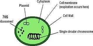
Inclusion Bodies in Prokaryotic Cells
The video below explains this:
Inclusion Bodies Detailed Video Explanation:
Properties of Inclusion Bodies
Below mentioned are the various features of the inclusion bodies.
- Inclusion bodies act as reserve deposits.
- With the help of cells, plenty of nutrients can be stored in them and it is utilised when the environment is deficient.
- Inclusion bodies are made up of virus antigens that are found at the site where the synthesis of the virus takes place.
- They are generally acidophilic.
- The Inclusion Bodies can also be present as crystalline aggregates of virions.
- They represent degenerative changes that are produced by a viral infection.
- When stained with gypsum on methylene blue dye, inclusion bodies can also be observed as ping structures under a microscope.
Classification of Inclusion Bodies
Below mentioned are the classification of Inclusion Bodies.
- Metachromatic Inclusions- Few of the large cell inclusions that sometimes stain red with blue dyes like methylene blue are termed metachromatic inclusions and consist of granules that consist of starch and glycogen. These glycogen granules appear reddish-brown in colour and the starch granules appear blue in the presence of iodine.
- Lipid Inclusions- Lipid Inclusions can be observed when cells are dyed with fat-soluble dyes like Sudan dyes and appear in various species of Bacillus, Mycobacterium, Azotobacter, and other genera and act as lipid storage material.
- Sulphur Granules- By oxidizing sulphur, Sulphur Granules derive energy and sulphur-containing compounds and may deposit sulphur granules in the cell that serve as an energy reserve.
- Carboxysomes- Carboxysomes inclusions contain the enzyme ribulose 1, -5 diphosphate carboxylase and the bacterias use these as a source of carbon for carbon dioxide fixation during photosynthesis.
- Magnetosomes- Magnetosomes are intracellular inclusion bodies or particles of iron oxide mineral called magnetite (Fe3O4) and some bacteria orient themselves within a magnetic field due to the presence of magnetosomes. A magnetosome is enclosed by a thin membrane composed of protein, phospholipid and glycoprotein and imparts a magnetic dipole on a cell allowing itself to exhibit magnetotaxis which is the process of migrating along the earth’s magnetic field. It is found in many aquatic organisms.

Cell Inclusion Bodies
We can classify inclusion bodies in the following categories based on the location, either at the nucleus or cytoplasm or both of these cell organelles.
- Intranuclear Cell Inclusions
- Infection Inclusion Bodies
- Intracytoplasmic Inclusion Bodies
- Physiological Inclusion Bodies
Inclusion bodies can be present in a bacterium or eukaryotic cell in the form of cystic lesions, fungal infections, virus-infected cells, bacterial infections, autoimmune diseases, neoplasms and blood dyscrasias.
Examples of Viral Inclusion Bodies
- Acidophilic Intracytoplasmic Inclusion Bodies (eosinophilic)
Eg: Negri bodies in Rabies, Paschen bodies in Smallpox, Bollinger bodies in fowlpox
- Acidophilic Intranuclear Inclusion Bodies (Eosinophilic)
Eg: Torres bodies in Yellow Fever, Cowdry type A in Herpes simplex virus, Cowdry type B in Polio and Adenovirus
- Intranuclear Basophilic Inclusions
Eg: Cowdry type B in Adenovirus
Owl’s eye appearance - Cytomegalovirus
- Both Intranuclear and Intracytoplasmic
Eg: Warthin-Finkeldey bodies in Measles
- Viral Inclusion Bodies in Plants
Eg: Virus particles in Cucumber mosaic virus
Diseases that Involve Inclusion Bodies
- Inclusion bodies myositis which affects muscle cells.
- Amyotrophic lateral sclerosis affects motor neurons.
- Dementia with Lewy inclusions that affect cerebral neurons.
How to Prevent Formation of Inclusion Bodies?
The cell inclusions are made of denatured aggregates of proteins (inactive), and several techniques have been developed to prevent the inclusion bodies formation and they help in the solubilisation and recovery of active proteins. Below mentioned are the techniques of it:
- To slow down the rate of protein expression, we can use weaker promoters.
- By using low copy number plasmids.
- Co-expression of a chaperone.
- By using specific E.coli strains.
- Fusing the target protein with a soluble partner.
- Lowering of the expression temperature.
Things to Remember
- Inclusion bodies are cytoplasmic or nuclear aggregates of stainable substances.
- Bacteria using hydrogen sulphide as an electron source contain sulphur granules.
- When the genes from one organism are expressed in some other organism, the proteins synthesised form inclusion bodies.
- A few bacteria store excess carbon in the form of glycogen or polyhydroxyalkanoates.
Read More: Cilia and Fagella
Sample Questions
Ques. What is the function of Inclusion Bodies? (1 Mark)
Ans. Inclusion bodies serve as storage vessels and the glycogen is stored as a reservation of carbohydrates and energy.
Ques. Can ribosomes be considered inclusion bodies? (1 Mark)
Ans. ribosomes are macromolecules that are made up of RNA and proteins and are non-membranous structures. They play an important role in protein formation and thus can be considered as an intermediate between inclusion bodies and cell organelles.
Ques. How are the inclusion bodies formed? (1 Mark)
Ans. Inclusion bodies are formed with increasing PH and pool of soluble fusion proteins within the cell and are formed during infectious disease or within the virus-infected cells such as rabies, herps, etc.
Ques. Give some examples of Acidophilic Intracytoplasmic Inclusion Bodies. (1 Mark)
Ans. Negri Bodies in rabies, Paschen Bodies in fowlpox are some of the examples of Acidophilic Intracytoplasmic Inclusion Bodies.
Ques. Name some diseases that involve inclusion bodies. (1 Mark)
Ans. Inclusion body Myositis, Amyotrophic lateral sclerosis, Dementia are some of the diseases that involve Inclusion Bodies.
Ques. What are Ribosomes? (2 Marks)
Ans. Ribosomes are the site of protein synthesis and are attached to the plasma membrane of the cell. It consists of two subunits 50 S and 30 S that form 70 S prokaryotic ribosomes. These are 15 to 20 nm in size, non-membrane bound bodies and several ribosomes get attached to the mRNA to form a chain called polyribosomes or polysomes.
Ques. What are the Inclusion Bodies? (2 Marks)
Ans. The tiny free particles that are freely suspended in the cytoplasmic matrix are referred to as inclusion bodies. They are referred to as Cytoplasmic Inclusions. Some of the examples of inclusion bodies include gas vacuoles, inorganic inclusions present as granules like iron and sulphur granules, food reserve inclusion bodies that are responsible for food storage (eg: lipid globules and protein granules).
Ques. What is the difference between Cell Organelles and Cell Inclusions? (2 Marks)
Ans. Cell organelles are the living components that are present in the cytoplasm of a cell and some of its examples include mitochondria, ribosome, chloroplast, ER, Golgi body, etc. Cell inclusions are the non-living components of the cytoplasm and its examples include reserve food, excretory/ secretory products and mineral matter.



Comments