



Content Curator
Sexually reproducing creatures generate gametes such as sperm and egg cells by this type of cell division. It then divides twice again, resulting in the creation of four daughter cells. The number of chromosomes is decreased to half in this type of division, which results in four gamete cells.
What is Meiosis?
Meiosis is a cell division process that describes the division of germ cells, which comprises two nucleus fissions. The nucleus's two fissions, meiosis 1 and meiosis 2, produce four sex cells known as gametes. The number of chromosomes in each sex cell is half that of the parent cell. This kind of cell division happens during reproduction, when one cell divides into four haploid daughter cells. The nucleus splits into four daughter cells (nuclei). Each generated cell has a unique set of chromosomes and half of the parent cell's genetic information. In eukaryotes, meiosis is involved in two important elements of the sexual life cycle. The first represents the transition from a diploid to a haploid state, and the second is the line of new allele combinations. Only the germline undergoes meiosis.

Meiosis
Functions of Meiosis?
Many sexually reproducing creatures require meiosis to guarantee that their progeny have the same number of chromosomes as their parents. Fertilization is the process of two cells coming together to form a new zygote. There will be four copies of each gene in the child if the number of alleles of each gene is not decreased to one in the gametes that create the zygote. This would result in a variety of developmental abnormalities in various species.
Polyploidy is prevalent in other species, and they can have several copies of the same gene. Meiosis must take place before reproduction if the organism cannot live if it is polyploidy. Meiosis is divided into two halves, each with its own set of stages.
Phases of Meiosis
DNA is duplicated before meiosis, just as it is before mitosis. Meiosis is then divided into two stages, known as meiosis I and meiosis II. The duplicated DNA is divided into daughter cells during the first division, which consists of many stages. The two alleles of each gene are split into distinct cells in the division that immediately follows the first.
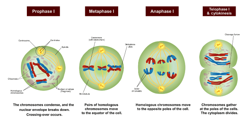
Phases of Meiosis
The two divisions, as well as the numerous phases or stages of each meiosis, are described below. Remember that the usually diploid DNA has been replicated before meiosis begins. This means that each gene has four copies, each of which is present in two complete sets of DNA, each of which has two alleles. The red chromosomes in the figure below are inherited from the mother, whereas the blue chromosomes are inherited from the father.
The DNA has already been duplicated at the start of the next diagram, which is why the red and blue chromosomes resemble the letter "X." Two sister chromatids - cloned DNA from replication – make up each of these "X" chromosomes. For storage, they are linked at the centromere, although they can split into independent chromosomes.
Phases of Meiosis I
Prophase I
Prophase I, the first stage of meiosis, is similar to prophase in mitosis in that the chromosomes condense and migrate toward the cell's center. The nuclear envelope breaks down, allowing microtubules from the cell's centrioles on each side to connect to the kinetochores in each chromosome's centromeres. The chromosomes couple with their homologous partner, unlike in mitosis. The red and blue chromosomes that join together in the figure demonstrate this. In mitosis, this step does not occur. Homologous chromosomes are prepared for crossing-over at the conclusion of prophase I and the start of metaphase I.

Prophase I
Homologous chromosomes can switch portions of themselves that house the same genes between prophase I and metaphase I. This is known as crossing-over, and it is accountable for the law of independent assortment, which is another rule of genetics. According to this rule, characteristics are inherited independently of one another. This is absolutely true for characteristics on separate chromosomes all of the time. Crossing-over allows maternal and paternal DNA to recombine for characteristics on the same chromosome, allowing traits to be inherited in an almost limitless number of ways.
Metaphase I
The homologous pairs of chromosomes align up on the metaphase plate, towards the cell's center, during metaphase I of meiosis I. A reductional division is the term for this phase. The two distinct alleles for each gene are lined up to be separated on homologous chromosomes. While the chromosomes line up on the metaphase plate with their homologous pair, there is no order in which the maternal or paternal chromosomes line up, as seen in the diagram above. The molecular rationale for the law of segregation is this mechanism.

Metaphase I
Each allele has the same probability of being handed on to children, according to the rule of segregation. The alleles are split during metaphase I of meiosis, which allows for this occurrence. They'll be divided into individual gametes during meiosis II. All chromosomes line up on their centromeres during mitosis, and each chromosome's sister chromatids split into new cells. In mitosis, the homologous pairs do not couple up and are divided in half, resulting in two distinct alleles for each gene in the new cells. These alleles originated from a maternal and paternal source, even though they were the same allele. The lining up of homologous chromosomes in meiosis results in the appearance of two alleles in the final cells, although they are on sister chromatids and are clones of the same DNA source.
Anaphase I
The chromosomes are now being pushed towards the centrioles on each side of the cell, similar to anaphase in mitosis. In anaphase I of meiosis, however, the centrosomes that keep sister chromatids together do not disintegrate, implying that only homologous chromosomes, not sister chromatids, are split.

Anaphase I
Telophase I
The chromosomes are fully separated in telophase I, and new nuclear envelopes develop. Cytokinesis efficiently separates the plasma membrane, resulting in the formation of two new cells.

Telophase I
Result of Meiosis I
Meiosis I produces two new cells, each of which is haploid in DNA but has two copies. Although each gene has two alleles, they are on sister chromatid copies of each other. As a result, these cells are classified as haploid. Before starting the second division of meiosis, meiosis II, these cells take a short break.

Result of Meiosis I
Phases of Meiosis II
Prophase II
Prophase II is quite similar to prophase I. The nuclear envelopes vanish, and centrioles emerge. Microtubules run the length of a cell, connecting to the kinetochores of individual chromatids through centromeres. The chromosomes start to move towards the metaphase plate.
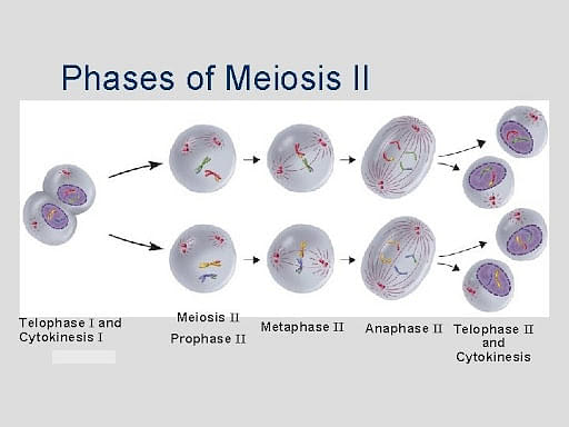
Prophase II
Metaphase II
The chromosomes align up with their centromeres on the metaphase plate, simulating mitosis. Each side of the metaphase plate has one sister chromatid. The protein cohesin is still linked to the centromeres at this stage.
Anaphase II
Sister chromatids split apart. Sister chromosomes are now named such because they are pushed toward the centrioles. The last division of the DNA occurs at this point. This division is known as an equational division because each cell finishes up with the same number of chromosomes as when the division began, but no copies.
Telophase II
The cell is now divided in two and the chromosomes are on opposing ends of the cell, as in the preceding telophase I. New nuclear envelopes are produced surrounding the chromosomes during cytokinesis, or plasma division.
Result of Meiosis II
There are four haploid cells at the end of meiosis II, each with only one copy of the genome. Gametes, or eggs in females and sperm in men, may now be produced from these cells.
Things to Remember
- Meiosis is a process in which a single cell divides twice to generate four cells with half the amount of genetic material as the original cell. These cells are called sperm and eggs, respectively, in men and females.
- Meiosis is divided into two phases.
- Haploids are organisms or gametes that have just one copy of each gene in each cell.
- If each cell has two copies of each gene, then it is known as Diploid.
Sample Questions
Ques: If a cell is going through Meiosis. The sister chromatids are lined up on the metaphase phase. So what is the phase of this meiosis?
Ans: This is the second step of the process. The homologous chromosomes are lined up on the metaphase plate in metaphase I. This is a significant distinction between the two. Metaphase II is identical to mitosis, but metaphase I resulted in ploidy decrease.
Ques: Give an example of Human Meiosis?
Ans: In humans, meiosis takes place in the sex organs. Female ovaries create eggs, whereas male testicles generate sperm. However, before these gametes can be created, the DNA must be decreased. Humans have 46 chromosomes, with 23 different chromosomes that occur in homologous pairings between maternal and paternal DNA. The DNA in the cell is duplicated before meiosis, resulting in 46 chromosomes in 92 sister chromatids. Each pair of sister chromatids has a matching set of sister chromosomes (maternal or paternal). Homologous chromosomes are these pairs of chromosomes. These homologous chromosomes align up and split during meiosis I. Each cell now has 23 chromosomes, each of which is made up of sister chromatids. Crossing-over may have happened during metaphase I of meiosis I, and these chromatids are no longer similar. Finally, sister chromatids are split into distinct cells during meiosis II. This leaves four haploid cells, each with 23 chromosomes.
Ques: How is Meiosis I different from Meiosis II?
Ans: In meiosis I, the homologous chromosome is separated, whereas in meiosis II, sister chromatids are separated. In meiosis I, two daughter cells are created, but in meiosis II, four daughter cells are formed.
Ques: Explain for which two reasons, we get genetically different gametes by meiosis division?
Ans: Crossing over: This is the moment at which homologous chromosomes cross over and genetic material is exchanged at random, and this process is unique to each cell that goes through meiosis.
Random orientation of homologous pairings: Random orientation of homologous pairs occurs in metaphase 1. This results in the generation of gametes with a wide range of homologous chromosomal combinations.
Ques: 60 chromosomes or 30 homologous chromosomes make up an adult organism. 30 are derived from the mother, and 30 are derived from the father. After mitosis, how many chromosomes are in each cell?
Ans: Mitosis yields the same number of chromosomes as meiosis. Mitosis, in essence, creates an identical clone of the parent cell. So after mitosis there are 60 chromosomes, and 30 homologs.



Comments