



Education Journalist | Study Abroad Lead
Microfilaments or actin filaments comprise two intertwined strands of actin, a globular protein. The cytoskeleton anchors organelles in place; aids in endocytosis, the ingestion of foreign materials by a cell, and cytokinesis, the separation of daughter cells following cell division; and moves portions of the cell in growth and mobility processes. The prokaryotic cytoskeleton is involved in maintaining cell shape and polarity. There are different types of fibers present in the Eukaryotic Cytoplasm: Microfilaments, Intermediate filaments, and Microtubules. In this article, we will study microfilaments, their role in cytoplasm, and their structure and functions.
Read More:- Binary fission
| Table of Contents |
Keyterms: Actin, Microfilaments, Protein, Cytoskeleton, Cell, Cytoplasm, Microtubules, Intermediate filaments, Fibers, Cell division, Cytokinesis
What are microfilaments?
Microfilaments are double-stranded polymerized fibrous actin filaments; the monomeric form of the protein is globular (G) actin, and the two conditions coexist in the cell. They are lengthy chains of G-actin produced into two parallel polymers that have been twisted around each other into a helical orientation with a diameter of 6 to 8nm. The microfilaments are found in bundles, forming a three-dimensional (3D) intracellular meshwork.

Microfilaments
These are found in all eukaryotic cells. They are primarily structural and are a vital component of the cytoskeleton, along with microtubules and, in some cases, intermediate filaments. They are the cytoskeleton's tiniest filaments. They perform various functions like cytokinesis, cell contractility, amoeboid movement, cell motility, endocytosis, exocytosis, change in cell shape, and mechanical stability.
Read Also:-
Role of Cytoplasm
The Cytoplasm is a rich semifluid that fills the spaces between cells. Except for the nucleus, the Cytoplasm in a eukaryotic cell mixes with all the rest cell contents. However, this does not occur in prokaryotic cells because they lack a distinct nuclear membrane, the Cytoplasm, containing the cell's genetic material. Compared to eukaryotes, the cells are smaller and have a more straightforward cytoplasm structure.
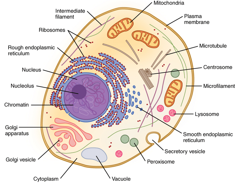
Microfilament and Cytoplasm
Structure of Microfilaments
Microfilaments are predominantly made up of actin polymers. Actin appears globular when it is first generated by the cell. On the other hand, microfilaments appear as long polymerized chains of molecules interwoven in a helix, resulting in a filamentous form of the protein, i.e. F-actin. They are made up of two strands of actin subunits coiled in a spiral. Actin subunits that come together to create a microfilament are known as globular actin (G-actin). Once connected, they are known as filamentous actin (F-actin). They are the thinnest filaments, typically 7 nm in diameter. These polymers are rigid yet have a flexible framework. Microfilaments determine the shape and mobility of the cell's surface.

Structure of Microfilaments
Self-assembling of microfilaments
When three G-actin proteins join together to create a trimer, a microfilament begins to form. More actin then bonds to the barbed end. Autoclampin proteins, which operate as motors to assist construct the lengthy strands that makeup microfilaments, aid in the self-assembly process. A microfilament is formed by two long strands of actin arranged in a spiral.
Read More About Difference Between Cytoplasm and Protoplasm
Function of Microfilaments
Microfilaments perform various functions such as:
- As we know, Microfilaments are flexible; thus, in collaboration with myosin, they help to generate the forces required for cellular contraction and basic cell motions.
- The integrity of actin filaments helps the eukaryotic cells survive environmental stress.
- Microfilaments help in cell division during mitosis (cell division). Microfilaments aid in the process of cytokinesis, which occurs when a cell "pinches off" and physically divides into two daughter cells.
- The microfilaments are also involved in amoeboid movements in some of the organisms.
- Microfilaments, which are a component of the cytoskeleton, hold organelles in position within the cell. They are responsible for cell stiffness and form.
- Microfilaments enable a cell to change its shape and move.

Function of Microfilaments
Location of Microfilaments
Microfilaments are found at the cell's periphery, where they travel from the plasma membrane to the microvilli. They are present in bundles in this area, forming a three-dimensional intracellular meshwork. They are found in the pericanalicular zone, constructing pericanalicular web/meshwork.
Read Also:- Asexual Reproduction
Things to remember
- Microfilaments, also known as actin filaments, are protein filaments that comprise part of the cytoskeleton in the cytoplasm of eukaryotic cells. They are mostly made up of actin polymers.
- Microfilaments, intermediate filaments, and microtubules make up the eukaryotic cytoskeleton.
- The cytoskeleton organizes and maintains the form of the cell.
- Microfilaments are found in all eukaryotic cells.
- The microfilaments are found in bundles, forming a three-dimensional (3D) intracellular meshwork.
- The polymers of microfilaments are simple to use and flexible but at the same time robust, resisting crushing and buckling while providing support to the cell.
- They are the thinnest filaments, typically 7 nm in diameter.
- They perform various functions like cytokinesis, cell contractility, amoeboid movement, cell motility, endocytosis, exocytosis, change in cell shape, and mechanical stability.
- The integrity of actin filaments helps the eukaryotic cells survive environmental stress.
- Microfilaments enable a cell to change its shape and move.
Sample Questions
Ques. What are microfilaments? (3 marks)
Ans. Microfilaments, also known as actin filaments, are protein filaments that comprise part of the cytoskeleton in the cytoplasm of eukaryotic cells. They are essentially made up of actin polymers. Still, they are changed by and interact with various other proteins in the cell. Microfilaments are typically 7 nm in diameter and composed of two actin strands. Cytokinesis, cell motility, endocytosis and exocytosis, amoeboid movement, cell contractility, changes in cell shape, and mechanical stability are all activities of microfilaments. Microfilaments are reasonably robust and flexible, resisting buckling by multi-piconewton compressive stresses and filament breaking by nano-newton tensile forces.
Ques. Differentiate between microfilaments and microtubules. (3 marks)
Ans. Microtubules and microfilaments both are components of a cell's cytoskeleton.
The polymerization of tubulin protein (alpha and beta) results in the formation of microtubules. They support the cell mechanically and help in intracellular transport. Polymerization of actin protein monomers results in the construction of microfilaments. They contribute to the movement of a cell on a surface. Microtubules and microfilaments are distinguished by the fact that microtubules are long, hollow cylinders composed of tubulin protein units. In contrast, microfilaments are double-stranded helical polymers composed of actin proteins.
Ques. Explain Cytoplasm's role in microfilaments. (3 marks)
Ans. The Cytoplasm is a rich semifluid that fills the spaces between cells. It is made up of the cytosol, which is filled with filaments, ions, proteins, macromolecular structures, and other organelles suspended in the cytosol. Except for the nucleus, the Cytoplasm in a eukaryotic cell mixes with the cell contents. However, this does not occur in prokaryotic cells because they lack a distinct nuclear membrane, the Cytoplasm, containing its genetic material. Compared to eukaryotes, the cells are smaller and have a more straightforward cytoplasm structure.
Ques. Explain the microfilament structure. (3 marks)
Ans. Microfilaments are predominantly made up of actin polymers. Actin appears globular when it is first generated by the cell. On the other hand, microfilaments appear as long polymerized chains of molecules interwoven in a helix, resulting in a filamentous form of the protein, i.e. F-actin. They are made up of two strands of actin subunits coiled in a spiral. Actin subunits that come together to create a microfilament are known as globular actin (G-actin). Once connected, they are known as filamentous actin (F-actin). They are the thinnest filaments, typically 7 nm in diameter. These polymers are rigid yet have a flexible framework. Microfilaments determine the shape and mobility of the cell's surface.
Ques. Describe the functions of microfilament. (5 marks)
Ans. The functions of microfilaments are:
- Microfilaments are flexible; thus, in collaboration with myosin, they help to generate the forces required for cellular contraction and basic cell motions.
- The integrity of actin filaments helps the eukaryotic cells survive environmental stress.
- Microfilaments help in cell division during mitosis (cell division). Microfilaments aid in the process of cytokinesis, which occurs when a cell "pinches off" and physically divides into two daughter cells.
- The microfilaments are also involved in amoeboid movements in some of the organisms.
- Microfilaments, which are a component of the cytoskeleton, hold organelles in position within the cell. They are responsible for cell stiffness and form.
- Microfilaments enable a cell to change its shape and move.
Ques. What is the diameter of the microfilament? (1 mark)
Ans. The diameter of the microfilament is 7nm.
Read Also:-



Comments