



Content Curator
Heart is a muscular organ and is responsible for pumping blood through a medium also known as the cardiovascular system. Functioning of the heart is absolutely necessary in order to keep the secondary activities of the body going.
- The heart has a total of four chambers through which blood passes one by one and converts into oxygenated blood.
- The heart is about the size of a fist and it expands and contracts about 100,000 times per day on an average.
- The muscular organ squeezes to push blood and relaxes to refill blood through a chain of events.
- The heart can be termed as the central organ that is responsible for blood flow.
Key Takeaways: Arteries, Veins, Valves, Chambers, Heart walls, Blood
Parts of the human heart
[Click Here for Sample Questions]
The heart is located under the ribcage, between the lungs and above the diaphragm. It weighs about 10.5 ounces and is cone shaped in structure. It consists of the following parts:
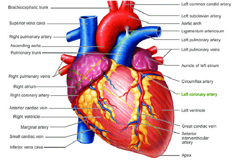
Heart Detailed Diagram
Heart - Chambers
There are four chambers of the heart.
- The upper two chambers are the auricles and the lower two are called ventricles.
- The two atria are thin-walled chambers that receive blood from the veins.
- The two ventricles are in contrast thick-walled which forcefully pump blood out of the heart.
- The amount of myocardium in the chambers causes the difference in the thickness which is a reflection of the amount of force each chamber needs to generate.
- The right atrium receives blood from systemic veins and it is deoxygenated blood. And the left atrium receives oxygenated blood from pulmonary veins.
Heart - Outer Wall
The organ is covered by a sack called the pericardium or the pericardial sac. There are three layers of the heart wall- the outer layer or the epicardium, the middle layer or the myocardium and the inner layer or the endocardium.
Heart - Valves
There are four main valves of the human heart- aortic valve, mitral valve, pulmonary valve and tricuspid valve.
- The aortic valve is mainly responsible for stopping the backflow of the blood when it is pumped to the aorta from the left ventricle.
- The mitral valve helps to prevent backflow when blood is pumped from left atrium to left ventricle.
- The pulmonary valve helps to prevent backflow of blood from the right ventricle to the pulmonary artery.
- The tricuspid valve helps prevent backflow from the right atrium to the right ventricle.
Heart - Arteries
- The right coronary artery is responsible for supplying the right atrium and right ventricle with blood.
- The left coronary artery divides into two different arteries called the circumflex artery and the left anterior artery which provide the left ventricles with blood.
- The anterior artery also supplies the front of the septum with blood.
- The pulmonary artery carries deoxygenated blood to the lungs for purifying it.
Heart - Veins
The main veins of the cardiac system are coronary sinus, anterior interventricular veins, left marginal veins, posterior veins of the left ventricle, and the posterior interventricular veins.
- The veins carry blood which is impure for purification from different parts of the body to the heart. They carry metabolic waste products from myocardium to the right atrium.
- The blood then flows back to the lungs for reoxygenation and removal of carbon dioxide.
- However, the pulmonary vein carries oxygenated blood to the heart.
Things to Remember
- The human heart is a muscular organ and can be called the most vital organ present in the human body.
- The heart pumps blood through a network consisting of arteries and veins, a medium also known as the cardiovascular system.
- The human heart is about the size of a fist, it weighs about 10.5 ounces and it expands and contracts about 100,000 times per day on an average.
- There are four chambers of the heart. The upper two chambers are the auricles and the lower two are called ventricles.
- There are four main valves of the human heart- aortic valve, mitral valve, pulmonary valve and tricuspid valve. They help prevent backflow of the blood.
- After going through the chain of events in the two auricles and ventricles, the blood enters the lungs for oxygenation.
Also Read:
Sample Questions
Ques. What are the main functions of the heart? (2 Marks)
Ans. There are four main functions of the human heart- pumping oxygenated blood to other body parts, pumping hormones and vital substances to other substances to other body parts, receiving deoxygenated blood to make it oxygenated in the lungs and maintaining blood pressure.
Ques. What are the different parts of the heart? (2 Marks)
Ans. The different parts of the heart consist of left atrium, left auricle, right atrium, right auricle, interventricular septum and septal papillary muscles, right ventricle, left ventricle, various arteries and veins which all function to maintain blood circulation in the body.
Ques. What is the heart made of? Where is the heart located? (2 Marks)
Ans. The heart is made up of three layers of tissue. Endocardium is the inner thin lining of the heart, myocardium is the thick middle layer of muscle and pericardium is the sac which covers the heart.
The heart is located in the front and middle of the chest, behind and slightly to the left of the breastbone.
Ques. What is the size of the heart? What are the chambers of the heart?(2 Marks)
Ans. The heart weighs a little between 7 to 15 ounces which is 200 to 425 grams and it is a little larger than the size of the individual’s fist. A human heart has four chambers- two auricles and two ventricles. The upper chambers receive the incoming blood and the lower 2 chambers pump out the blood of the heart.
Ques. What are the types of valves present in the human heart? What will happen if the heart stops beating? (2 Marks)
Ans. There are mainly 4 types of valves present in the human heart- tricuspid valve, mitral valve, pulmonary valve and aortic valve. If the heart stops beating, the lack of oxygen rich blood can cause death or permanent brain damage within a few minutes. The heart should function at all times to make sure blood is pumped out throughout the whole body.
Ques. State the functions of heart valves. (5 marks)
Ans. The heart valves mainly work to keep the blood from flowing in the wrong direction. The endocardium consists of epithelial cells which line the heart chamber and valves. The endocardium also contains muscle cells which cause muscle contraction and collagen fibres in order to make the endocardium elastic. A group of cells are clustered in the upper right atrium which generate significant electrical impulses. These impulses move down towards the atrioventricular node which is located between the bottom of the right atria and the top of the ventricles. The atrioventricular node squeezes blood into the ventricles and transmits impulses to them. Ventricular contractions generate the heart rate and the node impulse is generated by the autonomic nervous system.
Each valve consists of a set of flaps called leaflets or cusps. The leaflets of the valves are attached and supported by a ring of tough, fibrous tissue called the annulus which maintains the exact shape of the valve. The mitral and tricuspid valve leaflets are supported by tough and fibrous strings known as chordae tendineae. Papillary muscles are the small muscles which hold the valve leaflets and are part of the inside walls of the ventricles. The mitral valve has two leaflets while others have three.
In a state of relaxation or rest, the heart beats 50 to 99 times per minute. Exercise, medications or stress can force the heart to beat faster, which can be over 100 times per minute.



Comments