




The NCERT Class 11 Biotechnology Handwritten Notes Chapter 2 Cellular Organelles are scanned notebook pages written for one sitting revision against the 2026-27 syllabus. They cover the prokaryotic against eukaryotic split, the fluid mosaic model of the plasma membrane, Gram positive and Gram negative cell walls, the endomembrane system, mitochondria, plastids, ribosomes, microbodies, the cytoskeleton, cilia and flagella, the nucleus and the chromosome, with every labelled figure copied by hand.
- Length: 34 handwritten pages covering all twelve sections of the chapter, every textbook figure and all thirteen exercise questions.
- Must-learn pair: single membrane against double membrane against non membrane bound organelles, the classification that exercise question 8 asks for directly.
- Also on this page: an organelle classification tree, a prokaryotic against eukaryotic comparison, a quick recall table and links to sibling cell biology chapters.
Every page in these Cellular Organelles notes is written by hand, checked against the 2026-27 NCERT Biotechnology print, and matched to the way Class 11 papers ask about membrane transport, organelle structure and the 9+2 axoneme.
Student Feedback: In a Collegedunia survey of 8,720 Class 11 Biotechnology students, 71% of students said Cellular Organelles feels easy while reading and turns hard in the exam, because the paper asks for structure with numbers rather than a general description. Toppers reported one fix that works: for every organelle write down its membrane count, its size, its internal compartments and one function, and revise that four line card instead of the paragraph.
Source: 2026-27 Class 11 Biotechnology student survey. Sample of 8,720 students from schools across 14 states.
Why the Cell Is the Starting Point of Cellular Organelles
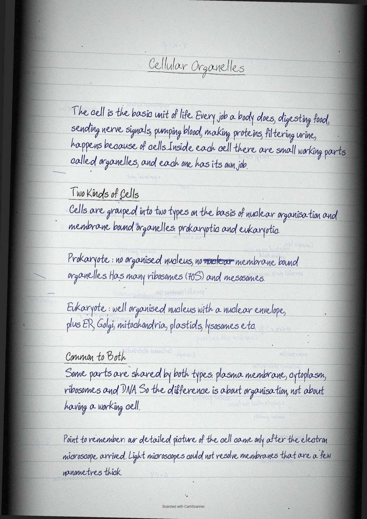
The chapter opens with a list of jobs the body does at any given moment, digesting food, sending electrical messages through nerves, pumping blood, circulating nutrients, synthesising proteins and filtering urine. All of it happens because of cells, which the textbook calls the basic unit of life. Each cell is fitted with different machineries called organelles, and each organelle handles a different job.
The exam rarely asks what a cell is. It asks which organelle does what, how many membranes bound it, and what happens when it fails. That is why the notebook builds the chapter as a set of organelle cards rather than as continuous prose.
- Structure first: shape, size, membrane count and internal compartments.
- Function second: the one process the organelle is famous for.
- Named example third: the organism, disease or scientist the textbook attaches to it.
Advancement in microscopic technique is credited in the opening paragraph for making all of this visible. Without the electron microscope, the internal detail of cristae, thylakoids and nuclear pores could never have been described.
Prokaryotic Against Eukaryotic Cells: The Split That Runs Through the Chapter
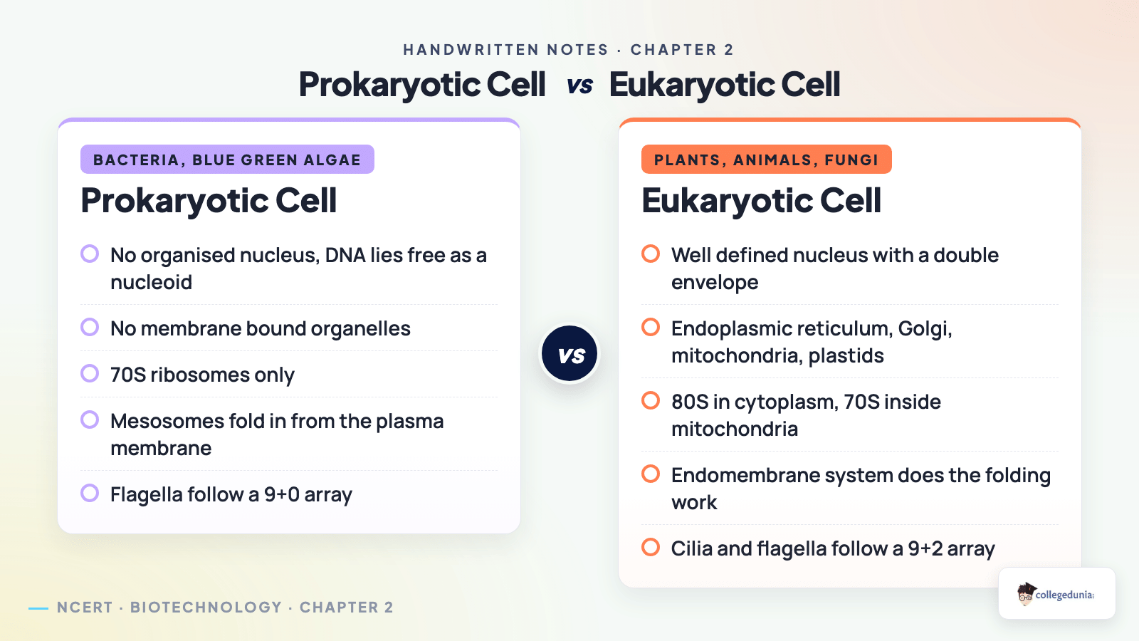
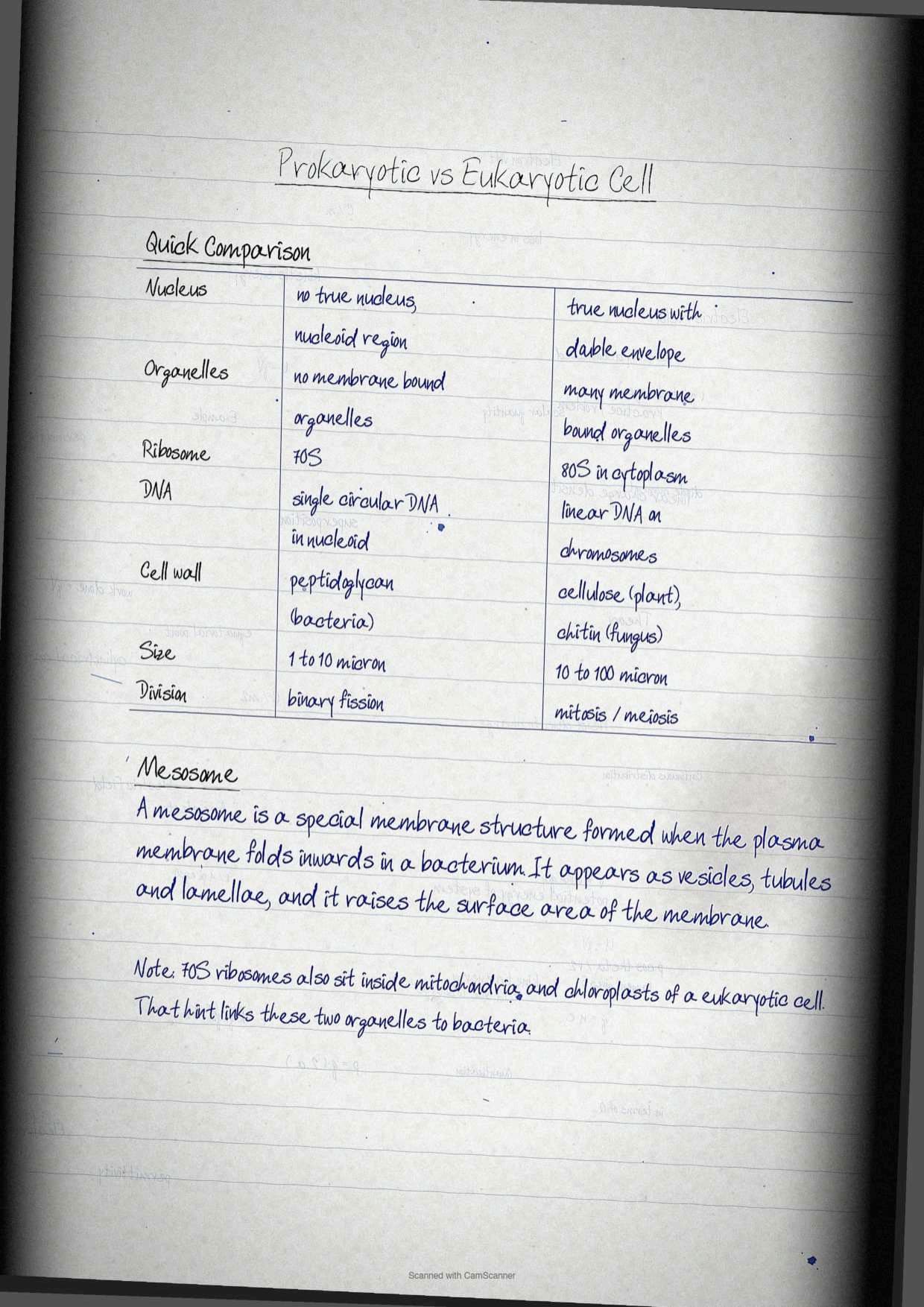
Cells in unicellular and multicellular organisms are broadly grouped into two categories on the basis of nuclear organisation and the presence of membrane bound cell organelles. Getting this split right at the start makes every later section easier, because the notebook keeps returning to it.
| Feature | Prokaryotic cell | Eukaryotic cell |
|---|---|---|
| Nucleus | No organised nucleus, genetic material lies free as a nucleoid | Well organised nucleus bounded by a nuclear envelope |
| Membrane bound organelles | Absent | Present, including endoplasmic reticulum, Golgi apparatus, mitochondria, plastids, vacuole, lysosomes and peroxisomes |
| Ribosomes | Numerous, 70S | 80S in the cytoplasm, 70S inside mitochondria and chloroplast |
| Special membrane folds | Mesosomes, folds of the plasma membrane | Absent, the endomembrane system does the equivalent work |
| Chromosome | Usually a single circular chromosome, more than one in Vibrio cholerae | Several linear chromosomes inside the nucleus |
| Flagellar array | 9+0 arrangement | 9+2 arrangement |
Some components are common to both. Plasma membrane, cytoplasm, ribosomes and DNA appear in every cell, prokaryotic or eukaryotic, and the textbook says so in the overview. That single sentence answers a very common one mark question.
Tip: when a question asks for differences, do not write that prokaryotes have no organelles. They have ribosomes, which are organelles, just not membrane bound ones. Write that they lack membrane bound organelles, and the mark is safe.
Plasma Membrane and the Fluid Mosaic Model
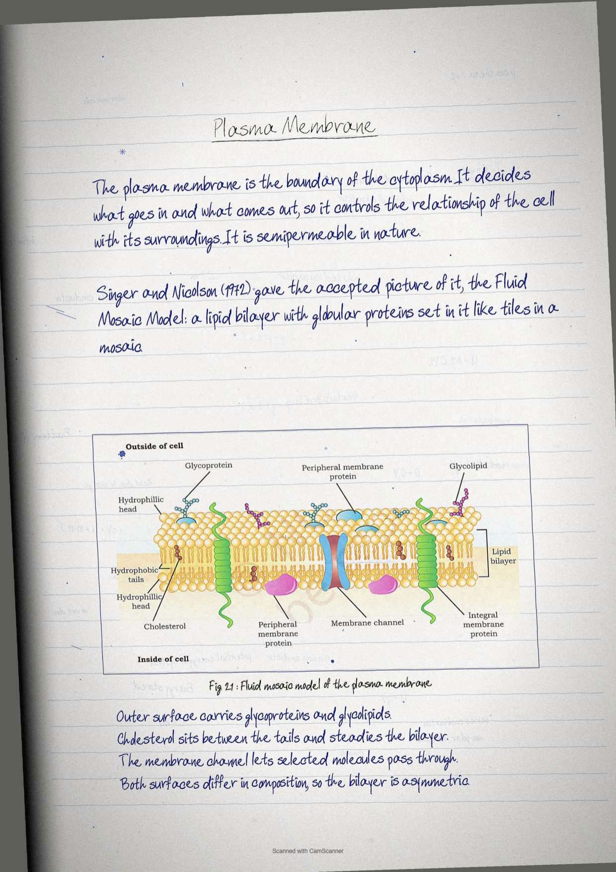
The plasma membrane forms the boundary of the cytoplasm and is guarded from outside by the extracellular matrix. It is semipermeable in nature and is responsible for the relationship of the cell with its surroundings. The detailed structure only became clear once the chemical composition, mainly lipid and protein, was known and the electron microscope was invented.
The widely accepted model was proposed by Seymour Jonathan Singer and Garth L. Nicolson in 1972 and is called the Fluid Mosaic Model. It describes the membrane as a lipid bilayer surrounding the cell with a mosaic of globular proteins set into it.
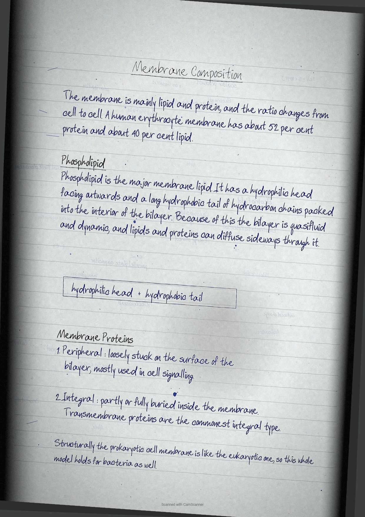
- Phospholipids are the major membrane lipid. The hydrophilic head faces the exterior and the long hydrophobic hydrocarbon tails occupy the interior of the bilayer.
- Peripheral membrane proteins are superficially attached to the bilayer and are mainly involved in cell signalling.
- Integral membrane proteins are partially or fully buried in the membrane, and transmembrane proteins are the most abundant type of integral protein.
- Cholesterol, glycoproteins and glycolipids also sit in the bilayer, as Figure 2.1 of the chapter shows.
Composition varies between cell types. The human erythrocyte membrane contains roughly 52 per cent protein and 40 per cent lipids, a figure worth memorising because it turns up as a direct one mark question. The bilayer keeps the cell boundary in a quasifluid state, so lipids and proteins can diffuse freely in the lateral plane.
Fluidity is not a decorative detail. Cell division, cell growth, communication at intercellular junctions, cell secretion and endocytosis all depend on it. Structurally, the prokaryotic cell membrane is similar to the eukaryotic one, which is why the model is presented as universal.
The historical box is examinable too. Edwin Gorter and F. Grendel, in 1925, collected chromocytes from mammalian artery or vein, washed them with saline, extracted the lipids with acetone and found enough lipid to cover the cell surface exactly twice. That is how the bilayer, rather than a monolayer, was proposed, using mammalian red blood cells as the model. High magnification electron micrographs later showed the membrane as a railroad track, two densely stained lines of polar head groups with a lightly stained hydrophobic core between them.
A mesosome is a special membranous structure formed by extension of the plasma membrane in prokaryotic cells, in the form of vesicles, tubules and lamellae. Its purpose is simple, it increases the surface area of the plasma membrane.
Transport Across the Plasma Membrane: Passive, Facilitated and Active
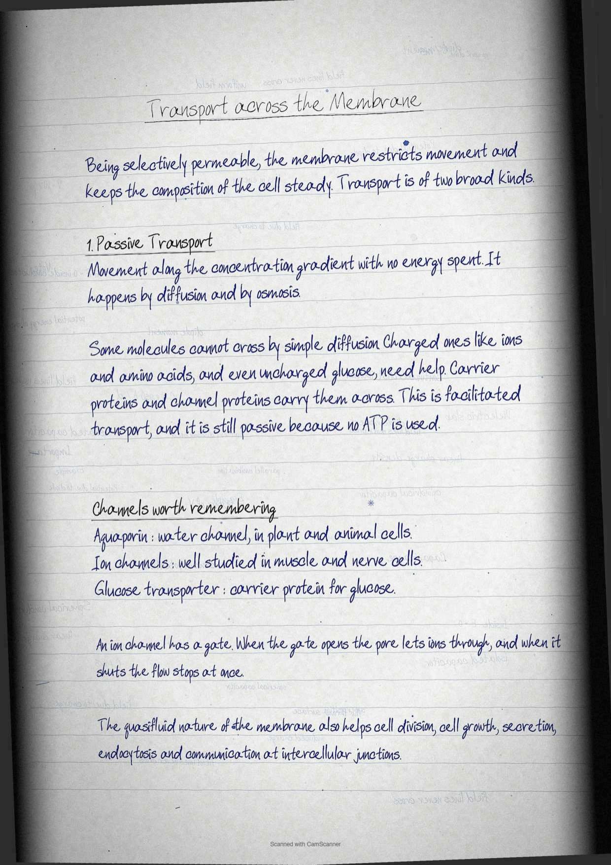
Exercise question 4 asks students to describe the various mechanisms of transport across the plasma membrane with labelled diagrams, so this section is worth full marks on its own. Being selectively permeable, the membrane restricts molecular movement and maintains cell composition.
| Mechanism | Energy needed | Direction | Example |
|---|---|---|---|
| Passive transport | None | Along the concentration gradient | Diffusion and osmosis |
| Facilitated transport | None, but a protein is required | Along the concentration gradient | Glucose transporter, ion gated channels, aquaporins for water |
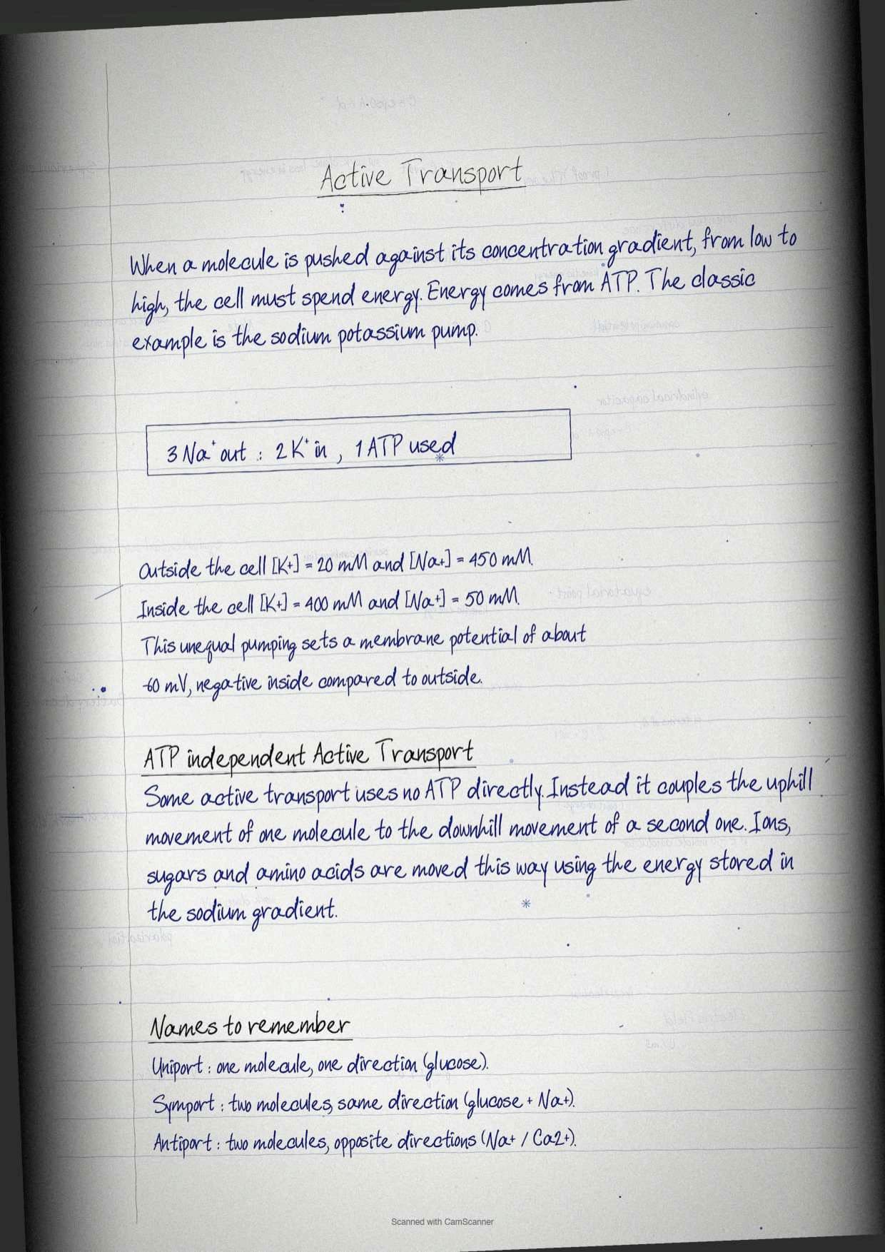
| Active transport | ATP hydrolysis | Against the concentration gradient | Na+ and K+ pump |
| Coupled or ATP independent active transport | Energy from an existing ion gradient | Against the gradient for one molecule | Sugars and amino acids moved using the Na+ gradient |
Charged molecules such as ions and amino acids, and even uncharged ones such as glucose, cannot cross the bilayer by simple diffusion. Their movement is facilitated by carrier proteins and channel proteins. Aquaporins are the critical channel proteins for water transport in both plant and animal cells, and the best studied channel proteins in muscle and nerve cell membranes are the ion channels.
Coupled transport has three named forms, and the paper likes asking for the difference between them.
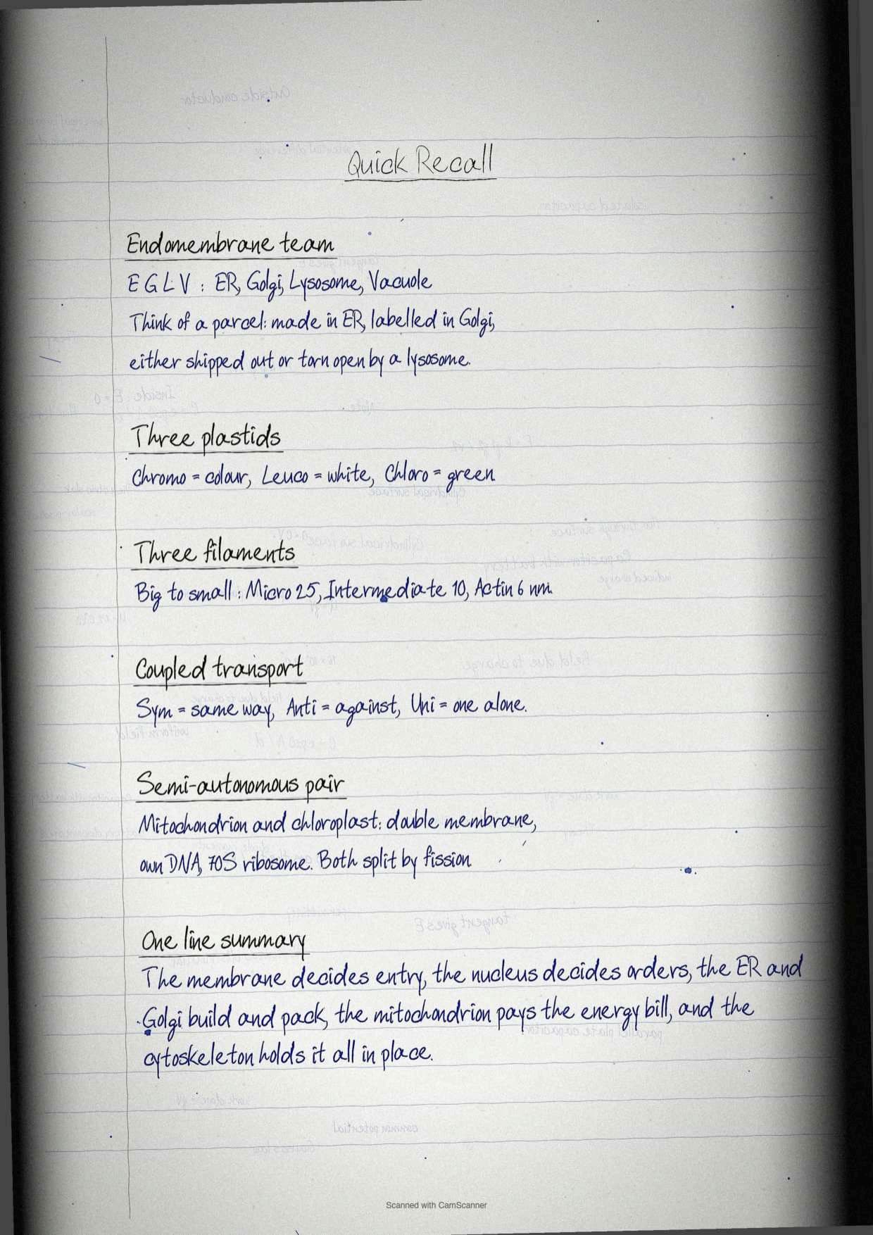
- Symport: two molecules transported in the same direction, for example the uptake of glucose along with Na+.
- Antiport: two molecules transported in opposite directions, for example Na+ and Ca2+ by the Na+ and Ca2+ antiporter.
- Uniport: facilitated diffusion of a single molecule, for example glucose.
Tip: hold the three by their prefixes. Sym means together, anti means against, uni means one. Students who wrote the prefix meaning beside each term stopped losing this mark.
Cell Wall in Bacteria: Gram Positive Against Gram Negative
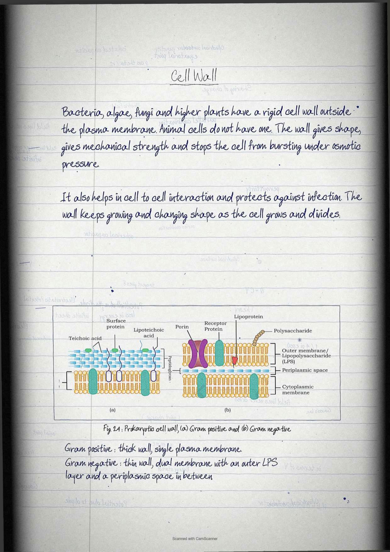
Cells of bacteria, algae, fungi and higher plants carry a rigid cell wall outside the plasma membrane. It is not found in animal cells, and it differs structurally between bacteria and eukaryotes. In bacteria it is built of polysaccharide cross linked by small peptides, which gives rigidity, shape and protection from osmotic pressure.
Exercise question 7 asks for the importance of the cell wall in prokaryotic cells, so learn the function list as four separate points.
- Determines the cell shape.
- Prevents the cell bursting under osmotic pressure.
- Helps in cell to cell interaction.
- Provides mechanical strength and protection from infection.
| Point of difference | Gram positive bacteria | Gram negative bacteria |
|---|---|---|
| Cell wall thickness | Thick cell wall | Thin cell wall |
| Membrane arrangement | Single plasma membrane | Dual plasma membrane, an inner cytoplasmic membrane and an outer membrane |
| Characteristic wall molecules | Teichoic acid and lipoteichoic acid | Lipopolysaccharide, porins and lipoprotein in the outer membrane |
| Periplasmic space | Not prominent | Present between the two membranes |
| Figure reference | Figure 2.4 (a) | Figure 2.4 (b) |
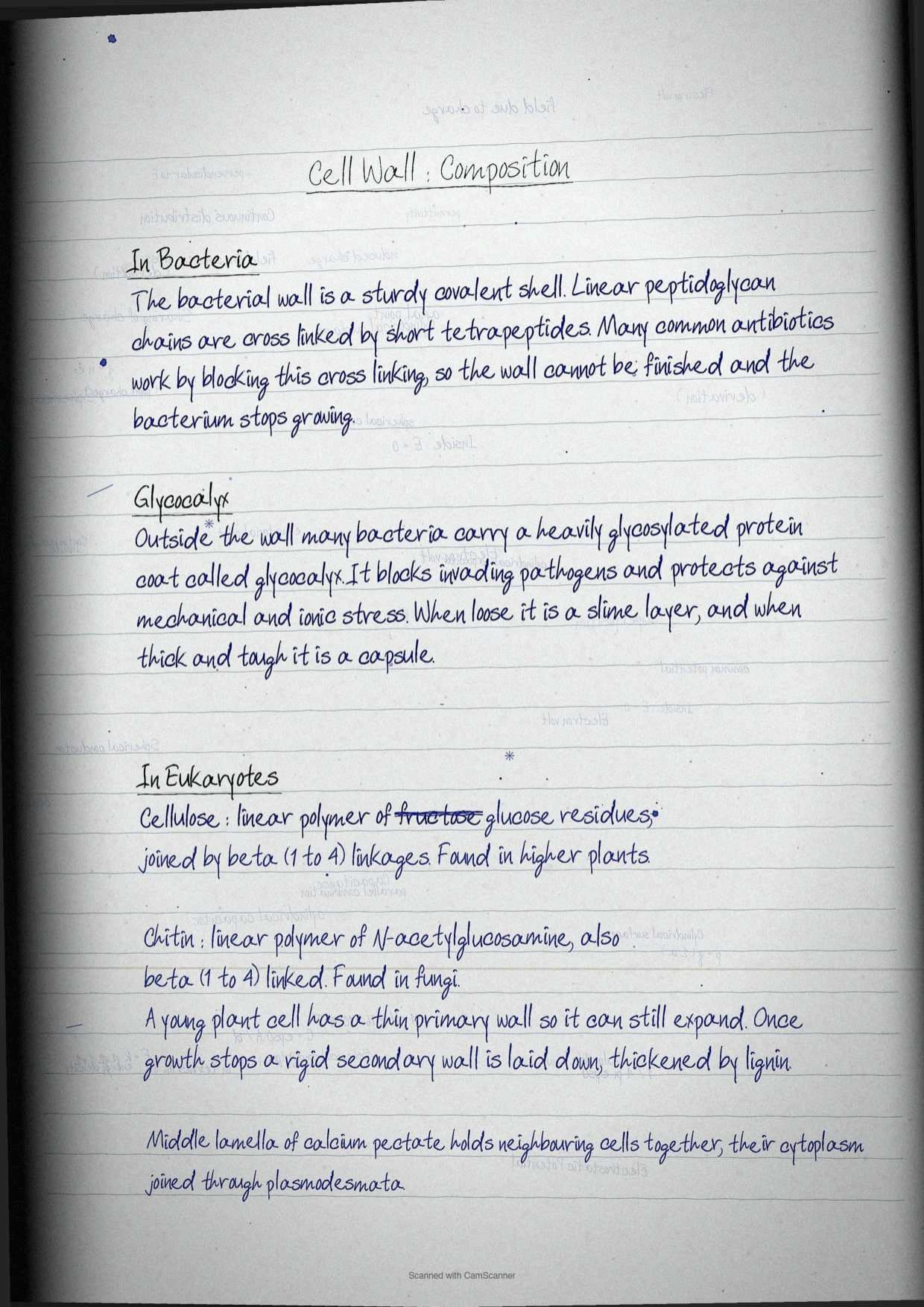
Structurally the bacterial cell wall is a sturdy covalent shell of linear peptidoglycan chains cross linked by tetrapeptides. Commonly used antibiotics work by inhibiting this cross linking of peptidoglycan strands, which interferes with bacterial growth. That single sentence links this chapter to pharmacology and is a favourite short note.
Outside the wall, prokaryotic cells carry a heavily glycosylated protein layer called the glycocalyx. It acts as a barrier to invading pathogens, protects the cell from mechanical and ionic stress and takes part in cell to cell interaction. When it is loose it is called a slime layer, and when it is thick and tough it is called a capsule. The wall itself grows and changes shape continuously as the bacterium grows and divides.
Cell Wall in Plants and Fungi: Cellulose, Chitin and the Middle Lamella
Among eukaryotes the wall is mainly polysaccharide, and the polysaccharide identity is the examinable part. In most higher plants it is cellulose, a linear polymer of glucose residues, and in fungi it is chitin, a linear polymer of N-acetylglucosamine. Both are joined by beta 1 to 4 glycosidic linkages, as Figure 2.5 shows.
| Point of difference | Primary cell wall | Secondary cell wall |
|---|---|---|
| When it forms | Around a growing cell | After the cell ceases growing |
| Position | Outermost, next to the middle lamella | Between the primary wall and the plasma membrane |
| Thickness | Comparatively thin | Very thick and rigid |
| Special deposit | None | Lignin |
| Scope for expansion | Yes, the cell can still expand | No |
Neighbouring plant cells are held together by a layer of calcium pectate known as the middle lamella, and their cytoplasms stay connected through channels called plasmodesmata. Exercise question 13 (b) asks for the primary against secondary wall difference directly, so the table above is written out in full in the notebook.
The Endomembrane System: Four Organelles Working as One
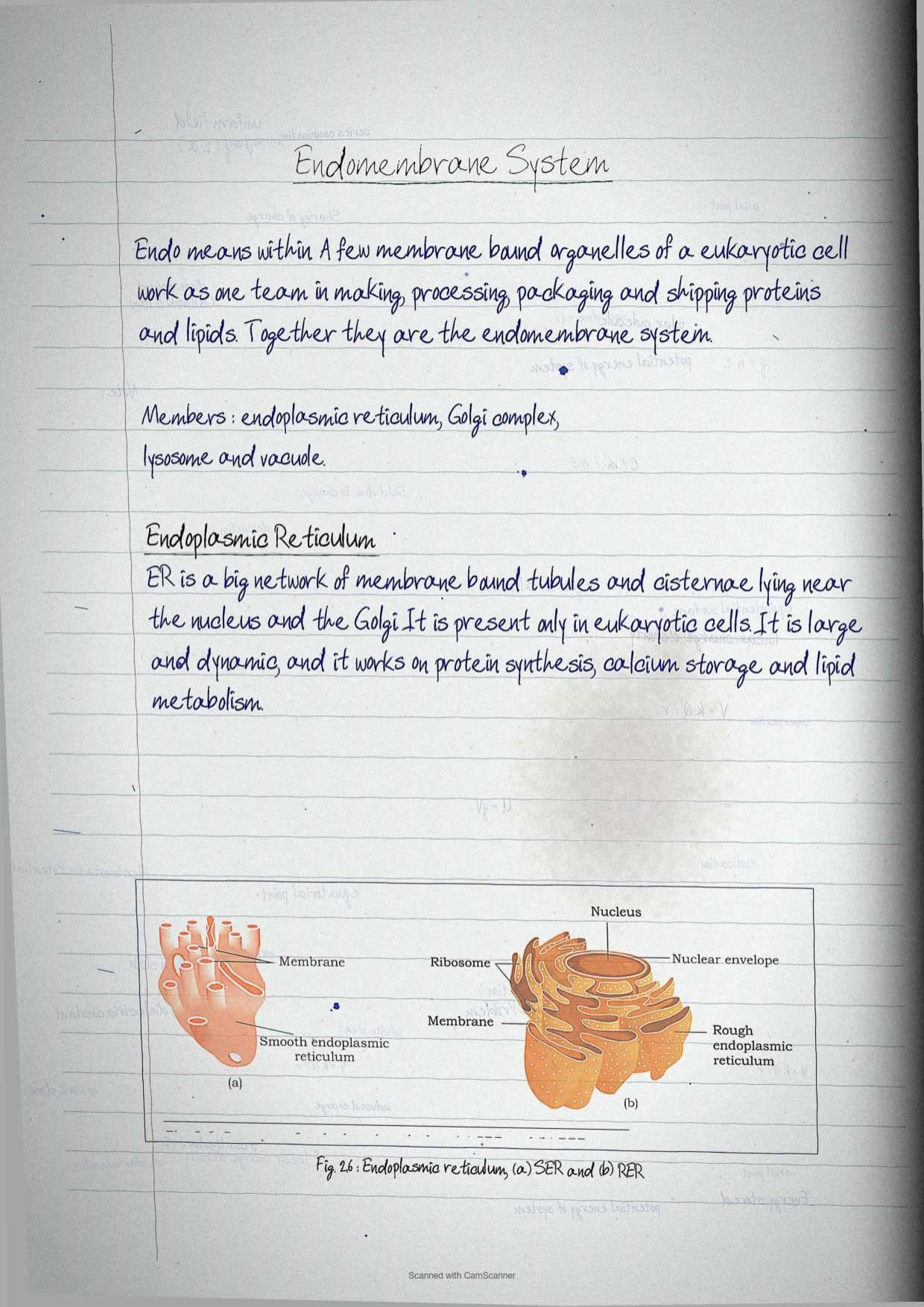
Many eukaryotic organelles are bound by a membrane similar to the cell membrane and are distinct in structure and function. Some of them, however, work together as a coordinated system called the endomembrane system, where endo means within. The system handles protein and lipid synthesis, along with the processing, packaging and transport of those products to their destinations inside the cell.
Only four organelles belong to it, and adding a fifth is the commonest error in this chapter.
- Endoplasmic reticulum
- Golgi complex
- Lysosomes
- Vacuoles
Mitochondria, chloroplast and peroxisomes are membrane bound but are not part of the endomembrane system, because their functions are not coordinated with the protein secretion pathway.
The pathway itself was worked out in the 1960s by George Palade and colleagues, who traced the route of a secretory protein as rough endoplasmic reticulum, then Golgi apparatus, then secretory vesicles, then the cell exterior. Learning that four step chain answers several questions at once.
Endoplasmic Reticulum: Rough Against Smooth
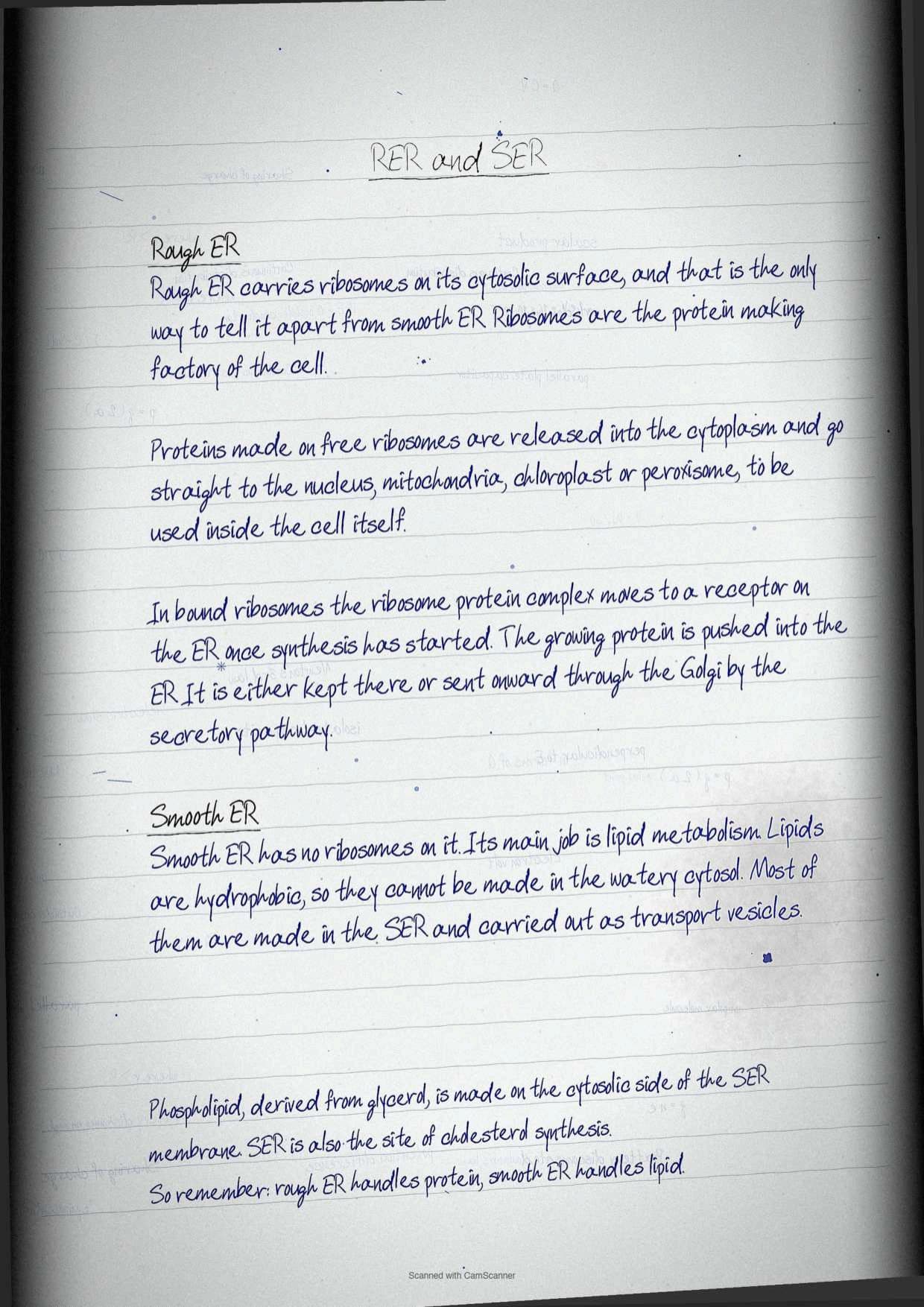
The endoplasmic reticulum is an extensive network of membrane enclosed tubules and cisternae located near the nucleus and the Golgi apparatus. It is present exclusively in eukaryotic cells, and it is large, dynamic and continuously involved in protein synthesis, calcium storage and lipid metabolism. It is classified on one criterion alone, the presence or absence of ribosomes on its surface.
| Feature | Rough endoplasmic reticulum | Smooth endoplasmic reticulum |
|---|---|---|
| Ribosomes on the cytosolic surface | Present | Absent |
| Main role | Synthesis and trafficking of secretory proteins | Lipid metabolism |
| Named products | Proteins destined for Golgi, lysosomes and the plasma membrane | Phospholipids and cholesterol |
| Figure reference | Figure 2.6 (b) | Figure 2.6 (a) |
Protein sorting, shown in Figure 2.7, splits into two routes and the paper likes asking for both. Proteins made on free ribosomes are released into the cytoplasm and go directly to the nucleus, mitochondria, chloroplast and peroxisomes to be used within the cell. Proteins made on bound ribosomes take a different route: after protein synthesis begins, the ribosome and protein complex is transferred to a receptor on the endoplasmic reticulum, and the nascent protein is inserted into the reticulum. From there the protein is either retained in the reticulum or transported to its destination through the Golgi complex by the secretory pathway.
Smooth endoplasmic reticulum earns its own answer. Because lipids are hydrophobic they cannot be synthesised in the cytosol, so most lipids are made in the smooth reticulum and moved out as transport vesicles. Phospholipids are derived from glycerol and are synthesised on the outer, cytosolic side of the smooth reticulum membrane, and the same organelle is an essential site for cholesterol synthesis.
Golgi Apparatus: The Receiving and Shipping Department
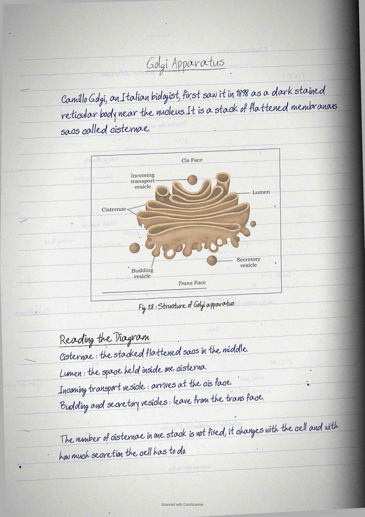
The Golgi apparatus was first observed by Camillo Golgi, an Italian biologist, in 1898, as a dark stained reticular structure near the cell nucleus. It was later found in other cell types and renamed the Golgi apparatus or Golgi complex. It is a membrane bound organelle made of a series of flattened membranous sacs that look like stacked pouches, called cisternae.
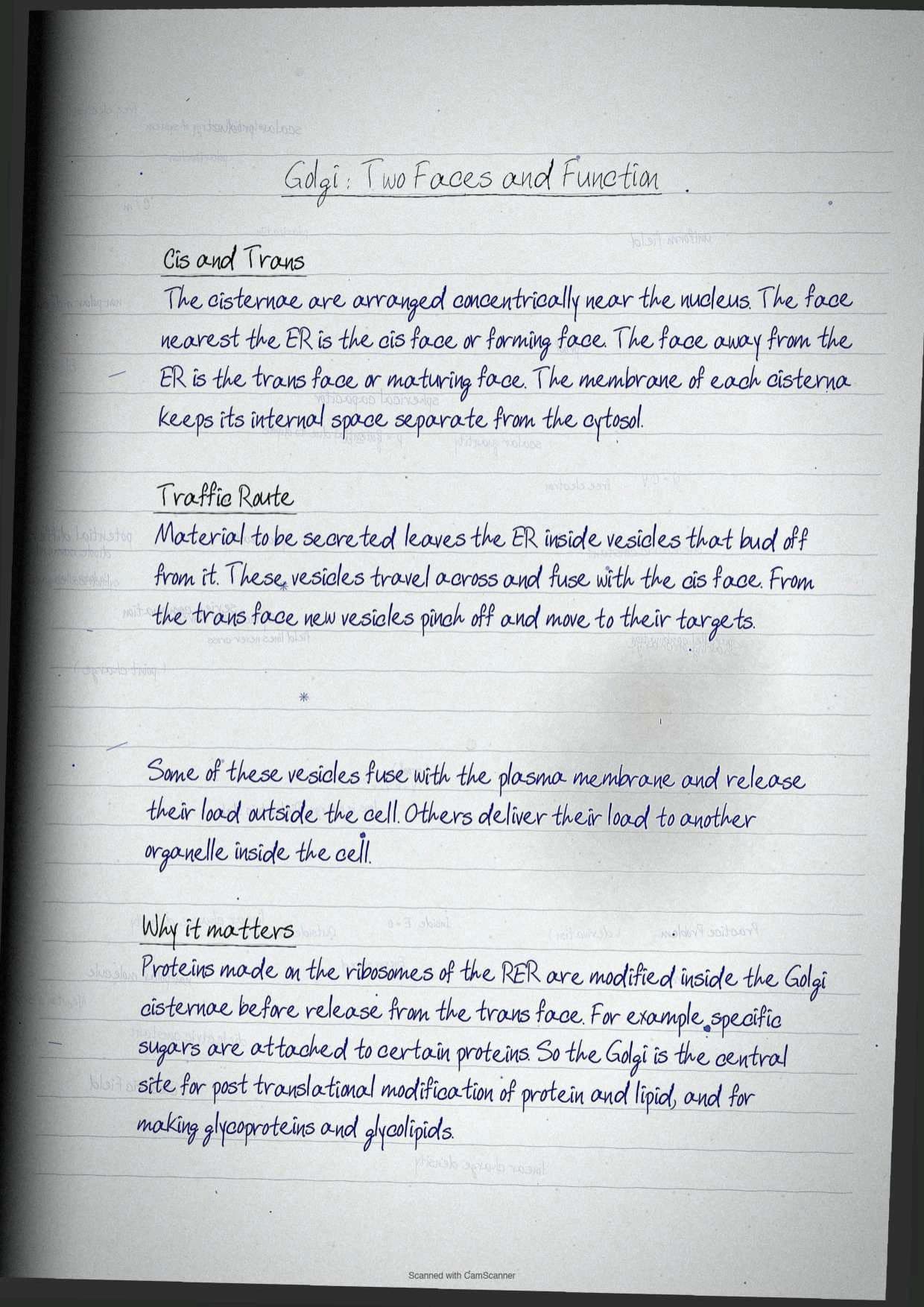
- A varied number of cisternae sit in one stack, concentrically arranged near the nucleus.
- The cis face is the set of cisternae nearest the endoplasmic reticulum, and is also called the forming face.
- The trans face is the set away from the endoplasmic reticulum, and is also called the maturing face.
- The membrane of each cisterna separates its internal space from the cytosol.
Traffic runs in one direction. Material to be secreted leaves the endoplasmic reticulum in vesicles that bud off, travel to the Golgi apparatus and fuse with the cis face. Vesicles pinch off from the trans face and travel to other sites, where they either fuse with the plasma membrane and release their contents outside the cell, or deliver their contents to another organelle.
Proteins made by ribosomes of the rough endoplasmic reticulum are modified in the Golgi cisternae before release from the trans face. For example, specific sugars are attached to some proteins before they leave the cell. The Golgi apparatus is therefore the central organelle for trafficking and post translational modification of protein and lipid, and the main site of glycoprotein and glycolipid formation.
Lysosomes: Acid Hydrolases and Tay-Sachs Disease
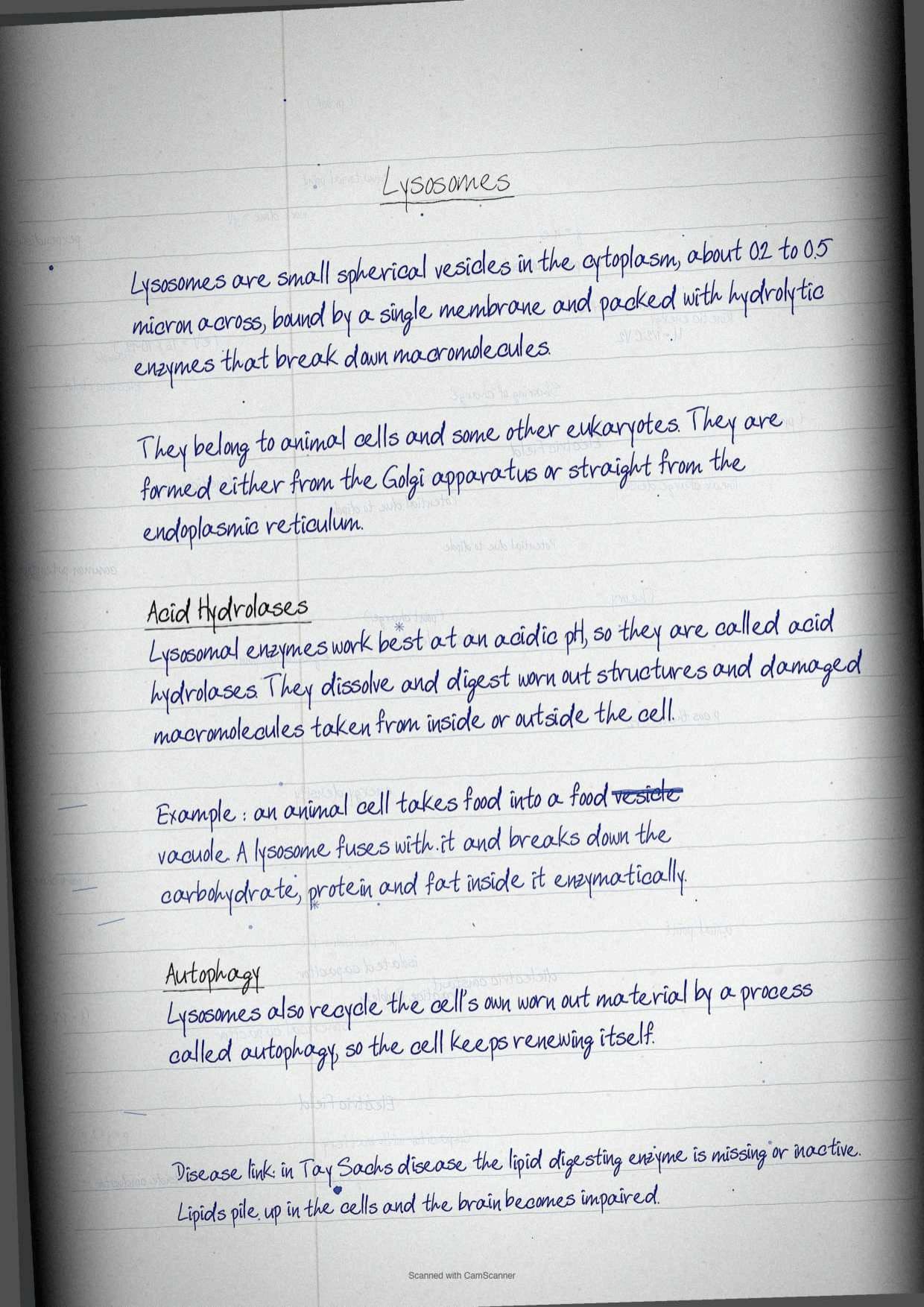
Lysosomes are small spherical vesicles in the cytoplasm, roughly 0.2 to 0.5 micron in diameter, bound by a single membrane and packed with hydrolytic enzymes that break down macromolecules. They occur in animal cells and some other eukaryotes, and they are formed either from the Golgi apparatus or directly from the endoplasmic reticulum.
Their enzymes show optimal activity at an acidic pH, which is why they are called acid hydrolases. They dissolve and digest redundant structures or damaged macromolecules from inside or outside the cell.
- Intracellular digestion: when an animal cell ingests food into a food vacuole, lysosomes fuse with the vacuole and enzymatically break down carbohydrates, proteins, fats and other components.
- Autophagy: lysosomes use their hydrolytic enzymes to recycle the cell's own organic material, so the cell continually renews itself.
- Disease link: in Tay-Sachs disease the brain becomes impaired because lipids accumulate in cells, due to absence or inactivation of the lipid digesting enzyme.
Tip: Tay-Sachs is the only named human disease in this chapter, so it is the easiest place for a paper to test whether the chapter was actually read. Write the enzyme defect, not just the name.
Vacuoles: Storage, Turgor and the Tonoplast
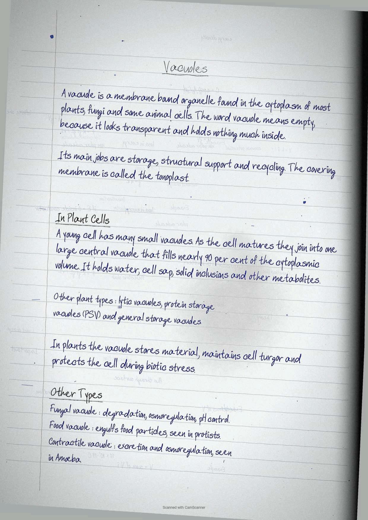
Vacuoles are membrane bound intracellular organelles found in the cytoplasm of most plants and fungi and in some animal cells. The name comes from the Latin sense of empty, because of the transparent look and the lack of cytoplasmic material. Their broad functions are storage, structural support and recycling, and the membrane that covers them is called the tonoplast.
The number and size of vacuoles varies with the age of a plant cell. Young cells carry a large number of small vacuoles. As the cell matures those vacuoles amalgamate into one large central vacuole that occupies almost 90 per cent of the cytoplasmic volume, and it holds water, cell sap, solid inclusions and other metabolites.
Exercise question 9 asks for the different types of vacuoles, so the notebook writes the list out cleanly.
| Type of vacuole | Where it occurs | What it does |
|---|---|---|
| Central vacuole | Mature plant cells | Storage of water, cell sap and metabolites, maintains turgor |
| Lytic vacuole | Plant cells | Degradative work |
| Protein storage vacuole | Plant cells, notably seeds | Stores protein reserves |
| Fungal vacuole | Fungi | Storage, degradation, osmoregulation and control of intracellular pH |
| Food vacuole | Protists and animal cells | Engulfs food particles |
| Contractile vacuole | Amoeba and other protists | Excretion and osmoregulation |
In plant cells the major functions are storage, maintaining cell turgor and protecting cells during biotic stress. Fungal vacuoles are comparatively complex and do more than store material. Exercise question 13 (c) asks students to distinguish lysosomes from vacuoles, and the cleanest contrast is enzyme content: lysosomes are defined by their acid hydrolases, vacuoles by their storage role.
Mitochondria: Cristae, Matrix and the Organelle Genome
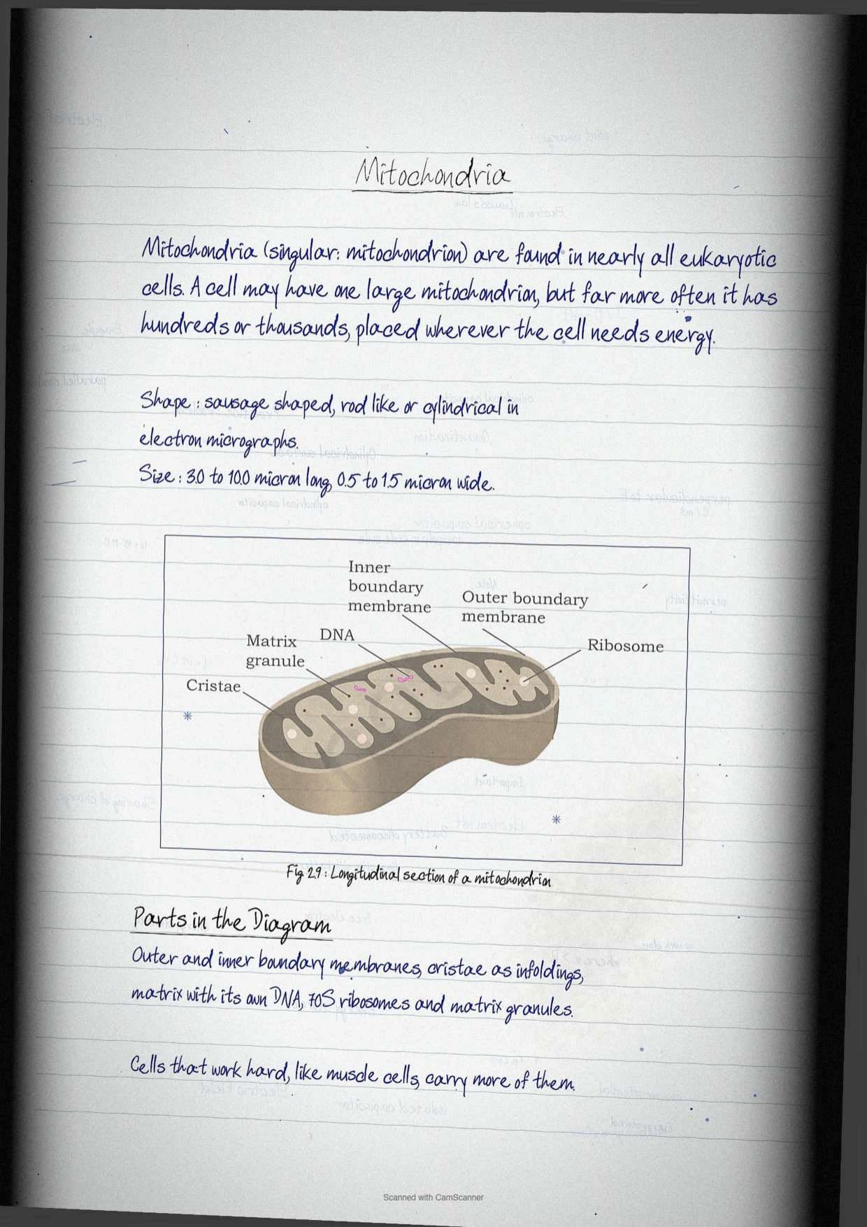
Mitochondria, singular mitochondrion, are found in nearly all eukaryotic cells. Some cells carry a single large mitochondrion, but more often a cell has hundreds or even thousands of them, positioned according to what the cell does. A typical mitochondrion is sausage shaped and appears rod shaped or cylindrical in electron micrographs.
- Size: 3.0 to 10.0 μm long and 0.5 to 1.5 μm wide.
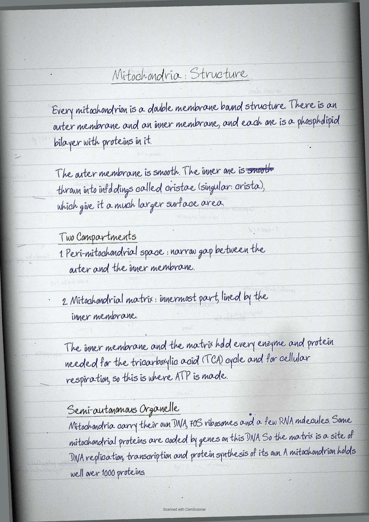
- Membranes: double membrane bound, an outer and an inner membrane, each a phospholipid bilayer with proteins.
- Outer membrane: smooth.
- Inner membrane: thrown into infoldings called cristae, singular crista, which greatly increase its surface area.
The inner membrane divides the organelle into two internal compartments. The narrow region between the inner and outer membranes is the peri-mitochondrial space, and the innermost compartment lined by the inner membrane is the mitochondrial matrix.
The inner membrane and matrix carry all the enzymes and proteins of the tricarboxylic acid cycle and cellular respiration, working towards ATP synthesis. Mitochondria also contain their own DNA molecules, 70S ribosomes and a few RNA molecules, and some mitochondrial proteins are made by genes on that mitochondrial DNA. The matrix is therefore the site of organellar DNA replication, transcription and protein synthesis, which is why mitochondria are called semi-autonomous. The organelle is known to hold well over 1,000 proteins, varying within and between species according to the needs of the organism.
Plastids and the Chloroplast: Grana, Stroma and Thylakoids
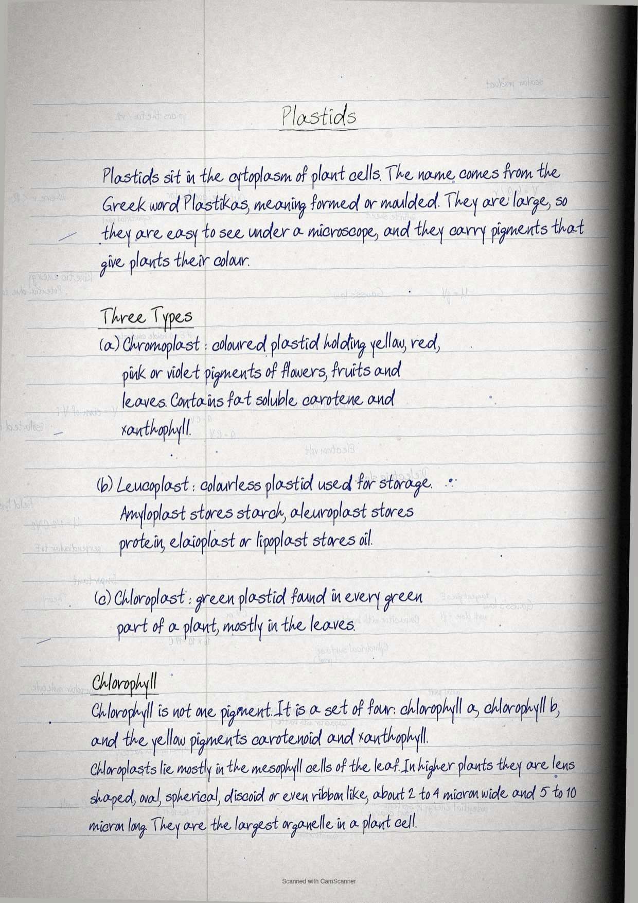
Plastids occur in the cytoplasm of plant cells. The name comes from the Greek word Plastikas, meaning formed or moulded. They are large enough to be seen easily under a microscope and they carry pigments that give plants their colour. Three types are recognised on the basis of pigmentation.
| Plastid | Pigment status | What it stores or does |
|---|---|---|
| Chromoplasts | Coloured, with fat soluble pigments such as carotene and xanthophylls | Yellow, red, pink and violet colours of flowers, fruits and leaves |
| Leucoplasts | Colourless | Store reserve food, as amyloplasts for starch, aleuroplasts for protein and elaioplasts or lipoplasts for oil |
| Chloroplasts | Green, containing chlorophyll | Photosynthesis in all green parts of the plant |
Chlorophyll is not one pigment but a collection of four, chlorophyll a, chlorophyll b, carotenoids and xanthophylls. Chloroplasts sit predominantly in the mesophyll cells of leaves, are generally lens shaped, oval, spherical, discoid or even ribbon like, measure about 2 to 4 μm wide and 5 to 10 μm long, and are the largest organelles in the plant cell.
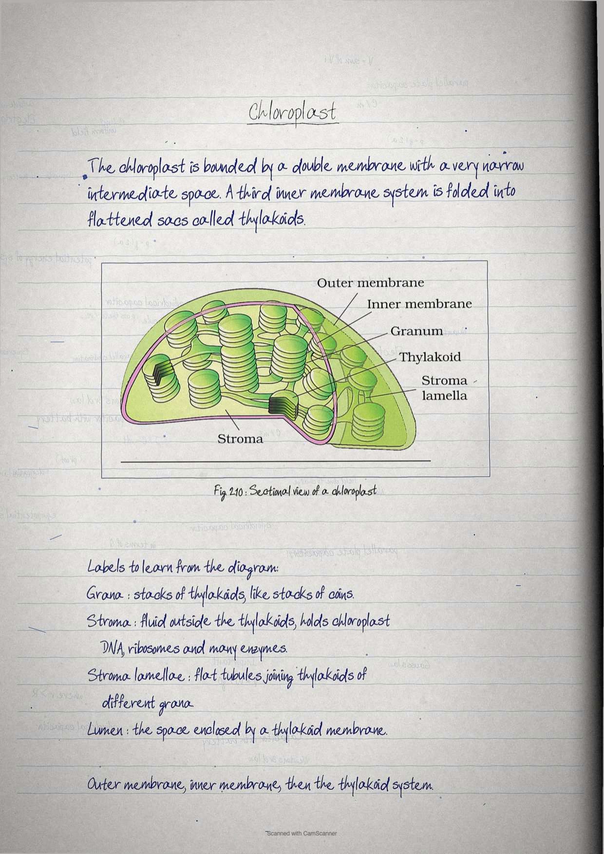
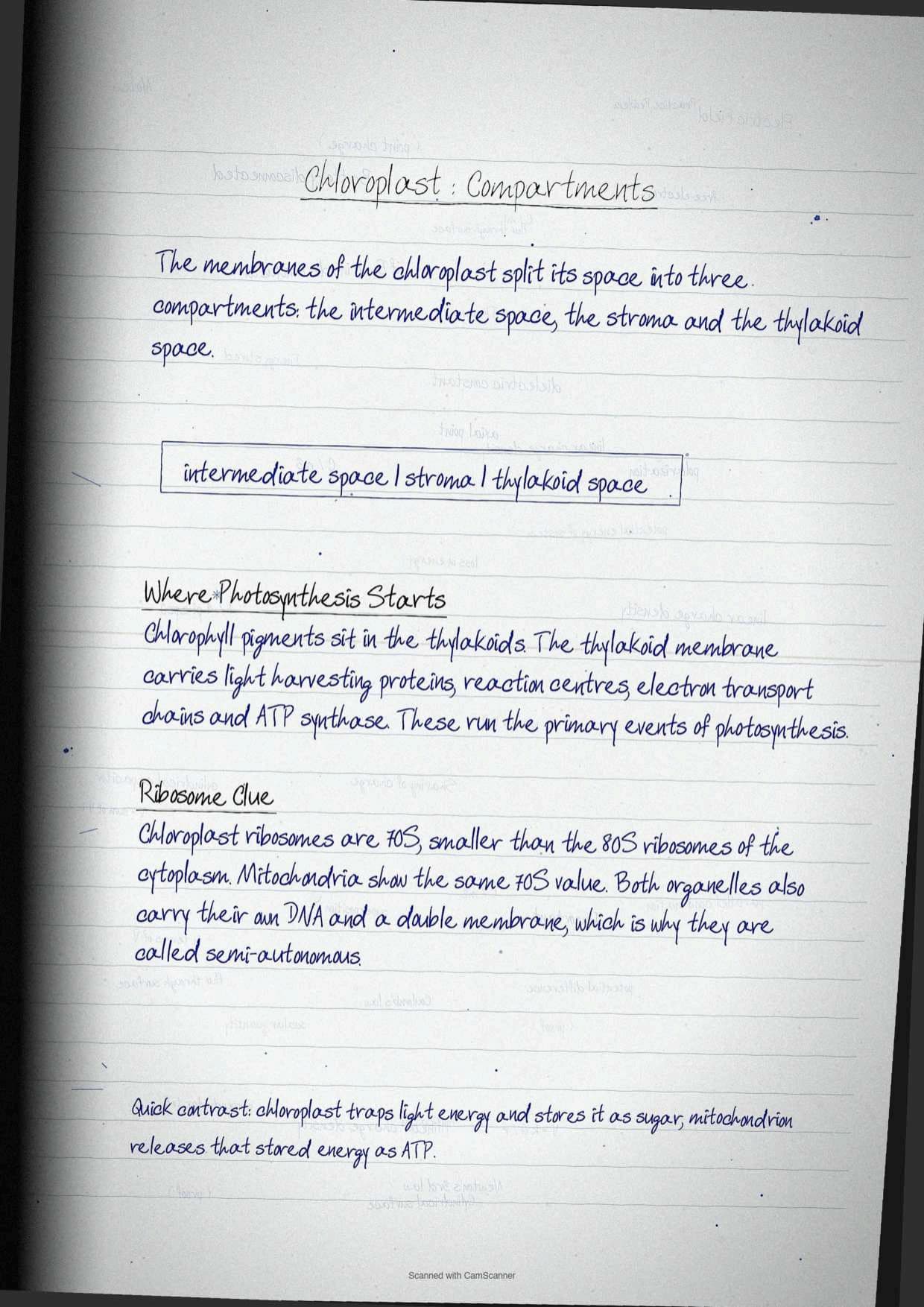
The internal architecture is where marks are won and lost, so learn it as three compartments.
- A double membrane envelope separated by a narrow intermediate space.
- The stroma, the fluid outside the thylakoids and within the envelope, holding chloroplast DNA, ribosomes and many enzymes.
- The thylakoid space or lumen, enclosed by the thylakoid membrane.
Thylakoids are flattened membranous sacs formed by a third inner membrane, stacked in orderly piles called grana, singular granum, which resemble stacks of coins. Flat membranous tubules called the stroma lamellae connect the thylakoids of different grana. Chlorophyll pigments sit in the thylakoids, and the thylakoid membrane holds light harvesting proteins, reaction centres, electron transport chains and ATP synthase, which drive the primary events of photosynthesis. The ribosomes of chloroplasts are 70S, smaller than the 80S cytoplasmic ribosomes.
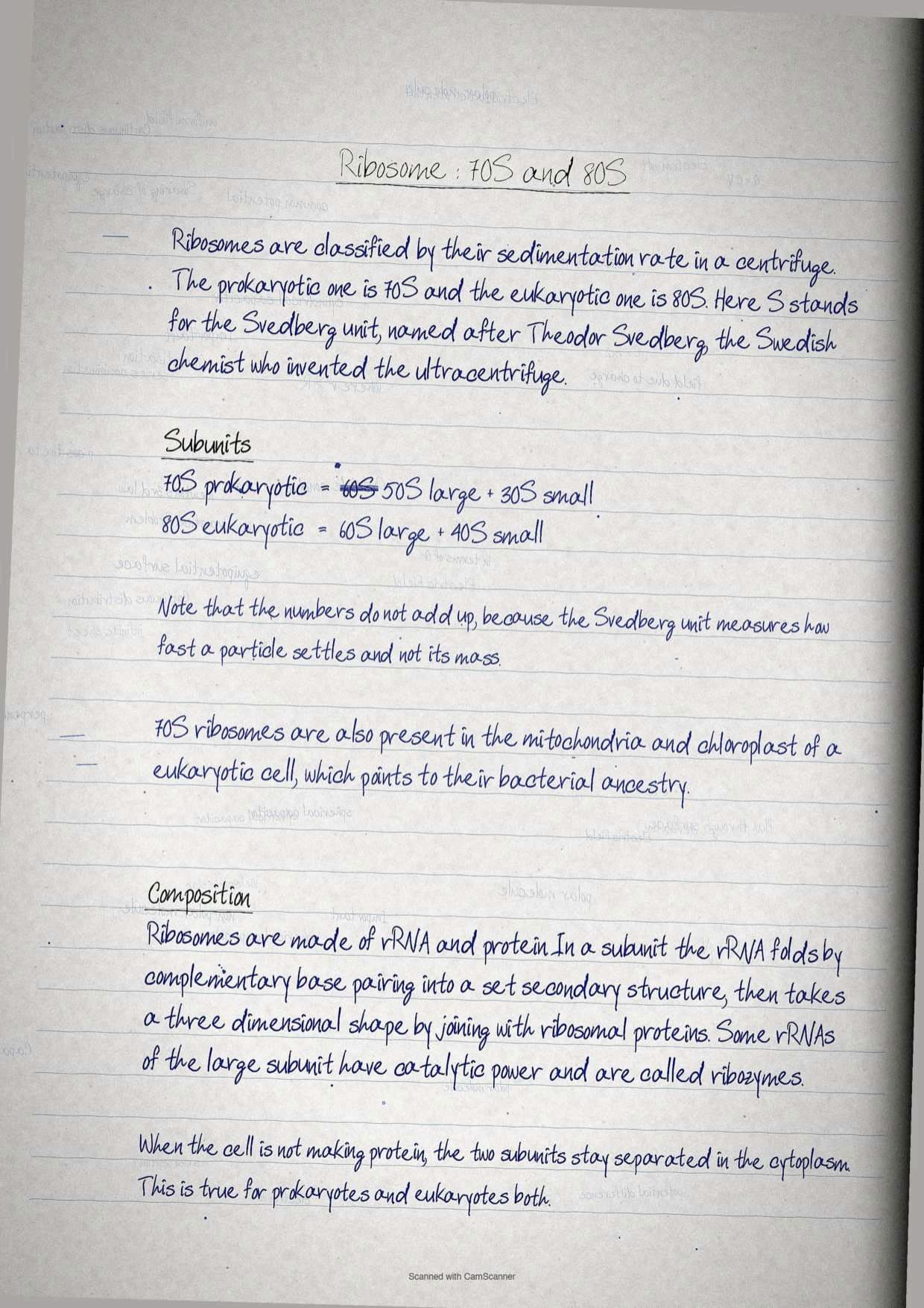
Ribosomes: 70S Against 80S Protein Factories
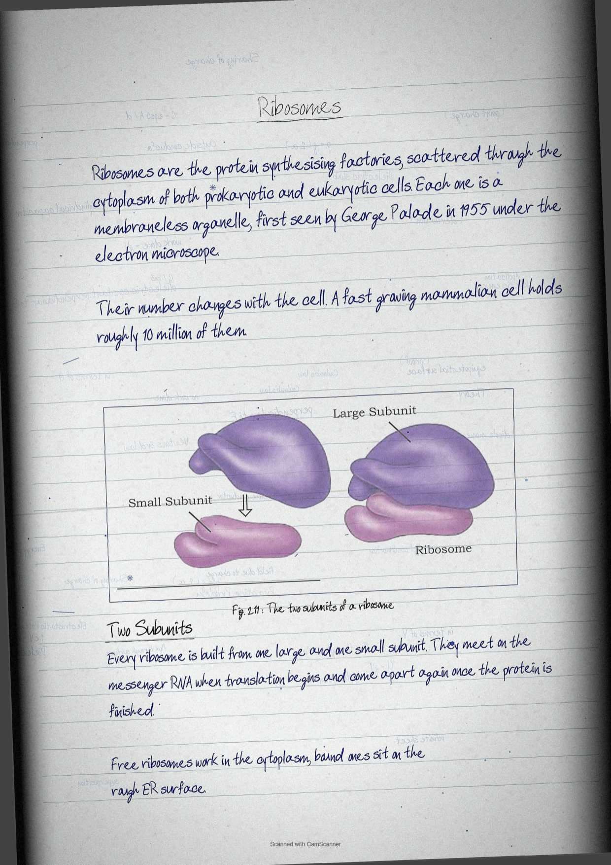
Ribosomes are the protein synthesising factories of the cell, scattered through the cytoplasm of both prokaryotic and eukaryotic cells. Each ribosome is a membraneless organelle, first observed by George Palade in 1955 under the electron microscope. A rapidly growing mammalian cell holds roughly 10 million ribosomes.
| Ribosome type | Where it occurs | Large subunit | Small subunit |
|---|---|---|---|
| 70S | Prokaryotes, and inside mitochondria and chloroplast | 50S | 30S |
| 80S | Eukaryotic cytoplasm | 60S | 40S |
Ribosomes are classified by their sedimentation rate in a centrifuge, and the S stands for the Svedberg unit, named after Theodor Svedberg, the Swedish chemist who won the Nobel Prize in chemistry for inventing the ultracentrifuge. Sedimentation rate measures the speed at which a particle sediments under the gravitational field induced by a centrifuge.
Composition is asked directly in the multiple choice section: ribosomes are made of rRNA and proteins, with no DNA and no lipid. In both prokaryotes and eukaryotes the two subunits stay dissociated in the cytoplasm when the ribosome is not engaged in protein synthesis. Inside a subunit, rRNAs fold by complementary base pairing into a characteristic secondary structure and then a three dimensional shape by associating with ribosomal proteins. Some rRNAs in the large subunit have catalytic activity and are called ribozymes.
The presence of 70S ribosomes inside mitochondria and chloroplast of a eukaryotic cell is a clue the chapter draws attention to, since it suggests those organelles are related to prokaryotic cells.
Microbodies: Peroxisomes and Glyoxysomes
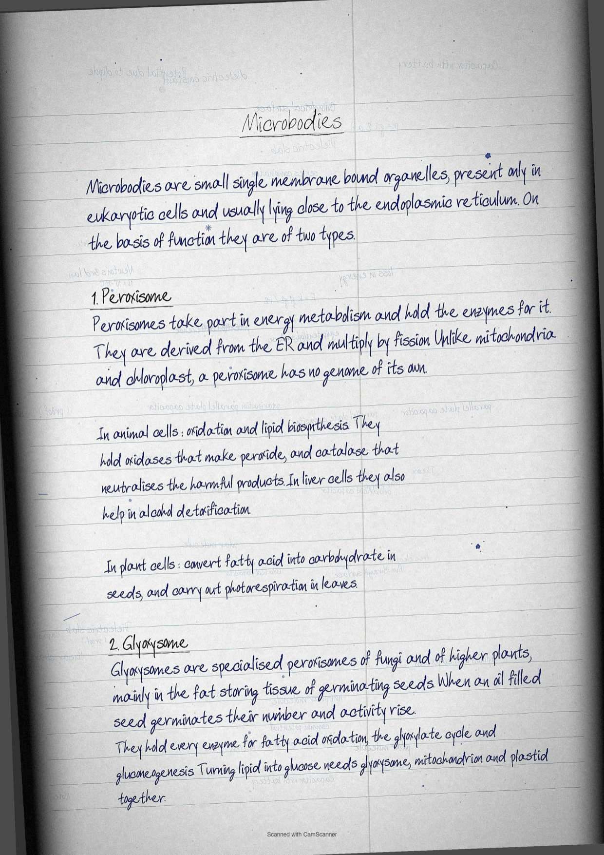
Microbodies are small, single membrane bound organelles present only in eukaryotic cells, usually located near the endoplasmic reticulum. On functional grounds they are classified into two types, peroxisomes and glyoxysomes.
Peroxisomes take part in energy metabolism and act as a site for enzymes involved in metabolic reactions. They are derived from the endoplasmic reticulum and replicate by fission. Unlike mitochondria and chloroplast, a peroxisome lacks its own genome, which is exactly the comparison exercise question 10 asks for.
| Cell type | What peroxisomes do there |
|---|---|
| Animal cells | Oxidation and lipid biosynthesis, with oxidases producing peroxide and catalase neutralising the harmful products of oxidation |
| Liver cells | Alcohol detoxification |
| Plant cells | Conversion of fatty acids into carbohydrates in seeds, and photorespiration in leaves |
Glyoxysomes are specialised peroxisomes found in fungi and higher plants, especially in the fat storage tissue of germinating seeds. When oil filled seeds germinate, the number and activity of glyoxysomes rise. They contain all the enzymes needed for fatty acid oxidation, the glyoxylate cycle and gluconeogenesis, so the seedling can live on sugars made from stored fat until it is mature enough to photosynthesise. Converting lipid into glucose needs the coordinated work of glyoxysomes, mitochondria and plastids together, and exercise question 11 expects that three organelle answer.
Cellular Organelles Explained in Simple Language
Source: Magnet Brains on YouTube
Cytoskeleton: Microtubules, Actin Filaments and Intermediate Filaments
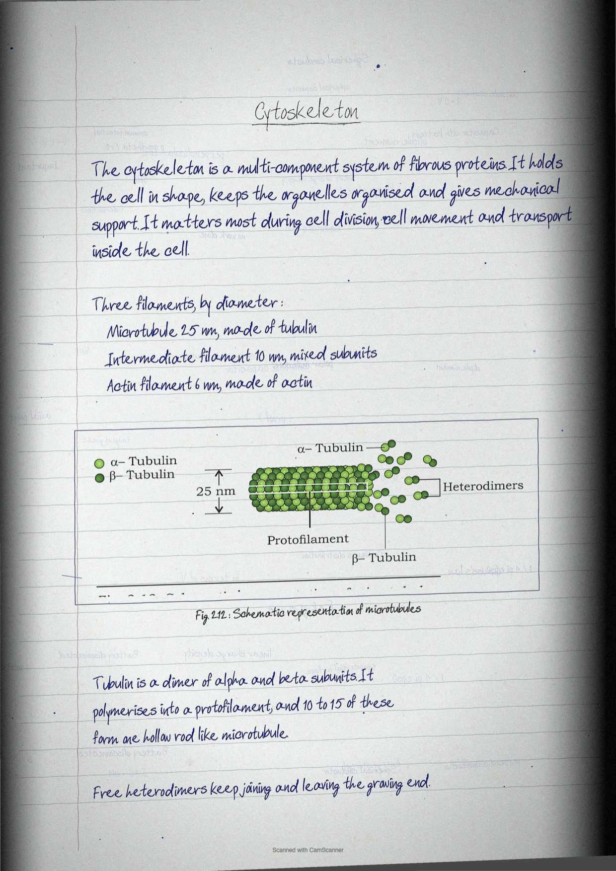
The cytoskeleton is a multi component system made of fibrous proteins that maintains cell organisation and shape. It provides mechanical support and is crucial during cell division, cell movement and intracellular transport. It is built from three major filament types that differ in protein composition and diameter, and the diameters are examinable numbers.
| Filament | Diameter | Protein | Main function |
|---|---|---|---|
| Microtubules | 25 nm | Tubulin, a dimer of alpha and beta subunits | Hollow rods of 10 to 15 protofilaments, responsible for the rhythmic movement of cilia and flagella |
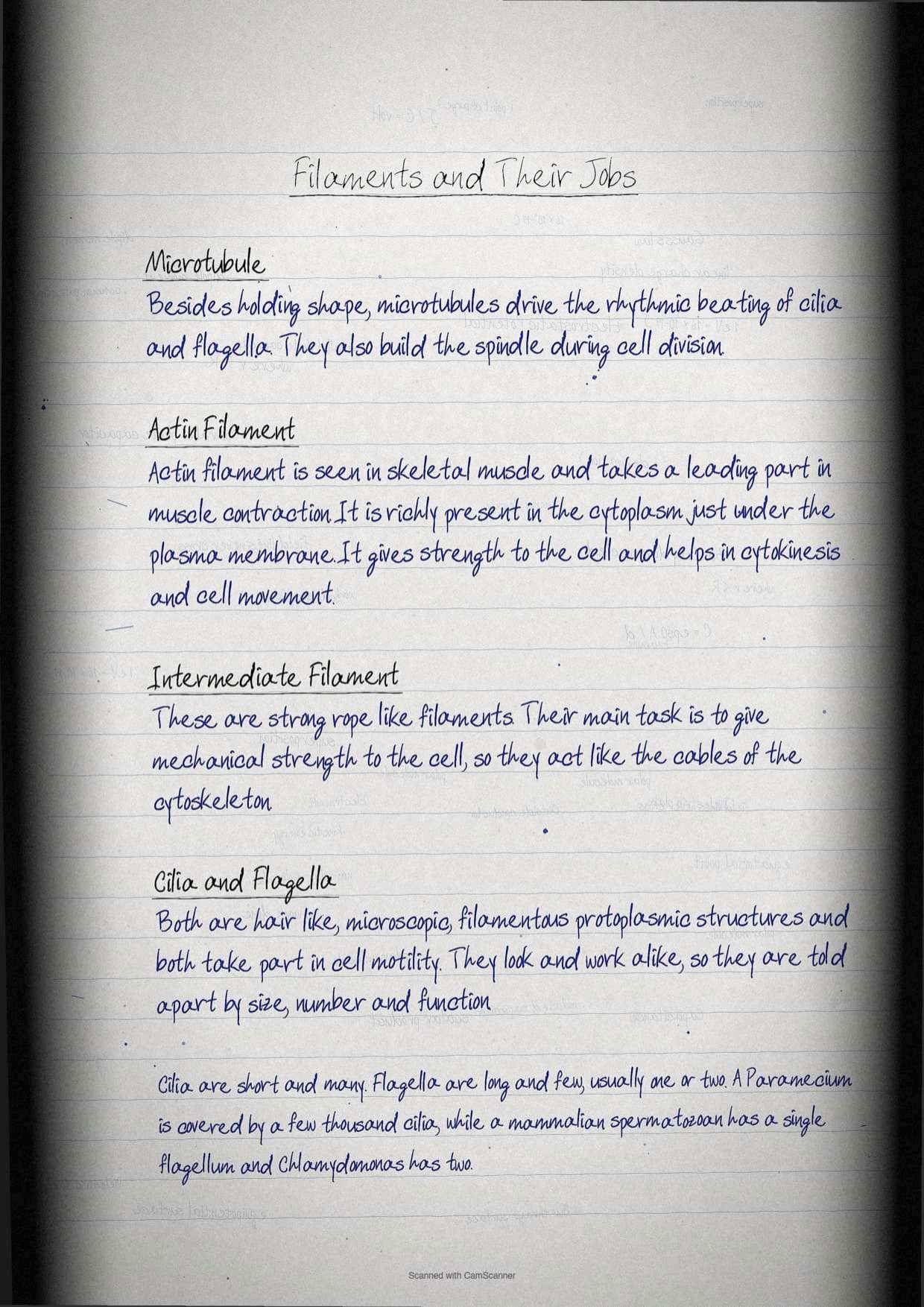
| Actin filaments | 6 nm | Actin | Muscle contraction, strength to the cell, cytokinesis and cell movement |
| Intermediate filaments | 10 nm | A combination of different protein subunits | Rope like strength, mechanical support to the cell |
Microtubules form when tubulin proteins polymerise into a protofilament, and several protofilaments assemble into the hollow rod shown in Figure 2.12. Actin filaments are seen in skeletal muscle and lie richly in the cytoplasm close to the plasma membrane. Exercise question 13 (d) asks students to distinguish microtubules from actin filaments, and diameter plus protein plus function is the three point answer that scores.
Cilia and Flagella: The 9+2 Axoneme
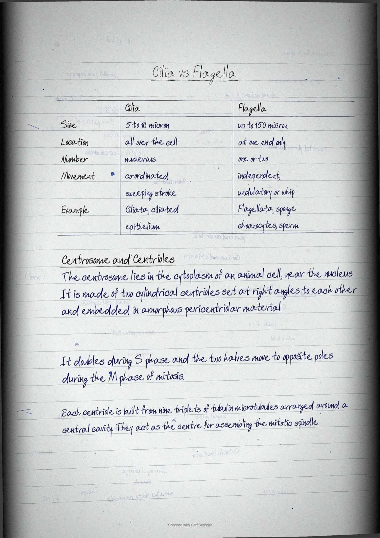
Cilia, singular cilium, and flagella, singular flagellum, are hair like, microscopic, filamentous protoplasmic structures involved in cell motility. They are morphologically and physiologically identical, so they are told apart by size, number and function rather than by build.
| Characteristic | Cilia | Flagella |
|---|---|---|
| Size | Smaller, up to 5 to 10 μm | Larger, up to 150 μm |
| Location | Throughout the surface of the cell | At one end of the cell |
| Number | Numerous | One or two |
| Movement | Coordinated rhythm, sweeping or perpendicular stroke | Independent, undulatory or whiplash movement |
| Examples | Protozoans of class Ciliata, ciliated epithelium of metazoans, larvae of Platyhelminthes, Annelids, Mollusca and Echinodermata | Protozoans of class Flagellata, choanocyte cells of sponges, spermatozoa of Metazoa, algae and gamete cells of plants |
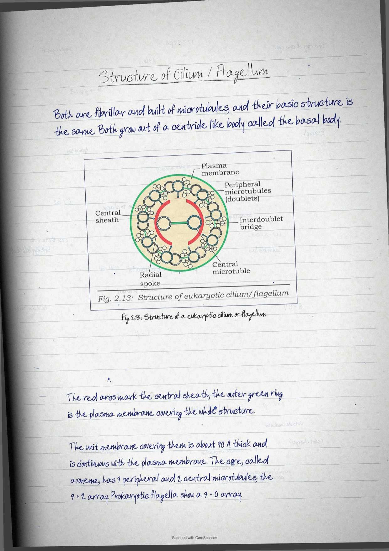
Both are fibrillar and made of microtubules, and both arise from a centriole like structure called the basal body. Under the electron microscope they are bounded by a unit membrane about 90 Å thick that is continuous with the plasma membrane. Their core is called the axoneme, and it holds nine peripheral doublets and two central microtubules, an arrangement referred to as the 9+2 array. The central fibrils are enclosed by a central sheath, and radial spokes and interdoublet bridges hold the structure together, as Figure 2.13 shows.
Prokaryotes also carry cilia and flagella, but theirs are structurally different and follow a 9+0 array. The body of Paramecium, a unicellular protozoan, is covered by a few thousand cilia. Cells of the upper respiratory tract of mammals carry cilia that expel particulate matter from inhaled air. A mammalian spermatozoan has a single flagellum, while Chlamydomonas, a unicellular green alga, has two.
Centrosome and Centrioles: Nine Triplets Around a Central Cavity
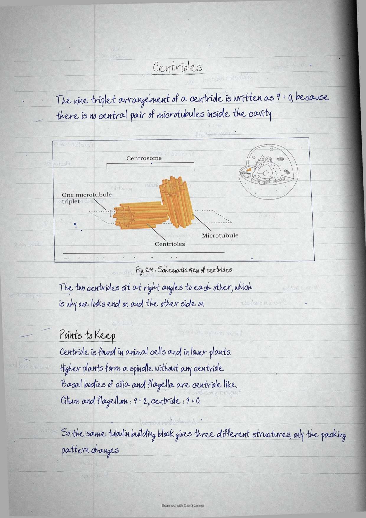
The centrosome sits in the cytoplasm of animal cells near the nucleus. It consists of two cylindrical centrioles placed perpendicular to each other and embedded in amorphous pericentriolar material. During the cell cycle it duplicates in the S phase and the copies separate towards opposite poles during the M phase of mitosis.
- Each centriole is built from nine triplets of microtubules of tubulin arranged around a central cavity.
- Centrioles act as the centre of mitotic spindle assembly during cell division.
- Centrosome and centrioles also form the basal body of cilia and flagella, which is how the chapter links locomotion back to cell division machinery.
Tip: keep the three microtubule arrangements apart. Centriole is nine triplets, eukaryotic cilium is nine doublets plus two central, prokaryotic flagellum is 9+0. Mixing them is the single most common structural error in this chapter.
Nucleus: Envelope, Nuclear Pores, Nucleoplasm and Nucleolus
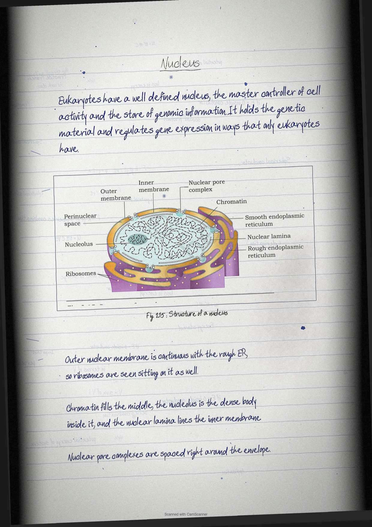
Compared with prokaryotes, eukaryotes have a well defined nucleus, described in the chapter as the master controller of cell activities and a vast repository of genomic information. It holds the genetic material and regulates gene expression through mechanisms exclusive to eukaryotes. The notebook splits it into three named parts because the textbook does.
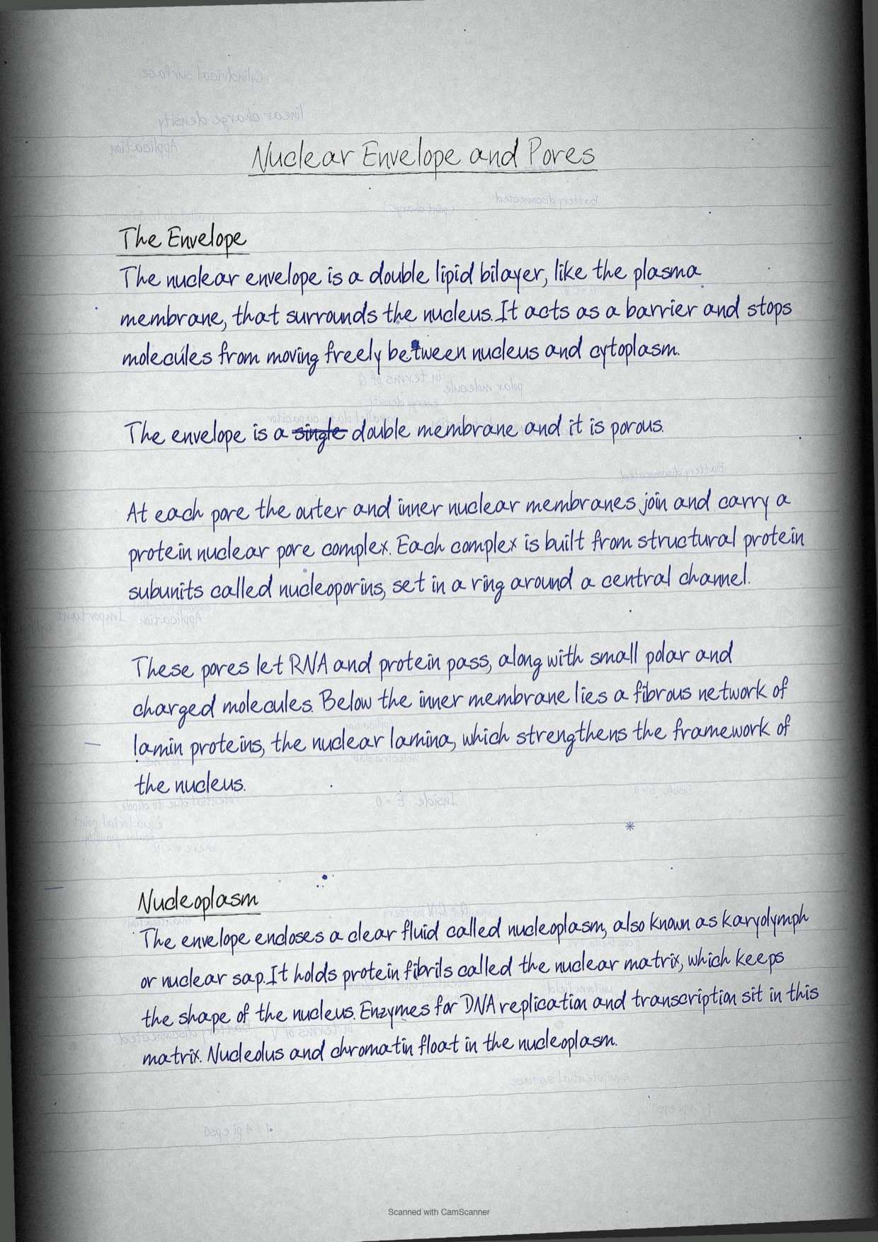
The nuclear envelope, also called the nuclear membrane, is a double lipid bilayer similar to the plasma membrane that surrounds the nucleus and acts as a barrier to free movement of molecules between nucleus and cytoplasm.
- The envelope is porous, carrying openings called nuclear pores, where the outer and inner membranes are continuous.
- Each pore is fitted with a proteinaceous nuclear pore complex made of structural protein subunits called nucleoporins, arranged in a ring around a central channel.
- The pore complex allows trafficking of RNA and proteins along with small polar and charged molecules.
- The outer nuclear membrane is continuous with the endoplasmic reticulum, which is a favourite one mark fact.
- A fibrous network of lamin proteins below the inner membrane forms the nuclear lamina, which strengthens the structural framework of the nucleus.
Inside the envelope lies the nucleoplasm, also called karyolymph or nuclear sap, a clear fluid containing protein fibrils known as the nuclear matrix. The matrix helps maintain the shape of the nucleus and holds the enzymes associated with DNA replication and transcription. Both the nucleolus and the chromatin are suspended in the nucleoplasm.
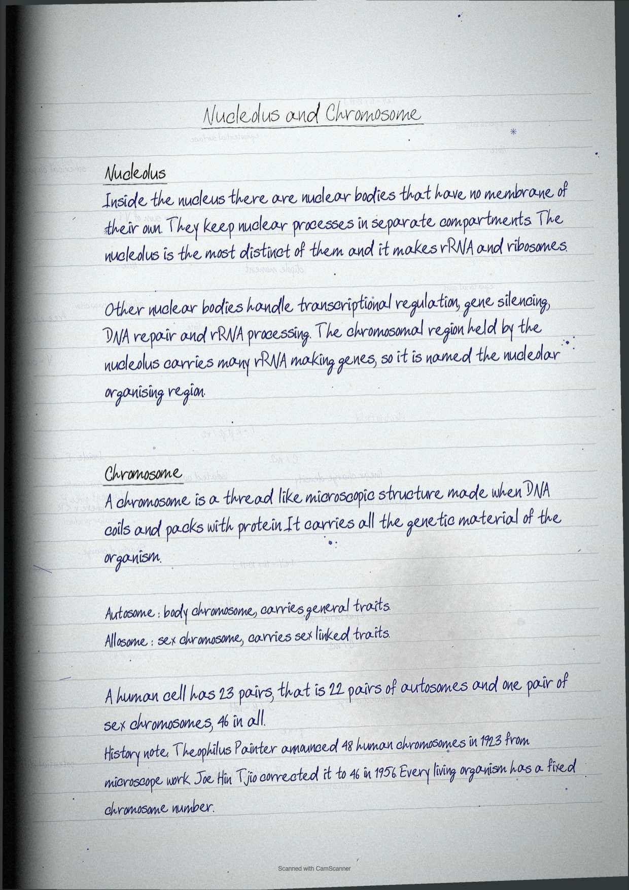
The nucleolus is the most distinct of the nuclear bodies, a set of structures that lack a well defined membrane and help compartmentalise nuclear processes. Its job is the synthesis of rRNA and ribosomes. Other nuclear bodies handle transcriptional regulation, gene silencing, DNA repair, and rRNA transcription and processing. The chromosomal region occupied by the nucleolus carries a large number of rRNA synthesising genes, which is why it is called the nucleolar organising region.
Chromosomes: Chromatin, Nucleosomes and the Numbers to Remember
A chromosome is a thread like microscopic structure formed by the coiling of DNA packaged with protein, and it carries all the genetic material of an organism. Chromosomes fall into two categories, autosomes or body chromosomes and allosomes or sex chromosomes. Certain hereditary traits are linked to a person's sex and are passed on through the sex chromosomes, while autosomes carry the rest of the genetic information.
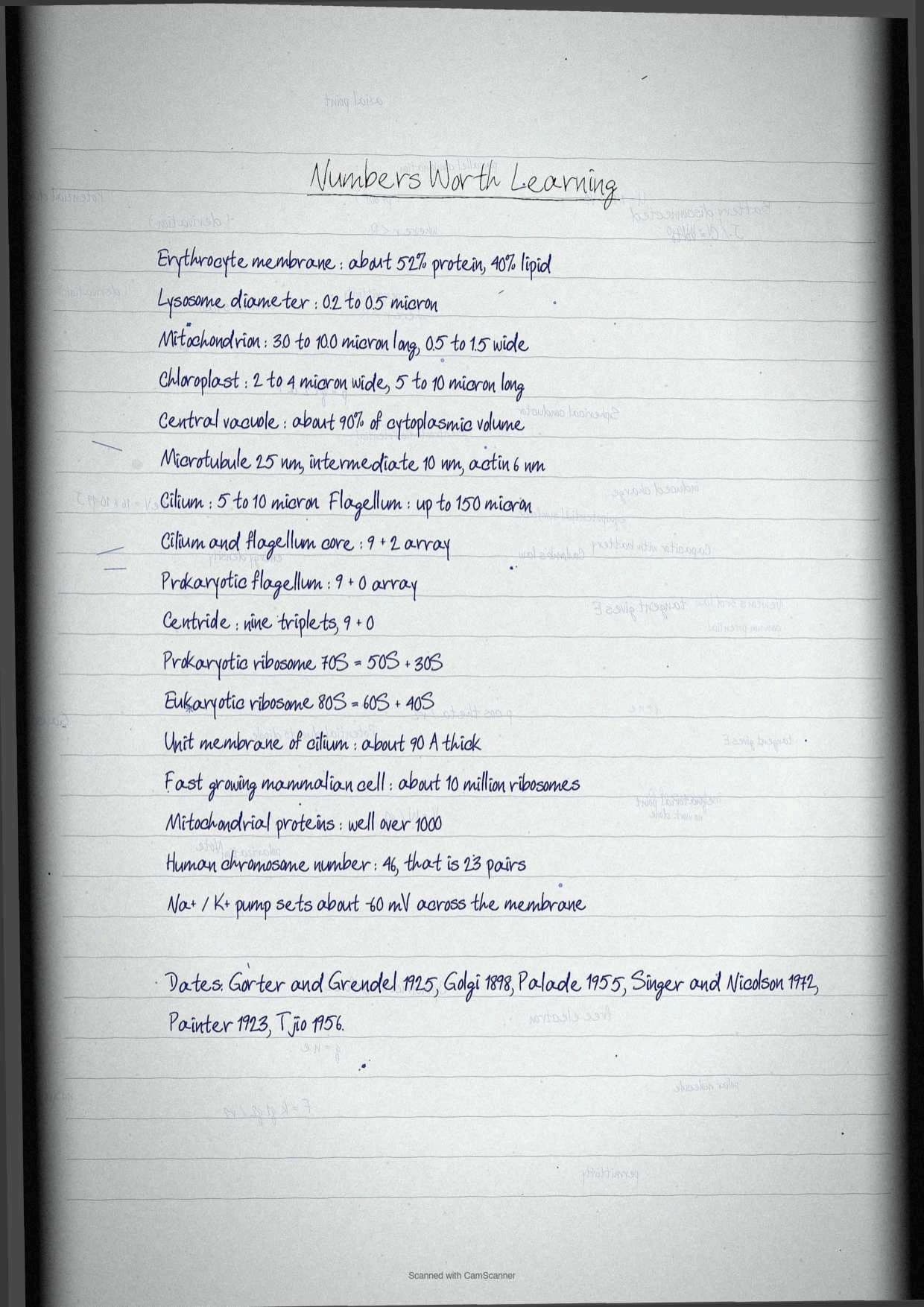
Human cells hold 23 pairs of chromosomes, made of 22 pairs of autosomes and one pair of sex chromosomes, a total of 46 per cell. Table 2.2 of the chapter lists counts for other organisms, and a handful of them are worth memorising.
| Organism | Number of chromosomes |
|---|---|
| Arabidopsis thaliana (diploid) | 10 |
| Common fruit fly (diploid) | 8 |
| Maize (diploid) | 20 |
| Wheat (hexaploid) | 42 |
| Mouse (diploid) | 40 |
| Human (diploid) | 46 |
| Dog (diploid) | 78 |
| Gold fish (diploid) | 100 |
The history box is examinable. Theophilus Painter, an American zoologist, declared the human chromosome number as 24 pairs or 48 in 1923 on the basis of microscopic study, and that was wrong. It was corrected by Joe Hin Tjio, an Indonesia born American cytogeneticist, in 1956, who put the total at 46. Every living organism has a fixed number of chromosomes.
Prokaryotes such as bacteria and blue green algae usually carry a single circular chromosome called the nucleoid in the cytoplasm, since they have no well defined nucleus, although some prokaryotes such as Vibrio cholerae carry more than one. In eukaryotes the chromosomes sit inside the nucleus, and during interphase they exist as long threads called chromatin fibres. A chromatin fibre is composed of nucleosomes, which are DNA wrapped around histone proteins. Chromatin is what lets very long DNA fit inside the nucleus: if all the DNA in a single human cell were unwound from the histones and laid end to end, it would stretch to about 6 feet. During cell division the chromatin condenses further into microscopically visible, long and slender chromosomes, which are replicated, divided and passed to daughter cells. Errors sometimes change chromosome number or structure in the new cells, and those changes can cause serious problems.
Classifying Eukaryotic Organelles by Membrane Count
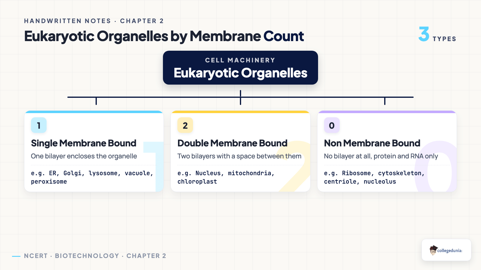
Exercise question 8 asks students to classify the various eukaryotic organelles into single membrane, double membrane and non membrane bound types. It is worth three or four marks, it is fully predictable, and it is the fastest thing to revise the night before.
| Category | Organelles |
|---|---|
| Single membrane bound | Endoplasmic reticulum, Golgi apparatus, lysosomes, vacuoles, peroxisomes, glyoxysomes |
| Double membrane bound | Nucleus, mitochondria, chloroplast and other plastids |
| Non membrane bound | Ribosomes, cytoskeleton filaments, centrioles and the centrosome, nucleolus |
Two traps sit inside this table. The nucleolus is not membrane bound even though it lives inside a double membrane bound nucleus, and cilia and flagella are not organelles in this classification, they are microtubule based extensions covered by the plasma membrane.
Common Mistakes Students Make in Cellular Organelles
Six traps that cost easy marks in the Class 11 Biotechnology paper:
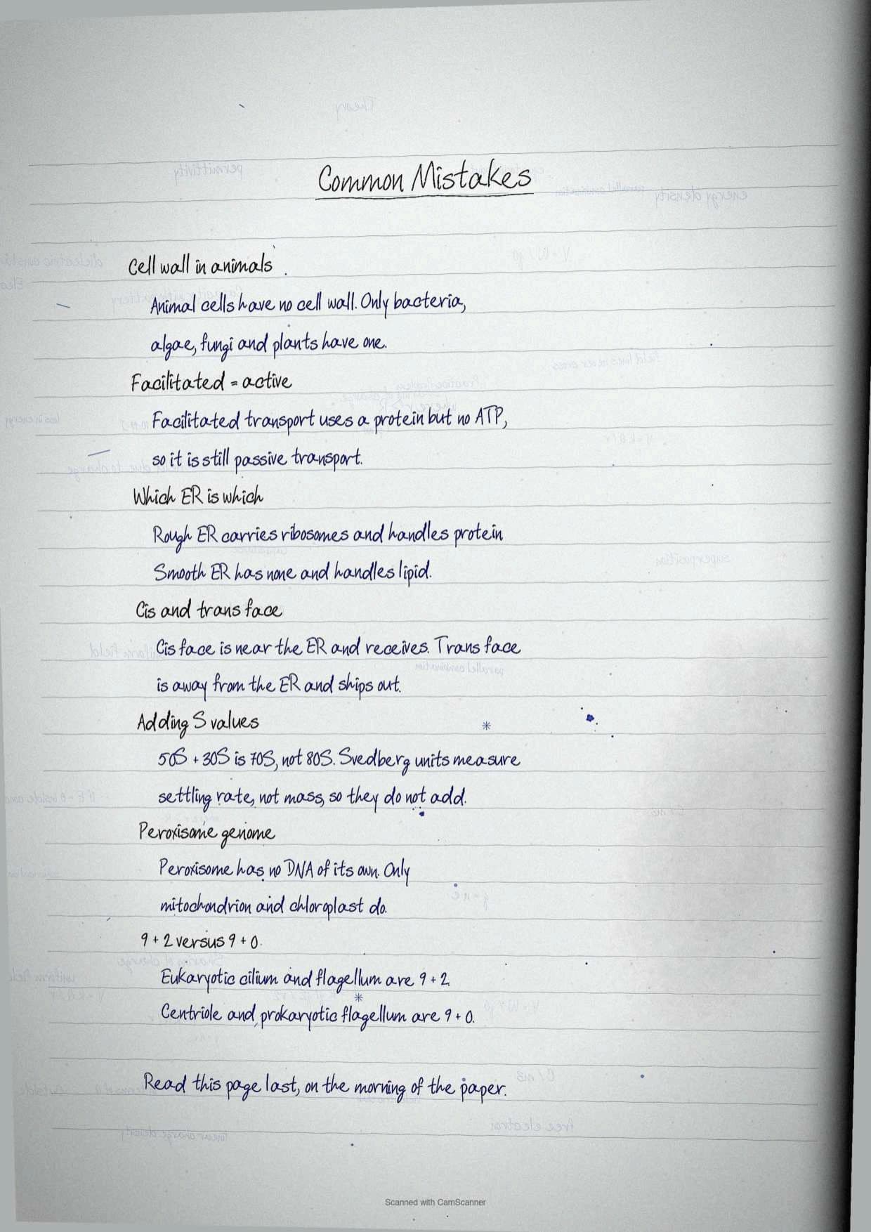
- Crediting the fluid mosaic model to the wrong scientists. It is Singer and Nicolson, 1972. Gorter and Grendel, 1925, proposed the bilayer, and they are two different answers.
- Putting mitochondria inside the endomembrane system. Only endoplasmic reticulum, Golgi complex, lysosomes and vacuoles belong to it.
- Reversing the cis and trans faces. Cis faces the endoplasmic reticulum and forms, trans faces away and matures.
- Confusing the microtubule arrays. Centriole is nine triplets, eukaryotic cilium is 9+2, prokaryotic flagellum is 9+0.
- Calling the tonoplast a plastid membrane. The tonoplast covers the vacuole, and the multiple choice question in the chapter tests exactly that.
- Giving peroxisomes a genome. Mitochondria and chloroplast have DNA, peroxisomes do not.
Tick each trap off only after you have answered it correctly once in a written attempt. Students who did this reported the structure questions stopped costing them marks within two revision rounds.
Quick Recall Sheet for Cellular Organelles
This is the page to read the night before and again in the morning. Every term the chapter can ask sits in one place.
| Term | One-line meaning |
|---|---|
| Fluid mosaic model | Singer and Nicolson, 1972, lipid bilayer with a mosaic of globular proteins |
| Mesosome | Membranous extension of the prokaryotic plasma membrane that increases surface area |
| Aquaporin | Channel protein for water transport across the plasma membrane |
| Symport, antiport, uniport | Same direction, opposite direction, single molecule transport |
| Peptidoglycan | Linear chain cross linked by tetrapeptides, the core of the bacterial wall |
| Glycocalyx | Glycosylated protein coat, loose as a slime layer or thick as a capsule |
| Middle lamella | Calcium pectate layer holding neighbouring plant cells together |
| Plasmodesmata | Cytoplasmic connections between adjacent plant cells |
| Cisternae | Flattened membranous sacs of the endoplasmic reticulum and Golgi apparatus |
| Cis and trans face | Forming face near the reticulum and maturing face away from it |
| Acid hydrolase | Lysosomal enzyme active at acidic pH |
| Autophagy | Recycling of the cell's own material by lysosomal enzymes |
| Tonoplast | Membrane covering the vacuole |
| Cristae | Infoldings of the inner mitochondrial membrane |
| Grana and stroma lamellae | Stacks of thylakoids and the tubules connecting different grana |
| Ribozyme | Catalytically active rRNA in the large ribosomal subunit |
| Glyoxysome | Specialised peroxisome of fat storing seed tissue |
| Axoneme | Microtubule core of a cilium or flagellum in the 9+2 array |
| Nucleoporin | Structural protein subunit of the nuclear pore complex |
| Nucleosome | DNA wrapped around histone proteins, the repeating unit of chromatin |
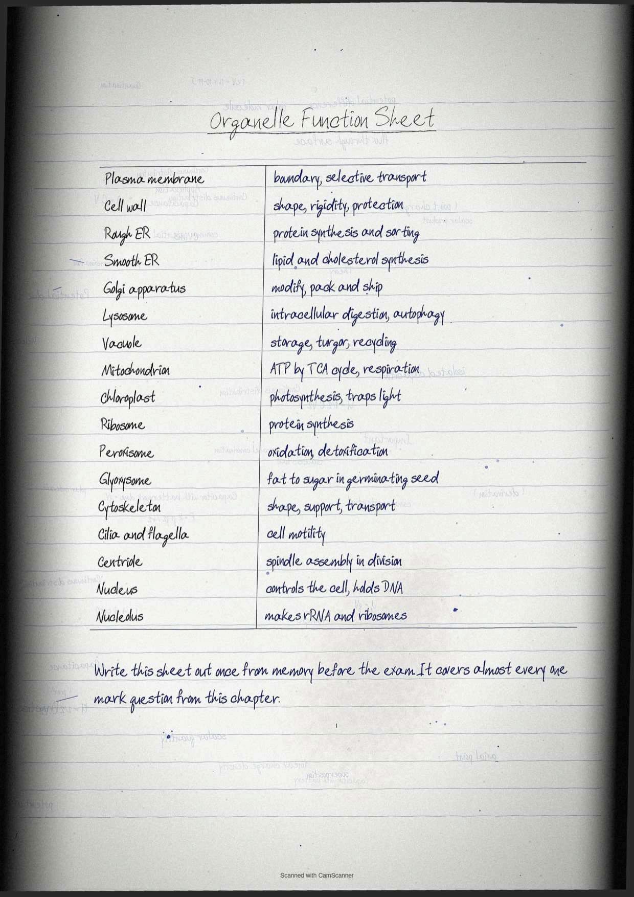
The notebook also carries a mnemonic for the running order of the chapter: My Wall Ends Motion, standing for Membrane, Wall, Endomembrane system, Mitochondria and plastids, Organelles without membranes, Transport structures, Interphase nucleus, Otherwise chromosome, Numbers to memorise.
What the Cellular Organelles Handwritten Notes PDF Contains
The file is a scanned notebook written on ruled paper, with the textbook figures redrawn where the chapter uses them. Nothing is typed, so it reads the way a classmate's notes read.
- Pages 1 to 7: cover, the prokaryotic against eukaryotic split, the fluid mosaic model and the Gorter and Grendel experiment.
- Pages 8 to 14: membrane transport in full, with the Na+ and K+ pump diagram, and both bacterial and eukaryotic cell walls.
- Pages 15 to 22: the endomembrane system, endoplasmic reticulum, Golgi apparatus, lysosomes and vacuoles with the protein sorting flow.
- Pages 23 to 29: mitochondria, plastids, ribosomes, microbodies, cytoskeleton, cilia and flagella and the centrosome.
- Pages 30 to 34: nucleus, chromosome, the membrane count classification, the quick recall sheet and every exercise question worked out.
Textbook Exercise Questions and How These Notes Answer Them
The chapter closes with thirteen exercises, three of them multiple choice and one a matching set. The notebook writes out a model answer for each, and the table below shows where the material for each answer sits.
| Exercise question | Where the answer is built |
|---|---|
| Who proposed the fluid mosaic model | Singer and Nicolson, 1972, in the plasma membrane section |
| What ribosomes are composed of | rRNA and proteins, in the ribosomes section |
| What the tonoplast is | The membrane covering the vacuole, in the vacuoles section |
| Mechanisms of transport across the plasma membrane with diagrams | The passive, facilitated and active transport table plus symport, antiport and uniport |
| Matching set on nucleolus, mesosome, vacuoles, cristae, ribosomes, thylakoid, peroxisomes and cisternae | The quick recall sheet, which carries all eight terms in one place |
| Significance of the protein to lipid ratio in membranes | The erythrocyte figure of 52 per cent protein and 40 per cent lipid, and the effect of fluidity on function |
| Importance of the cell wall in prokaryotic cells | The four point function list in the bacterial cell wall section |
| Classify organelles as single, double or non membrane bound | The membrane count classification table |
| Different types of vacuoles | The six row vacuole table |
| Similarities and differences of peroxisomes with mitochondria and chloroplast | The microbodies section, where the missing genome is the key contrast |
| What glyoxysomes are, where they occur and what they do | The glyoxysome paragraph with the glyoxylate cycle and gluconeogenesis |
| Justify that the cell is the structural and functional unit of life | The opening section plus one function drawn from every organelle card |
| Distinguish cilia and flagella, primary and secondary cell wall, lysosomes and vacuoles, microtubules and actin filaments, active and passive transport | Five separate comparison tables spread through the notes |
How to Use These Handwritten Notes Most Effectively
This chapter is not conceptually hard, it is dense with structures and numbers, so drawing beats reading. Students who split it into four blocks reported finishing it in about three hours.
- Block 1, 45 minutes. Prokaryotic against eukaryotic, the fluid mosaic model and all four transport mechanisms. Draw the membrane from memory before moving on.
- Block 2, 45 minutes. Both cell walls, then the whole endomembrane system in the order reticulum, Golgi, lysosome, vacuole.
- Block 3, 50 minutes. Mitochondria and chloroplast, drawn twice each with all compartments labelled, then ribosomes and microbodies.
- Block 4, 40 minutes. Cytoskeleton, cilia and flagella, centrioles, nucleus and chromosome, then attempt every distinguish question in writing.
- Night before, 15 minutes. The membrane count table, the quick recall sheet and the six common mistakes, nothing else.
How These Handwritten Notes Pair with Other Cell Biology Resources
Also Check: the same organelles are taught again in Class 11 Biology and were introduced in Class 9 Science. Read this chapter for the biotechnology framing, then use the sibling pages below for extra worked questions on the same structures.
| Resource | Best used for | Open it |
|---|---|---|
| Class 11 Biology Notes | The same organelles taught in the biology stream, with extra diagrams and terminology | Cell The Unit of Life Class 11 Notes |
| Class 11 Biology Handwritten Notes | A second scanned revision pass on membranes, organelles and the nucleus | Cell The Unit of Life Class 11 Handwritten Notes |
| Class 11 Biology Solutions | Worked answers on cell structure that overlap almost every section here | Cell The Unit of Life Class 11 Solutions |
| Class 9 Science Notes | The basic organelle vocabulary if the plasma membrane section feels new | Cell The Building Block of Life Class 9 Notes |
| Class 11 Biotechnology Handwritten Notes | The previous chapter, which sets up the vocabulary this one builds on | An Introduction to Biotechnology Class 11 Handwritten Notes |
| Class 11 Biotechnology Notes | Typed revision notes for this chapter with every term explained | Cellular Organelles Class 11 Notes (coming soon) |
| Class 11 Biotechnology Book PDF | The official Class 11 Biotechnology chapter text with all original figures | Cellular Organelles Class 11 Book PDF (coming soon) |
NCERT Handwritten Notes for Class 11 Biotechnology: All Chapters
Related Links: handwritten revision notes for every chapter of the Class 11 Biotechnology textbook, written to the 2026-27 syllabus.
| Chapter | Class 11 Biotechnology Handwritten Notes |
|---|---|
| Chapter 1 | An Introduction to Biotechnology Class 11 Handwritten Notes |
| Chapter 2 | Cellular Organelles Class 11 Handwritten Notes (this page) |
| Chapter 3 | Biomolecules Class 11 Handwritten Notes (coming soon) |
| Chapter 4 | Enzymes and Bioenergetics Class 11 Handwritten Notes (coming soon) |
| Chapter 5 | Cellular Processes Class 11 Handwritten Notes (coming soon) |
| Chapter 6 | Basic Principles of Inheritance Class 11 Handwritten Notes (coming soon) |
| Chapter 7 | Basic Processes of DNA Class 11 Handwritten Notes (coming soon) |
| Chapter 8 | Genetic Disorder Class 11 Handwritten Notes (coming soon) |
| Chapter 9 | Introduction to Bioinformatics Class 11 Handwritten Notes (coming soon) |
| Chapter 10 | Protein Informatics and Cheminformatics Class 11 Handwritten Notes (coming soon) |
| Chapter 11 | Programming and Systems Biology Class 11 Handwritten Notes (coming soon) |
Cellular Organelles Class 11 Handwritten Notes FAQs
Questions Students Ask Before Downloading These Notes
Ques. Who proposed the fluid mosaic model of the plasma membrane and in which year?
Ans. Seymour Jonathan Singer and Garth L. Nicolson proposed the fluid mosaic model in 1972. It describes the plasma membrane as a lipid bilayer surrounding the cell with a mosaic of globular proteins set into it, held in a quasifluid state so that lipids and proteins can diffuse laterally. The earlier bilayer proposal, from Edwin Gorter and F. Grendel in 1925, is a separate answer and should not be confused with it.
Ques. What is the difference between a prokaryotic and a eukaryotic cell?
Ans. A prokaryotic cell has no organised nucleus and no membrane bound organelles, carries 70S ribosomes, mesosomes and usually a single circular chromosome called the nucleoid. A eukaryotic cell has a well defined nucleus and membrane bound organelles such as endoplasmic reticulum, Golgi apparatus, mitochondria, plastids, vacuoles, lysosomes and peroxisomes, with 80S ribosomes in the cytoplasm. Plasma membrane, cytoplasm, ribosomes and DNA are common to both.
Ques. Which organelles make up the endomembrane system?
Ans. The endomembrane system includes the endoplasmic reticulum, the Golgi complex, lysosomes and vacuoles. These four are grouped together because their functions are coordinated in the synthesis, processing, packaging and transport of proteins and lipids. Mitochondria, chloroplast and peroxisomes are membrane bound as well but are not part of the system, since they do not work in that shared pathway.
Ques. How do Gram positive and Gram negative bacterial cell walls differ?
Ans. Gram positive bacteria have a thick cell wall with a single plasma membrane, and their wall carries teichoic acid and lipoteichoic acid. Gram negative bacteria have a thin cell wall surrounded by a dual membrane arrangement, with an outer membrane carrying lipopolysaccharide, porins and lipoprotein, and a periplasmic space between the two membranes. Both walls are built on peptidoglycan chains cross linked by tetrapeptides.
Ques. What is the 9+2 arrangement in cilia and flagella?
Ans. The core of a eukaryotic cilium or flagellum is called the axoneme, and it contains nine peripheral microtubule doublets arranged around two central microtubules, which is written as the 9+2 array. The central fibrils are enclosed by a central sheath, and radial spokes and interdoublet bridges hold the structure together. Prokaryotic cilia and flagella are structurally different and follow a 9+0 array.
Ques. How are eukaryotic organelles classified by the number of membranes?
Ans. Single membrane bound organelles are the endoplasmic reticulum, Golgi apparatus, lysosomes, vacuoles, peroxisomes and glyoxysomes. Double membrane bound organelles are the nucleus, mitochondria and plastids including the chloroplast. Non membrane bound structures are ribosomes, the cytoskeleton filaments, centrioles and the centrosome, and the nucleolus. This is the classification exercise question 8 of the chapter asks for.
Ques. What is the difference between 70S and 80S ribosomes?
Ans. The 70S ribosome is prokaryotic and is built from a 50S large subunit and a 30S small subunit, while the 80S ribosome is eukaryotic and is built from a 60S large subunit and a 40S small subunit. The S stands for the Svedberg unit of sedimentation rate. The 70S type is also found inside mitochondria and chloroplast of eukaryotic cells, which points to their relatedness to prokaryotic cells.
Ques. Why do peroxisomes differ from mitochondria and chloroplast?
Ans. Peroxisomes are single membrane bound, are derived from the endoplasmic reticulum, replicate by fission and lack their own genome, while mitochondria and chloroplast are double membrane bound and carry their own DNA and 70S ribosomes. All three take part in energy related metabolism, which is the similarity the chapter points to, but only mitochondria and chloroplast are semi-autonomous.
Ques. What does the nucleolus do and is it membrane bound?
Ans. The nucleolus is the most distinct of the nuclear bodies and is involved in the synthesis of rRNA and ribosomes. It is not membrane bound, even though it sits inside the double membrane bound nucleus. The chromosomal region it occupies carries a large number of rRNA synthesising genes, which is why that region is called the nucleolar organising region.
Ques. Where can I download the Class 11 Biotechnology Cellular Organelles handwritten notes PDF?
Ans. Use the download button at the top of this page. The file is free and printable, runs to 34 handwritten pages, and follows the 2026-27 chapter order, so it can sit beside the textbook while you revise every organelle in sequence.



Comments