



Education Journalist | Study Abroad Lead
Leg Anatomy refers to the study of a human’s leg. Being one of the most essential parts of the human body, the legs do much more than help us in our daily movement. They support our body and are essential to carry out recreational activities like swimming, jumping, climbing, dancing, running and so on. Our legs consist of many different types of muscles and bones, that work together to help you carry out daily activities with ease. The movement or the ability to move from one place to another is called locomotion. Locomotion is a major need not only for mammals but also many other different organisms. Amoeba, paramecium, insects, reptiles, all of them need legs, mainly to search for food. Although the methods of locomotion may vary with different habitats of these organisms, it is generally used for search of food, shelter, mate, suitable breeding grounds, favourable climatic conditions or to escape from enemies/predators.
Table of Contents
| Table of Content |
Key Terms: Leg Anatomy, Bones, Locomotion, Movement, Femur, Tibia, Fibula, Upper Leg Muscles, Lower Leg Muscles, Quadriceps, Hamstrings, Adductors, Gastrocnemius, Soleus, Plantaris, Tibialis, Peroneus
Also Read: Reproductive Health
Introduction to Leg Anatomy
[Click Here for Sample Questions]
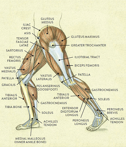
The leg is the entire lower limb of the human body including the foot, thigh and even the hip or gluteal region. It is the longest part of our body. The leg basically consists of five main parts. They are the Upper leg, knee, lower leg, ankle and foot. Our legs comprise of three types of bones and several muscles, all based on different parts like thigh, knee, ankle, feet and so on. All four appendages of four-limbed vertebrates are commonly called legs, but in the case of bipedal animals, including humans, only the lower two are so-called.
Legs have gone through many adaptations according to the needs and the habitats of the organisms. For example, the foreleg of birds and bats evolved into wings. Similarly, the appendages of moles and kangaroos evolved according to their needs of swimming, running, digging etc.
Leg Anatomy
Bones of the Leg
[Click Here for Sample Questions]
The human leg consists of mainly three types of bones. The various bones of the leg are as follows:
- Femur: Also known as the thighbone, the femur is the longest bone of the human body. Forming a ball-and-socket joint with the hip, the femur connects the pelvic bone to the lower leg bones. It is the strongest and the heaviest bone of our body. Due to this, the femur has the ability to bear extreme forces which act upon it.
- Tibia: Also called the shank bone or the shinbone, the tibia is one of the two bones present in the lower leg region. It is the second-largest bone in the human body. Tibia connects the knee and the ankle joints and is a key weight-bearing structure.
- Fibula: Commonly known as the calf bone, the fibula is another bone present in the lower leg region. The fibula is much smaller and thinner when compared to the tibia. Being a slender long bone, it bears very little body weight. The fibula provides stability to the lower leg region and also acts as a tie rod to increase the range of motion for the ankle. The average fibula is about 390 millimetres (mm) long in adult men and about 360 mm long in adult women. It runs laterally to the tibia but is smaller in size.

Bones of the Leg
Leg Muscles
[Click Here for Sample Questions]
Having the ability to stretch distances, the leg muscles mainly consist of long muscles. Many different kinds of muscles make up the upper and the lower leg. Together these muscles help us in carrying out our daily activities like walking, jumping, running and so on. Our leg muscles work with our bones, tendons and ligaments to stabilize our body, support our weight and help us move. The structure of our different muscles completely depends on the part or the region of the leg where it is found.
Read More:
Parts of the Leg Muscles
[Click Here for Sample Questions]
Our leg muscles can be broadly divided into two types: Upper leg muscles and lower leg muscles.
Upper Leg Muscles
Hamstrings, Quadriceps and Adductors make up the three main muscles in the upper leg region.
- Hamstrings: These are the three skeletal muscles that are located at the back of the outer thighs. Since these are voluntary muscles, we have complete control over them. Hamstrings allow our knees to bend and flex. These muscles are commonly used to walk, climb stairs, do squats and perform many other leg movements. Semimembranosus, semitendinosus and biceps femoris are the three hamstring muscles.
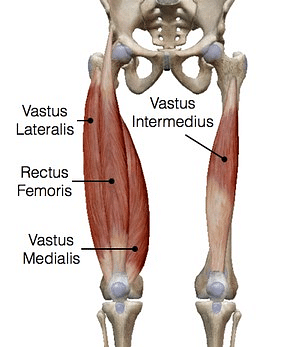
- Quadriceps: Quadriceps are the four muscles found at the front of our thighs. These four individual muscles are vastus lateralis, vastus medialis, vastus intermedius and rectus femoris. These are some of the largest and strongest muscles present in our body. Vastus lateralis is the largest of the four muscles that run down the outside of our thigh, connecting our femur to our kneecap. Running along the inner part of our thigh, Vastus medialis also connects the femur and the kneecap. As the name suggests, the Vastus intermedius muscle is located in between the other two vastus muscles in our thigh. It is the deepest of all the four quadriceps. Partially covering the three vastus muscles, the Rectus femoris originates in our hip bone and attaches to our kneecap.
Quadriceps
- Adductors: Binding the thighs together, Adductors also consist of four muscles located inside the thighs. They are Adductor Magnus, Adductor Longus, Adductor Brevis, Obturator Externus and Gracilis. We are able to carry out activities like walking, sprinting, etc. due to Adductor Magnus which contracts and pulls the hip towards the body’s midline. The Adductor Longus is a skeletal muscle located in the thigh. Its primary function is to adduct the thigh at the hip joint. Along with helping the thigh to rotate and flex, the Adductor Brevis also helps to adduct the thigh at the hip joint. Obturator Externus is a flat, triangular muscle in the medial compartment of the thigh. Together with other short muscles around the hip joint, it contributes to joint stability. Gracilis is a long and slender muscle found in the groin and is responsible for hip adduction and knee flexion.

Adductors
Knee
A connection between the upper and the lower leg, the knee is one of the largest and most complex joints in the body. The femur and the tibia are joined together by the knee joint. Knee ligaments, knee bones and knee tendons are the main components. Tendons connect the knee bones to the leg muscles that move the knee joint while the Ligaments join the knee bones and provide stability to the knee.
Also Read: Difference Between Ligaments and Tendons
Lower Leg Muscles
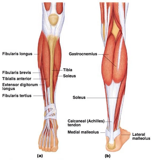
Gastrocnemius, Soleus, Plantaris, Tibialis muscles and Peroneus muscles are the five muscles that together form the lower leg.
- Gastrocnemius: Also called triceps, gastrocnemius is a very powerful muscle that forms a major portion at the back of our lower leg. It can be easily seen and felt by us at the back of our legs. Gastrocnemius provides a significant amount of propulsive force when running, walking or jumping. A powerful knee plantar flexor, these muscles allow a movement that helps the toes to point downwards.
- Soleus: The gastrocnemius with the soleus, is the main flexor of the ankle joint. The soleus muscle is a wide flat leg muscle found on the posterior leg that runs from just below the knee to the heel and lays immediately deep to the gastrocnemius. These muscles help in walking and standing.
- Plantaris: Plantaris is long, thin muscle extending behind the knee and into the calf of the posterior leg. Together with gastrocnemius and soleus, it forms the superficial group of the posterior compartment of the leg. While there is an ongoing debate about its functions, it is found that in around 10% of the population, plantaris is absent altogether.
- Tibialis: It is the most central and deepest muscle located in the posterior aspect of the leg. It helps in elevating our heel thus resulting in walking, running and various fitness exercises, such as calf raises.
- Peroneus: Present in the front part of the lower leg, the Peroneus muscle is a major mover and stabilizer of our ankle. It is a superficial muscle that can easily be seen and felt.
Lower Leg Muscles
Check More:
Things to Remember
- Leg anatomy refers to the study of a human’s leg.
- The movement or the ability to move from one place to another is called locomotion.
- Locomotion is a major need not only for mammals but also many other different organisms like Amoeba, insects etc.
- Locomotion is generally used for search of food, shelter, mate, suitable breeding grounds, favourable climatic conditions or to escape from enemies/predators.
- The human leg consists of mainly three types of bones: Femur, Tibia and Fibula.
- Our leg muscles can be broadly divided into two types: Upper leg muscles and lower leg muscles.
- Hamstrings, Quadriceps and Adductors make up the three main muscles in the upper leg region.
- Gastrocnemius, Soleus, Plantaris, Tibialis muscles and Peroneus muscles are the five muscles that together form the lower leg.
Read More:
Sample Questions
Ques. Briefly explain the five types of adductors that help bind the thighs together. (3 Marks)
Ans. Adductors consist of four muscles located inside the thighs. They are-
- Adductor Magnus: We are able to carry out activities like walking, sprinting, etc. due to Adductor Magnus which contracts and pulls the hip towards the body’s midline.
- Adductor Longus: The Adductor Longus is a skeletal muscle located in the thigh. Its primary function is to adduct the thigh at the hip joint.
- Adductor Brevis: Along with helping the thigh to rotate and flex, the Adductor Brevis also helps to adduct the thigh at the hip joint.
- Obturator Externus: Obturator Externus is a flat, triangular muscle in the medial compartment of the thigh. Together with other short muscles around the hip joint, it contributes to joint stability.
- Gracilis: Gracilis is a long and slender muscle found in the groin and is responsible for hip adduction and knee flexion.
Ques. Which two bones are the longest bones in the human body? Explain. (3 Marks)
Ans. The two longest bones in the human body are the Femur bone and Tibia.
- Also known as the thighbone, the femur is the longest bone of the human body. Forming a ball-and-socket joint with the hip, the femur connects the pelvic bone to the lower leg bones. It is the strongest and the heaviest bone of our body. Due to this, the femur has the ability to bear extreme forces which act upon it.
- The tibia is the second largest bone in the human body. It is also called the shank bone or the shinbone. Tibia connects the knee and the ankle joints and is a key weight-bearing structure.
Ques. What are the four Quadricep muscles found at the front of our thighs? (3 Marks)
Ans. Quadriceps are the four muscles found at the front of our thighs. These four individual muscles are-
- Vastus Lateralis: Vastus lateralis is the largest of the four muscles that run down the outside of our thigh, connecting our femur to our kneecap.
- Vastus Medialis: Running along the inner part of our thigh, Vastus medialis also connects the femur and the kneecap
- Vastus Intermedius: As the name suggests, the Vastus intermedius muscle is located in between the other two vastus muscles in our thigh. It is the deepest of all the four quadriceps.
- Rectus Femoris: Partially covering the three vastus muscles, the Rectus femoris originates in our hip bone and attaches to our kneecap.
Ques. Explain all the muscles found in the lower leg region. (3 Marks)
Ans. There is a total of five muscles that together form the lower leg. They are as follows:
- Gastrocnemius
- Soleus
- Plantaris
- Tibialis
- Peroneus
Ques. Briefly explain the functions of Gastrocnemius muscles. (3 Marks)
Ans. The gastrocnemius is a very powerful muscle that forms a major portion at the back of our lower leg and is also known as the triceps. It can be easily seen and felt by us at the back of our legs. Gastrocnemius provides a significant amount of propulsive force when running, walking or jumping. A powerful knee plantar flexor, these muscles allow a movement that helps the toes to point downwards.
Ques. Which leg bone runs laterally to Tibia but is smaller in size? Explain the same. (3 Marks)
Ans. Commonly known as the calf bone, the fibula is another bone present in the lower leg region that runs laterally to Tibia. The fibula is much smaller and thinner when compared to the tibia. Being a slender long bone, it bears very little body weight. The fibula provides stability to the lower leg region and also acts as a tie rod to increase the range of motion for the ankle. The average fibula is about 390 millimetres (mm) long in adult men and about 360 mm long in adult women.
Ques. What is the knee? Explain briefly. (3 Marks)
Ans. A connection between the upper and the lower leg, the knee is one of the largest and most complex joints in the body.
- The femur and the tibia are joined together by the knee joint.
- It consists of knee ligaments, knee bones and knee tendons.
- Tendons are the connective tissues that connect the knee bones to the leg muscles that move the knee joint.
- Ligaments are the connective tissues that join the knee bones and provide stability to the knee.
Read More:



Comments