



Exams Prep Master
Gram-negative bacteria belong to the genus Eubacteria and are included in the phylum Firmicutes. Eubacteria are broadly classified into two groups depending upon the result obtained from the gram staining procedure. The bacteria culture that does not retain the violet color and appears pink under the microscope are gram-negative bacteria. The gram-negative bacteria are disease-causing in humans as they have high pathogenicity and are highly resistant to antibiotics. They affect almost all systems in an organism- the digestive system, nervous system, urinary system, and bloodstream, causing diarrheal gastroenteritis or severe meningitis. The two major disease-causing groups of the gram-negative bacteria are the Enterobactericiae and non-fermenters, of which Enterobactericiae accounts for 80% of gram-negative bacteria. Here, we will learn more about the features, cell structure, and gram staining procedure.
| Table of Content |
Key Takeaways: Gram-negative bacteria, cell wall, disease, nagative, cell culture, alcohol
Also Read: Reproductive Health
Characteristic Features of Gram-negative Bacteria
[Click Here for Sample Questions]
- Gram-negative bacteria include enterococci, salmonella species, and Pseudomonas species.
- Gram-negative bacteria cell shapes range from rods (Escherichia coli) to bacillus (Streptobacillus moniliformis), from filamentous (Nocardia spp) to spiral (Treponema pallidum).
- The chemical structure of the outer membrane of lipopolysaccharides is often unique to specific bacterial strains (i.e. subspecies) and is responsible for many of the antigenic properties of these strains.
- Many species of gram-negative bacteria are pathogenic in nature. The pathogenicity of gram-negative bacteria is usually attributed to the lipopolysaccharide (LPS) layer of the Gram-negative cell envelope.

- The cell envelope of gram-negative bacteria protects it from a wide variety of antibiotics (penicillin, etc).
- After cell lysis, it releases many endotoxins which contribute to the severity of symptoms during infections.
- Gram-negative bacteria when tested by gram staining procedure does not retain the violet color after treatment with acetone or alcohol.
- Gram-negative bacteria can cause many severe infections such as pneumonia, peritonitis (inflammation of the membrane of the abdominal cavity), urinary tract infections, bloodstream infections, wound or surgical infections, and meningitis.
Read more:
Cell Structure of Gram-negative Bacteria
[Click Here for Sample Questions]
- The gram-negative bacteria have a distinct outer wall, the outer membrane, which makes them completely different from gram-positive bacteria. This outer membrane is made up of lipopolysaccharides which are large and complex molecules of lipids and carbohydrates.
- The region between the outer membrane and the cell membrane is known as periplasmic space.
- The cell membrane along with the outer membrane and the middle layer of periplasmic space is called the cell envelope.
- The presence of a negative charge on lipopolysaccharides is responsible for imparting a negative charge to the cell envelope.
- The outer membrane is present just above the periplasm and is composed of Braun’s lipoprotein which is covalently bound to the peptidoglycan and embedded in the outer membrane by hydrophobic ends. Braun's lipoprotein strongly links the outer membrane and the peptidoglycan layer.
- The gram-negative cell wall is also strengthened by the adhesion sites on the outer membrane. Substances enter the cell through these adhesion sites. The cell membrane is thin and bilayered composed of main peptidoglycans.
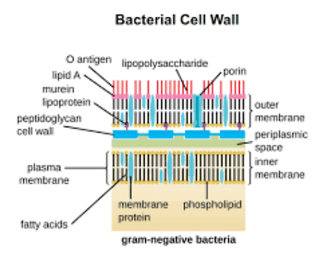
- The lipopolysaccharide is made up of 3 units: Lipid A, the core polysaccharides, and the O side chain. Lipid A is the toxic element of the Lipopolysaccharides, hence it acts as an endotoxin. These O antigens are also responsible for bacterial evading antibody responses. To sum up, the outer membrane functions to prevent the entry of antibodies, certain antibiotics, bile salts, and other similar toxins into the cell. It also evades the antibody responses of the host.
- In the Gram-negative wall, the region between the cytoplasmic and the outer membrane is filled with a gel-like fluid called periplasm. Periplasm is composed of peptidoglycan chains called peptidoglycan layers. Periplasm consists of many proteins associated with many cellular functions.
- Consequently, the outer membrane is made up of Porins which are proteins that make it permeable allowing the entry of small molecules such as glucose.
- To transport large molecules like Vit B 12 through the cell, specific carrier proteins are present on the outer membrane.
Read more:
Diseases Caused
[Click Here for Sample Questions]
Though a lot of strains of Gram-negative bacteria are harmless to humans, there are several diseases caused by some strains of Gram-negative bacteria. Some of them are listed below:-
| Diseases/Symptoms | Name of Bacteria |
|---|---|
| Gonorrhea- Causes painful and abnormal urination and discharge from the vagina or penis. | Neisseria gonorrhoeae |
| Meningitis- invades the brain barrier causing purulent meningitis with fever and severe headaches, joint aches, and a purpuric rash. | Neisseria meningitidis |
| Salmonellosis (gastroenteritis)- characterized by nausea, vomiting, and non-bloody diarrhea. | Salmonella spp. |
| Shigellosis (Bacillary dysentery)- characterized by intense abdominal pain and bloody mucus in stool. | Salmonella dysentery |
| Cholera- associated with profuse watery diarrhea and dehydration. | Vibrio cholerae |
Gram Staining Procedure
[Click Here for Sample Questions]
Scientist Christian Gram of Denmark first demonstrated the method of Gram staining in 1884. This is a kind of differential staining procedure. Based on the reaction of Gram stain, bacteria are classified into two groups-(i) Gram-positive and (ii) Gram-negative forms.
- Gram-positive bacteria: Bacteria retaining the violet color of crystal violet are called Gram positive. The deep violet color could not be washed by treatment with a solvent like acetone or alcohol. Examples of common Gram-positive bacteria are Staphylococcus, Bacillus, Streptococcus.
- Gram-negative bacteria: Bacteria that could not retain the violet color after treatment with acetone or alcohol are called Gram-negative bacteria. After acetone or alcohol treatment bacterial culture is counterstained with Safranin that imparts red color. Examples are E.coli, Salmonella, Neisseria, etc.
Procedure:
- A small amount of bacterial culture is smeared in a clean grease-free slide.
- The smear is then stained with a dilute alkaline solution of crystal violet for a minute.
- The slide is rinsed in water.
- The smear is then treated with a 0,5% solution of iodine and then is rinsed with water.
- The slide is then treated with 90% alcohol.
- Drops of safranin or eosin solution are added to the smear.
- The slide is washed in water and dried in air.
- Bacteria retaining the violet color of crystal violet are Gram-positive and those that could not retain the violet color and took the red color of safranin are Gram-negative.
Things to Remember
- Bacteria can be classified into two broad categories based on the result given in the Gram Staining procedure- the bacteria culture which retains the crystal violet color of iodine are Gram-positive bacteria while the ones that retain the crystal violet are Gram-negative.
- The cell envelope is made up of three layers- cell membrane, periplasm, and the outer membrane. Periplasm has a network of peptidoglycan chains and the outer membrane is made up of lipopolysaccharides.
- The lipopolysaccharides in the outer membrane are specific in specific strains of Gram-negative bacteria. The lipopolysaccharides cause a negative charge to the cell envelope to provide stability to it.
- Porins are special proteins that help to transport small molecules in and out of the bacteria, while specific carrier proteins transport larger molecules.
- The gram-negative bacteria cell envelope is more resistant to toxins, antibiotics, and antibodies than Gram-positive bacteria, which makes it more pathogenic. It also causes several serious diseases in humans- cholera, Bacillary dysentery, gastroenteritis, UTIs, meningitis, etc.
Read More: Heterotrophic Bacteria
Sample Questions
Ques. What is the process of Gram Staining? (2 marks)
Ans. Gram Staining is the process that is used to find out whether the given bacterial culture is Gram-negative or Gram-positive. To find this, the bacterial culture is first stained with crystal violet and then treated with 90% alcohol. Lastly, it is stained with safranin. If the culture retains the violet color, then it is Gram-positive. If the culture does not retain the violet color then it is Gram-negative bacteria.
Ques. Name some diseases caused by gram-negative bacteria. (2 marks)
Ans. Some of the strains of Gram-negative Bacteria are highly pathogenic in humans, such as cholera (Vibrio cholera), Bacillary dysentery (Salmonella dysenteriae), meningitis (Neisseria Meningitidis), Gonorrhea (Neisseria gonorrhoeae), Salmonellosis (Salmonella spp).
Ques. What is the main difference between Gram-positive and Gram-negative bacteria? (2 marks)
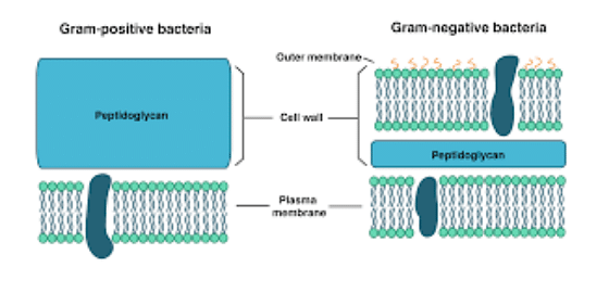
Ans. The cell envelope of Gram-negative bacteria has three-layer- cell membranes, periplasm, and the outer membrane. The cell membrane is thin.
While in the Gram-positive bacteria there is no outer membrane or periplasm and the cell membrane is thick.
Structure of Gram-negative and Gram-positive bacteria
Ques. Why are gram-negative bacteria more pathogenic than gram-positive bacteria? (2 marks)
Ans. The outer membrane of Gram-negative bacteria is highly resistant to the attack of toxins, antibiotics, bile salts, or any antibody reactions. Hence, saves the bacterial cell from various immune responses along with providing resistance to antibodies.
Ques. How are substances transported in and out of a gram-negative cell wall? (2 marks)
Ans. The Gram-negative cell has porins in the outer membrane; these are made up of specialized proteins which transport small particles and molecules in and out of the cell. To transport larger molecules, there are specific carrier proteins.
Ques. What is the cell envelope made up of in gram-negative bacteria? (2 marks)
Ans. The cell envelope of gram-negative bacteria has three layers- cell membrane, periplasm, and the outer membrane.
Ques. What is the function of Periplasm and what is it made up of? (2 marks)
Ans. The periplasm is made up of a complex network of peptidoglycan chains called the peptidoglycan layer. The periplasm is present in the periplasmic space between the outer membrane and the cell membrane. It is concerned with various cellular functions of bacteria.
Ques. Mention the three subunits of lipopolysaccharide and their functions. (2 marks)
Ans. The lipopolysaccharide is made up of 3 units: Lipid A, the core polysaccharides, and the O side chain. Lipid A is the toxic element of the Lipopolysaccharides, hence it acts as an endotoxin. When the cell dies these endotoxins are released contributing to the severity of the infection. The O antigens are also responsible for bacterial evading antibody responses.
Related Links:



Comments