



Content Curator
Various types of microscopes are nowadays being used for various scientific purposes. The microscope is a valuable instrument. There are many small items or information of gadgets that cannot be visible by using the unaided human eye.
- The microscope magnifies the image of such gadgets hence making them visible to the human eye.
- Microscopes are used to examine the shape of bacteria, fungi, parasites, and host cells in various stained and unstained arrangements.
- Over the course of the microscope’s history, technological innovations are made the microscope easier to use and improved the quality of the images produced.
Read More: Ray Optics and Optical Instruments
| Table of Content |
Key Terms: Microscope, Virtual, Magnified, Object lens, Eye Piece.
What are the Different Types of Microscopes?
[Click Here for Sample Questions]
There are special forms of microscopes and each of these has different use.
- Some are appropriate for biological programs, even as others are used in educational institutions.
- There are also microscope kinds that find application in metallurgy and reading 3D samples.
- There are 5 microscope types that are mentioned along with their diagram, working process, and applications.
- These 5 varieties of microscopes are
- Simple microscope
- Compound microscope
- Electron microscope
- Stereomicroscope
- Scanning probe microscope
Read More: NCERT Solutions for Class 12 Physics
Simple Microscope
[Click Here for Sample Questions]
A simple microscope is defined as a type of microscope that has one lens for the magnification of the sample. It is a convex lens with a small focal length. The magnifying power is given as
Where, m = 1 + (D/F)
- D is the least distinct vision
- F is the focal length
Simple Microscope Diagram

Simple Microscope Diagram
Principle of Simple Microscope
The working principle of a simple microscope is that a sample is placed within the focus of the microscope, erect, a virtual, and magnified image is obtained at the least distance of distinct vision from the eye to the lens.
- An easy microscope's mechanism uses a convex lens with a short focal length.
- In the discipline of microbiology, it is frequently employed.
- Typically, a thin, transparent item is used to make the light from the light source flow through it.
- The lens has to be closer to the sample in order to improve the picture.
- Additionally, the sample may be contrasted.
- To get the desired results, the sample must first be stained before the size and intensity of the light beam are changed with the use of a condenser or a diaphragm.
Application of Simple Microscope
A few applications of a simple microscope are–
- It is common among watchmakers as they could view a magnified image of the smallest parts.
- It is also used by jewelers for obtaining a magnified image of the jewelry.
- Educational institutions such as schools and colleges use a simple microscope in their laboratories mostly .
- Dermatologists (pores and skin specialists) they use simple microscopes to identify different pores and skin diseases.
Read More: Total Internal Reflection
Compound Microscope
[Click Here for Sample Questions]
A compound microscope is a type of microscope it has more than one lens. It has a combination of lenses and two optical parts which are known as the objective lens and eyepiece. The magnifying power is given as
m = (D/fo) × (L/fe)
Where
- D = least distance of distinct vision
- fo = focal length of the objective lens
- L = length of the microscope tube
- fe = focal length of the eyepiece
Compound Microscope Diagram

Compound Microscope Diagram
Principle of Compound Microscope
The working principle of the compound microscope is that the mixture of lenses enhances the magnification of the sample . The order can be first considered as a primary image in the tube and viewed again at that eyepiece.
- Commonly, the specimen or object to be studied is mounted on a transparent glass slide and positioned on the specimen stage between the condenser and objective lenses.
- A beam of visible light from the base is focused on the specimen by a condenser lens.
- The specimen's light is captured by the objective lens, which amplifies it within the body tube to create the main picture. This picture is once more magnified by the ocular lens or eyepiece.
- In order to line the higher magnification objective (often 45X) with the lighted portion of the slide when a greater magnification is required, the compound microscope's nose piece must be rotated after low-power focusing.
- Sometimes it was required to utilize a very high magnification (for example, to observe bacterial cells). In this case, an oil immersion objective lens (typically 100X) is employed.
- The ordinary light microscope is sometimes referred to as a bright-field microscope since the picture is produced inside a highly lit field.
Applications of Compound Microscope
A few applications of a compound microscope are–

Uses of Compound Microscope
- Viruses and Bacteria are possible by using a compound microscope.
- It can find applications in forensic laboratories.
- It is used in metallurgy.
Read More: Optical Density
Electron Microscope
[Click Here for Sample Questions]
An electron microscope is defined because the sort of microscope in which the supply of illumination is the beam of elevated electrons. It is a special kind of microscope with an excessive resolution image as the images can be magnified in nanometers.
There are 2 types of electron microscopes:
- The transmission electron microscope (TEM)
- The scanning electron microscope (SEM)
Electron Microscope Diagram

Electron Microscope Diagram
Principle of Electron Microscope
In an electron microscope the metal used is tungsten.
- A high voltage cutting-edge is carried out which ends up in the excitation of the electrons in the shape of a non-stop movement this is used as a beam of light.
- The lenses used in this microscope are magnetic coils.
- These magnetic coils are able to focus the electron beam on the sample such that the pattern receives illumination.
- As the strength of the magnetic lens increases , the flow of current increases.
- The electron beam flow is designed such that it can't pass through the glass lens.
Application of Electron Microscope
A few applications of an electron microscope are–
- Quality manipulation and failure evaluation in industries are accomplished with the help an electron microscope.
- The images obtained in that microscope can be captured as electron micrographs by using specialized cameras.
- The study of metals and crystals became easy with the electron microscope.
Read More: Dual Nature of Radiation and Matter MCQ
Stereo Microscope
[Click Here for Sample Questions]
A stereo microscope is defined as a form of microscope that provides a 3D view of a specimen. It is also called a dissecting microscope. In a stereo microscope, there are separate eyepiece and objective lenses and there are separate optical paths for each eye.
Stereo Microscope Diagram
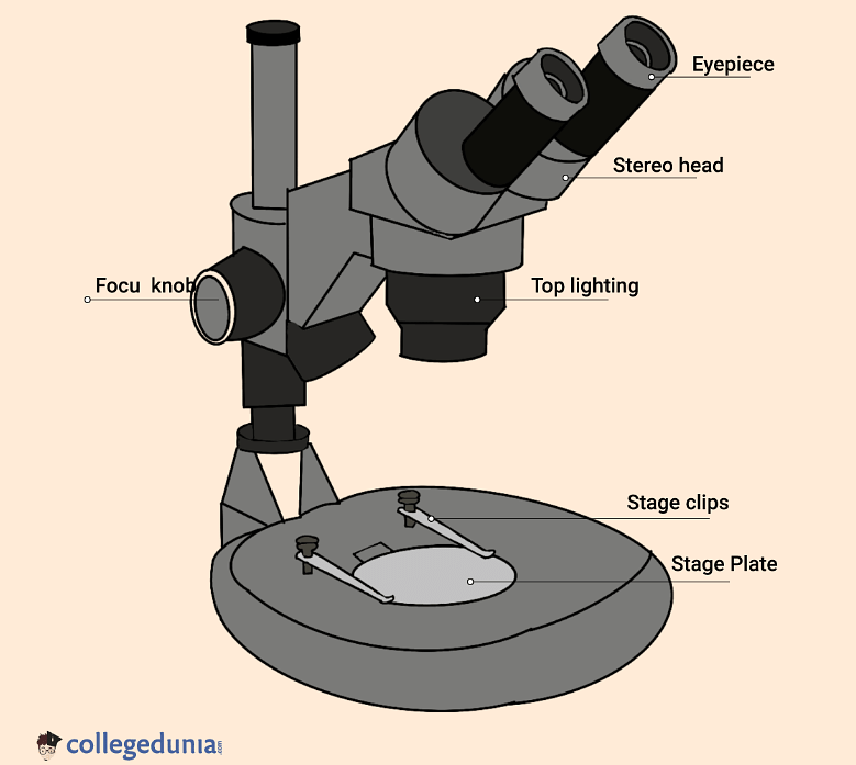
Stereo Microscope Diagram
Principle of Stereo Microscope
A stereo microscope works at the reflected mild from the sample.
- The magnification of the microscope takes vicinity at low power and subsequently, it's far appropriate for magnifying opaque objects.
- It is appropriate for thick and stable samples because it uses light meditated from the sample.
- The magnification in this microscope is between 20x and 50x.
Applications of Stereo Microscope
A few applications of a stereo microscope are–
- Examination of historic coins is possible with the help of the stereomicroscope.
- It finds application in microsurgery.
- Viewing crystals became easy with the help of a stereo microscope.
Read More: Kaleidoscope
Scanning Probe Microscope
[Click Here for Sample Questions]
The scanning probe microscope is described because a form of microscope that finds applications in industries in which the examination of the specimen is carried out on the nanoscale levels. The study of a specimen’s properties , its reaction time, and its behavior while stimulated may be carried out with the help of a scanning probe microscope.
Scanning Probe Microscope Diagram

Scanning Probe Microscope Diagram
Principle of Scanning Probe Microscope
The scanning probe microscope has a probe tip this is installed at the top of a cantilever.
- The tip is so sharp that it could move precisely and as it should be across the surface of the pattern scanning every atom.
- The tip is located close to the surface of the sample, such that the cantilever reports a deflection due to forces.
- The deflection distance is measured with the help of a laser.
- The final photo after scanning is received on the computer.
Application of Scanning Probe Microscope
A few applications of scanning probe microscopes are–
- It can be helpful for studying different properties of the sample like electrical properties.
- The magnetic property of the sample is studied by using a probe microscope.
- The transferring of information on the sample can be done with the help of this microscope.
Things to Remember
- By enlarging the picture of such devices, the microscope makes them visible to the human eye.
- The form of bacteria, fungi, parasites, and host cells is examined under a microscope in both stained and unstained configurations.
- The schematic, operational steps, and uses for each of the five different types of microscopes are also explained.
- Five types of microscopes are there– Simple microscope, Compound microscope, Electron microscope, Stereomicroscope, and Scanning probe microscope.
- The magnifying power of a simple microscope is given as m = 1 + (D/F), here, D is the least distinct vision and F is the focal length.
- The electron microscope made it simple to investigate metals and crystals.
- With the use of a scanning probe microscope, information about the sample may be transferred.
Sample Questions
Ques. What are the types of microscopes? (2 Marks)
Ans.
- Simple microscope
- Compound microscope
- Electron microscope
- Stereomicroscope
- Scanning probe microscope
Ques. What is Simple Microscope? (1 Mark)
Ans. A simple microscope is defined as a type of microscope that has one lens for the magnification of the sample. It is a convex lens with a small focal length.
Ques. What is Compound Microscope? (1 Mark)
Ans. A compound microscope is a type of microscope it has more than one lens. It has a combination of lenses and two optical parts which are known as the objective lens and eyepiece.
Ques. What is Electron Microscope? (1 Mark)
Ans. An electron microscope is defined because the sort of microscope in which the supply of illumination is the beam of elevated electrons. It is a special kind of microscope with an excessive resolution image as the images can be magnified in nanometers.
Ques. What is Stereo Microscope? (2 Marks)
Ans. A stereo microscope is defined as a form of microscope that provides a 3D view of a specimen. It is also called a dissecting microscope. In a stereo microscope, there are separate eyepiece and objective lenses and there are separate optical paths for each eye.
Ques. What is Scanning Probe Microscope? (2 Marks)
Ans. The scanning probe microscope is described because the form of microscope that finds applications in industries in which the examination of the specimen is carried out on the nanoscale levels. The study of a specimen’s properties and reaction time and its behavior while stimulated may be carried out with the help of a scanning probe microscope.
Ques. What are the applications of Stereo microscopes? (2 Marks)
Ans.
- Examination of historic coins is possible with the help of the stereomicroscope.
- It finds applications in microsurgery.
- Viewing crystals became easy with the help of a stereo microscope.
Ques. What are the Precautions have to take when Using Microscopes? (2 Marks)
Ans.
- Do not contact the glass a part of the lenses together with your fingers. Use special lens paper to clean the lenses.
- Always preserve your microscope and cover it while not in use.
- Always deliver a microscope with both hands. Grasp the arm with one hand and region the opposite hand underneath the bottom for support.
For Latest Updates on Upcoming Board Exams, Click Here: https://t.me/class_10_12_board_updates
Check-Out:



Comments