



Content Curator
Cardiac muscle tissue, often known as myocardium, is a kind of muscular tissue that forms the heart. This muscular tissue, which contracts and relaxes automatically, maintains the heart and the pumping of blood throughout the body.
- Muscle tissue in the human body is classified into three types: skeletal, smooth, and cardiac.
- The heart contains cardiac muscle tissue, which is made up of cells called myocytes.
- The characteristic of the heart is its contractility, which is the pumping action, and the rhythmicity of the contraction.
- The amount of blood that is pumped by the heart per minute (also called the cardiac output) varies in order to meet the metabolic needs of peripheral tissues, especially the kidneys, brain, skeletal muscles, skin, liver, heart, and gastrointestinal tract.
Key Terms: Cardiac Muscle, Heart, Muscle, Myocardium, Fibres, Cardiomyocytes, Cells, Membrane
What is Cardiac Muscle?
[Click Here for Sample Questions]
Cardiomyocyte cells make up the cardiac muscle. These cells are also known as contractile myofibrils because they are long and cylindrical (like fibrils) and contract at a regular rate to maintain blood flow through the heart.
- The sarcomere is the contractile functional unit of the myofibril.
- Cardiomyocytes are the cells that make up the ventricles and atria of the heart.
- Cardiomyocyte shape is critical because, being a kind of muscle cell, they rely on actin and myosin for contraction.
- Cardiomyocytes are surrounded by a membrane containing salt ions, which allows them to contract by generating an electrical signal known as action potentials.
- Cardiac muscle has many mitochondria that produce energy for it to function properly.
- Since cardiac muscle fibres lack cross-bands (T-tubules), they cannot store or release calcium ions.
![What is Cardiac Muscle]()

Cardiac muscle
Read Also:
Function of Cardiac Muscle
[Click Here for Previous Year Questions]
The heart muscle is made up of specialised cells for contraction and pumping. Thus,
- They contract at regular intervals to transport blood from the heart to the rest of the body.
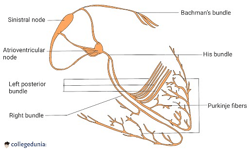
- Purkinje fibres are specialised cells found in cardiac muscles that assist the transfer of electrical impulses to and from heart muscle cells, making them a vital component in the heart's functioning.
![Function of Cardiac Muscle]()

Purkinje Fibres Diagram
- There are also specific forms of cardiac tissue in the heart, including "pacemaker" cells. These constrict and expand in response to the nervous system's electrical impulses.
- Pacemaker cells provide electrical impulses, or action potentials, to heart muscle cells, instructing them to contract and relax. Pacemaker cells regulate heart rate and how quickly the heart circulates blood.
Structure of Cardiac Muscle
[Click Here for Sample Questions]
The linked cardiac muscle cells, or fibres, provide strength and flexibility to cardiac muscle tissue.
- The cardiac muscle cells have one nucleus, but some have two. The nucleus contains all of the genetic material in the cell.
- Cardiac muscle cells also have mitochondria, which are referred to as "the powerhouses of the cell" by many. These are the organelles that convert oxygen and glucose into adenosine triphosphate energy.
- Under a microscope, cardiac muscle cells seem striated or striped. These stripes are caused by alternating filaments of myosin and actin proteins.
- The dark stripes represent thick filaments made up of myosin proteins. Actin is found in thin or lighter filaments.
- When a heart muscle cell contracts, the myosin filament attracts the actin filaments, causing the cell to shrink. This contraction is powered by ATP in the cell.
- On either side, a single myosin filament joins two actin filaments. This results in a single piece of muscle tissue known as a sarcomere.
- Intercalated discs are structures that link cardiac muscle cells. Nerve signals are relayed from one cardiac muscle cell to another via gap junctions inside the intercalated discs.
- Desmosome is another type of structure seen in the intercalated discs. These aid in the bonding of heart muscle fibres.
Read Also: Difference between Red Muscle Fibres and White Muscle Fibres
Difference Between Skeletal Muscle and Cardiac Muscle
[Click Here for Previous Year Questions]
Some of the differences between Cardiac muscle and Skeletal muscle are:
| Basis of Difference | Cardiac muscles | Skeletal muscle |
|---|---|---|
| Define | Cardiac muscles are situated in the heart and are self-stimulating and are responsible for conducting involuntary muscular actions. | Skeletal muscles are connected to bones throughout the body and are responsible for conducting voluntary muscular movements. |
| Major function | Coordinates cardiac contractions to pump blood throughout the body and maintain blood pressure. | Facilitates locomotion, facial expression, posture, and other voluntary movements of the body. |
| Movement | Involuntary | Voluntary |
| Number of nuclei present | One or two nuclei. | Multiple nuclei. |
| Shape | Semi-spindle | Cylindrical and long |
| Controlled by | Autonomic nervous system | Somatic nervous system |
| Fibres | Thicker ranging from 20 to 200 micrometres wide. | Long and thin ranging from 10 to 100 micrometres wide. |
Read Also: Difference between Cardiac Muscle and Skeletal Muscle
Cardiac Cycle
[Click Here for Sample Questions]
The cardiac cycle describes the functioning of the human heart from the start of one heartbeat to the start of the next.
- It is divided into two parts: diastole, which occurs when the heart muscle relaxes and refills with blood, and systole, which occurs when the heart muscle contracts and pumps blood.
- After emptying, the heart instantly relaxes and expands to aid another inflow of blood returning from the lungs and other body systems, before contracting again to pump blood to the lungs and other body systems.
- A regularly functioning heart must be fully enlarged before it can pump efficiently again.
Phases of Cardiac Cycle
There are two phases of cardiac cycle:
Atrial Event: Herein, the duration of systole and diastole is as follows:
- Systole: 0.1 s.
- Diastole: 0.7 s.
Ventricular Event: Herein, the duration of systole and diastole is as follows:
- Systole: 0.3 s.
- Diastole: 0.5 s.
![Cardiac Cycle Diagram]()

Cardiac Cycle Diagram
Clinical Significance (Diseases related to the heart)
Some diseases of the heart include:
- "Cardiovascular disease" refers to chronic heart or blood vessel illness, including heart muscle disorders, valves, and arteries. This phrase surrounds coronary artery disease (CAD) in which the heart's blood supply is limited.
- The growth of atherosclerotic plaques narrows the coronary arteries. The symptoms of angina pectoris may emerge if these narrowing’s become acute enough to partially obstruct blood flow. congestive heart failure, hypertension, and other conditions.
- Chronic diseases are frequently associated with anomalies in the anatomy of the cardiopulmonary system or congenital abnormalities.
- They vary from common disorders such as coronary artery disease to uncommon hereditary diseases affecting several organs.
- Atrial septal defect (ASD) and patent ductus arteriosus are two examples.
- Despite a regular blood supply, the heart muscle can be injured.
- Myocarditis is an inflammation of the heart muscle that is most usually caused by a viral infection but can also be caused by the body's immune system.
- Drugs such as alcohol can potentially harm the heart muscle, as can long-term high blood pressure or hypertension. Untreated myocarditis leads to dilated cardiomyopathy and heart failure in the long run.
Read More:
Things to Remember
- Cardiac muscle, commonly termed heart muscle, is the layer of muscular tissue between the endocardium and the epicardium.
- A heart attack caused by a clogged artery can cut off blood flow to specific parts of the heart.
- When the heart's oxygen demand exceeds its oxygen supply, cardiac muscle tissue might die. This results in the release of cardiac proteins into the circulation, such as troponin.
- Aerobic exercise regularly can assist build cardiac muscle tissue and reduce the risk of heart attack, stroke, and other cardiovascular disorders.
Previous Year Questions
- Blood pressure in the mammalian aorta is maximum during...[NEET 2015]
- Blood pressure in the pulmonary artery is...[NEET 2016]
- Blood capillaries are made of...[NEET 1993]
- Blood group AB has...[NEET 1991]
- An adult human with average health has systolic and diastolic pressures as… [NEET 1998]
- Arteries are best defined as vessels which....[NEET 2018]
- Arteries carry oxygenated blood except...[NEET 1989]
- Breakdown product of haemoglobin is...[NEET 1988]
- Bundle of His is a network of...[NEET 2003]
- Cells formed in bone marrow include...[NEET 1992]
Sample Questions
Ques. What is Cardiac Muscle? Where can you find cardiac muscles? (1 mark)
Ans. Cardiac muscle tissue, which is also known by the name myocardium, is a kind of muscular tissue that forms the heart. Cardiac muscles are present in the heart's lining.
Ques. What are the two special features of cardiac muscle? (1 mark)
Ans. Cardiac muscle cells (cardiomyocytes) are known to be striated, and branched, which contain several mitochondria, and are under involuntary control.
Ques. What are some heart diseases? (1 mark)
Ans. Some heart diseases include coronary artery disease (CAD), atrial septal defect (ASD), congestive heart failure, hypertension, and patent ductus arteriosus (PDA).
Ques. What Is the Distinction Between Skeletal Muscle and Cardiac Muscle? (2 marks)
Ans. Skeletal muscles connect to bones and contract to move the body. Cardiac muscles are situated in the heart and contract to help the heart pump blood. The fibres of skeletal muscle are long and thin. Cardiac muscle fibres are dense.
Ques. What are some causes of cardiovascular heart diseases? (2 marks)
Ans. The primary causes of cardiovascular heart disease include:
- Atherosclerosis
- Low-fat conditions
- Embolism
- Aneurysms
Ques. Define fibroblasts. (2 marks)
Ans. Heart fibroblasts are the important supporting cells found inside the cardiac muscle. These, unlike cardiomyocytes, are unable to cause strong contraction of the heart, but they are significantly important for both building and maintaining the extracellular matrix that provides the mortar in which cardiomyocyte bricks are embedded. Fibroblasts are critical in the response to damage, such as a myocardial infarction.
Ques. What is the duration of the Ventricular Event? (2 marks)
Ans. Ventricular Event’s duration of systole and diastole is:
- Systole: 0.3 s.
- Diastole: 0.5 s.
Ques. What are Purkinje fibres? What happens if they don't work? (3 marks)
Ans. Purkinje fibres are an essential component of the cardiac conduction system. It is primarily responsible for assisting the heart in contracting and successfully pumping blood. If the Purkinje fibres fail to do so, the ventricles lose their ability to convey blood. As a result, blood circulation in the body is disrupted, which can lead to major problems.
Purkinje Fibres
Ques. Distinguish between Atria and Ventricles? (3 marks)
Ans. The atria and ventricles of the heart are formed by cardiac muscle. Although the muscular tissue in the heart chambers is the same, there are a few variances.
The myocardium in the ventricles is thick to allow for powerful cardiac contraction, whereas the myocardium in the atria is thinner.
Individual myocytes, which make up the myocardium, differ between heart chambers as well. Ventricular cardiomyocytes have a denser T-tubule network and are broader and longer.
Ques. What exactly are cardiac muscles? What are its purposes? (4 marks)
Ans. Cardiac muscles are a form of striated muscle that can only be found in the heart.
- They are controlled by the autonomic nervous system, which means they are involuntary and operate independently.
- Cardiac muscles are made up mostly of cells termed cardiomyocytes, which generate contractile force as well as structural and functional support for the cardiac muscle tissue.
- They also have blood arteries that deliver nutrients to heart muscle tissue and eliminate waste.
- Nerve cells or neurons, which begin impulses, are also found in cardiac muscles.
Also read:



Comments