



Education Journalist | Study Abroad Lead
Plants are divided into two categories: flowering and non-flowering. Angiosperms are flowering plants, while gymnosperms are plants that do not blossom. With over 200,000 species, angiosperms are the most diverse group of plants in the kingdom. Herbs, shrubs, and trees that reproduce sexually by seeds fall within this category. Angiosperms are divided into Monocotyledonous and Dicotyledonous plants based on the number of cotyledons in the seed and the nature of the embryo in the seed. Dicotyledonous plants have seeds with two cotyledons and are thus referred to as dicots. Sunflower and mango are two examples. Monocotyledonous (monocot) plants have only one cotyledon. Sugarcane and maize are two examples. Let's discuss dicotyledonous and monocotyledonous plants and their anatomy.
Read Also: Double Fertilization in Angiosperms
| Table of Content |
Key Takeaways: Dicot Root, Monocot Root, Dicot Stem, Monocot Stem, Dicot Leaf, Monocot Leaf, Dicot Seed, Monocot Seed
Dicotyledonous Root
[Click Here for Sample Questions]
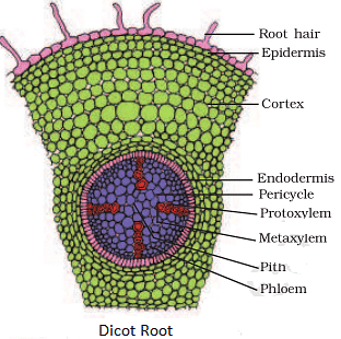
The transverse section of the sunflower root is depicted in the diagram. The following is the internal tissue organization:
- Epiblema is the topmost layer. Epiblema cells protrude in the form of unicellular root hairs on many occasions.
- The cortex is made up of many layers of parenchyma cells with thin walls and intercellular gaps.
- Endodermis refers to the cortex's deepest layer and is made up of a single layer of barrel-shaped cells with no intercellular gaps between them.
- Endodermal cells have a deposition of water-impermeable waxy substance suberin in the form of Casparian strips on their tangential and radial walls.
- The pericycle is a layer of thick-walled parenchymatous cells that is next to the endodermis.
- During secondary growth, these cells initiate the formation of lateral roots and vascular cambium. The pith is tiny and unnoticeable.
- Conjunctive tissue comprises parenchymatous cells that are found between the xylem and the phloem.
- Patches of xylem and phloem are normally two to four in number.
- A cambium ring forms later between the xylem and the phloem.
- The stele is made up of tissues on the inner side of the endodermis, such as the pericycle, vascular bundles, and pith.
Check Important Difference Between Dicot and Monocot Root
Monocotyledonous Root
[Click Here for Sample Questions]
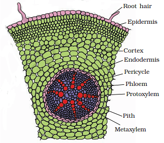
In many ways, the anatomy of a monocot root is identical to that of a dicot root (Figure). Epidermis, cortex, endodermis, pericycle, vascular bundles, and pith make up the epidermis. In comparison to the dicot root, which has fewer xylem bundles, the monocot root frequently has more than six (polyarch) xylem bundles. The pith is substantial and well-developed. Secondary growth does not occur in monocotyledonous roots.
Dicotyledonous Stem
[Click Here for Sample Questions]
The epidermis is the stem's outermost protective covering, as shown in the transverse section of a typical immature dicotyledonous stem.
- It may have trichomes and a few stomata and is covered in a thin layer of cuticle.
- The cortex is made up of cells grouped in many layers between the epidermis and the pericycle.
- It is divided into three zones.
- Just below the epidermis, the outer hypodermis consists of a few layers of collenchymatous cells that offer mechanical support to the embryonic stem.
Round thin-walled parenchymatous cells with visible intercellular gaps make up the cortical layers beneath the hypodermis. The endodermis is the deepest layer of the brain. Because the endodermis cells are densely packed with starch grains, the layer is also known as the starch sheath. Pericycle appears as semi-lunar patches of sclerenchyma on the inner side of the endodermis and above the phloem. The medullary rays are a few layers of radially placed parenchymatous cells that lie between the vascular bundles.
A high number of vascular bundles are arranged in a ring; dicot stems are known for their 'ring' arrangement of vascular bundles. Each vascular bundle is joined, open, and has a protoxylem at the end. The pith is made up of a large number of spherical, parenchymatous cells with extensive intercellular gaps that occupy the middle region of the stem.

Check Important Difference Between Monocot and Dicot Stem
Monocotyledonous Stem
[Click Here for Sample Questions]
A sclerenchymatous hypodermis, numerous distributed vascular bundles, each enclosed by a sclerenchymatous bundle sheath, and a broad, prominent parenchymatous ground tissue characterize the monocot stem.
- The vascular bundles are joined and closed together.
- Vascular bundles at the periphery are typically smaller than those in the center.
- The phloem parenchyma is missing, and there are water-filled holes inside the vascular bundles.

Read More: Regions of Root
Dorsiventral (Dicotyledonous) Leaf
[Click Here for Sample Questions]
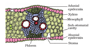
The epidermis, mesophyll, and vascular system are visible in a vertical piece of a dorsiventral leaf through the lamina. The epidermis of the leaf has a prominent cuticle that covers both the upper (adaxial epidermis) and bottom (abaxial epidermis). In general, the abaxial epidermis has more stomata than the adaxial epidermis. Stomata may even be absent in the latter.
- Mesophyll is the tissue that lies between the top and lower epidermis. The mesophyll is made up of parenchyma, which contains chloroplasts and performs photosynthesis.
- The palisade parenchyma and the spongy parenchyma are the two types of cells that make up this tissue.
- The elongated cells that make up the adaxial palisade parenchyma are organized vertically and parallel to each other.
- The spongy parenchyma, which is oval or spherical and loosely organized, is found underneath the palisade cells and reaches the lower epidermis.
- Between these cells, there are various huge gaps and air cavities. Vascular bundles, which can be seen in veins and the midrib, are part of the vascular system.
- The size of the vascular bundles is determined by the veins' size. The veins in the dicot leaves' reticulate venation vary in thickness.
- A layer of thick-walled bundle sheath cells surrounds the vascular bundles.

Check Important Difference Between Monocot and Dicot Leaf
Isobilateral (Monocotyledonous) Leaf
[Click Here for Sample Questions]
In many ways, the anatomy of the isobilateral leaf is identical to that of the dorsiventral leaf. It demonstrates the following distinguishing characteristics. Stomata are present on both epidermal surfaces in an isobilateral leaf, and the mesophyll is not divided into palisade and spongy parenchyma.
A few adaxial epidermal cells along veins in grasses can transform into huge, empty, colorless cells. They are known as Bulliform cells.
- The leaf surface is exposed when the bulliform cells in the leaves have absorbed water and are turgid.
- They curl the leaves inwards for reducing water loss when they are flaccid due to less water content.
- In vertical slices of monocot leaves, the parallel venation is represented in the near comparable diameters of vascular bundles (save in principal veins).
Check More: Anatomy of Flowering Plants
Things to Remember
[Click Here for Sample Questions]
- Monocot and dicot plants differ in their roots, stem, leaves, flowers, and seeds.
- Monocot comprises a single cotyledon in its embryo whereas dicot has two cotyledons in its embryo.
- Monocot plants have narrow, elongated leaves with parallel venation, which is used to distinguish them from dicots.
- Dicot leaves are rounded with reticulate venation and are structurally and anatomically distinct from monocot leaves.
- Maize and orchid are examples of Monocot Root; banyan tree roots and roots of green peas are examples of Dicot Root.
- Palm tree stem is a Monocot Stem whereas cactus stem is a Dicot Stem.
- Maize leaves and grass are Monocot leaves whereas mint leaves and mustard leaves are dicot leaves.
- Seeds with a single (mono) embryonic leaf or cotyledon are known as monocot seeds, whereas seeds with two embryonic leaves or cotyledons are known as dicot seeds.
- Monocot seeds include maize, bamboo, palm, banana, ginger, and onion.
- Dicot seeds include Neem seeds, Night Jasmine seeds, Papaya seeds, and Tamarind seeds, to name a few.
Also Check:
Sample Questions
Ques. State 5 Key Differences between Monocot and Dicot Root(5 marks)
Click here for the answer
Ans. The main difference between monocot and dicot roots are as given below:
| Characteristics | Monocot Root | Dicot Root |
| Definition | Monocotyledonous roots are fibrous or adventitious roots that grow from the stem and consist of a large network of thin roots and root fibers. | Taproots consisting of a single primary root from which secondary and tertiary roots form and extend vertically downwards through the soil are known as dicotyledonous roots. |
| Root system | Plants with a fibrous or adventitious root structure are known as monocots. | Plants that are dicots have a tap root system. |
| Primary root | During the postembryonic growth of the roots, the primary root development comes to a halt. | Taproot, which is the major root, continues to grow throughout the plant's life. |
| Epidermal Covering | After the epidermis is peeled away, the monocot roots are covered by a cork cambium. | Exodermis, which is a modified epidermis, covers the dicot roots. |
| Cortex | Monocot roots have a broad cortex made up entirely of parenchymatous cells. | Dicot roots have a thin cortex made up of both parenchymatous and sclerenchymatous cells. |
Ques. Explain 5 Key Differences between Monocot and Dicot Stem.(5 marks)
Click here for the answer
Ans. The main differences between monocot and dicot stem are as given below:
| Characteristics | Monocot Stem | Dicot Stem |
|---|---|---|
| Definition | The monocot stem is a hollow axial section of the plant. It produces leaves, nodes, internodes, branches, and flowers, as well as roots at the base. | The dicot stem is a plant's solid cylindrical axial component made up of nodes and internodes that give rise to leaves, branches, and flowers. |
| Internodes | The monocot stem's internodes are hollow. | The dicot stem's internodes are solid. |
| Transverse section | In the transverse section of a monocot stem, the epidermis, hypodermis, and ground material are distinguished. The monocot stem's interior tissues are arranged in no particular order. | The epidermis, cortex, and stele are the three layers of the dicot stem. The dicot stem's interior tissues are arranged in concentric layers or rings. |
| Epidermal hairs | The monocot stem is devoid of epidermal hairs. | The dicot stem contains epidermal hairs. |
| Cortex | The hypodermis represents the cortex of the monocot stem, which is less developed. | The hypodermis, endodermis, and general cortex of the dicot stem are well developed and differentiated. |
Ques. State 5 Key Differences between Monocot and Dicot Leaf.(5 marks)
Click here for the answer
Ans. The key differences between monocot and dicot leaf are:
| Characteristics | Monocot Leaf | Dicot Leaf |
|---|---|---|
| Definition | Monocotyledonous plants have narrow, elongated leaves with parallel venation, which is used to distinguish them from dicots. | Dicot leaves are rounded with reticulate venation and are structurally and anatomically distinct from monocotyledonous leaves. |
| Shape | The leaves of monocots are narrower, slenderer, and longer than those of dicots. | The leaves are wider and smaller than monocots |
| Symmetry | The symmetry of monocot leaves is isobilateral. | As the upper and lower surfaces are distinct, leaves are dorsoventral. |
| Stomata | Monocot leaves are also known as amphistomatous because the number of stomata on the upper and lower surfaces of the leaves is equal. | The lower surface has more stomata than the top surface. Some leaves lack stomata on the upper surface and these plants are known as hypostomatous. |
| Mesophyll | Monocot leaves have two types of mesophyll: spongy mesophyll and palisade mesophyll. | Dicot leaves have no distinct mesophyll. |
Ques. Give 2 examples each of Monocot and Dicot Roots.(5 marks)
Click here for the answer
Ans. Monocot Root
- Maize root
- The fibrous or adventitious root system of maize is composed of multiple short-borne roots that are present above the soil surface.
- Like other monocot seeds, maize seeds have 3-7 seminal rootlets that start growing laterally and vertically.
- Orchid roots
- Orchids are monocot plants that are commonly utilized as beautiful plants. Orchids can grow on the ground or in the air.
- The roots of terrestrial orchids are thick and meaty, and they act as a storage system.
- Epiphytic orchids, on the other hand, have modified aerial roots that are lengthy and contain a feature known as velamen.
Dicot Root
- Banyan tree roots
- Banyan trees have distinctive tap root systems, which are frequently accompanied by aerial prop roots that grow into thick, woody trunks.
- The roots of the trees become indistinguishable from the primary root as they grow older. The lateral roots begin to extend in all directions, covering a large area.
- Roots of garden peas
- Garden peas have a basic tap root system with a heavily branching primary root that only extends about 6 inches below the soil's surface.
- Pea secondary roots have a symbiotic connection with a variety of bacteria. These bacteria can be found in the root nodules of plants, where they help in nitrogen fixation.
Ques. Give 1 example each of Monocot and Dicot Stem.(5 marks)
Click here for the answer
Ans. Monocot Stem: Palm tree stem
- The stem's surface can vary from smooth to rough and knobby, with sharp spines. Leaf scars may be visible on some stems, revealing where the leaves were linked to the stem.
- The stem's interior structure is similar to that of most monocots' internal structure.
- Pseudobark is the tissue of the hypodermis and epidermis of the stem that is found outside the central cylinder of the stem.
Dicot Stem: Cactus stem
- The cactus stem is a modified dicot stem with fleshy tissue that allows it to store enormous amounts of water.
- The epidermis is made up of cells with thin walls that are placed in close proximity to one another. The hydrophobic cuticle is located outside the epidermis and aids in the prevention of water loss.
- The cortex, which is made up of long-lived thin-walled parenchymal cells, is the most visible part of the stem. Mucilage cells and laticifers are two types of cells found in the cortex.
Ques. Give 2 examples each of Monocot and Dicot Leaves.(5 marks)
Click here for the answer
Ans. Monocot Leaves
- Maize leaves
- Maize leaves are the most common monocot leaves, as they have simple and tidy architecture.
- The top blade, auricle, lower sheath are the 3 parts of a maize leaf. Leaf cells are arranged in such a way that they produce parallel lines and an elongated shape.
- Grass
- A grass leaf is a monocot leaf with an extended shape that develops from a node that includes a basal cylindrical sheath that encircles the stem and other younger leaves.
- The leaf has an auricle, which can be an ear-like projection or a hairy edge near the base of the leaf blade.
Dicot Leaves
- Mustard leaves
- Mustard is a dicot plant that is mainly utilized in dicotyledonous plant research.
- Green leaves with reticulate venation are green and broad. The leaves are flattened dorsoventrally, with the ventral surface being lighter than the dorsal.
- Mint leaves
- Mint is a plant that grows rapidly with round lanceolate leaflets that are placed in opposing pairs on a stem.
- Reticulate venation is observed on the leaves, with a central midrib through which branched veins emerge.
Ques. Explain the structure of a Dicot Seed.(3 marks)
Click here for the answer
Ans. A seed coat protects a dicotyledonous seed. There are two layers to a seed coat: an exterior testa and an inner tegmen. On the seed coating, there is a scar known as hilum. The growing seeds are linked to the fruit via the hilum. The little pore right above the hilum is known as a micropyle. The seed coat protects the embryo. The embryo has two cotyledons and an embryonal axis. The cotyledons are fleshy and serve as food reserves for the embryo as it develops. The radicle and the plumule are located at the two ends of the embryonal axis. Examples of a dicot seed include Rice, wheat, maize, bamboo, palm, banana, ginger, onion, garlic, lilies, and others.

Ques. Explain the structure of a Monocot Seed.(3 marks)
Click here for the answer
Ans. The seed coat is membranous and attached to the cell wall, and it covers the seed. The endosperm is a thick substance that acts as a food reserve. The aleurone layer is a proteinous layer that covers the endosperm's outer surface. At one end of the endosperm, a tiny embryo is found in a groove. The scutellum, a huge shield-shaped cotyledon, is present. With the radicle and plumule, the embryo has a short axis. Coleorhiza and coleoptile sheaths surround the radicle and plumule, respectively. Examples of a monocot seed include Bitter gourd seeds, Castor seeds, Mango seeds, Neem Seeds, Night Jasmine seeds, Papaya seeds, and others.

Ques. What are the differences between Stomata and Lenticels?(5 marks)
Click here for the answer
Ans. The main differences between stomata and lenticels are:
| Stomata | Lenticels |
|---|---|
| On the epidermis of the leaves, there are tiny pores. | Pores are found on the tree trunk or the plant's stem. |
| Stomata can be controlled in their opening and shutting. | The lenticels remain open at all times. |
| Guard cells are present in these. | These don't have any guard cells. |
| They make it easier to breathe and exchange gases. | Only the exchange of gases is handled by them. |
| During the day, stomata are active. | Active at all hours of the night. |
| Roots and fruits are devoid of this substance. | Roots and fruits contain this substance. |
Ques. Explain the difference between vascular bundles of monocot and dicot root.(5 marks)
Click here for the answer
Ans. The difference between vascular bundles of monocot and dicot root are:
| Monocot Root | Dicot Root |
|---|---|
| Polyarch vascular bundles | Radial and exarch vascular bundles |
| There are 6 vascular bundles | There are 2 to 6 vascular bundles |
| The vascular cambium is not formed by conjunctive tissue. | Between the xylem and the phloem is a parenchymatous tissue termed conjunctive tissue. |
| Bundles of xylem and phloem alternate with each other. | The vascular cambium is formed later by the conjunctive tissue. |
| Between the xylem and the phloem is a parenchymatous tissue termed conjunctive tissue. | Protoxylem is in contact with the pericycle, whereas Metaxylem is found towards the root's center. |
Biology Related Links:



Comments