



Education Journalist | Study Abroad Lead
Villus are small finger-like folding that is responsible for increasing the surface area of a membrane. They are thin, slender, vascular projections in anatomy. They are mainly present in the mucous membrane lining of the small intestine as well as in the placenta. It increases the surface area for absorption of food in the intestinal cavity. Intestinal villi are also responsible for adding digestive secretions. Intestinal villus length ranges from approximately 0.5 mm to 1 mm. They are around 10 to 40 per square millimetre of tissue in the human body. They are most abundant at the beginning of the small intestine but they become very less in number at the end of the intestinal tract. Each villus consists of many Microvilli which emerges from the enterocytes of its epithelium giving the brush border appearance. Unlike any other circular folds in the intestine, the villus in the intestine is much smaller. Villus increases the surface area of absorption so that the nutrients like monosaccharides and amino acids pass into the semipermeable membrane of Villi through diffusion. Villi are well supplied with blood vessels for the circulation of these nutrients through the blood.
| Table of Contents |
Key Terms: Villus, Location of Villus,Structural features of Villius, Nutrient Absorption and Waste Elimination
What is Villus?
[Click Here for Sample Questions]
Villus is highly vascularized finger-like projections present on the mucosal surface of the small intestine. They are present in greater number in duodenum and jejunum part but ileum consists of less number of Villi. They appear tall and broad in the initial part of the small intestine but gradually shorten toward the end of the small intestine. They give a velvety appearance to the lining of the small intestine and increase the surface area of the lumen by about many folds. They mainly take part in the absorption of nutrients from digested food.
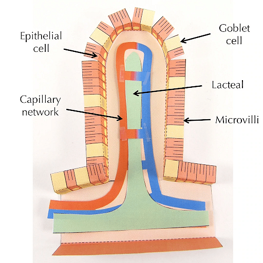
Villus
Location of Villus
[Click Here for Sample Questions]
Villus is present in the small intestine. They are formed from the mucosa layer of the alimentary canal. The mucosa is the innermost layer of the alimentary canal. Mucosa also forms irregular folds (rugae) in the stomach. Villi are also formed in the placenta during pregnancy as chorionic villi.

Villi Location
Structure of Villus
[Click Here for Sample Questions]
Each villus consists of a central core which further contains one vein, one artery, a muscle strand and a centrally located lymphatic capillary (lacteal). It also consists of connective tissues as it also requires proper structure and support. When villi are present in a great number, it gives the intestinal wall a velvety appearance.
Protein and Carbohydrates ingested by the Villi cells are assumed to be carried out by blood vessels. Emulsified fat (chyle) are eliminated by the lymphatic capillary. Contraction and expansion of Villi take place by muscle strand. Lacteal material empties into the larger lymphatic vessels by these contractions.

Structure of Villus
Structural features of Villius
Intestinal Villi consists of several structural features which play a significant role in the absorption of digested products. Structural features of Villi are as follows-
-
Microvilli-
Epithelial membrane is further ruffled for an increase in surface area. A wide number of functions such as absorption, secretion, cellular adhesion, etc are also carried out by microvilli.
-
Membrane proteins-
Digested materials get transported into the epithelial cells by these proteins. These proteins generally act as carriers for those materials which cannot be transported by diffusion.
-
Rich blood supply-
Villi consists of a dense network of capillaries so that products get rapidly absorbed. Except for fats and fat-soluble vitamins, the majority of the nutrients are absorbed by the blood capillaries.
-
Glands of Intestine-
Digestive juices for digestion of food material are also released by exocrine pits. The intestinal gland (also known as the crypt of Liberkuhn and intestinal crypt) are present in the intestinal epithelium lining in between the villi of the intestinal wall which are responsible for the secretion of digestive juices.
-
Lacteals-
Lipids are absorbed from the intestine by lacteals into the lymphatic system. Fluids such as electrolytes and proteins present in interstitial space are also collected by lacteals.
-
Presence of epithelium as a single layer-
It is present as a single layer so that distance between the lumen and blood decreases and diffusion gets minimised for efficient exchange of material across this layer.
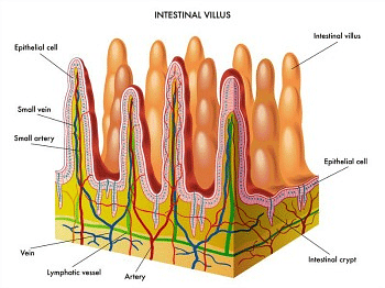
Intestinal Villus
Structural features of the epithelial lining of Villus
Epithelial lining of villus exhibits several structural features. These features are responsible for optimum absorption by intestinal Villi. These are as follows-
-
Tight junctions-
Tight junctions are the connections between the plasma membrane of two adjacent cells. They also create an impermeable barrier between the cells. They are responsible for one way movement of the material. Tight junctions keep digestive fluids separated from the tissues. They are also responsible for maintaining a concentration gradient.
-
Microvilli–
They allow more absorption of nutrients in blood capillaries as they significantly cause an increase in the surface area of the plasma membrane of the cell. Immobilised digestive enzymes are embedded within the membrane of microvilli. Several proteins which act as channels are also present for smooth uptake of material. They further cause cellular adhesion.
-
Mitochondria-
- A large number of mitochondria are present in the epithelial cells of the intestinal Villi. They provide energy in the form of ATP for active transport mechanisms.
- Primary active transport which takes place against the concentration gradient requires ATP. Secondary active transport (co-transport) or pinocytic also needs ATP. Due to the greater requirement of energy, a greater number of mitochondria are present in the villi and microvilli.
-
Pinocytotic vesicles–
When the uptake of non-specific fluids as well as dissolved solutes takes place by the cell then this process is known as pinocytosis commonly known as the cell drinking process.
Pinocytosis is a quick way to translocate material in bulk quantity. It is one type of endocytosis. When breaking and reforming of the membrane take place, then this material is ingested. They are further contained within a vesicle known as a pinocytotic vesicle. This process is primarily responsible for the absorption of fat droplets. Pinocytosis takes place in cells that form the lining of the small intestine in microvilli to absorb nutrients from the food.

Structure of Villus
Functions of Villus
[Click Here for Sample Questions]
The functions performed by villus are as follows-
- Villus provides an effective nutrient absorption in the lumen of the small intestine. Surface area is increased by approximately 30- folds and 600-folds by Villi along with the microvilli respectively.
- They are also involved in the secretion of digestive enzymes (enterocyte digestive enzymes). Villus capillaries collect amino acids and simple sugars in the bloodstream.
- They have single-cell thick walls which carry out efficient diffusion. Villus is responsible for the absorption of nutrients in the small intestine.
- They have a rich blood supply for sustaining a concentration gradient. For effective absorption of fatty acids and glycerol, they have a wide surface area.
- Digestion of protein in the last digestive stage is achieved by the enterocytes of the intestinal lumen.
Nutrient absorption and waste elimination
[Click Here for Sample Questions]
Oesophagus transports the food from the mouth into the stomach where it receives the gastric juices from the stomach. The stomach churns the food into chyme by mixing it with gastric secretions. Chyme is further transported to the duodenum where bile juice from the liver & pancreatic juice from the pancreas acts on it and disintegrate the nutrients. Villi absorb the nutrients from the digested food as Villi form the entire lining of the small intestine.
- Nutrients are absorbed by the blood capillaries and lacteals (lymphatic vessels) present in the villus. Fatty acids from the digested food are passed down to the lacteals.
- From the small intestine, chyme is transferred to the large intestine through the ileocaecal valve. In ascending and transverse parts of the large intestine, some water and residual nutrients are absorbed.
It leads to the compaction of chyme known as faeces.
- Faecal matter is temporarily stored in the rectum and anus. It is eliminated from the body through the process known as defecation. Defecation is carried out by rectal muscles contractions and internal anal sphincter relaxation. External anal sphincter is voluntary muscle while under the autonomic nervous system, defecation is often involuntary
Villous Atrophy
Due to effects of inflammation, the villi may get flattened and can often get disappear. This condition is known as Villous atrophy. It is often a characteristic of coeliac disease. The main causes of Villous atrophy are immune deficiencies, allergy to certain food and other infections. A person with Villous atrophy should consult a specialist for proper medications and treatment.
Things to Remember
- The finger-like projections made up of cells lining the entire length of the small intestine is known as Villus.
- Villus is mainly responsible for the absorption of nutrients from the food we eat.
- Villus alternates with the depressions known as crypts. Crypts manufacture the cells that form the Villi.
- Amino acids and Simple sugars are collected by Villus capillaries and transported into the bloodstream.
- Villus have a thin wall that is one cell thick for shortening the diffusion path.
- Efficient absorption of fatty acids and glycerol occurs due to a large surface area.
- Lacteals of Villus collect lipoproteins which get transported to the rest of the body through the lymph fluid.
- Villous atrophy is a condition when the villus gets flattened and even get disappear.
Sample questions
Ques. What is the reason for the high vascularity of Intestinal Villius? (3 mark)
Ans. The Intestinal Villus are highly vascularized due to the following reasons-
- Villus is mainly responsible for the absorption of nutrients from the food we eat.
- For efficient absorption of nutrients, Villi are supplied with a dense network of blood capillaries.
- As soon as food gets digested, its nutrients get absorbed by the villi due to its greater surface area.
- Absorbed nutrients from the Villus get transported into the bloodstream through the blood capillaries. The nutrients are then utilised by the cells and tissues.
Ques. What are the functions of lacteal in Villus? (5 marks)
Ans. The functions performed by the lacteals are as follows-
- The lymphatic capillaries found in the Villus of the small intestine are known as lacteals.
- Fats, lipids and other large molecules are mainly absorbed and transported as lipoproteins by lacteals.
- Chyle is the combination of fat and lymph in lacteals that is milky in appearance.
- Lacteals carry the Chyle to the lymph vessels in the intestinal wall.
- Lacteals get merged with the larger lymphatic vessels for the transportation of fats.
Ques. How the path of diffusion decreases across the Intestinal Villus? (3 marks)
Ans. The path of diffusion across the Intestinal Villi decreases in the following ways –
- The wall of the Intestinal Villus is made up of epithelium which is one cell thick only.
- Such structure of the Intestinal Villus makes its wall very thin for efficient exchange of material across the Villus. It decreases the distance between lumen and blood and hence maximise diffusion.
- The thin wall of epithelium along with a rich supply of blood vessels are mainly responsible for the efficient exchange and absorption of nutrients.
Ques. How does active transport take place in Intestinal Villus? (3 marks)
Ans. Both Primary and Secondary Active transport takes place in Intestinal Villi in the following ways-
- For active transport which takes place against the concentration gradient, energy is required.
- To compensate the requirement of energy, villi possess a large number of mitochondria in the epithelial cells of the intestinal Villi.
- Mitochondria produces ATP which is the energy currency of the cell. These ATPs are responsible for active transport of material.
- Certain carrier proteins present on the membrane also facilitate such transport.
Ques. Describe the Structure, Location and Function of microvilli? (5 marks)
Ans. The structure, location and function of microvilli are as follows-
- Structure = Microvilli are microscopic cellular membrane protrusions. They are covered in plasma membrane enclosing cytoplasm and microfilaments. Although they are cells but don't contain any cell organelles.
- Location = Microvilli are mainly found on the apical surface of some epithelial cells such as the Villi of the small intestine. They are also present on the plasma surface of eggs, white blood cells, etc.
- Function = They are mainly responsible for the absorption of nutrients in the gastrointestinal tract. They are also packed with enzymes for the breakdown of complex nutrients into simple compounds. They are also present in immune cells and hence plays a significant role in immunity.
Ques. What is the length of the intestinal Villus? (5 marks)
Ans. Villus is very thin finger-like projections forming the lining of the small intestine.
- They could be as small as 0.5 millimetres in length and cannot exceed more than around 1.6 millimetres in length.
- For convenient understanding, take the example of the ballpoint pen. When the tip of the fine ballpoint pen forms a line, the diameter of the ink line is 1.6 millimetres while 0.5 millimetres is even smaller.
- Villi often overlap with crypts which are depressions between Villi. Cells forming Villi and other parts of the intestinal lining are formed by crypts.
- In a healthy state, crypts are generally one-third to one-fifth of the length of Villi.
Ques. State various conditions that can cause damage to Intestinal Villus? (5 marks)
Ans. Villous atrophy is the condition in which Villi gets damaged or flattened. It is the characteristic of Coeliac disease. Certain conditions that can cause damage to Villi are as follows-
- Certain food which is rich in gluten can cause the immune system to attack the intestinal Villi and slow them down.
- Vitamin and mineral deficiency can also cause the impairment of intestinal villus
- Erosion of Villi can also be caused by inflammatory bowel disease.
- Certain medicines can also cause serious damage to the intestinal villus.
Ques. Describe the functions performed by the Villi? (5 marks)
Ans. The various functions performed by the Villi are as follows-
- Villi increases the surface area of the lumen of the Small intestine up to many folds. Greater surface area ensures better absorption of nutrients.
- Enterocyte digestive enzymes are present on the surface of Villi for proper digestion of food.
- Villi are highly vascularized for the absorption of simple sugars and amino acids.
- Absorbed chylomicrons are collected by the lacteals in the Villi. It is supplied to the rest of the body through the lymph fluid. Chylomicrons are lipoproteins composed of triglycerides, cholesterol and amphipathic proteins.
- The wall of Villus is one cell thick for decreasing the diffusion path. Fatty acids and glycerol are also absorbed efficiently due to greater surface area.
Ques. What features of Intestinal Villus makes it suitable for the absorption of nutrients? (5 marks)
Ans. The various structural features which increase the efficiency of absorption by intestinal Villi are as follows-
- The wall of Villi is single-cell thick which makes it a semi-permeable membrane and thereby increases the diffusion.
- Villi also consists of microvilli which gives it a velvety appearance. They play a significant role in absorption.
- Villi are richly supplied with blood vessels so that the absorbed nutrients are immediately circulated within the body.
- Lacteals collect the chylomicrons, proteins and electrolytes. It further transports them through the lymph fluid.
- Intestinal crypts present in between the Villi are responsible for the secretion of digestive enzymes.
Check-Out:



Comments