



Education Journalist | Study Abroad Lead
Synovial joints are the kind of joints that are characterised by the presence of fluid filled synovial cavity between the articulating surface of two adjoining bones. These joints allow considerable movement in vertebrates. They help in locomotion and many such movements.
Some examples of such joints are ball and socket joint (in between humerus and pectoral girdle), hinge joint (knee joint), pivot joint (between atlas and axis), gliding joint (between the carpals), and saddle joint (between carpal and metacarpal of thumb).
| Table of Content |
Key Terms: Synovial Joints, Synovial Fluid, Cartilage, Movement, Bones, Joints, Body, Tendons, Fibrous Joint, Ligament
What is a Joint?
[Click Here for Sample Questions]
A joint can be defined as a region of the body where two or more bones meet to ensure movement. Joint basically means a junction and is necessary for flexible movement of the body. Joints are further classified into 3 categories, namely, a cartilaginous joint, a fibrous joint and a synovial joint. Following mentioned are the types of joints in our body:
- Immovable: These types of joints are fixed onto each other and cannot be moved around. It doesn’t allow any kind of movement. Examples, the suture joint of the skull.
- Slightly Movable: These kinds of joints only allow certain kinds of movement or little movement. This is also known as amphiarthrosis. Examples of such kind of movement are intervertebral disks present in the spine and the pubic symphysis located in the hip and lower portion of the human body.
- Freely Movable: These kinds of joints allow full movement or the highest degree of movement in the body. It is known as the diarthrosis joints. Examples of such joints are any joints of the elbow, the wrist, shoulder, and knee.

Joint
Synovial Joint
[Click Here for Sample Questions]
A synovial joint is a diarthrosis which joins cartilage or bone with the fibrous joint. These joints allow rotation of the bone around each other and slide the bone onto one another. This easy movement is possible due to the synovial fluid present in between the capsule of the joint. The synovial fluid is secreted by the synovial membrane (synovium), which lines the articular capsule. This fluid filled cavity is at the meeting point of articulating surfaces of the bones contacting each other.
- Synovial Joint is one of the most usable and common joints.
- The synovial joint along with its synovial fluids helps to connect bone with the help of tendons and cartilages.
- The membrane lining the capsule helps in lubricating the joint by producing oily liquid which helps in reducing friction and wear and tear. It is also known as the diarthrosis, meaning joining cartilage or bones with a fibrous joint.
- Few of the synovial joints of the body have a fibrocartilage structure located between the articulating bones. This is known as an articular disc, which is small and oval-shaped, or a meniscus, which is large in shape and C-shaped.

Synovial Joint
Structural Features of Synovial Joint
[Click Here for Sample Questions]
Mentioned below are the few important features of Synovial Joint:
- Presence of synovial membrane with articular capsule.
- Presence of synovial fluid in between synovial cavity.
- The hyaline cartilage, which acts as a bridge between two bones, allows articulating bones to move freely and smoothly against each other without damaging the underlying tissue.
- Presence of fat pads, which acts as a cushion between bones.
- Bursae, which is a small fluid filled sac lined by a synovial membrane with an inner capillary layer of synovial fluid is seen. It provides a cushion between the bone and tendon or/and muscles around a joint. This reduces the friction between the bones and allows free movement.
- There is a strong band of fibrous connective tissue which strengthens and supports the joint by anchoring bones together and preventing their separation. Ligament allows movement of joints but limits abnormal movement.
Read More:
Types of Synovial Joint
[Click Here for Sample Questions]
The different types of synovial joints are mentioned below:
- Ball and Socket Joint: Ball and socket joints are also known as spheroidal joints. It is a joint where the rounded portion of the bone moves within the depression on the other bone. This allows great freedom which is not seen in any other kind of joints.

- Hinge Joint: Hinge Joint acts like the hinge of the door which allows the bone to move in one direction back and forth with very limited motion along other planes.

Hinge Joint
- Gliding Joint: Gliding joint is also known as arthrodial joint. It is the type of joint where a structure is formed between two bones in which the articulating or the free surface of the bones are flat or nearly flat which enables bones to slide over each other.
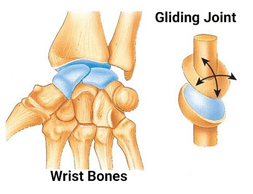
Gliding Joint
- Pivot Joint: Pivot joint is also known as trochoid joint. A joint where there is free movement only in a single axis. It only allows rotatory movement in one direction.

Pivot Joint
- Saddle Joint: The saddle joint is named so because the bone at one part of the structure is concave in shape, which means it’s inward, looking like a saddle. The other bone is convex, it is outward, looking like a rider on a saddle.

Saddle Joint
Additional Features Within Some Synovial Joints
[Click Here for Sample Questions]
Some of the additional features of Synovial Joints are:
- Presence of fibrocartilage structure between the articulating bones: The Articular disc generally which is small and oval-shaped, or a Meniscus, which is larger and C-shaped is seen. These structures serve many functions, depending upon the specific joint. Example of such a joint is the temporomandibular joint.
- Presence of intrinsic ligament which is fused to or incorporated into the wall of the articular capsule
- Presence of intracapsular ligament which is located inside of the articular capsule.
- Presence of Intra-capsular tendons eg. popliteus tendon within the knee joint.
- Presence of intra-articular tendons eg. Long head of biceps tendon within the shoulder joint
Read More:
Additional Features Surrounding Some Synovial Joints
[Click Here for Sample Questions]
Some of the additional features surrounding Synovial Joints are:
- Presence of bursae which is a small fluid-filled sac lined by a synovial membrane with an inner capillary layer of synovial fluid. It acts as a cushion between bones and tendons or in the muscles around a joint. It reduces friction between the bones and allows free movement.
- Presence of extracapsular ligaments allow normal movements at a joint, but with limited range of these motions, thereby preventing excessive/abnormal joint movements.
- Presence of tendon sesamoid bones which alter the direction of the tendon and modify pressure thereby reducing friction.
Things To Remember
- Synovial joints are the type of joints which have fluid filled synovial cavity between the articulating surface of two adjoining bones. These joints allow considerable movement in vertebrates.
- They help in locomotion and various other movements. It is the most usable and common Joints.
- The membrane lining the capsule lubricates the joint by producing oily liquid reducing friction and wear and tear. It is also known as the diarthrosis, meaning joining cartilage or bones with a fibrous joint.
- There are 3 kinds of joints- immovable, slightly movable and freely movable. Immovable joints do not allow any movement, slightly movable allow only movement till a certain limit and freely Movable joints allow movement to the highest degree.
- There is a presence of fat pads, which acts as a cushion between bones. There is a bursae, which helps in reducing the friction between two bones by secreting oily fluid.
Sample Questions
Ques. Discuss the joints involved and movements required for you to move your feet and hand. (2 marks)
Ans. The type of joint found in feet and hands is a gliding joint, also known as an arthrodial joint.
It is the type of joint where a structure is formed between two bones in which the articulating or the free surface of the bones are flat or nearly flat which enables bones to slide over each other.
Ques. What is the use of the synovial fluid? (2 marks)
Ans. The synovial joint along with its synovial fluids helps to connect bone with the help of tendons and cartilages. The membrane lining the capsule helps in lubricating the joint by producing oily liquid which helps in reducing friction and wear and tear. The fluid also acts as a cushion between bones for easily gliding of bone to each other.
Ques. Which joint has the greatest range of motion? Give examples. (2 marks)
Ans. Freely movable joints allow full movement or the highest degree of movement in the body. It is known as the diarthrosis joints. Examples of such joints are any joints of the elbow, the wrist, shoulder, and knee.
Ques. Which synovial joint allows you to turn your head side to side? (2 marks)
Ans. Pivot joint helps to move the head from side to side. It is a joint where there is free movement only in a single axis. It only allows rotatory movement in one direction. For example, between the atlas and the axis (first and second cervical vertebrae), directly under the skull, which allows for turning of the head from side to side.
Ques. What is bursae? What is its function? (2 marks)
Ans. Bursae is a small fluid filled sac lined by a synovial membrane with an inner capillary layer of synovial fluid.
- It provides a cushion between the bone and tendon or/and muscles around a joint.
- This reduces the friction between the bones and allows free movement.
Ques. Why is the saddle joint called so? (2 marks)
Ans. The saddle joint is named so because the bone at one part of the structure is concave in shape, which means it’s inward, looking like a saddle. The other bone is convex, it is outward, looking like a rider on a saddle. Due to this resemblance, it is known as a saddle joint.
Ques. What is the use of fibrous tissue? (2 marks)
Ans. Fibrous connective tissue are strong bands which strengthen and support the joint by anchoring bones together and preventing their separation. Ligament allows movement of joints but limits abnormal movement. Fibrous joints unites the adjacent bones and thereby provide protection to internal organs, strength to body regions, or weight-bearing stability.
Ques. How do you provide additional support to synovial joints? (2 marks)
Ans. Many tendons provide additional support to the synovial joints. Tendons are dense connective tissue structures attaching a muscle to bone. When force increases, the body automatically increases the overall strength of contraction of the muscles thereby allowing the muscle and its tendon to resist forces and support the joint. This type of indirect support by muscles is very important at the shoulder joint where the ligaments are weak for support.
Ques. Explain the mechanism of blood supply to Synovial Joints. (3 marks)
Ans. Following are the details:
- Synovial Joints receive a range of blood supply through a rich anastomosis of arteries extending from either side of the joint.
- The articular cartilage, which is also known as the avascular hyaline cartilage, is nourished by the synovial fluid.
- Lymphatic vessels for every joint follow the lymph drainage of the surrounding tissue. Some tissues have lymph nodes, for example, the popliteal lymph nodes in the popliteal fossa of the knee.
Ques. Explain the mechanism of nerve supply to synovial joints. (3 marks)
Ans. Following are the details:
- There is a presence of Sensory and autonomic fibres in synovial joints.
- The autonomic nerves are vasomotores in function. They control the dilation or constriction of blood vessels.
- The sensory nerves of the articular capsule and ligaments provide feedback from Ruffini endings and Pacinian corpuscles. The joints permit reflex control of posture, locomotion, and movement and the Free nerve endings convey pain sensation that appears to be diffuse and poorly localised.
- The articular cartilage has no nerve supply.
Read More:



Comments