



Content Writer
Centromere is defined as a structure in the chromosome that is responsible for the linking of two chromatids. It plays an essential role in chromosome segregation during the cell cycle process in eukaryotic cells. A centromere is either a constricted region in DNA or a structure in a chromosome.
- It ensures the equal division of genetic material between daughter cells during the cell division.
- Centromeres are characterised by the function of binding or holding together the sister chromatids in the chromosome.
- The chromatids are also the points which attach the spindle fibre.
- The spindle is responsible for pulling the chromatids to opposite ends of the cell.
- In the process, elements of the cell’s cytoskeleton assemble and attach to each other.
- It is the DNA region of the eukaryotic cell responsible for the sister chromatid's cohesion.
Read More: Difference Between Gene and DNA
Key Terms: Centromere, Chromosome, Daughter Cells, Mitosis, Chromatids, DNA, Cell Division, Eukaryotic, Cells, Adhension, Prophase
What is Centromere?
[Click Here for Sample Questions]
Centromere is defined as the specific region inside the DNA sequence in the chromosome which links the sister chromatids during the period of cell prophase. During the cell division, chromosomal regions are physically connected to the spindle.
- At the same time, centromeres ensure the equal division of genetic material between the daughter cells.
- Centromeres are not located exactly at the centre of the chromosome.
- They are located at the end of chromosomes.
- Centromeres separate chromosomes into two main sections: short arm (p) and long arm (q).
- The two copies of daughter cells remain stuck together until they are separated.
- Before continuing the process of reproduction, cells first make a complete copy of each of its chromosomes.
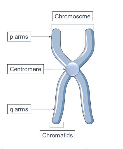
Centromere Diagram
Structure of Centromeres
[Click Here for Sample Questions]
There are some interesting facts about the structure of the centromere in a chromosome, which are mentioned below:
Kinetochore
Kinetochore is the primary element in the centromere. The centromere is the genetic locus which specifies the site of kinetochore assembly.
- It is responsible for the events of chromosome segregation.
- Kinetochore is a highly complex protein structure.
- Also, the chromosome is attached to the kinetochore microtubule.
- In metaphase, the pericentromeric, which is a physical region, is responsible for the geometry of the bi-oriented sister kinetochore.
Chromatids
The chromatids are referred to as one of the usually paired and parallel strands of a replicated chromosome, which are joined by a single centromere. It is required that DNA should be in a heterochromatin state for the attachment of sister chromatids mediated by cohesin complex as well as for the separation during anaphase.
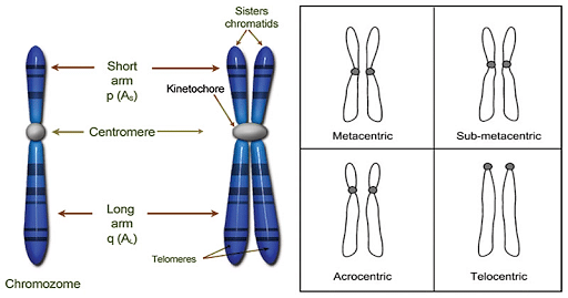
Structure of Centromere Diagram
Read More: Difference between Chromosome and Chromatid
Types of Centromeres
[Click Here for Sample Questions]
Centromeres are mainly classified into two main categories, which are discussed below:
Point Centromeres
Point centromeres are those centromeres that bind to specific proteins which recognize the particular DNA sequence very efficiently.
- In this, spindle fibres are attached to the specific DNA sequence.
- Moreover, the mitotic spindle fibre is possible when proteins are attached to the individual DNA sequence.
Regional Centromeres
Regional centromeres are those centromeres which are formed on regions of preferred DNA sequence. However, they are also capable of forming on other DNA sequences as well.
- Epigenetic signals are used to form regional centromeres.
- The regional centromeres are found in humans and most eukaryotic cells.
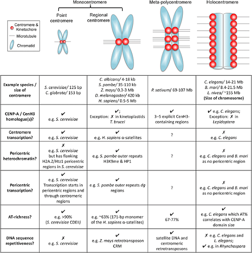
Types of Centromeres
Read More: Difference between DNA and Gene
Function of Centromeres
[Click Here for Sample Questions]
Centromeres perform many functions inside the chromosome, which are mentioned below:
- In eukaryotic cells, centromeres help in the proper alignment as well as the segregation of the chromosomes during the cell division.
- It acts as a binding site for the two duplicated chromosomes, which are known as sister chromatids.
- The structure of chromosomes acts as the signal processing centers that are responsible for the regulation of cell cycle progression.
- The centromeres are the genetic locus in the spindle fibre responsible for sister chromatids' attachment.
- It is the attachment point of the kinetochore.
- Other functions include adhesion and separation of sister chromatids and movement of chromosomes.
- It is also responsible for the attachment of microtubules, heterochromatin establishment, and control of mitotic checkpoint.
Read More:
Things to Remember
- The centromere is a constricted region within the chromosome or inside a DNA sequence.
- The equal division of genetic material between the daughter cells is ensured by the centromeres.
- The sister chromatids are bound together because of centromeres within the chromosome.
- Spindle fibres are attached through the kinetochore during mitosis.
- During the cell division, it links a pair of sister chromatids together.
- Each chromosome has two main positions or arms(short arm(p) and long arm(q)).
- Many organisms, such as humans and all eukaryotic cells, have regional structures.
Sample Questions
Ques. What do you mean by centromeres? [2 marks]
Ans.
Centromeres are defined as the constricted regions of chromosomes that connect the sister chromatids. Also, they are the special region which contains the specific DNA sequence.- They are also crucial for the production of new cells.
- Centromeres are usually a genetic locus which specifies the site of kinetochore.

Centromere DIagram
Ques. What is a chromosome? [2 marks]
Ans.
Chromosome is defined as the thread-like structure which is found inside the nucleus of a cell. They are composed of proteins and DNA organised into genes.- Each cell of the nucleus contains 23 pairs of chromosomes.
- Also, each chromosome has a constriction point known as a centromere.
- The main function of the centromere is to divide the chromosomes into two sections, i.e. arms.

Chromosomes Diagram
Ques. What are the kinds of centromeres on the basis of their position in the chromosome? [2 marks]
Ans. There are four main types of centromeres based on the position in the chromosome such as:
- Metacentric
- Submetacentric
- Acrocentric
- Telocentric

Types of Different Centromeres Diagram
Ques. Which part of the chromosome is critically responsible for even distribution of parental DNA during cell division ? [2 marks]
a) Spindle fibre
b) Centromere
c) Telomere
d) Centroils
Ans: b) Centromere.
Explanation for an answer: Centromere is responsible for the equal division of genetic material between the daughter cells in the chromosome. This is happening because centromere is the constricted region within the chromosomes which binds the sister chromatids together as well as attaches the chromosome to the spindle fibres during the metaphase of division.
Ques. Define eukaryotic cells? [2 marks]
Ans. Eukaryotic cells are those cells which have a defined nucleus. Also, these cells contain a nuclear membrane which surrounds the nucleus.
- The chromosome is located inside this membrane.
- Eukaryotic cells are capable of carrying out complex metabolic reactions.
- Moreover, the DNA is surrounded by the membrane which directs the synthesis of proteins as well as ribosomes.
Ques. Choose the appropriate option which does not relate to Telomere? [2 marks]
a) Maintenance
b) Chromosome degradation
c) Replication
d) Division
Ans: d) Division.
Explanation for an answer: Telomere is not related to the division. Telomeres usually refers to the sequences at the ends of eukaryotic chromosomes which plays an essential role in the maintenance and replication of DNA. Also, telomeres are helpful in chromosomal degradation which leads to ageing.
Ques. Centromeric DNA was initially defined in —----------- [2 marks]
a) Bacteria
b) Yeast
c) Fungi
d) Human
Ans: b) Yeast.
Explanation for an answer: Centromeric DNA was initially defined in the Yeast. Their functions were assessed by following the segregation of plasmids at mitosis. Also, the plasmids which consist functional centromeres segregate like chromosomes as well as equally distributed to the daughter cells following mitosis.
Ques. What do you understand from mitosis? Name its different phases. [3 marks]
Ans. Mitosis is defined as the process where a single cell divides into two identical daughter cells. In the cell cycle, replicated chromosomes are separated into two new nuclei.
- The primary function of mitosis is for growth and to replace the worn out cells.
- Moreover, the mitosis process is also known as equational division.
- The Mitosis process is very critical, that's why it is controlled by certain genes.
There are four main phases of mitosis processes which are mentioned below:
- Prophase
- Metaphase
- Anaphase
- Telophase
Ques. What are the functions of centromeres? [3 marks]
Ans. The functions of centromeres are as follows:
- The primary function of centromeres includes the attachment of daughter cells or sister chromatids.
- It is responsible for the segregation of chromosomes during the process of cell division.
- It is involved in the progression of the cell cycle as it acts as a signal processing centre.
- The functions of centromeres include the alignment of cells and microtubule attachment.
Ques. What are the side effects of centromeres on the human health? [3 marks]
Ans. The side effects of centromeres on the human health are as follows:
- The dysfunction of centromeres increases the instances of miscarriage among women.
- It will cause an increase in the number of cancer cells in the body, which are helpful for any individual.
- The growth of an individual will be hindered, and he may face genetic issues.
- Sometimes, an imbalance of centromeres may lead to death.
Ques. In how many categories centromeres is divided? [1 mark]
Ans. Centromeres are divided into two categories namely Point Centromeres and Regional Centromeres.
Ques. Explain the type of chromosomes based on the location of the centromere? [3 marks]
Ans. The type of chromosomes based on location of the centromere are as follows:
- Metacentric Chromosome: In this category, the centromere is placed exactly at the midpoint of the chromosome and looks like alphabet V.
- Sub-metacentric Chromosome: In this category, the centromere is placed some place near the midpoint of the chromosome and looks like alphabet L.
- Acrocentric: In this category, the centromere is placed at one end of the chromosome and looks like the alphabet J.
- Telocentric: In this category, the centromere is placed at the right end of the chromosome and looks like the alphabet i.
Ques. Explain the structure of centromere? [3 marks]
Ans. The structure of centromere is divided into two categories which are as follows:
- Kinetochore: It is the primary element in the structure of centromere. The centromere is responsible for specifying the site of the kinetochore. Kinetochore function includes the segregation of chromosomes within the body. It is a highly complex compound which is attached to the microtubule. During the centromere process, the kinetochore structure is explained in the metaphase.
- Chromatids: Chromatids are defined as one of the identical halves of chromosomes. They usually occur in pairs and are popularly known as homozygous. It may be the sister or non-sister form of chromatids. At the stage of cell division, chromatids are separated into individual chromosomes.
Check-Out:



Comments