



Content Curator
Duodenum is basically the first part of the small intestine which is about a 10-12 inch long hollow tube. The absorption of nutrients starts in the duodenum, the first of the small intestine's three sections that receive partly digested food from the stomach.
- It is directly connected to the stomach's pylorus.
- It is shaped like a C, has four sections—superior, descending, horizontal, and ascending—and is intimately associated with the head of the pancreas.
- The duodenum has the same three layers that other hollow gastrointestinal organs have.
- It also possesses Brunner's glands, which are unique to the duodenum.
Read More: Circulatory system
| Table of Content |
Key Terms: Duodenum, Intestine, GI tract, Stomach, Blood supply, Digestion.
What is Duodenum?
[Click Here for Sample Questions]
Duodenum is closely situated to the head of the pancreas and is fixed to the pylorus of the stomach (It is basically the first part of the small intestine, the pylorus is the valve that opens and closes during digestion). It is roughly horse-shaped. The duodenum receives the partially digested food and this is the starting part from where the absorption of nutrients happens.
- The duodenum has the same three layers that other hollow gastrointestinal organs have, but it also possesses Brunner's glands, which are unique to the duodenum.
- It has the Brunner's glands (Brunner's glands are located in the submucosa of the duodenum.
- They secrete an alkaline fluid containing mucin, which protects the mucosa from the acidic stomach contents entering the duodenum), which is the characteristic feature of the duodenum.
- The Brunner’s gland secretes mucus which contains various bicarbonates, it as a secretion helps to neutralize the acidic effect of the gastric juices.
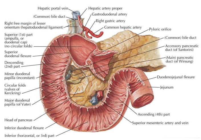
Duodenum
| Major facts about the Duodenum | |
|---|---|
| Location | 25-30 cm long, C-shaped around the head of the pancreas, and L1 – L3 level |
| Sections | Superior, Descending, Horizontal, and Ascending |
| Blood supply | Inferior pancreaticoduodenal arteries and Superior (anterior, posterior) |
| Innervation | Celiac plexus and vagus nerve |
| Histology |
|
| Functions | Neutralisation of the acidic gastric juice, mixing of bile and pancreatic enzymes, mechanical digestion of chyme, absorption of water, electrolytes, and nutrients |
Read More: Antibodies
Anatomy of The Duodenum
[Click Here for Previous Year Questions]
The Duodenum, which marks the starting of the small intestine, is a C-shaped tube which is 25-30 cm long and hollow. It is located in the upper abdomen part at the level of L1 – L3. It is lying adjacent to the stomach. There are various parts of the Duodenum which are discussed below:
- Superior Part of The Duodenum - It is the very first part and lies inside the peritoneum (the peritoneum is the tissue that lines the abdominal wall.
- It covers most of the organs in a person's abdomen) lies.
- This part is having a proximal enlarged part known as the duodenal bulb.
- The hepatoduodenal ligament is a substantial anatomical component that is encased in the peritoneum.
- It connects it to the liver and is a component of the smaller omentum.
- The first segment of the duodenum ends at the superior duodenal flexure.
- Descending Part of The Duodenum - It is also known as the second part of the duodenum.
- The Ampulla of Vater contains secretions from the common bile (It is a yellowish-brown fluid produced by the liver of most vertebrates.
- It helps in the digestion of lipids within the intestine) duct, and the hepatopancreatic duct empties in this part.
- The inferior duodenal flexure marks the transition from the descending part to something called the horizontal part of the duodenum.
- Horizontal Part of The Duodenum - It is the third part of the duodenum that runs ventrally from the abdominal aorta and the inferior vena cava (the inferior vena cava (IVC), the largest vein of the human body located at the posterior abdominal wall on the right side of the aorta.)
- Ascending Part of The Duodenum - This part runs cranially along the vertebral column and joins the jejunum (It is the middle part of the small intestine) at the duodenojejunal flexure.
- The descending portion is suspended against the abdominal wall by the Treitz ligament.
- It follows the Horizontal and is part of their progression.
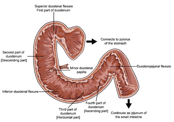
Anatomy of The Duodenum
Read More: Heart
Histology of Duodenum
[Click Here for Sample Questions]
If one talks about the histology of the duodenum, it is very similar to other hollow gastrointestinal tracts. Similar to the others, duodenum also has mucosa ,submucosa and the muscularis coverings.
- The mucosal layer is composed of lamina epithelial, lamina propria, and lamina muscularis.
- The submucosal layer which comes after this contains loose connective tissue (tissue that supports, connects, binds, or separates other tissues or organs), Meissner’s plexus, and the blood vessels.
- The muscularis layer comprises inner circular and outer longitudinal muscles with the Aurbach’s plexus (Auerbach plexus is one of two significant components of the enteric nervous system, it can be called a collection of interconnected neurons that spans from the esophagus to the rectum) lying in between them.
- The duodenum is rich in goblet cells, enterocytes, and endocrine cells. The microvilli contain the Crypts of Paneth cells and Lieberkuhn.
Read More: Symptoms of Heart Attack
Blood Supply and Innervation of Duodenum
[Click Here for Previous Year Questions]
The anterior (at the front) and posterior (at the back) superior pancreaticoduodenal arteries, and the inferior pancreaticoduodenal artery are responsible for the blood supply in Duodenum.
- The corresponding veins around it take care of the venous system of the duodenum.
- The nerves of the Coeliac plexus (the celiac plexus is part of the nervous system like a bundle of nerves in the upper abdomen that sits behind the pancreas which is close to the aorta, the body's largest blood vessel ) are responsible for the sympathetic innervation of the duodenum.
- The Vagus nerve carries out the parasympathetic innervation.
Read More: Heart Diseases
Functions of Duodenum
[Click Here for Sample Questions]
The duodenum is crucial for the process of digestion in the small intestine in many ways. The duodenum neutralizes the acidic gastric juice by producing alkaline (which are basic in nature) secrets. It is also responsible for the proper mixing of bile and the various pancreatic enzymes which helps to break down fats, proteins, and carbohydrates.
- Apart from these the main function of the duodenum includes absorption of nutrients and mechanical processing of chyme (partly digested food).
- The absorption of water, electrolytes, and other nutrients such as Vitamin A, fatty acids, Vitamin B1, and calcium takes place in the duodenum.
- The chyme gets broken down by the bodily fluids in the duodenum for easy absorption of nutrients after it passes through the Ampulla of Vater which stores the secretory juices produced by the hepatopancreatic duct and common bile duct.
Read More: Cancer
Inflammation of the Duodenum
[Click Here for Previous Year Questions]
Inflammation of the duodenum is known as duodenitis. It has various causes, one of the marked among them is the infection by the bacterium Helicobacter pylori.
- H. pylori increases the susceptibility of the duodenal mucosa to cause damage from unneutralized digestive acids and is a major cause of peptic ulcers which is the most common health problem affecting the duodenum.
- Other conditions that may be associated with duodenitis include celiac disease, Crohn's disease, and Whipple disease.
- Due to its proximity to the liver, pancreas, and major blood veins, the horizontal duodenum can get crushed by those organs in excessively thin individuals, necessitating surgical release to stop painful duodenal dilatation, nausea, and vomiting.
Read More: Tuberculosis
Things to Remember
- Duodenum is closely situated to the head of the pancreas and is fixed to the pylorus of the stomach.
- The duodenum receives the partially digested food and this is the starting part from where the absorption of nutrients happens.
- Duodenum has Brunner's glands, which is the characteristic feature of the duodenum.
- The Duodenum, which marks the starting of the small intestine, is a C-shaped tube that is 25 – 30 cm long and hollow.
- It is located in the upper abdomen part at the level of L1 – L3. It is lying adjacent to the stomach.
- Duodenum is divided into four parts, these are Superior Part, Descending Part, Horizontal Part, and Ascending Part.
- Duodenum Neutralisation of the acidic gastric juice, mixing of bile and pancreatic enzymes, mechanical digestion of chyme, absorption of water, electrolytes, and nutrients.
- Inflammation of the duodenum caused by the bacterium Helicobacter pylori, is known as duodenitis.
Previous Year Questions
- Electric flux at a point in an electric field is..
- The correct order of acid strength of the following carboxylic acids is..[Jee Advanced 2019]
- The measurement of voltmeter in the following circuit is...[AIIMS 2017]
- Angular velocity of minute hand of a clock is...[MP PMT 2004]
- Highly pure dilute solution of sodium in liquid ammonia...[Jee Advanced 1998]
- A gas mixture consists of 22 moles of oxygen and 44 moles of Argon at temperature T. Neglecting all vibrational modes, the total internal energy of the system is..[NEET UG 2017]
- Alkali halides do not show Frenkel defect because..
- Let R = {(1,3),(4,2),(2,4),(2,3),(3,1)} be a relation on the set A = {1,2,3,4}. The relation R is...[AIEEE 2004]
- Degree of freedom for polyatomic gas...[AIIMS 2012]
- In pyrrole, the electron density is maximum on...[NEET UG 2016]
Sample Questions
Ques: What is Duodenum in the digestive system? (2 Marks)
Ans: Duodenum is closely situated to the head of the pancreas, and is fixed to the pylorus of the stomach. It is roughly horse-shaped. The duodenum receives the partially digested food and this is the starting part from where the absorption of nutrients happens.
Ques: What is the Brunner's Glands? (2 Marks)
Ans: Brunner's glands (Brunner's glands are located in the submucosa of the duodenum. They secrete an alkaline fluid containing mucin, which protects the mucosa from the acidic stomach contents entering the duodenum), which is the characteristic feature of the duodenum. The Brunner’s gland secretes mucus which contains various bicarbonates, it as a secretion helps to neutralize the acidic effect of the gastric juices.
Ques: If we talk about anatomy, what is the Superior part of the Duodenum? (2 Marks)
Ans: It is the very first part and lies inside the peritoneum (The peritoneum is the tissue that lines the abdominal wall. It covers most of the organs in a person's abdomen) lies. This part is having a proximal enlarged part known as the duodenal bulb. The hepatoduodenal ligament (The hepatoduodenal ligament is a thick anatomical structure wrapped in the peritoneum. It constitutes part of the lesser omentum) connects it to the liver. The superior duodenal flexure marks the end of the first part of the duodenum.
Ques: What is the Ascending Part of The Duodenum? (2 Marks)
Ans: This part runs cranially along the vertebral column and joins the jejunum (It is the middle part of the small intestine) at the duodenojejunal flexure. The ligament of Treitz suspends the descending part against the abdominal wall. It is in the continuation with the Horizontal, as it comes after it.
Ques: What are the various layers/ coverings in the Duodenum? (3 Marks)
Ans: Similar to the others, the duodenum also has mucosa, submucosa, and muscularis coverings.
If we talk about the mucosal layer, it is composed of lamina epithelial, lamina propria, and lamina muscularis. The submucosal layer which comes after this contains loose connective tissue (tissue that supports, connects, binds, or separates other tissues or organs), Meissner’s plexus, and the blood vessels. The muscularis layer comprises inner circular and outer longitudinal muscles with the Aurbach’s plexus (Auerbach plexus is one of two significant components of the enteric nervous system, it can be called a collection of interconnected neurons that spans from the esophagus to the rectum) lying in between them. The duodenum is rich in goblet cells, enterocytes, and endocrine cells. The microvilli contain the Crypts of Paneth cells and Lieberkuhn.
Ques: What are the Various Functions of the Duodenum? (3 Marks)
Ans: The duodenum is crucial for the process of digestion in the small intestine in many ways. The duodenum neutralizes the acidic gastric juice by producing alkaline (which are basic in nature) secrets. It is also responsible for the proper mixing of bile and the various pancreatic enzymes which helps to break down fats, proteins, and carbohydrates. Apart from these the main function of the duodenum includes absorption of nutrients and mechanical processing of chyme (partly digested food). The absorption of water, electrolytes, and other nutrients such as Vitamin A, fatty acids, Vitamin B1, and calcium take place in the duodenum.
Ques: What is Inflammation of the Duodenum? (2 Marks)
Ans: Inflammation of the duodenum is known as duodenitis. It has various causes, one of the marked among them is the infection by the bacterium Helicobacter pylori. Other conditions that may be associated with duodenitis include celiac disease, Crohn's disease, and Whipple disease. The horizontal duodenum, because of its location between the liver, pancreas, and major blood vessels, can become compressed by those structures in people who are severely thin, requiring surgical release to eliminate painful duodenal dilatation, nausea, and vomiting.
Ques: Explain the Descending Part of The Duodenum. (2 Marks)
Ans: It is also known as the second part of the duodenum. The Ampulla of Vater contains secretions from the common bile (It is a yellowish-brown fluid produced by the liver of most vertebrates. It helps in the digestion of lipids within the intestine) duct, and the hepatopancreatic duct empties in this part. The inferior duodenal flexure marks the transition from the descending part to something called the horizontal part of the duodenum.
Ques: What is the Blood Supply and Innervation of Duodenum? (2 Marks)
Ans: The anterior (at the front) and posterior (at the back) superior pancreaticoduodenal arteries and the inferior pancreaticoduodenal artery are responsible for the blood supply in Duodenum. The corresponding veins around it take care of the venous system of the duodenum. The nerves of the Coeliac plexus are responsible for the sympathetic innervation of the duodenum. The Vagus nerve carries out the parasympathetic innervation.
For Latest Updates on Upcoming Board Exams, Click Here: https://t.me/class_10_12_board_updates
Check-Out:



Comments