



Content Writer
The ears in the human body are the most sensitive organs that perform vital functions, such as providing the ability to hear and maintaining body balance. It helps detect different kinds of sound and then transmit and transmit them.
- Ear can be bifurcated into three smaller sections, starting from the outer and middle ear and ending at the inner ear.
- The middle ear is divided into three parts namley tympanic cavity, the eustachian tube, and the ear ossicles.
- The ossicles are composed of three tiniest bones in the human body, namely the malleus, the incus and the stapes.
- These three bones are attached to each other, almost resembling a chain.
- The ear ossicles are used to transmit sound waves from the eardrum to the middle ear.
Key Terms: Human Ear, Ear Ossicles, Malleus, Incus, Stapes, Human Body, Auditory Ossicles, Functions of Ear Ossicles, Structure of Ear Ossicles
What are Ear Ossicles?
[Click Here for Sample Questions]
Ear ossicles are among the smallest bones in the human body. They are one of the three tiny articulating bones in the middle ear. The literal meaning of the term ossicle is tiny bone.
- Ear ossicles are also known as Auditory Ossicles.
- It forms a chain in the middle ear for conduction and amplification of sound waves from the tympanic membrane to the inner ear.
- This bone is divided into three parts: the malleus, the incus and the stapes.
- They were named after their resemblance to a hammer, anvil, and stirrup.
- Ear ossicles are used to connect the eardrum of the human body to the cochlea and inner ear.
Read More:
Structure of Ear Ossicles
[Click Here for Sample Questions]
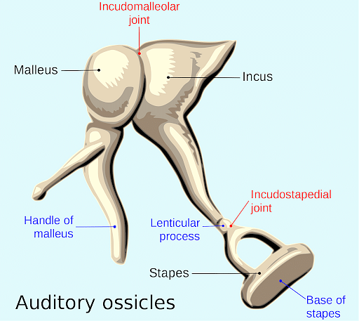
If we talk about the structure of ear ossicles, the outermost part is the malleus, followed by the incus and the stapes. The three parts are connected in a form that will allow the bones to move in response to the motion of the eardrum.
Malleus
The malleus is the first and largest of the ear ossicles. It is composed of a head, neck, handle called manubrium and two extensions, namely the anterior and lateral processes.
- On the anterior side, the oval-shaped head is connected to the incus through a small, saddle-shaped facet joint.
- The neck is responsible for connecting the head to the handle and forms the incudo-malleolar joint
- The lateral part is a cone-shaped projection that moves upward from the root of the handle.
Incus
Incus is the upper portion of ear ossicles, supported by the superior ligament running from the top of the ear canal to the bone. It is made of two projections, short and long limbs.
- The short limb is used to provide stability to the ear.
- The long limb is set at an angle of 90 degrees to form the lenticular process.
Stapes
The stapes, also known as stirrups, are the smallest ear ossicles bone, measuring 3.2 mm in height and 2.8 mm in width. They are divided into four parts: the head, base, anterior, and posterior limbs.
- The anterior and posterior limbs are used to give ossicles the stirrup shape.
- It is used for articulating the oval window of the inner ear.
Location of Ear Ossicles
[Click Here for Sample Questions]
When dissecting the three smallest bones of the ear, it is important to know their particular locations.
- The malleus in the human ear is attached to the tympanic membrane.
- Stapes, however, are situated in the oval window of the cochlea.
- The incus is located between and is also connected by delicate articulations.
Function of Ear Ossicles
[Click Here for Sample Questions]
The elementary function of the auditory system is to help transmit sound waves through a consistent chain reaction of vibrations. In a nutshell, the ear ossicles present in the human body help increase the transmission of every particular sound wave to the inner ear.
- In simple words, the auditory ossicles also protect the human ear when it is exposed to continuous loud noises.
- It intensifies the vibrations of sound level and passes them on to the inner ear
Working of Ear Ossicles
The working of ear ossicles are as follows:
- First, sound waves will enter the ear canal, which is channelled to the eardrum, causing it to vibrate.
- Next, the malleus of the ear ossicles is attached to the eardrum, and other ossicles will form a chain-like sequence.
- When the malleus starts vibrating, another part of the ossicles starts vibrating.
- This, in return, will send the signal to the inner ear.
- Now, after the vibrations reach the cochlea of the human ear, a chamber-like space filled with fluid, the brain processes them into several nerve impulses that are processed as sounds.
- The auditory chain reaction initiates when reaching the tympanic membrane (known as the eardrum).
- This results in vibrational pressure moving to the malleus and articulated bone situated at one of the two incudomalleolar joints.
- Later, the vibration smoothly passes on to the incus before finally transferring to the stapes.
- As a backdrop, the role of ear ossicles comes to an end after the vibrational input is transferred to the cochlea via the oval window.
- The motion of the stapes will produce waves, which are selected up by the receptor cells.
- This will convert mechanical information into electrical signals and transmit information to the brain via the vestibulocochlear nerve.
Also Read:
Things to Remember
- Ear ossicles, the tiniest bones in the body, consist of three bones: the malleus, incus, and stapes.
- It plays a pivotal role in helping achieve the capability of hearing.
- The eustachian tube helps connect the middle ear cavity with the pharynx.
- Human ear also helps in the maintenance of the body's balance.
- The malleus resembles a hammer, the incus resembles an anvil, and the stapes resemble a stirrup.
Sample Questions
Ques. What is meant by Ear Ossicles? (3 marks)
Ans. Ear ossicles can be dissected into three main parts, the malleus, incus and stapes, which are also the smallest bones of the human body. Ear ossicles work in a very particular way, helping amplify sound waves and transmitting them to the inner ear.
- The malleus is attached to the tympanic membrane (which is basically the eardrum).
- The stapes is usually attached to the oval window of the cochlea.
- Lastly, the incus is situated and connected by delicate articulations.
Second Answer
Ans. Ear ossicles have three tiny bones, the malleus, incus, and stapes, hold the title of the smallest bones in the human body. They play a critical role in hearing by amplifying sound waves and transmitting them to the inner ear.
- Malleus is the first ossicle that is directly attached to the eardrum (tympanic membrane), which vibrates when sound waves hit it.
- The incus acts as the middle runner, connected to both the malleus and the third ossicle, the stapes. It acts as a bridge, ensuring the vibrations are passed on efficiently.
- Finally, the stapes transmits the vibrations, causing ripples in the cochlear fluid.
Ques. What is eustachian tube? (2 marks)
Ans. The Eustachian Tube is a narrow passageway that helps in connecting the middle ear cavity with the pharynx. In general, the Eustachian Tube helps to equalise the pressure on every side of the eardrum.
Second Answer
Ans. The Eustachian tube is a narrow passageway that acts as a bridge, connecting the middle ear cavity to the back of your throat (pharynx).
Its main function is to maintain equal air pressure on both sides of your eardrum. This pressure balance is crucial for optimal hearing and the proper functioning of the eardrum.
Ques. Name the part of our body that helps in maintaining the body's balance? (2 marks)
Ans. Vestibular apparatus, which is situated in the internal ear, right above the cochlea, generally helps in maintaining the body's balance of a human. Widely speaking, the crista and macula are both sensory spots that are part of the vestibular apparatus that helps in controlling dynamic equilibrium.
Second Answer
Ans. The vestibular apparatus, located just above the cochlea in the internal ear, normally aids in preserving a person's body balance. Broadly speaking, the vestibular apparatus, which aids in maintaining dynamic equilibrium, includes the macula and crista as sensory locations.
Ques. Explain briefly the mechanism through which a sound is able to produce a nerve impulse in the inner ear? (2 marks)
Ans. Sound waves usually generate vibrations in the eardrum that are further processed to the ear ossicles by means of an oval window of the cochlea to the inner ear. In general, nerve impulses are produced and transmitted to the auditory cortex of the brain by different fibers.
Second Answer
Ans. The process of hearing begins with sound waves entering your outer ear. These waves travel down the ear canal and cause the eardrum (tympanic membrane) to vibrate. The vibrations are then passed on to a chain of tiny bones in the middle ear called the ossicles. Finally, the incus passes the vibrations to the stapes, the smallest bone in the human body
Ques. Name the part of the ear that helps in determining the pitch of a sound? (1 mark)
Ans. The cochlea of the ear is generally responsible for determining the pitch of a sound. This leads to equalising the pressure on every side of the eardrum.
Second Answer
Ans. The cochlea part of the ear helps in determining the pitch of a sound. This leads to equalising the pressure on every side of the eardrum.
Ques. What are the general functions of ear ossicles? (2 marks)
Ans. The main function of auditory ossicles (also widely known as the ossicular chain) is to help transmit sound waves through a vibratory chain reaction occurring to connect the eardrum to the cochlea and inner ear. The ear ossicles usually help in increasing the regulation of transmission of every sound wave to the inner ear.
Second Answer
Ans. Auditory ossicles, sometimes referred to as the ossicular chain, are primarily responsible for transmitting sound waves via a vibratory chain reaction that connects the eardrum to the cochlea and inner ear. Generally speaking, the ear ossicles aid in improving the regulation of each sound wave's passage to the inner ear.
Ques. In which part do the ear ossicles are located? (1 mark)
Ans. The ear ossicles, also called auditory ossicles, are found to be in the middle ear. They are a composition of the tiniest bones present in the human body.
Second Answer
Ans. Dissecting the insides of the middle ear, the ear ossicles are the three tiniest bones in the human body, named as, the malleus, the incus and the stapes. These three bones are attached to each other, almost resembling a chain.
Ques. Name the parts of the ear ossicles and define their locations briefly? (2 marks)
Ans.The three main parts of the ear ossicles are the malleus, incus, and stapes which are also the smallest bones of the human body. The malleus is attached to the tympanic membrane (which is the eardrum), and the stapes is found attached to the oval window of the cochlea. The incus, moreover, is situated and connected by delicate articulations.
Second Answer
Ans. The tiniest bones in the human body, the malleus, incus, and stapes, are the three primary components of the ear ossicles. The stapes is connected to the oval window of the cochlea, while the malleus is joined to the tympanic membrane, or the eardrum. Furthermore, the incus is positioned and joined by fine articulations.
Ques. What are structural malformations of ear ossicles? (4 marks)
Ans. The structural malformations of ear ossicles are as follows:
- Lack of stapes: When the stapes are absent then hearing of an individual is impaired.
- Lack of the long limb: When limbs which is connecting to the lenticular process are absent, then it will lead to significant hearing loss
- Stapes Aplasia: Stapes aplasia are conditions in which the stape is not properly formed and the base is not connected to the rest of the bone of the ear, which leads to hearing loss.
- Stapes Hyperplasia: Stapes hyperplasia is a form of disability that will cause deafness in an individual.
Second Answer
Ans. The tiny ear ossicles are vital for hearing, but sometimes these bones can develop abnormally. These malformations can lead to varying degrees of hearing loss. Here are some common types:
- Missing Stapes (Stapes Aplasia): In this condition, the stapes bone is entirely absent from birth. This malformation significantly impacts hearing.
- Incomplete Stapes (Stapes Dysplasia): The stapes bone may be malformed, with an abnormally shaped base that doesn't connect properly to the rest of the ossicular chain. This can also cause hearing loss.
- Missing Long Incus Leg: The incus bone has a long leg that connects to another ossicle. If this long leg is absent, the transmission of sound waves is disrupted, leading to hearing loss.
Ques. Which muscles are integrated with ear ossicles? (3 marks)
Ans. Two muscles are integrated with ear ossicles, which are as follows:
- Tensor Tympani: First is the tensor tympani, which gets attached to the malleus. It is used to reduce the vibrations from the tympanic membrane. This muscle will protect the ear from loud and damaging sounds.
- Stapedius: The second is the stapedius muscle, which is located in the posterior portion of the tympanic membrane. It attaches to the stapes and moves them posteriorly. It reduces the ear's oscillatory range.
Second Answer
Ans. Two muscles are integrated with ear ossicles, which are as follows:
- Tensor Tympani: This muscle acts like a volume control knob. Attached to the malleus (the first ossicle), it contracts to dampen vibrations from the eardrum. This helps protect the inner ear from loud or potentially damaging sounds.
- Stapedius Muscle: Located near the stapes (the smallest ossicle), this muscle plays a similar role. When it contracts, it pulls the stapes bone slightly backward, reducing the movement of the entire ossicular chain. This, in turn, reduces the intensity of vibrations transmitted to the inner ear.
Ques. What is Otosclerosis? (2 marks)
Ans. Otosclerosis is a type of abnormal growth of bone that starts in the bony labyrinth. The oval window will move toward the auditory ossicles, which will lead to conductive hearing loss. The hardening of muscle will result in loss of flexibility of the ear. This will ultimately hinder the stapes footplate from oscillating the oval window.
Second Answer
Ans. Otosclerosis is a condition characterized by abnormal bone growth in the middle ear. This growth typically starts in the bony labyrinth, a chamber surrounding the inner ear. It can cause mild to severe hearing loss depending on the extent of bone growth. Treatment options are available, including medication and surgery, to improve hearing in individuals with otosclerosis.
Do Check Out:
| Class 11 Biology Study Guides | ||
|---|---|---|
| Biology MCQs | Biology Study Notes | Class 11 Notes |
| Comparison Topics in Biology | NCERT Solutions for Class 11 Biology | NCERT Class 11 Biology Book |



Comments