



Education Content Expert
Cochlea is a hollow, spiral-shaped bone present in the inner ear, which enables the hearing process. It is a spiral tube that is wrapped around the modiolus, a hollow central pillar. It is stuffed with endolymph and receives vibrations from the middle ear, which causes the millions of hair cells inside the ear to feel motion and move.
- Cochlea is a long and coil outgrowth of the sacculus.
- It is the main hearing organ in mammals.
- It consists of three membranes.
- The organ of Corti (a hearing organ) is located on the basilar membrane that contains hair cells.
The word “cochlea” is derived from the Greek word “Kokhlias” which means spiral or snail shell.
Read More: NCERT Solutions for Class 11 Biology Neural Control and Coordination
Key Terms: Cochlea, Human Ear, Inner Ear, Endolymph, Hearing, Sacculus, Utricle, Sound Waves, Scala Tympani
What is Cochlea?
[Click Here for Sample Questions]
Cochlea is a spiral-shaped cavity in the inner ear. It is a hollow bone that plays a crucial role in the transmission of auditory signals. The modiolus, a central hollow axis of the cochlea, houses the cochlear arteries and veins as well as cochlear nerve fibers. Two apertures, the oval window, and the round window connect the cochlea to the middle ear. Two membranes and three fluid-filled channels make up the cochlea itself.
Inner Ear of Mammals are present in the baby labyrinth and consists of
- Semicircular Canals
- Saccule
- Utricle
- Cochlea
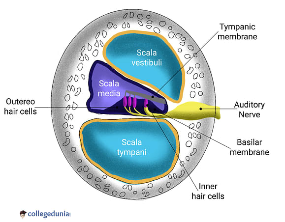
Cochlea
Read More:
| Relevant Concepts | ||
|---|---|---|
| Structure of Human Ear | Ear Ossicles | Sense Organs |
| Nerve Impulse | Human Eye | Neuroscience |
Structure of Cochlea
[Click Here for Previous Year Questions]
The structure of Cochlea is explained as follows:
- The spiral, the conical, hollow cochlea is where sound waves travel from the ear's base to its tip.
- The inner ear's bone labyrinth contains the cochlea's spiral canal, which is roughly 30 mm long and makes around 234 turns around the modiolus.
- Cochlea consists of three canals namely the top vestibular canal, also known as the scala vestibule, the middle cochlear duct, also known as the scala media, and the lower tympanic canal, also known as the scala tympani.
- The oval window is touched by the Scala Vestibuli, which are located above the cochlear duct and contain perilymph.
- Perilymph is also present in the Scala Tympani, where it is located inferior to the cochlear duct and ends at the round window.
- Endolymph is present in the Scala Media, where the hair cells extend into an area with a high potassium ion concentration.
- Helicotrema is the location where the vestibular duct and tympanic duct converge. It can be found at the cochlea's apex.

Structure of Cochlea
- Reissner's membrane separates the Scala media and the scala vestibuli.
- The osseous Spiral lamina is the main structural element of the cochlea that separates the tympanic and cochlear ducts.
- The auditory organ, also known as the organ of Corti, is located in the scala media and is filled with endolymph.
- Corti has about 18000 hair cells per organ. Scala media and scala tympani are divided by the basilar membrane, which contains hair cells.
- The cochlear duct is reached by stereocilia that extend from the hair cells.
- Above hair cells lies a different membrane called the tectorial membrane.
- The hair cells in the cochlea sense pressure waves and sensory receptors (afferent nerves) at their bases transmit information to the brain.
Read More: A Well-Labeled Diagram of Ear With Explanation
Functions of Cochlea
[Click Here for Sample Questions]
Cochlea contains the auditory receptors. The functioning of the cochlea can be expressed as follows:
- The endolymph starts to vibrate as vibrations travel from the middle ear through the oval window to the inner ear.
- The sound vibrations convey pressure waves to the endolymph, which causes the basilar membrane to move.
- The Organ of the Corti moves as a result of this vibration, and the hair cells detect this movement.
- The afferent neuron located at the base, near the hair cells, is stimulated by the bending of the hair cells against the tectorial membrane, causing nerve impulses to be produced.
- After being transformed into action potentials by the auditory neurons, the signals are subsequently sent to the brain for additional processing.
The region of the basilar membrane that is triggered determines the pitch of the sound, which is another ability the human ear has.
- Cochlear hair cells located at the base and top of the cochlea may distinguish between high and low frequencies.
- The quantity of impulses sent by the cochlear nerve allows one to determine how loud something is.
Cochlea has a special ability that allows it to amplify very faint sounds. The OHCs use reverse transduction to augment weak sounds. The propagation of sound waves is also coupled by a protein known as progestin.
Cochlea in Other Animals
[Click Here for Previous Year Questions]
Mammal Cochleas are determined to be distinct from those of other animals.
- It can occasionally be detected as a cochlear duct, a blind-ended tube.
- The disparity between mammals' and other creatures' hearing frequency ranges is what causes this distinction to be observed.
- A mammal has unique hearing frequencies because the hair cell present in the mammal’s ear pre-amplify the sound.
- Bats and Marine mammals like dolphins can hear frequencies up to 100 kHz, whereas birds can hear frequencies not more than 4 - 5 kHz.
- For humans, the common hearing frequency range is 20 Hz to 20 kHz.
The reason for the high hearing frequencies of mammals is the presence of long and coiled cochlea which help in increasing the hearing range of mammals.
Check More:
Things to Remember
- Cochlea is a conical, hollow, spiral tube present in the human inner ear.
- Cochlea consists of three canals namely the scala vestibule, the scala media, and the scala tympani.
- The scala vestibule and scala media contain the perilymph while the scala tympani contain the endolymph.
- The cochlear duct and vestibular duct are separated by a membrane known as Reissner’s membrane.
- The auditory organ, also known as the organ of Corti, is located in the scala media and is filled with endolymph.
- Stereocilia, a hair-like structure is present in the sensory cells of the organ of Corti.
- Cochlea receives sound waves and enables us in hearing using the auditory organ.
- The hair cells convert the vibrations to electrochemical impulses which are sent to the auditory nerve and then transmitted to the brain for further processing.
For Latest Updates on Upcoming Board Exams, Click Here: https://t.me/class_10_12_board_updates
Previous Years’ Questions
- Cochlea of mammalian internal ear is concerned with… (WBJEE - 2007)
- The vestibular and tympanic canal of human cochlea are connected by… (Himachal Pradesh PMT - 2007)
- The part of internal ear responsible for hearing is…
- Glenoid cavity is found in… (DUET - 2007)
- Organ of Corti is located on the ________… (MHT CET - 2017)
- The organ of Corti is a structure present in…
- The organ of corti is situated on the… (AMUEEE - 2013)
- Vestibular apparatus helps us in maintaining balance of body…
- Internal ear is filled with…
- From outer to inside the sequence of three bones present in the middle ear… (JIPMER - 2008)
Sample Questions
Ques. Define Cochlea. (1 Mark)
Ans. Cochlea is a spiral-shaped, hollow bone that is a component of the inner ear. It takes part in auditory transduction as well as hearing.
Ques. What is the main function of Cochlea? (1 Mark)
Ans. Cochlea transforms auditory data into neural impulses, which are then transported to the brain via afferent nerve fibers and auditory nerves, where they are combined and translated into sound.
Ques. Apart from hearing, what are the other functions of the cochlea? (3 Marks)
Ans. Apart from hearing, the other functions of the cochlea are as follows:
- Cochlea can determine the pitch of the sound.
- Cochlear hair cells located at the base and top of the cochlea may distinguish between high and low frequencies.
- The quantity of impulses sent by the cochlear nerve allows one to determine how loud something is.
- Cochlea also can amplify very faint sounds.
- By reversing the electrical to mechanical signal conversion, they trigger a positive feedback mechanism.
- The propagation of sound waves is also coupled by a protein known as progestin.
Ques. How does the cochlea aid in hearing? (3 Marks)
Ans. Cochlea contains auditory receptors.
- The endolymph starts vibrating as vibrations travel from the middle ear to the inner ear through the oval window.
- These sound vibrations transport pressure waves to the endolymph, which results in the movement of the basilar membrane.
- As a result of these vibrations, the organ of the Coti moves and the hair cells detect this movement.
- The bending of the hair cells against the tectorial membrane stimulates the afferent neuron next to the hair cells, resulting in the production of nerve impulses.
- The auditory neurons transform the signals into action potentials, which are then sent to the brain for further processing.
Ques. Describe the anatomical features of the cochlea. (5 Marks)
Ans. Cochlea is a spiral tube that is wrapped around the modiolus, a hollow central pillar. It shapes a cone that is 5 mm tall with a base that is 9 mm (0.35 inch) in diameter. The inner ear's bone labyrinth contains the cochlea's spiral canal, which is roughly 30 mm long and makes around 234 turns around the modiolus.
The cochlea consists of three canals:
- The top vestibular canal, also known as the scala vestibule.
- The middle cochlear duct, also known as the scala media.
- The lower tympanic canal, also known as the scala tympani.
Functions of Cochlea
- The cochlea receives sound waves coming from the source. Also, its structure is designed in such a manner that it can amplify very faint sounds. The OHCs use reverse transduction to augment weak sounds.
- The propagation of sound waves is also coupled by a protein known as progestin.
Ques. Cochlea arises from
a. Sacculus
b. Utriculus
c. Semicircular canals
d. Middle ear (2 Marks)
Ans. (a) Sacculus
Explanation: Sacculus is a chamber in the inner ear from which the cochlea arises in mammals, birds, and reptiles. Sacculus supports patches of sensory epithelium perturbed with balance. It is also known as saccule.
Ques. Name three scalae of the cochlea with their features. (3 Marks)
Ans. The cochlea has three scalae or chambers. These are
- Scala Vestibuli
- Scala Tympani
- Scala Media
The scala vestibuli contains perilymph. It lies above the cochlea duct. The scala tympani also contains perilymph and it lies below the scala media and ends at the round window. The scala media contains endolymph also known as a cochlear duct. Scala media is a region rich in potassium ion concentration. In this region, the stereocilia of hair cell lie.
Ques. Why do mammals have unique hearing frequencies than other animals? (2 Marks)
Ans. Mammals have unique hearing frequencies because the hair cells present in their ears pre-amplify sound. The presence of a long, coiled cochlea rather than a small, straight one aids in extending the hearing range of mammals.
Ques. Why the design of the cochlea is coiled or spiral? (2 Marks)
Ans. The spiral cochlea aids in detecting low-frequency sound. Based on the calculations done by researchers, it is concluded that spiral or coiled shapes enhance the sensitivity of hearing. Because of the differences between the inner and outer edges of the spiral, the cochlea cells can easily respond to the vibration received.
Ques. Name all the parts of the cochlea. (3 Marks)
Ans. A fluid is present in the cochlea known as perilymph. The human cochlea has three scalae (chambers) as follows:
- Vestibular Duct
- Cochlear Duct
- Tympanic Duct
Cochlea also has three fluid sections among which two are the canals, and one is an organ of Coti. These canals are:
- Vestibular Canal
- Tympanic Canal
Check More:



Comments