



Content Curator
The hyoid bone is a small U-shaped or horseshoe-shaped bone that is found in the middle of the neck, at the mandible base anteriorly, and posteriorly at the fourth cervical vertebrae. It is 2.5 cm wide and 1 cm thick along with greater and lesser horns.
- The hyoid bone is the only bone in the body that is not connected to any other bone.
- It is only connected to other bones with the help of muscles or ligaments.
- The primary function of the hyoid bone is to support the tongue and muscles of the floor of the oral cavity.
- It helps in tongue movement, swallowing, and speech.
- The blood supply to the hyoid bone is via the lingual artery.
The suprahyoid branch of the lingual artery runs along the upper border of the hyoid bone and supplies the blood to the muscles that are attached.
| Table of Content |
Key Terms: Greater Cornua, Lesser Cornua, Pharyngeal Arch, Ossification, Hyoid Bone, Anterior Neck, Epiglottis
Hyoid Bone Anatomy
[Click Here for Sample Questions]
The hyoid bone is an irregular horseshoe-shaped bone. The hyoid bone location is the anterior neck region at the base of the mandible.
There is a bursa behind the hyoid bone, separating it from the epiglottis. While swallowing, the hyoid bone, tongue, and larynx move in an upward direction rapidly. Hyoid bone comprises of:
- Body: The body is the central part of the hyoid bone that provides origin and insertion to various muscles. It is irregular in shape and laterally stretched. The body is responsible for the U-shape of the hyoid bone. The anterior end of the body faces upward and the posterior end faces in a downward direction.
- Pair of larger horns called greater cornua: They project from each end of the body in the superior and lateral direction. Greater cornua are wide, narrow, and flat horizontally. They end in the tubercles. They are the site of attachment of various muscles.
- Pair smaller horns called lesser cornua: They project from the superior aspect of the body and near the origin of the greater cornua. They are conical in shape. Its apex provides attachment to the stylohyoid ligament.
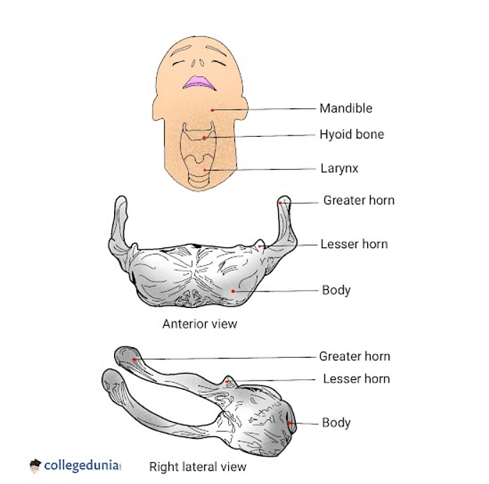
Hyoid Bone
Development of Hyoid Bone
[Click Here for Previous Year Questions]
The ways by which Hyoid Bone develops are:
- Second Pharyngeal Arch or the Hyoid arch: Gives origin to the lesser cornua of the hyoid bone and the upper part of the body of the hyoid bone
- The third pharyngeal arch: The cartilage of the third pharyngeal arch gives origin to the greater cornua of the hyoid bone and lower portion of the hyoid.
- Ossification: The hyoid bone ossification occurs from 6 centres in the body (two for the body and one for each cornua). The ossification starts in the greater cornua by the end of fetal development and in the body later. Ossification starts in lesser cornua in the first and second year after birth. The connection between the body and greater cornua is fibrous until the middle ages. The outer body of the body is connected to the greater horn by synchondroses and after middle life by bony union.
Read More:
| Topic Related Concepts | ||
|---|---|---|
| Deltoid Muscles | Ellipsoid Joints | Types of Movement |
| Bones of Legs | Saddle Joints | Humerus |
Hyoid Bone Attachments
[Click Here for Sample Questions]
The hyoid bone is itself not connected to any bone; however, it acts as a pivot to other bones through muscles and ligaments.
- Oral cavity and pharynx: In the oral cavity and pharynx, the following muscles are connected:
- Middle pharyngeal constrictor.
- Hyoglossus
- Genioglossus
- Suprahyoid: The following muscles are attached to the hyoid bone on the superior surface.
- Digastric muscle.
- Stylohyoid muscle.
- Geniohyiod muscle.
- Mylohyoid muscle.
- Infrahyoid: The following muscles are attached to the hyoid bone on the inferior surface.
- Thyrohyoid muscle.
- Omohyoid muscle.
- Sternohyoid muscle.
- Ligament attachments: There are 3 main ligaments that are attached to the hyoid bone.
- Stylohyoid ligament: It is attached from the styloid process of the temporal bone to the smaller horns of the bone.
- Thyrohyoid membrane: The membrane starts from the higher line of the thyroid cartilage and attaches itself to the posterior surface of the bone and to the horns.
- Hyoepiglottic ligament: It connects the hyoid bone to the anterior end of the epiglottis.
Hyoid Apparatus
Hyoid apparatus is the term used for the hyoid bone and the ligaments and muscles connected to it. The apparatus consists of stylohyoid, thyrohyoid, ceratohyoid bones, and a single basihyoid bone. The shape of the apparatus is in the form of an “H” or a trapeze.
Hyoid Bone Function
[Click Here for Previous Year Questions]
The functions of the hyoid bone are:
- It acts as an attachment point for the larynx and serves the task of holding it up.
- The hyoid bone provides a base and support for tongue movement.
- The hyoid bone helps in respiration by keeping the airway open.
- It helps in preventing sleeping disorders by keeping the upper airway open.
- The hyoid bone transmits the muscular forces to open the jaw.
- It helps in swallowing.
- The hyoid bone helps in speech articulation.
Also Read:
Things to Remember
- The hyoid bone is a U-shaped bone located at the anterior neck region at the base of the mandible.
- It consists of a body, greater cornua, and lesser cornua.
- Hyoid apparatus is the term used for the hyoid bone and the ligaments and muscles connected to it.
- The hyoid bone helps in speech articulation, swallowing, respiration, and preventing sleeping disorders.
Previous Year's Questions
- Blood pressure in the mammalian aorta is maximum during...[NEET 2015]
- Blood capillaries are made of...[NEET 1993]
- Blood group AB has...[NEET 1991]
- An adult human with average health has systolic and diastolic pressures as… [NEET 1998]
- Blood pressure in the pulmonary artery is...[NEET 2016]
- Which of the following is made up of a…[AIIMS 1994]
- Smooth muscles…[NEET 2016]
- The contractile protein of skeletal muscle…[NEET 2016]
- The number of floating ribs…[NEET 1995]
Sample Questions
Ques. How can you distinguish hyoid bone from other bones of the axial skeleton? (1 mark)
Ans. The hyoid bone is suspended by suprahyoid and infrahyoid muscles. It does not articulate with any other bone in the skeleton. It is “H” shaped and consists of smaller conical lesser cornua and larger greater cornua and a body.
Ques. Which organs are joined to the hyoid bone? (1 mark)
Ans. The hyoid bone is situated at the root of the tongue, at the anterior aspect of the neck, and between the lower jaw and larynx. The primary function of the hyoid bone is to serve as an attachment structure for the tongue and for muscles in the floor of the oral cavity.
Ques. Can posture affect the hyoid bone? (1 mark)
Ans. The high neck muscle tone due to inappropriate posture may encourage hyoid depression and increase the maximum elevated position of the hyoid bone, henceforth increasing the risk of aspiration.
Ques. What can contribute to hyoid bone pain? (1 mark)
Ans. There are small muscles that control the hyoid bone or Adam’s apple. They can develop trigger points and become overactive due to changes in jaw or neck motion. Injury to the upper neck with pain and loss of mobility can cause changes to the bone as seen in imaging.
Ques. Can hyoid bone cause breathing difficulties? (1 mark)
Ans. The hyoid bone is not anchored firmly to its position but is rather free to move around, this may allow the area to collapse and cause airway blockage during sleep.
Ques. What is a hyoid bone syndrome? (2 marks)
Ans. The hyoid bone syndrome is a type of cervicofacial pain caused by degeneration of the greater cornua of the hyoid at the attachment of the stylohyoid ligament. In the case of hyoid bone syndrome, the symptoms include pain on swallowing, that might radiate to the ear, face, and lower jaw or in the pharynx. The syndrome can be treated with oral NSAIDs or an NSAID ointment.
Ques. What is obstructive sleep apnea (OSA) and how can hyoid bone contribute to it? (2 marks)
Ans. The common cause of obstructive sleep apnea is obesity, which is associated with soft tissues of the mouth and throat. During sleep throat and tongue muscles are more relaxed which can cause the airway to become blocked. The OSA can lead to serious health issues like hypertension and heart issues if left untreated. It causes loud snoring, tiredness, etc.
Ques. What is hyoid apparatus and what comprises it? (2 marks)
Ans. Hyoid apparatus is the term used for the hyoid bone and the ligaments and muscles connected to it. It consists of 5 different bones that vary in length and size depending on the species. The 5 bones are: paired stylohyoid, epihyoid, ceratohyoid, thyrohyoid, and basihyoid bones. The hyoid apparatus is responsible for holding the larynx in place and supporting the pharynx and tongue from the skull.
Ques. What is the clinical significance of hyoid bone? (3 marks)
Ans. The hyoid bone is crucial as it assists in the following functions:
- Breathing.
- Swallowing.
- Speech.
- In the development and treatment of obstructive sleep apnea.
- Movement of the hyoid bone is important in modifying upper airway properties.
Ques. What are the types of hyoid bone? (3 marks)
Ans. There are six types of hyoid bones:
- U-shaped: 25%.
- H-shaped: 3%.
- B-shaped: 48%.
- D-shaped: 14%.
- V-shaped: 2%.
- HK-shaped: 8%.
Ques. What happens if you fracture your hyoid bone? (5 marks)
Ans. The hyoid bone is crucial for swallowing, speech, and respiration.
- Although hyoid bone is not easily susceptible to fracture; however, it might fracture due to direct trauma, strangulation, or blunt trauma.
- There are two types of complications as a result of fracture that is early and late complications.
- Early complications are subcutaneous emphysema, dyspnoea, pharyngeal tears, and thyroid cartilage injury.
- Late complications include dysphagia, stridor, pseudoaneurysm of the external carotid artery, and crepitus by neck flexion.
- All the patients with hyoid bone fractures survived and had excellent outcomes. Both conservative and surgical management yielded positive outcomes.
Read More:



Comments