



Content Curator
Sartorius Muscle is the longest muscle in the human body that belongs to the anterior muscles located in the thigh. They are connected together with the quadriceps femoris.
- Sartorius muscle location arises from the anterior superior lilac spine then crosses the medial side and finally inserts into the proximal tibia of the medial side.
- The Sartorius's muscle is also called the tailor’s muscle.
This muscle is known to have an oblique course throughout the entire thigh because of which this muscle flexes, abducts, and externally rotates the thigh and internally it helps to rotate the leg.
Read More: Largest Organ in Human Body
Key Terms: Oblique, Muscle, Sartorius, Thigh, Tibia, Hamstrings, Apex, Joints and Tendon
Sartorius Muscle
[Click Here for Sample Questions]
The sartorius muscle is a superficial, long, thin muscle located in the anterior thigh compartment.
- Sartorius Muscle is the longest muscle in the human body as it spans both the hip and knee joints and runs the entire length of the thigh.
- This muscle can reach lengths of more than 50 cm.
- Not all muscle fibres in such long muscles extend the entire length of the muscle belly.
- The muscular belly curves around the medial and anterior surfaces of the thigh like an S-shaped piece of tape.
- A tendon is where it ends after passing behind the medial condyle of the femur.
Also Check: Difference between Serum and Plasma
Sartorius Muscle Diagram
[Click Here for Previous Year Questions]
The Sartorius muscle runs across the upper and anterior part of the thigh slantly.
- It passes behind the medial of the femur towards the end of a tendon.
- Then it curves to join the semitendinosus and gracilis muscles, which attach at the upper surface of the tibia.
- The upper section of this muscle forms the lateral border of the female triangle and also crosses the apex of the triangle.
- The saphenous nerve, femoral artery and vein, and the nerve to vastus medialis pass through the depth of the Sartorius muscle.
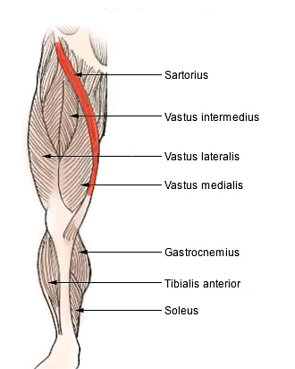
Sartorius Muscle Diagram
Also Read: Bones of Legs
Sartorius Origin and Insertion
[Click Here for Sample Questions]
The origin of Sartorius muscle and the sartorius insertion follows the procedure below:
- The sartorius muscle develops from tendinous fibres that originate from the anterior superior iliac spine and travel inferomedially across the top and anterior portion of the thigh.
- It descends behind the medial femoral condyle to the medial side of the knee, where it ends in a tendon. It performs an internal rotation and flexion action at the knee.
- The gracilis and semitendinosus connect it to two other tendons at its insertion, resulting in the pes anserinus, a group of connected tendons. This tendon turns anteriorly before insertion into the proximal section of the tibia on its medial surface.
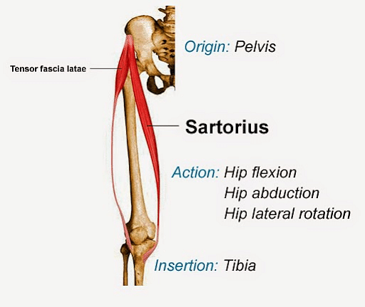
Sartorius Muscle Origin and Insertion
Also Read:
Sartorius Muscle Function
[Click Here for Previous Year Questions]
Sartorius muscle acts both as hip and knee joints, by producing movement in both of the parts.
Sartorius Muscle is very long and it crosses two joints (the knee and the hip), it has many functions in the lower extremities of the human body. The movement combination allows one to sit in a cross-legged position.
The movement which happens due to the Sartorius are:
- Flexion of Hip: While marching, this muscle bends our hips along with other muscles such as the psoas muscle that helps to flex the hip up.
- Rotation (external) of Hip: The portion of Sartorius towards the front of the thigh has a line of pull which helps in the rotation of the hip outwards. While in a standing position and lifting the foot up and looking at the bottom of the shoe, the Sartorius muscle will be active and will rotate the hip externally.
- Flexion of Knee: The Sartorius which is situated towards the inner side of the knee and attaches with the front of the shin bone contracts and bends our knees. Sartorius is most active while bending the knees while the hip is flexed and rotated and the hamstrings also bend.
Also Check-out: Spinal Cord: Diagram, Anatomy, Structure and Function
Sartorius Muscle Blood Supply
[Click Here for Sample Questions]
Long Muscles like the Sartorius's muscle require extensive vascular supply from several sources:
- The vascular supply from the branch femoral, deep femoral branches, lateral femoral arteries and quadriceps artery that is received from the third proximal
- The femoral artery branches provide the middle third of the Sartorius muscle
- Blood supply is received to the femoral artery and the descending genicular artery due to the third of the distal.
Sartorius Muscle Conditions
[Click Here for Previous Year Questions]
Several conditions are involved with the functional mobility of the human body which include:
- Pes anserine Tendonitis
- Strain in Sartorius muscle
- Tear of Sartorius muscle
- Sartorius Paralysis (due to injury in the lumbar spine)
Rehabilitation of Sartorius Muscle
In case of any injury in the Sartorius muscle, one might be benefitted from working with a physical therapist (PT) during rehabilitation. The injuries may receive:
- Sartorius Tendonitis: In sartorius tendonitis, the pain and limited mobility of the muscle can typically cause tendonitis due to pain felt during rotating it during walking and flexing. It may help decrease muscle inflammation and ability to improve the muscle's ability to help to regain mobility.
- Tear in Sartorius: Tear in the Sartorius may require a significant period for immobility and rest to let the muscle heal. Once healing is done, PT may work to improve muscle flexibility, scar tissue mobility, and improve muscle strength.
- Pes Anserine Tendonitis: In “pes anserine tendonitis”, the pain takes place in the front and medial aspect of your upper shin bone just below the knee portion. With therapy, it can decrease pain and inflammation, improve Sartorius flexibility and improvement in strength.
- Nerve injury due to weakness: Compression of nerve in the lower back due to herniated disc or spinal foraminal stenosis that can produce weakness and pain in the front of the thigh. Treatments including exercises are helpful to relieve nerve compression and correct posture.
Another injury occurs in conjunction with injury in the other thigh muscle like the quadriceps.
Most of the problems related to Sartorius and associated muscles heal within the time period of four to six weeks. Treatment will be shorter or longer depending on the severity of the injury to one muscle that might affect the others.
Things to Remember
- Sartorius muscle lies in the thigh, having only fascia and skin over the surface of it.
- Sartorius muscle originates from the anterior superior iliac spine by a round tendon and the upper half consists of a notch. The thin, flat muscle fibers extend across the anterior surface of the thigh.
- Sartorius is the longest muscle in the human body. It belongs together with the quadriceps in the thigh and has an oblique course in the overall thigh.
- Sartorius helps in various body movements; it is capable of flexion, external rotation as well as the abduction of the leg; rotation of the thigh.
- Sartorius is highly responsible in functioning for climbing.
Read More:
Previous Year Questions
- Calcium is important in skeletal muscle contraction because it...[NEET 2018]
- Colle's fracture is associated with...[BHU UET 2009]
- In human body which one of the following is an atomically correct ?...[NEET 2007]
- A deltoid ridge occurs in...[NEET 1990]
- Number of cervical vertebrae in camel is...[NEET 1990]
- Stimulation of a muscle fiber by a motor neuron occurs at….[NEET 2014]
- Which one of the following items gives its correct total number?...[NEET 2008]
- ATPase enzyme needed for muscle contraction is located in -[NEET 2004]
- Inter-articulated disc is found in...[BHU UET 2005]
- During strenuous exercise glucose is converted into...[BHU UET 2005]
Sample Questions
Ques. What are the variations of the Sartorius muscle? (3 marks)
Ans. The Sartorius muscle has the following variations.
- It might emerge from the inguinal ligament's outer end, the ilium's notch, the ilio-pectineal line, or the pubis.
- The muscle can be divided into two pieces, with each piece being placed into a different structure, such as the semitendinosus tendon, patellar ligament, femur, fascia lata, or patella ligament.
- The insertion tendon may stop in the fascia lata, the knee capsule, or the fascia of the leg.
- In certain individuals, the muscle may not exist.
Ques. Why is Sartorius muscle important? (2 marks)
Ans. Sartorius is the longest muscle in the body. The Sartorius muscle acts as a rotator of the thigh; as a flexor at the hip joint. Knee and thigh movements are dependent on this muscle.
Ques. Does a tight Sartorius cause knee pain? (2marks)
Ans. Sartorius muscle can become tight due to an acute muscle tear, overuse of a muscle, or staying in a specific position for a long time. The bursa which lies deep in these muscles gets irritated which causes knee pain or weakness, especially in runners.
Ques. How can Sartorius muscle pain be relieved? (1 mark)
Ans. The techniques like massage are used to relieve the pain from Sartorius's muscles. The occurrence of the pain is due to the twisting of a foot, twisting fall or maintaining a contorted position for a long time and holding legs up in a sitting or sleeping position.
Ques. Explain the anatomy of the sartorius muscle in detail. (3 marks)
Ans. The Sartorius muscle originates from the anterior superior iliac spine and descends obliquely across the thigh.
- The medial edge of the sartorius creates the lateral margin of the femoral triangle in the upper portion of the thigh.
- The sartorius creates the adductor canal's anterior wall in the middle section of the thigh.
- The majority of the femoral artery's muscular branches make up the blood supply to the sartorius muscle.The femoral nerve innervates the sartorius.
Ques. What is unique regarding the Sartorius muscle? (2 marks)
Ans. The Sartorius muscle has a unique feature in that it can serve as a hip as well as a knee flexor. The Sartorius originates from the anterior superior iliac which shares its origin with tensor fascia lata. It flexes at the hip as well as rotates externally.
Ques. What problems can arise if the sartorius muscle is damaged? (3 marks)
Ans. Chronic overuse of the sartorius, gracilis, and semitendinosus muscles can cause inflammation at the insertion point of their conjoined tendon. This inflammation can irritate the surrounding tissue, including the bursa, resulting in pes anserine bursitis. This is most common in male athletes in their forties.
Pain in the sartorius muscle region can also indicate more serious issues, such as tears in the anterior or posterior cruciate ligaments, knee problems such as a floating patella or knee sprain, or even osteoporosis.
Ques. What is the function of the sartorius muscle? (5 marks)
Ans. The sartorius muscle, which crosses two joints (the hip and the knee), is incredibly lengthy and performs a variety of functions for your body and lower limbs.
The sartorius performs the following functions:
- The sartorius helps in flexing your hip. This is when your hip bends up as though you were marching. Other muscles, such as your psoas, flex your hip up as well.
- The sartorius muscle has a line of pull across the front of your thigh that helps to rotate your hip outwards. When you stand up and bring your foot up and in to inspect the bottom of your shoe, the sartorius muscle is actively raising and externally rotating your hip.
- The sartorius helps in flexing your knee this happens because the sartorius crosses to the inside side of the knee and connects to the front of the shin bone. When you flex the muscle, it bends your knee. Because your hamstrings also bend your knee, the sartorius is most active when your knee is bent and your hip is flexed and rotated.
Ques. What do you understand by the femoral triangle? (5 marks)
Ans. The sartorius medial edge forms the lateral border of a significant anatomical space identified as the femoral triangle. The inguinal ligament and the medial margin of the adductor longus muscle complete the triangle superiorly and medially.
- The femoral artery, vein, and nerve are thus located medial to the sartorius. Deep to the sartorius, the femoral artery continues inferiorly.
- The adductor longus and pectineus form the floor of the femoral triangle from the medial side, and the iliopsoas from the lateral side.
- Skin, subcutaneous tissue, superficial fascia, and deep fascia, collectively known as the fascia lata, make up the superficial to deep portions of the roof of the femoral triangle.
The borders of the femoral triangle:
- Sartorius
- Adductor longus
- Inguinal Ligament
Ques. How is blood supplied in the sartorius muscle? (5 marks)
Ans. The sartorius is supplied by 2-4 arteries, perhaps up to 9, that branch from the middle and distal thirds of the femoral artery. It is the femoral artery from which the sartorius primarily gets the blood supply.
- When just the proximal or distal artery is intact, the sartorius obtains a sufficient supply. Because of this, the sartorius can be used as a proximally or distally based flap in abdominal wall reconstructive surgery, as well as to cover bone deformities in the hip, femur, and knee.
- Although these muscular branches of the femoral artery supply more than half of the blood supply, the collateral flow does occur elsewhere.
- The superficial circumflex iliac artery, lateral circumflex femoral artery, superficial femoral artery, descending genicular artery, and superior medial genicular artery were all found to have collateral blood flow to the sartorius in a study on blood flow to the sartorius.
Read Also:



Comments