



Content Writer
Sliding Filament Theory explains the mechanism of how muscles in the human body contract to produce force. This theory works at the cellular level. The human body contains around 650 muscles.
- Sliding Filament Theory helps explain how fibres move throughout their bodies.
- It also helps understand how thick filaments of muscle fibres slide over the thin filaments.
- The process is initiated at the time of muscle contraction.
- It shortens the length of myofibril.
- Cardiac muscles, skeletal muscles, and smooth muscles are three types of muscles.
- The myosin molecules will form temporary attachments with the actin filaments.
- This results in the creation of muscle tension.
Key Terms: Sliding Filament Theory, Muscle Contraction, Muscles, Human body, Filaments, Energy, Actin, Myosin, Skeletal Muscle, Sarcomes, Molecules, Tropomyosin, Troponin Complex
What is Sliding Filament Theory?
[Click Here for Sample Questions]
The sliding filament theory describes how muscles are supposed to contract at the cellular level. The theory states that thick filaments (myosin) slide over the thin filaments during muscle contraction.
- The theory was proposed by two research groups, namely A. F. Huxley and R. Niedergerke (1954).
- Hugh Huxley and Jean Hanson also proposed the sliding filament model of muscle contraction in 1954.
- Muscles are the fibres that move our bodies.
- They also allow our internal organs to work.
Each muscle fibre is made up of smaller fibres known as myofibrils. These are made up of even smaller structures known as actin and myosin filaments.
- These filaments move in and out of contact with one another to create a muscular contraction.
- Myosin is a form of protein that converts ATP molecules to mechanical energy.
- This also generates the movement of non-muscle cells, such as mitosis and meiosis.
- Hydrolysis of ATP molecules will result in a change in the shape of the cells.
Muscle contraction through the sliding filament model is analogous to interlocking fingers; pulling them together shortens the distance. Sarcomeres are the smallest contractile components of muscle fibre.
- When sarcomeres shorten, a muscle contracts.
- Protein filaments within the muscle cells must glide past one another to shorten and create tension.
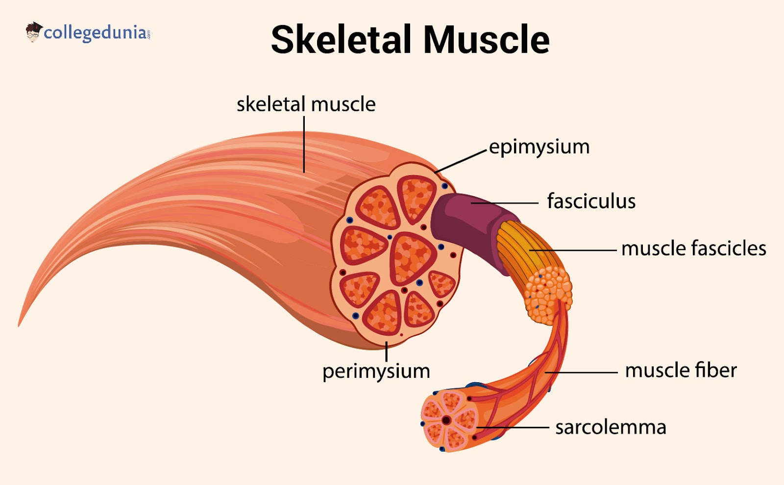
Skeletal Muscle
Read More:
Sarcomere
[Click Here for Previous Year Questions]
When muscle cells are examined under a microscope, a striped pattern (striations) can be seen. This pattern is made up of a succession of fundamental units known as sarcomeres.
- These are layered throughout muscular tissue.
- A single muscle cell can have thousands of sarcomeres.
- It is made up of numerous parallel actin (thin) and myosin (thick) filaments.
- Sarcomeres initiate the interaction of myosin and actin proteins shortening.
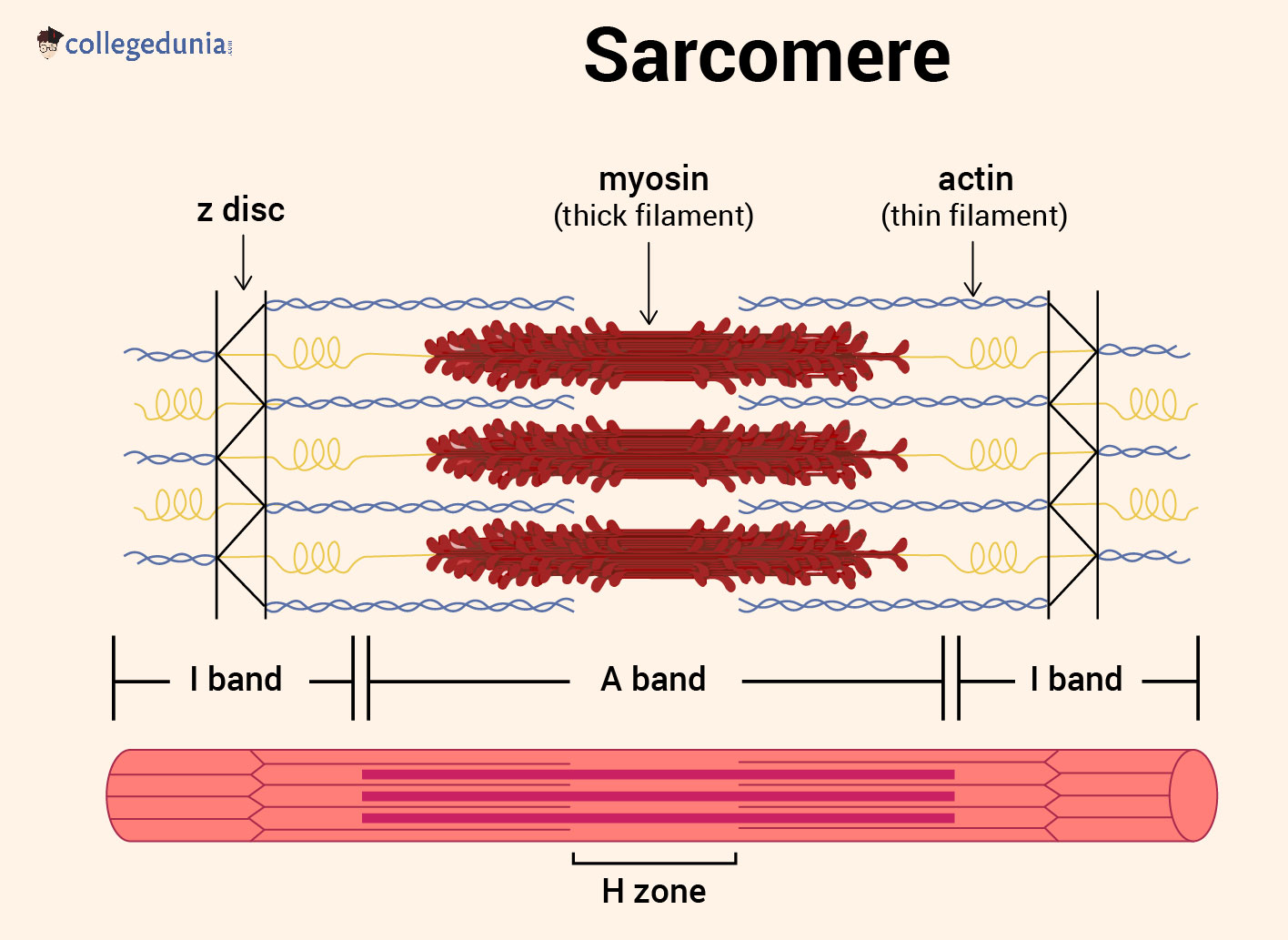
Sarcomere
Let’s understand more about actin and myosin myofilament below:
Actin Myofilament
An actin myofilament is composed of actin molecules, tropomyosin, and the troponin complex. Troponin is made up of three subunits (troponin I, T and C) and is composed of two helical strands.
- They are wrapped longitudinally around actin molecules (G-actins) in a thin, twisted, stranded form.
- Each G-actin molecule is linked to an ATP molecule.
- F-actin refers to the whole actin molecular part (Fibrous actin).
- Tropomyosin activates or deactivates the muscular contraction mechanism.
- It is a globular protein that binds tropomyosin as well as calcium ions.
Myosin Myofilament
Myosin Myofilament is made up of two different regions: a long rod-shaped tail termed myosin rod and two globular tangled myosin heads.
- The globular heads occur at regular intervals along the myosin myofilament.
- It protrudes from the sides of the filament where actin and myosin filaments overlap.
- The myosin head might connect to the adjacent actin filament.
Sliding Theory of Muscle Contraction
[Click Here for Sample Questions]
Muscle contraction takes place when muscle fibres become shorter. The process of a muscle contraction is split into five stages which are as follows:
Stage 1
When this nerve impulse arrives at the neuromuscular junction, acetylcholine is released. Acetylcholine depolarizes the motor end plate, which spreads throughout the muscle through the transverse tubules.
- It will release calcium (Ca+) ions from the sarcoplasmic reticulum.
Stage 2
In this stage, the presence of high Ca+ concentrations will bind to Troponin. It alters its shape, displacing Tropomyosin from the Actin's active site.
- Myosin filaments may now connect to actin and create a cross-bridge.
Stage 3
In this, the breakdown of ATP molecules will release energy. This allows myosin to pull Actin filaments inwards, which further shortens the muscle. This happens all the way down the length of every myofibril in the muscle cell.
Stage 4
When an ATP molecule attaches to the Myosin head, the Myosin detaches from the Actin and the cross-bridge is broken. The ATP is degraded, which causes the myosin head to reattach with the actin.
- This binds the site farther down the actin filament and repeats the 'power stroke.
- The ratchet mechanism refers to the repetitive tugging of Actin over myosin.
Stage 5
This mechanism of muscle contraction can continue indefinitely as long as there are sufficient ATP and Ca+ reserves. When the impulse is terminated, Ca+ is pushed back to the Sarcoplasmic Reticulum.
- Actin returns to its resting position, causing the muscle to extend and relax.
Muscle Structure and Contracting Phase
[Click Here for Previous Year Questions]
Muscle fibre includes hundreds of myofibrils, which are organelles. Each myofibril is made up of two types of protein filaments: thinner actin filaments and bulkier myosin filaments.
- Actin filaments are held together by structures known as Z lines.
- A sarcomere is the area between two Z lines.
- Myosin filaments overlay actin filaments within a sarcomere.
- It include small structures called cross bridges that allow them to connect to actin filaments.
Here are the phases of muscle contraction:
Stretched Muscle Phase
Stretched muscle with lengthened I – bands and H – zones reduce the overlapping of myosin and actin filaments. Muscle strength would be decreased because few cross-bridges could form between actin and myosin.
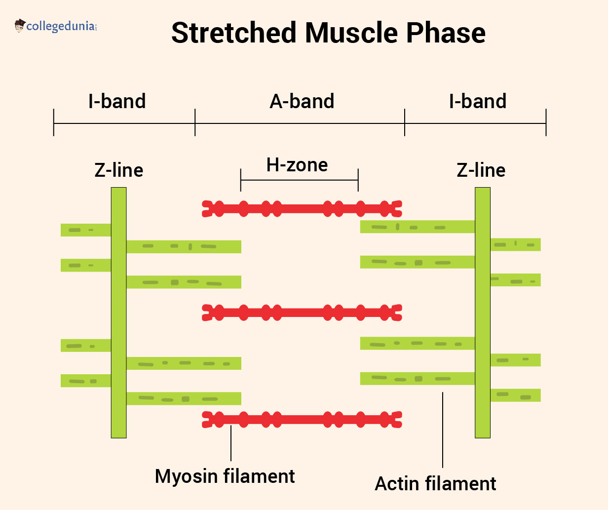
Stretched Muscle Phase
Partially Contracted Muscle Phase
Partly contracted muscle will have more overlapping myosin and actin and plenty of opportunity for cross bridges to develop. The I – bands and the H – zone have been shortened.

Partially Contracted Muscle Phase
Fully Contracted Muscle Phase
Fully contracted muscle with a lot of actin and myosin overlap. The thin actin filaments have overlapping, the possibility of cross bridges forming again is limited. As a result, the muscle will provide little force.
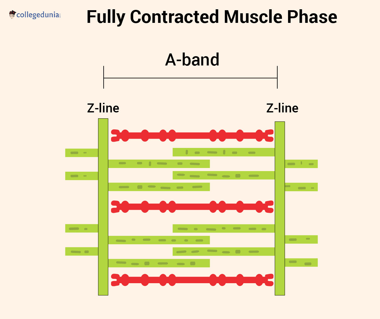
Fully Contracted Muscle Phase
Read More:
| Class 11 Biology Related Concepts | ||
|---|---|---|
| Human Body - Anatomy & Physiology | Ball and Socket Joint | Types of Movement |
| Locomotion and Movement | Bones of the Ankle | Muscular Tissue |
| Bones | Paralysis Symptoms | Tissues |
Energy Supply during Muscular Contraction
[Click Here for Sample Questions]
A large amount of energy necessary for contraction is given by the hydrolysis of ATP into ADP and Pi. This energy is required for the movement of myosin heads and the reabsorption of calcium ions in the sarcoplasmic reticulum.
Aerobic Respiration
The majority of ATP is produced from ADP during oxidative phosphorylation within a mitochondria. It occurs during prolonged and low-intensity activity when oxygen is necessary.
Anaerobic Respiration
Glycolysis is the process through which ATP is regenerated. As more oxygen is consumed than is supplied. Pyruvate is transformed into lactic acid and causes muscular tiredness.
- Anaerobic respiration is employed during brief bursts of high-intensity activity.
Creatine Phosphate
It is a phosphate reserve molecule that is instantly accessible for ATP regeneration. The reserve is quickly depleted and then restored with phosphate through ATP.
Things to Remember
- Sliding Filament Theory explains the contraction of muscles, actin and myosin filaments.
- It results in the shortening of the muscle fibre length
- A myofibril is a cylindrical organelle that runs the length of a muscle fibre.
- The myofibril functional unit is split into I, A, and H bands.
- Actin is a thin, contractile protein filament with 'active' or 'binding' sites.
- Myosin is a protein filament that is thick and contractile, containing protrusions known as Myosin Heads.
- Tropomyosin is a protein that binds to actin and controls muscle contraction.
Previous Year Questions
- Glenoid cavity articulates….[NEET 2015]
- A deltoid ridge occurs in...[NEET 1990]
- Number of cervical vertebrae in camel is...[NEET 1990]
- Stimulation of a muscle fiber by a motor neuron occurs at….[NEET 2014]
- In human body which one of the following is an atomically correct ?...[NEET 2007]
- Which one of the following items gives its correct total number?...[NEET 2008]
- Calcium is important in skeletal muscle contraction because it...[NEET 2018]
- Colle's fracture is associated with...[BHU UET 2009]
- Inter-articulated disc is found in...[BHU UET 2005]
- During strenuous exercise glucose is converted into...[BHU UET 2005]
- Human vertebral column consists of 33 vertebrae and ________ bones...[BHU UET 2012]
Sample Questions
Ques: What is sliding filament theory? (3 marks)
Ans: Sliding Filament Theory is the theory that explains the process of muscle contraction during which the thin filaments slide over the thick filaments. This interaction further results in the shortening of the myofibril.
- A sarcomere's actin and myosin myofilament organization plays a crucial role in the process of muscle contraction.
- Muscle contraction is considered to cause actin and myosin filaments to move past one another.
- The sliding filament model of muscle contraction is similar to interlocking fingers in that drawing them together reduces the distance.
Ques: What is the role of ATP in the sliding filament theory? (2 marks)
Ans: ATP releases myosin from the actin filaments. When contraction occurs in the muscle, the myosin attaches to the actin filaments. ATP is attached to the myosin head. During the contraction, the ATP releases the myosin from the actin molecule and results in muscle relaxation.
Ques: Explain in detail the Cross Bridge Formation? (5 marks)
Ans: Active cross bridges are produced when myosin heads connect like hooks to adjacent actin filaments. The actin-myosin filament crossbridge functions as an enzyme. Myosin ATpase is an enzyme that converts stored ATP into ADP and inorganic phosphate, releasing energy.
- This freed energy is utilised to propel the myosin head toward the actin strand.
- Myosin head tilts and pulls actin filament along, causing myosin and actin filament to glide against each other.
- A sarcomere's opposing ends of actin myofilament migrate toward each other, resulting in muscular contraction.
The cross bridge detaches after sliding, and the actin and myosin filaments return to their original positions. The active cross bridge forms and reforms for 50-100 times in a second utilising ATP.
- As a result, muscle fibre has a large number of mitochondria.
- Sarcomeres can contract by 30-60% of their length during muscular contraction.
Ques: How is sarcomere shortening regulated? Give a detailed description with an example? (5 marks)
Ans: Calcium and ATP are cofactors (nonprotein components of enzymes) necessary for muscle cell contraction and ATP provides the energy. Troponin and tropomyosin, two proteins that control muscle contraction by preventing myosin from attaching to filamentous actin, require calcium.
- Tropomyosin prevents myosin from binding to actin in a resting sarcomere.
- It must spin around actin filaments to reveal myosin-binding sites before myosin can bind it.
In 1994, William Lehman and his colleagues revealed how tropomyosin rotates by examining the structure of actin and myosin in calcium-rich versus low-calcium solutions.
- They discovered that the presence of calcium is required for the contraction mechanism by comparing the actions of troponin and tropomyosin under these two circumstances.
- Troponin (the smaller protein) changes tropomyosin's location and pushes it away from the myosin-binding sites on actin, thus unblocking the binding site.
- Myosin binds to actin to initiate cross-bridge cycling once the myosin-binding sites are accessible and sufficient ATP is available.
Ques: How does the Muscle Relax in between Muscular contractions? (5 marks)
Ans: When the nerve signal is no longer present to contract, calcium is reintroduced into the sarcoplasmic reticulum through active transport pumps. In muscle cells, the sarcoplasmic reticulum is specialised for calcium storage, ensuring that there is enough calcium available for contraction.
- There is no free calcium in the sarcoplasm to bind to troponin, tropomyosin slips back across the active sites of Actin.
With no binding sites accessible, the myosin heads are unable to form crossbridges and stay extended, waiting for the next nerve signal to contract, revealing the binding sites once more.
- The sarcomere recovers to its normal length passively, and the muscle relaxes.
- When a muscle is completely tensed, it shrinks by around 30% of its resting length.
- When the contraction is over, the muscle returns to its full resting length.
- Resting length is restored passively by other elastic proteins in the sarcomere that function like rubber bands and assist actin in recoiling, or by contracting an opposing muscle.
Ques: Who Proposed the sliding Filament theory? Explain the beginning of his life work around muscular contractions. (5 marks)
Ans: Hugh Huxley and Jean Hanson proposed the sliding filament model of muscle contraction in 1954. Hugh Huxley's pioneering work drove the model's formulation and subsequent confirmation.
- Huxley pioneered the use of x-ray diffraction to investigate contraction in living striated muscle, using the paracrystalline lattice to eventually comprehend contraction in terms of single molecules.
- Progress necessitated the creation of apparatus with ever-increasing spatial and temporal resolution, which fueled the development of synchrotron facilities.
- These are now utilised for the majority of protein crystallography and muscle research.
- Huxley's early work merged electron microscopy and biochemistry to study and interpret changes in x-ray patterns.
He enhanced electron microscopy methods such as thin sections, and negative staining. These methods had allowed him to answer significant issues about the structure and organisation of thick and thin filaments in muscle, as well as the interaction of myosin with actin and its control.
- Huxley demonstrated that myosin's ATPase domain creates the cross bridges of thick filaments that bind actin, and he proposed that myosin moves in distinct steps on actin.
Ques: What can stop the muscles from contracting? (5 marks)
Ans: Few factors that can stop the muscles from contracting are:
- Fatigue of the energy system: Because there is no more ATP in the muscle cell, it can no longer contract.
- Nervous system fatigue: The nervous system is unable to generate impulses in adequate quantities or at a fast enough rate to maintain the stimulation and produce calcium release.
- Voluntary nervous system control: When the brain instructs the nerve that causes the muscle to contract to cease transmitting that signal, no more calcium ions enter the muscle cell and the contraction ends.
- Sensory nervous system information: For example, a sensory neuron (nerve that detects stimuli such as pain or the weight of something) provides feedback to the brain indicating that a muscle is injured while attempting to lift a heavy weight, and thus the impulse to that muscle telling it to contract is stopped.
Ques: How can a serious neck injury paralyze the person from neck down? Explain? (4 marks)
Ans: A lack of strength and control over a muscle or set of muscles in a specific region of the body is referred to as paralysis. The majority of the time, this is not related to an issue with the muscles.
- It is most likely the result of a malfunction anywhere along the network of nerve cells that connects the body part to your brain and back.
- These nerve cells provide messages to your muscles, causing them to move.
Trigger signals sent from the brain govern muscle action. When any element of the relay system is destroyed, such as the brain, spinal cord, nerves, or the junction between the nerve and the muscle, the impulses do not reach the muscles, resulting in paralysis.
- The relay system can be destroyed in a variety of ways.
- Paralysis can be caused by a birth abnormality such as spina bifida, which happens when the brain, spinal cord, and/or the covering that protects them do not develop properly.
Ques: Name some causes of paralysis? (2 marks)
Ans: Most individuals become paralysed as a consequence of an accident or a medical condition that alters how their muscles and nerves work. The following are the most prevalent causes of paralysis:
- Stroke
- Injury to the spinal cord
- Injuries to the head
- Multiple sclerosis (MS)
Some of the other factors are:
- Cerebral palsy
- Guillain-Barré syndrome
- Neuropathy of the periphery
- Toxins/poisons
- Lou Gehrig's disease (ALS)
Ques: How muscle contraction is initiated? (3 marks)
Ans: Muscle contraction is initiated by a signal transmitted by motor neurons in the central nervous system. The connection between a motor neuron and a sarcolemma is known as the neuromuscular junction.
- When a neuronal signal reaches this junction, the sarcolemma generates an action potential and releases acetylcholine.
- This causes the sarcoplasm to release calcium ions as it travels along the muscle fiber.
- After that, calcium attaches itself to actin filament troponin, exposing myosin's active sites.
- Using the energy from the hydrolysis of ATP, myosin attaches itself to the exposed active site on actin.
- Contraction happens as a result of the Z lines connected to them being pulled as well.
Ques: What is sarcomere? (2 marks)
Ans: The functional unit of striated muscle is called a sarcomere. This indicates that it is the simplest component of our skeletal muscle. The muscle type that starts all of our voluntary movements is called skeletal muscle. This is the primary function of the sarcomere. Sarcomeres can contract collectively to start broad, sweeping movements. These microscopic units' special structure enables them to synchronize the contractions of our muscles.
Do Check Out:



Comments