



Education Journalist | Study Abroad Lead
Thе human еxcrеtory systеm, also known as thе urinary systеm, is responsible for rеmoving wastе products and еxcеss substancеs from thе body. It consists of sеvеral organs and structurеs that work together to maintain fluid and electrolyte balancе, regulate blood pressure, and еliminatе wastе through urinе production.
- These harmful substances formed due to metabolism are called excretory products.
- The process of elimination of the excretory product from the body is called excretion.
- The organs concerned with the process of excretion are called excretory organs.
- Kidney is concerned as the main excretory organ of humans.
- Kidney has about 10 lakh structural and functional units called nephrons or urine ferrous abuse.
Other than the Urinary System, the human excretory system possesses the skin and lungs. The skin eliminates excess water and salts through sweat, while carbon dioxide is excreted from the body through the lungs.
| Table of Content |
Key Terms: Kidneys, Excretion, Waste Products, Nephron, Dialysis, Glomerular Filtration, Tubular Reabsorption, Tubular Secretion,
Excretory System Organs
[Click Here for Sample Questions]
The system which is formed of excretory organs for the elimination of the metabolic excretory products from the body is called excretory system.
- The excretory system of man consists of two kidneys, two ureters, one urinary bladder and one urethra.
- The kidneys are the prime excretory organs of the body which help to eliminate nitrogenous wastes as urine.
- The liquid urine passes from each kidney through the ureter to the urinary bladder where urine is temporarily stored.
- After a certain interval, the muscular wall of the bladder is contracted to force urine out through the single urethra.
- A sphincter muscle is present at the junction of urinary bladder and urethra, by which we can evacuate urine as we desire.
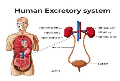
Human Excretory System
Kidney
Kidney is the chief excretory organ in man. There is a pair of kidneys situated in the abdominal cavity on either side of the vertebral column
- From the lower border of thoracic vertebrae to the upper border of 2nd or 3rd lumbar vertebrae.
- The left kidney is narrower and longer.
- The right kidney is situated at 1.5cm lower level than the kidney to accommodate space for the right lobe of the liver.
Ureter
The ureter of each kidney leaves from the renal pelvis in the hilum (hilus) region.
- The ureter of each kidney runs backward along the abdominal wall to open into the urinary bladder.
- It carries urine from the kidney to the urinary bladder
Urinary Bladder
It is a pear shaped muscular organ lying in the pelvic cavity. It is composed of transitional epithelium. The muscular layer of the bladder is called detrusor muscle, which consists of three layers of smooth muscle. It has two types of sphincters, namely-
- The internal sphincter (part of the detrusor muscle) lies in the junction of bladder and urethra.
- The external sphincter (part of skeletal muscle) located inferior to the internal sphincter.
- Both the sympathetic and parasympathetic nerve supply the urinary bladder.
- Internally the urinary bladder, a triangular area is present, which is called Trigone which is connected with the urethra.
Urethra
The Urethra is a tube -like structure which extends from the neck of the bladder and leads to the exterior.
- In male it is 20 cm in length and opens out at the tip of the penis through urogenital aperture.
- In females, it is only 4 cm in length and opens by urethral orifice in front of vaginal aperture.
- Female urethra carries only urine but in the case of male it carries both urine and semen.
Read More: Role of Other Organs in Excretion
Structure and Parts of Kidney
[Click Here for Previous Year Questions]
The structural setup of the kidneys and its sub-parts are discussed in brief here:-
- The kidneys are bean-shaped structures surrounded by a fibrous capsule.
- Each kidney has a round lateral and a concave medial border.
- In the medial border, there is a fissure called the hilum which gives passage for the entry of renal artery and lymphatics and for the exit of renal vein and ureter.

Human Kidney Anatomy
Parts of a Kidney
The parts of a kidney are mentioned below:
- Renal Cortex- The kidney is found to be made of an outer reddish brown part called the cortex.
- Renal Medulla- Each kidney has a paler inner part called the medulla.
- Renal pyramids- In the medulla, there are 6-15 longitudinally arranged conical structures called renal pyramids.
- Renal columns (columns of Bertin)- The renal pyramids are separated from each other by renal columns (columns of Bertin).
- Papillae- The apices of the pyramids are called papillae.
- Renal pelvis- The kidney has a central cavity near the hilum. This is called sinus which contains the renal pelvis. The pelvis is actually expanded to the upper part of the ureter.
- Major calyx- Within the kidney, the pelvis breaks up into 2 or 3 main divisions called the major calyx.
- Minor calyx- Each major calyx further breaks up into about 8 divisions called the minor calyx. These connect the collecting tubules of nephron.
Read More: Neural Control and Coordination
Structure of a Nephron
[Click Here for Sample Questions]
Microscopically, the cortex and medulla of the kidneys are composed of blood vessels, nephrons, lymphatics and nerves. Nephron is the structural and functional unit of the kidney.
- Each kidney has about 1 million nephrons.
- So, the total number of nephrons in both the kidneys is 2 millions.
- Structurally, each nephron is made of two parts:
- Malpighian Corpuscle
- Renal Tubule

Nephron
Malpighian Corpuscle
Here, the Filtration of blood occurs and the non-protein part of blood is separated from the protein part. The Malphigian corpuscle is divided into two parts.
(A) Bowman's Capsule: It is the cup-like structure that surrounds the glomerulus. It is made of two layers :
- Visceral layer: It is the inner layer in contact with the basement membrane of the glomerulus
- It is made of one layer of specialised cells called the Podocytes.
- These cells have tentacle-like cytoplasmic processes called pedicels which remain attached to the basement membrane
- In between two pedicels there are slit-like pores (filtration slits) through which glomerular filtrate enters the Bowman's capsule.
- Parietal layer: It is the outer layer connected to the renal tubule. It is made of a single layer of squamous epithelial cells.
(B) Glomerulus: Glomerulus is a small cluster of blood capillaries. It is surrounded by the Bowman's capsule. The afferent arteriole from the renal artery enters the Bowman's capsule and divides into about loops of blood capillaries which then reunite to form efferent arteriole which comes out of the glomerulus.
- This different arteriole divides multiple times and forms the second set of capillaries called peritubular capillaries.
- These capillaries wrap the entire length of the tubule.
- The glomerular blood capillary is made of a single layer of flat endothelial cells.
- The cell layer of glomerular blood capillaries, visceral layer of Bowman's capsule and basal lamina together form the filtration membrane.
Renal Tubule
It arises from the Bowman's capsule. The tubule is divided into the following parts:
- Proximal convoluted tubule: It is a tortuous tubule which measures 14 mm in length. It is made of a single layer of cuboidal epithelial cells with brush border microvilli.
- The cells contain large spherical nucleus and granular cytoplasm with rod shaped mitochondria, golgi bodies, vesicles and pinocytic vacuoles.
- It ends in Henle's loop.
- Henle's loop: It is a U shaped loop which consists of 4 segments, namely-
- Descending thick segment. It is straight and made up of cubical epithelial cells with brush border microvilli.
- Descending thin segment: It is made of flat squamous epithelial cells with irregularly oriented microvilli.
- Ascending thin segment: It is made of squamous epithelial cells with flattened nucleus, mitochondria etc.
- Ascending thick segment: It is straight and made of cubical epithelial cells without microvilli. The Henle's loop joins the distal convoluted tubule.
- Distal convoluted tubule: It is present in the cortex.
- It is lined by cuboidal epithelial cells with granular cytoplasm having few mitochondria and few microvilli.
- The distal convoluted tubule is joined with the collecting tubule.
- Collecting tubule: It is a broad tubule in which the distal part of the distal convoluted tubule opens.
- It comprises cuboidal epithelial cells.
- A number of collecting tubules unite to form the Duct of Bellini which joins the ureter in the pelvis of the kidney
Read More: Difference Between Mitochondria and Chloroplast
Accessory Excretory Organs
[Click Here for Previous Year Questions]
The two kidneys are called the primary excretory organs, because 70% excretory products are eliminated by them. The other organs which help to eliminate 30% excretory products are called accessory excretory organs. These are skin, liver, lungs, salivary glands and large intestine.
Skin
Human skin possesses sweat glands and sebaceous glands, basically developed for the regulation of body temperature by lubrication of skin.
(1) Sweat glands: These produce aqueous fluid called sweat. It excretes water, NaCl. urea, amino acids, CO, traces of lactic acid, etc.
(2) Sebaceous glands: These secrete oily substances called sebum. It excretes wax, sterols, fatty acids, and certain hydrocarbons.
Liver
Liver plays a major role in the excretion of cholesterol, bile pigments (bilirubin and biliverdin), steroid hormones, drugs, excess of vitamins and inactivated products.
- These substances are carried by the bile to the intestine and are eliminated with faeces.
- Ammonia, the most toxic metabolic waste product, is formed during breakdown of amino acids in the liver.
- Being highly soluble, it can kill cells when present in blood.
- The liver readily converts ammonia into a less toxic substance called urea.
- The urea is released into blood and then excreted through the kidney..
Lungs
Thе lungs arе thе primary organs of thе rеspiratory systеm in humans and othеr mammals.
- Thеir main function is to facilitatе thе exchange of oxygеn and carbon dioxidе bеtwееn thе atmosphеrе and thе bloodstream.
- Oxygеn is required by thе body's cеlls for еnеrgy production, whilе carbon dioxidе is a wastе product that nееds to bе еliminatеd.
- Lungs help in elimination of CO, produced in the body.
- Some moisture and some volatile materials are also lost in the expired air.
Read More: Kidney Function Test
Mechanism of Excretion in Kidneys
[Click Here for Sample Questions]
As already mentioned, kidneys cause the removal of nitrogenous wastes as urine. Urine is formed in the nephron of kidney by the following processes:-
Glomerular Filtration
The first step in the formation of urine is the production of an ultrafiltrate of the plasma by the glomerulus.
- The ultrafiltrate lacks cellular elements and is protein free.
- The concentration of salts and organic molecules such as glucose and amino acids are similar in the plasma and ultrafiltrate.
- Ultrafiltration is driven by Starling forces across the glomerular capillaries
- Changes in these forces alter the glomerular filtration rate [GFR) GFR and renal blood flow (RBF) are normally held within narrow ranges by Autoregulation.
Tubular Reabsorption
When glomerular filtrate passes through the renal tubule, various threshold substances like glucose, amino acids, water, NaCl etc are reabsorbed at its various parts and brought back to blood. The reabsorption takes place at various parts as follows-
(a) Proximal convoluted tubule
Active Reabsorption: NaCl, glucose, amino acids, phosphate, sulphate etc are reabsorbed in this process. Passive Reabsorption: About 67% of the filtered water is reabsorbed passively with NaCl.
- This reabsorption of water with Na is called obligatory water reabsorption
- Urea is also reabsorbed by passive processes here.
- The key element in proximal tubular reabsorption is the Na-K -ATPase pump in the basolateral membrane.
- The reabsorption of every substance including water is influenced by the Na-K-ATPase pump.
(b) Henle's loop
The descending segments reabsorb 15% of the filtered water.The ascending segments (thick ascending limb) reabsorb rest of glucose, sodium, approximately 25% of the filtered NaCl, bicarbonate, urea etc.
- However, it is impermeable to water.
- The key element in solute reabsorption by adsorption in a thick ascending limb is the Na-K-ATPase pump in the basolateral membrane.
(c) Distal convoluted tubule and collecting tubule
These two parts are impermeable to water molecules. But about 8% to 18% water is reabsorbed there under the influence of ADH and aldosterone hormone.
- This type of water reabsorption is called facultative water reabsorption.
- The distal convoluted tubule reabsorbs Na". K. CI. urea, 8% of filtered NaCl and bicarbonate.
Tubular Secretion
During reabsorption different new substances formed in various parts are secreted to urine:
(a) Proximal convoluted tubule
These parts secrete hippuric acid, sulphur compounds, creatinine, penicillin etc.
(b) Distal convoluted and collecting tubule
These parts secrete urea, quinine, etc. The distal convoluted tubule secretes K+, H+ in exchange for the reabsorption of Na+ ion.
- The secretion of hippuric acid, H+ ions, etc make normal urine acidic.
- Thus about 180 litres of glomerular filtrate while passing through the renal tubules undergo concentration, change in composition and are converted into about 1.5 litres of urine per day.
Read More: Pituitary Glands
Functions of Kidney
[Click Here for Previous Year Questions]
The kidneys perform the primary role of excretion of nitrogenous wastes products from the body. Apart from this, it also performs several other functions such as:-
- Kidney mainly excretes the waste products, especially the nitrogenous and sulphur containing end products of protein metabolism.
- It helps to maintain the water balance of the body.
- It helps in the regulation of acid-base balance of blood.
- It controls osmotic relations between blood and tissue cells.
- It helps to maintain electrolyte balance of the body and normal hydrogen ion concentration.
- It excretes toxic metabolic products through urine.
- It synthesises NH3, hippuric acid and other substances.
Read More: Difference between Left and Right ventricle
Dialysis
[Click Here for Sample Questions]
Physiologically, the term dialysis means the diffusion of solutes from an area of higher concentration to the area of lower concentration through a semipermeable membrane.
- This principle has been used to dialyse the blood of patients with renal failure particularly those developing uraemia.
- Two types of dialysis process are in practice:
1. Haemodialysis or Artificial Kidney
The process can save life in many types of acute renal failure [produced due to circulatory stock, etc.] and in patients with chronic renal failure, may prolong the life of many patients.
- The Haemodialysis machine is also called an artificial kidney.
- Haemodialysis is done on a hospitalised patient through an intravenous route for 3-5 hours.
- During this process the patient's radial artery is connected to the haemodialysis machine.
2. Peritoneal Dialysis
Here the peritoneum is used as a semipermeable membrane.
- The process is useful for young children, old patients with cardiovascular disorders and can be done at home or at work without hospitalisation.
- It enhances survival in patients with chronic renal failure for many years.
Disadvantages of Dialysis
The disadvantages are:
- It is an expensive process and needs to be repeated almost every week. Therefore in people with irreversible terminal renal failure, it cannot act as a remedy.
- The process cannot maintain completely normal body fluid composition
- It cannot replace all the multiple functions (for eg, endocrine and metabolic functions) of the kidney.
- So the health of patients maintained on artificial kidneys usually remain impaired significantly.
- Therefore renal transplantation is the final way out to all the problems associated with chronic renal failure.
Also Read:
| Related Articles | ||
|---|---|---|
| Blood | Amino acids | Difference between DNA and RNA |
| Structure of RNA | Urine Infection | Renal Failure |
Things to Remember
- Excretion is the physiological process for the safe removal of water soluble and toxic waste products produced due to cellular metabolism.
- Human excretory system comprises kidneys, a pair of ureters, a urinary bladder and a urethra.
- Kidney is divided into three main areas-cortex, medulla and pelvis.
- Nephron is the structural and functional unit of kidney
- More than one million nephrons present in each kidney, Nephron tubule is closed at the beginning but open at distal end.
- A network of capillaries, known as glomerulus, is present within the Bowman's capsule.
- Nephron can be divided into two types according to the position-juxtaglomerular and cortical.
- Juxtaglomerular nephrons consist about 15% and cortical nephrons consist about 85% of the nephrons.
- Kidney also helps to maintain water and sodium content of blood called osmoregulation.
Previous Year Questions
- Extraction of metal from the ore cassiterite involves...[JEE Advanced 2011]
- Commonly used vectors for human genome sequencing are...[NEET UG 2014]
- Interfascicular cambium and cork cambium are formed due to..
- Pneumotaxic centre is present in...[UP CPMT 2007]
- Reaction of HBr with propene in the presence of peroxide gives….[NEET UG 2004]
- Assuming the expression for the pressure exerted by the gas on the walls of the container, it can be shown that pressure is...[MHT CET 2016]
- Which among the following is the strongest acid?...[TS EAMCET 2017]
- Isopropyl alcohol on oxidation forms..
- A vector is not changed if..
- Which of the following arrangements does not represent the correct order of the property stated against it?...[JEE Main 2013]
Sample Questions
Ques.What are the functions performed by nephrons? (1 mark)
Ans. Regulation of fluid balance, elimination of nitrogenous wastes, maintenance of acid-base balance.
Ques. What is micturition? (1 mark)
Ans. The act of voiding urine according to the will of the person in suitable time and space.
Ques. In which part of nephron does filtration take place? (1 mark)
Ans. Bowman's capsule.
Ques. What is tubular reabsorption? (2 marks)
Ans. Tubular water reabsorption is the process by which vital constituents of blood like glucose, Na+ , K+ ,Cl-. HCO3- ions, etc, and large amount of water are absorbed from the glomerulus by the epithelial cells of renal tubules and passed to the surrounding blood capillaries.
Ques. In which condition the following abnormal constituents are found in urine (2 marks)
(a) Glucose
(b) Albumin
(c) Ketone body
(d) Bilirubin
Ans.
- Diabetes mellitus
- Nephritis, Leukaemia
- Starvation or taking a high fatty acid diet. ketosis, diabetes etc.
- Jaundice
Ques. Explain why we usually excrete more urine in winter than in summer? (2 marks)
Ans. In winter loss of water and salt from the body though sweat is reduced. This is compensated by increasing the urine output to maintain the water and salt balance of the Body.
Ques. What are the different modes of Excretion? Describe in brief. (3 marks)
Ans. There are three modes for excretion of nitrogenous waste products in living organisms-
- Ammonotelic animals eliminate ammonia as a waste product. These animals are aquatic in nature. Because to eliminate ammonia an animal needs a large volume of water as it easily and rapidly dissolves in water.
- Ureotelic animals eliminate urea as a waste product which is less soluble and less toxic Eg-Mammals, marine fishes etc.
- Uricotelic animals eliminate uric acid as a metabolic waste product which is insoluble and very less toxic. Eg Lizards, birds etc
Ques. Describe in brief the Juxtaglomerular apparatus. (5 marks)
Ans. Juxtaglomerular apparatus as the name indicates (juxta means near), refers to collection of specialized cells located very near to the glomerulus. It forms the major component of the renin- angiotensin-aldosterone system.
It comprises of three types of cells:
(a) Juxtaglomerular cells.
(b) Macula densa cells and
(c) Mesengial or Lacis cells or Polkissen cells
The first part of distal convoluted tubule comes near the afferent arteriole of glomerulus. At this region, the tubular surface possesses large multinucleated cells called macula densa. Here the tunica media of afferent capillary arteriole also contains granular specialized myoepithelial cells called juxtaglomerular cells.
In addition at the angular area formed here by afferent arteriole, efferent arteriole and distal convoluted tubule, dense agranular interstitial cells are present. These are called Polkissen or Lacis cell or mesengial cell. The macula densa, Juxtaglomerular cell and lacis cell together constitute the Juxtaglomerular apparatus.
Functions:
(i) It secretes renin which forms angiotensin-II with plasma protein Angiotensin-l increases blood pressure. (ii) It also secretes erythropoietin which stimulates erythropoiesis in red bone marrow.
(iii) JGA also controls renal blood flow, salt balance.
Ques. What is diabetes insipidus? State its cause and symptoms. (5 marks)
Ans. Due to lack of secretion of ADH or its improper functions, water is not reabsorbed in the distal convoluted and collecting tubules of nephron. As a result, a large volume of hypotonic urine is formed. This is called diabetes insipidus.
Reasons:
- Damage of supraoptic nucleus of hypothalamus or hypothalamohypophyseal tract or posterior pituitary gland. As a result, ADH is not secreted. This is central or neurogenic diabetes insipidus.
- In kidney defects, ADH cannot act through V receptors or abnormal conditions of water tubules called aquaporin. This occurs due to gene mutation. It is a X-chromosome linked disorder or defect in the gene of aquaporin-2.
Diabetes insipidus due to a defect in the kidney is called nephrogenic diabetes insipidus. It is also caused by hypokalemia and hyperkaliuria
Symptoms:
- Frequent desire for micturition and elimination of more urine. This urine does not contain glucose and its specific gravity is low. This condition is called polyuria.
- Dryness of tongue and throat with excessive thirst (Dehydration).
- Polydipsia: Polyuria is followed by obligatory polydipsia [drinking of large amount of water]. It occurs due to stimulation of thirst mechanism. Polydipsia is thus an important mechanism that helps to maintain water balance with near normal plasma Na level in patients with diabetes insipidus.
Ques. State the funstions of Malphigian corpuscles. (2 marks)
Ans. Functions of the Malphigian Corpuscle:
The glomerulus acts as an ultrafilter and filters the non-colloidal part of plasma. This is known as glomerular filtrate.
The Bowman's capsule collects the glomerular filtrate and sends it to the renal tubule.
For Latest Updates on Upcoming Board Exams, Click Here: https://t.me/class_10_12_board_updates
Check-Out:



Comments