



Content Curator
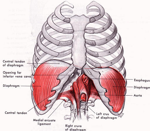
The thoracic cavity, also known as the chest cavity, is a vertebrate cavity bounded on the sides and top by the rib cage and on the bottom by the diaphragm. The thoracic vertebrae, which connect to the ribs that surround the cavity, surround the thoracic cavity. It comprises three sections: two pleural cavities and the mediastinum, located behind the sternum. The mediastinum is further compartmentalised into the superior and inferior mediastinum, the latter of which is again divided into anterior, middle and posterior.
Read about: Aerobic and Anaerobic Respiration
| Table of Content |
Key Terms: Thoracic Cavity, Mesothelium, pericardial cavity, mediastinum, oesophagus, diaphragm, endocrine glands
What is Thoracic Cavity?
[Click Here for Sample Questions]
The thoracic cavity, also known as the chest cavity, is a cavity enclosed by the ribs, the vertebral column, and the sternum or the breastbone. A muscular and membranous partition, the diaphragm, separates the thoracic cavity from the abdominal cavity. It houses the lungs and the bronchi, part of the oesophagus and trachea, as well as the heart and major blood vessels. The pleura lines the cavity (parietal pleura) and extends over the lung (visceral pleura) and the rest of the cavity's constituents, defining a space called the mediastinum.
Also Read: What are blood vessels?
Thoracic Cavity Functions
[Click Here for Sample Questions]
Thoracic cavity serves several functions – it provides protection and support to the body’s vital organs. Rib cage, as well as several layers of membranes, circumference the thoracic cavity, keeping the organs sheltered from environmental hazards. If a heart is to continue beating and providing pressure to the body’s vessels, it must be kept in a relatively safe and stable position. The rib cage, in addition to providing support, functions in unison with the diaphragm and other muscles to draw air in and out of the lungs.
Thoracic Cavity Organs
[Click Here for Sample Questions]
The thoracic cavity accommodates the heart and many of the main vessels of the circulatory system. The heart is enclosed within the pericardial cavity, which is separated from other cavities of the thoracic cavity.
Additionally, the lungs have their own cavities, the pleural cavities, and connect to the mouth via the trachea and thoracic inlet. Aside from these major vital organs, the thoracic cavity is a refuge for many nerves and vessels. Some run straight through the chest cavity, while others serve nerves and muscles in the cavity which assist in regulating breathing, the heart’s rhythm, and various muscle functions. The endocrine glands, lymph ducts, and the oesophagus are other minor organs, simply passing through the thoracic cavity.
Arrangement of organs in thoracic cavity
Also Read: NCERT Solutions For Class 11 Biology Chapter 6: Anatomy of Flowering Plants
Structure of Thoracic Cavity
[Click Here for Sample Questions]
The thoracic cavity can be categorized into two well-established compartments — the pleural cavities and the mediastinum. There exist two pleural cavities which respectively contain the left and the right lungs as well as pleura. The mediastinum is centrally located and can be found in between the two bilateral pleural cavities. The mediastinum extends to the inner border of the sternum anteriorly, the inner border of the thoracic vertebral bodies posteriorly, and spans the entire vertical length of the thoracic cavity.

Structure of Thoracic Cavity
The superior and inferior mediastinum are divided by the horizontal plane (also known as the thoracic plane) passing through the angle of the sternum (the junction of the manubrium and the body of the sternum) and crossing the T4-T5 vertebral junction. The anterior and posterior surfaces of the pericardium divide the inferior mediastinum into anterior, middle, and posterior compartments. The anterior mediastinum is located anterior to the pericardial sac, while the middle mediastinum houses the heart and pericardium. The posterior mediastinum is located posterior to the pericardial sac.
Also Read: NCERT Solutions For Class 11 Biology | Syllabus & Solutions
Diseases Related to Thoracic Cavity
[Click Here for Sample Questions]
A membrane, known as pleura, line the thoracic cavity and covers the lungs. The pleura is layered containing a small number of fluids that acts as a lubricant as you breathe in and out. The space between these layers is called the pleural cavity. Some of the diseases related to the thoracic cavity are outlined below:
- Pleural Effusion: Excess fluid in the pleural cavity is referred to as pleural effusion. Congestive heart failure is the most common cause. Lung cancer, pneumonia, tuberculosis, liver disease, pulmonary embolism, lupus, and drug reactions are some of the other causes.
- Pleurisy: Pleurisy is a type of pain caused by inflammation of the pleural cavity. A viral infection, such as influenza, is the most common cause. Bacterial and fungal infections, lung cancer, other diseases such as rheumatoid arthritis and mesothelioma, and drug reactions are some of the other causes.
- Pneumothorax: Pneumothorax is caused by an accumulation of air or gas in the pleural cavity around the lung, causing the lung to collapse. The most common causes are chronic obstructive pulmonary disease, tuberculosis, and trauma.
- Hemothorax: A hemothorax is an accumulation of blood in the pleural cavity. The most common cause is chest trauma, but lung and pleural cancer, as well as chest or heart surgery, can also result in a hemothorax.
- Pleural tumours: Pleural tumours are cancerous cells that grow in the pleural cavity. Occasionally, the cause is unknown. Pleural tumours are sometimes cancers that have spread from other parts of the body.
Things to Remember
- Thoracic cavity is lined with a serous membrane that excludes a thin fluid and is called the parietal pleura.
- The thoracic cavity provides protection and support to the body’s vital organs such as the heart and also helps in drawing air in and out of the lungs.
- The thoracic cavity has two openings, one from the top leading to the mouth and throat, and the other allowing the oesophagus and other chief vessels and tissues to pass between the thoracic cavity and the abdominal cavity.
- The thoracic cavity organs are the thymus gland, the heart, the lungs, the tracheobronchial tree, and the pleurae.
- Pleural effusion is the accumulation of fluid in the pleural cavity.
- Pleurisy is a painful condition resulting from inflammation of the pleural cavity.
Sample Questions
Ques: Earthworms have well-defined body cavities, some of which house the worm's heart. Is there a thoracic cavity in the worm? (3 marks)
(A) No, thoracic cavities are specific to vertebrates
(B) Yes, the thoracic cavity surrounds the heart
(C) Yes, it’s just different
Ans: A is the correct answer. The thoracic cavity gets its name from the thoracic vertebrae. While the development of a coelom, or body cavity, is shared by the worm and humans, this cavity develops in very different ways. There are no vertebrae or ribs in the worm. These features, which are unique to vertebrates, define the thoracic cavity of vertebrates.
Ques: Why are thoracic organs separated? (2 marks)
Ans: The thoracic wall actually encloses a cavity or space that is filled with various anatomical structures. Since there are so many of them, the thoracic cavity is divided into several compartments to aid their localization.
Ques: What are the thoracic cavity and its constituents? (3 marks)
Ans: The chest, also known as the thorax, is the superior part of the trunk that lies between the neck and the abdomen. The superior and inferior mediastinal cavities make up the mediastinum. The inferior mediastinal cavity is divided into three compartments: anterior, middle, and posterior.
Ques: Where is the thoracic cavity situated? (3 marks)
Ans: The thoracic cavity has two openings, one from the top that leads to the mouth and throat and the other that allows the oesophagus and other important vessels and tissues to pass between the thoracic cavity and the abdominal cavity. The thoracic inlet allows the trachea and oesophagus to enter the thoracic cavity.
Ques: What are thoracic cavity related disorders? (2 marks)
Ans: Blood (hemothorax) or air (pneumothorax, which can lead to atelectasis) in the pleural cavity, as well as pleural inflammation (pleurisy), are examples of thoracic cavity related disorders.
Ques: What protects the thoracic cavity? (3 marks)
Ans: The thoracic cavity (also known as the chest cavity) is a chamber of the vertebrate body that is protected by the thoracic wall (rib cage and associated skin, muscle, and fascia). The mediastinum is the thoracic cavity's central compartment. The thoracic cavity has two openings: a superior thoracic aperture known as the thoracic inlet and a lower inferior thoracic aperture known as the thoracic outlet.
The thoracic cavity contains the tendons as well as the cardiovascular system, both of which can be damaged by an injury to the back, spine, or neck.
Ques: What does the thoracic cavity include? (2 marks)
Ans: The thoracic cavity houses organs and tissues from the respiratory (lungs, bronchi, trachea, pleura), cardiovascular (heart, pericardium, great vessels, lymphatics), nervous (vagus nerve, sympathetic chain, phrenic nerve, recurrent laryngeal nerve), immune (thymus), and digestive (oesophagus) systems.
Ques: State the difference between the thoracic cage and the thoracic wall. (4 marks)
Ans:
The skeleton of the thoracic wall is the thoracic cage (rib cage). It is made up of the 12 thoracic vertebrae, 12 pairs of ribs and their associated costal cartilages, and the sternum. The vertical sternum or breastbone (anteriorly) and the 12 thoracic vertebrae support it (posteriorly).
On the other hand, the thoracic wall is made up of a bony framework that is held together by twelve thoracic vertebrae that give rise to ribs that encircle the lateral and anterior thoracic cavity. It develops apart from the ribs. In rare cases, the sternum does not fully form, exposing the underlying heart.
Ques: What are the types of respiratory disorders? (3 marks)
Ans: Airway diseases: These are the diseases that affect the tubes or airways or the passage which carries the oxygen and other gases into the lungs and out of the lungs.
Lung tissue diseases: These are the diseases that affect the structure of the lung tissues. It causes scarring or inflammation of the tissues.
Lung circulation diseases: These are the diseases that affect the blood vessels in the lungs. They also affect the working of the blood vessels.
Ques: What are the major functions of the human respiratory system? (3 marks)
Ans: The major functions of the human respiratory system are:
- It allows us to talk and also to smell things.
- It takes oxygen and supplies oxygen to the body cells.
- It releases carbon dioxide gas out of the body.
- Helps from the irritant gases.
Ques: What are the conditions affecting the respiratory system? (3 marks)
Ans: Some of the major conditions are:
- Allergies: This is due to inhaling dust, mold, chemicals.
- Asthma: This is due to inflammation in the airways.
- Infection: this is due to bacteria, fungus, and virus exposure.
- Ageing: As age increases the capability to work will decrease.
- Damage: Any damage to the respiratory system affects functioning.
Also read:



Comments