



Education Journalist | Study Abroad Lead
Rib Cage is a basket-like skeletal structure that is responsible for the formation of the chest or thorax. The rib cage is made up of bones which are commonly known as ribs. It is further attached to the sternum commonly known as breast bone as well as to the vertebral column. It provides bony protection to vital organs such as the lungs and heart. The rib cage plays a significant role in the function of the respiratory system. Respiratory muscles such as the diaphragm, intercostal muscles, etc are held by the ribcage intrinsically which are very important for active inhalation and forced exhalation. The ribcage is mainly present in the Vertebrate group of animals. The curvature becomes more open with each succeeding rib as we go down from the uppermost rib to the downward ribs. Although, ribs are semi-rigid they can get expanded and hence plays a significant role in breathing. The joilnts between the ribs and vertebrae aid in the movement of ribs while inhalation and exhalation.
| Table of Contents |
Key Takeaways: Rib cage, Thorax, Bones, Ribs, Vertebral Column, Joints, Breathing, Sternum
What is Rib cage?
[Click Here for Sample Questions]
Rib cage is the protective ‘cage` of the thorax formed from the twelve paired bones known as ribs. In ribcage, ribs are connected dorsally to the Vertebral Column and connected ventrally to the Sternum. Rib cage can be expanded to increase size because they are semi-rigid. Ribs can glide on the vertebrae due to the presence of small joints present between them and vertebrae while breathing and doing other activities.
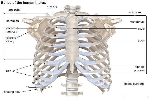
Rib Cage
Classification of Ribs
[Click Here for Sample Questions]
Ribs are classified into three groups on the basis of their attachment to the sternum. These are as follows-
- True ribs: Ribs 1-7 are true ribs. They are known as true ribs because they articulate with the sternum directly with their costal cartilage. Sterno costal joints are responsible for this articulation. However, there is an exception i.e. the first rib articulates with the clavicle by the costoclavicular joint.
- False Ribs: The ribs which are indirectly articulated with the sternum are known as false ribs. The costochondral joint connects its costal cartilages with the seventh costal cartilage. 8th 9th and 10th ribs are known as false ribs.
- Floating ribs: The ribs which do not articulate with the sternum are known as floating ribs. The 11th and 12th ribs are known as floating ribs.
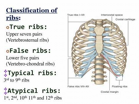
Classification of Ribs
Structure of Rib cage
[Click Here for Sample Questions]
The structure of the rib cage is as follows:
-
Parts of the Ribcage
On the basis of structure, ribs are classified as typical ribs and atypical ribs. Typical ribs are characterized by the presence of generalized structure while some variations are present on atypical ribs.
Typical ribs
Typical Ribs consists of a head, neck and body…
- Head: It has two articular facets which are separated by a wedge of a bone. The Head is generally wedge-shaped. Numerically corresponding vertebrae articulate with one facet while the other facet articulates with the vertebra above.
- Neck: It connects the body of the rib with the head. At the point of connection between the neck and the body, there is a rough tubercle. This tubercle has a facet with the transverse process through which the corresponding vertebra articulates with this facet.
- Body or Shaft: Shaft has a groove on the internal surface for the neurovascular supply of the thorax. It is responsible for the protection of vessels and nerves so that they can't get damaged. The body or Shaft of the rib is generally flat and curved.
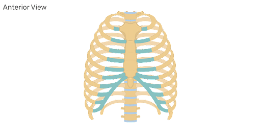
Structure of Rib Cage
Atypical ribs
The ribs whose features are uncommon to other ribs are known as atypical ribs. ‘Atypical’ ribs are 1st, 2nd,10th, 11th and 12th ribs.
- Rib 1: This rib has only one facet on its head through which it articulates with the corresponding vertebra. These grooves make way for the subclavian vessels. Unlike other ribs, it is shorter and wider.
- Rib 2: The head of this rib has two facets as normal. Its upper surface area is rough which acts as a source of origination of Serratus anterior muscle.
- Rib 3: It is characterized by the presence of one facet. This facet articulates with the numerically corresponding vertebra.
- Ribs 11th and 12th: These ribs have one facet through which the corresponding vertebra articulates with it. They do not consist of the neck.
-
Articulations
Anterior and posterior articulations are found in the majority of the ribs. These are as follows-
Posterior Articulation
The ribs are articulated to the vertebra of the spine posteriorly. Posterior articulation is found in all 12 ribs. Two joints are formed by each rib which is as follows-
- Costotransverse joint: This joint is present between the tubercle of the rib and the transversal costal facet of the corresponding vertebra.
- Costovertebral joint: The head of the rib is articulated with the superior costal facet of the corresponding vertebra as well as the inferior costal facet of the vertebra above.
Anterior articulations
They are as follows-
- Sternum is attached anteriorly by the 1-7 ribs independently.
- 8-10 ribs articulate anteriorly to the costal cartilages which are superior to them.
- Ribs 11 and 12 are also called floating ribs because they do not show an anterior attachment. They end in the abdominal musculature.
Ribcage:Bones
[Click Here for Sample Questions]
The uppermost rib of the ribcage is the shortest and most curved among all ribs. It is broad and flat. The second rib is also similar in curvature to the first but longer. It is connected to the vertebral column posteriorly by the second thoracic vertebra. It looks upward and outwards and its external surface is convex. The internal part is smooth and concave and a little inward.
- At the level of the same lumbar vertebrae, there lies the ninth rib. This level is known as the transpyloric plane.
- The tenth rib is directly attached to the T10 vertebra body. Therefore, it has completed the costal facet to the body on its body.
- The last two ribs are known as floating ribs and they have a single articular facet on their head. They do not have a neck and tubercle. The 11th rib has a shallow coastal and a slight angle which is absent in the 12th ribs. The 12th rib head is inclined downwards and is much shorter than the 11th rib.
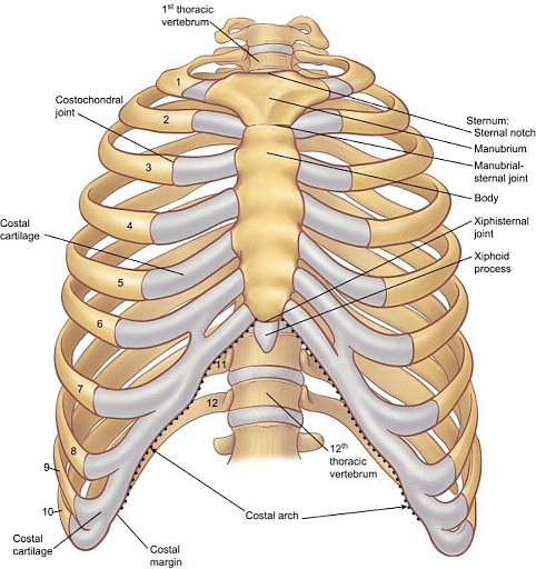
Ribcage:Bones
Muscle Attachments
[Click Here for Sample Questions]
The following muscles are related to ribs –
- The muscles which are present in between the intercostal spaces are known as intercostal muscles.
- On the 6th rib of costal cartilage, the diaphragm arises from the inner surface.
- From the 1st to 8th ribs, Serratus anterior originates anterolaterally.
- Superior anterior ribs serve as the point of origination for the Pectoralis major and minor muscles.
- On the first and second ribs, Scalenus anterior, posterior and medius muscles are attached.
- At the Xiphisternum and the 5th to 7th costal cartilages, rectus abdominalis is present.
Functions of Rib Cage
[Click Here for Sample Questions]
The functions performed by the ribcage are as follows-
- Contents of the thoracic medium such as lungs, heart are well protected by the ribcage.
- They play an important role in breathing as ribs have the flexibility so that they increase or decrease the size of the thoracic cavity. They can move anteriorly, posteriorly, inferiorly and superiorly to assist in breathing.
- They also serve as the place of attachment and origination for different muscles.
- They play a major role in erythropoiesis during development. Erythropoiesis is the process that is responsible for the production of Red Blood Cells.
Clinical Relevance
12% of total fractures are comprised of ribs fractures. It occurs mainly as a consequence of injuries or direct trauma. Middle ribs are commonly affected. Rib fractures can also lead to tissue injury and can damage the lungs, spleen or diaphragm. Such injuries should get immediately treated by specialised doctors. They diagnose such injuries by X-rays and give proper treatments.
Things to remember
Rib cage is a cage-like structure made up of twelve pairs of ribs enclosing the thoracic cavity.
- Delicate organs such as the lungs, heart are well protected by the Ribcage.
- Ribs are connected ventrally to the Sternum commonly known as the breastbone and dorsally to the vertebral column.
- Ribs are further classified as true ribs (articulates with the sternum directly), false ribs (indirectly articulates with the sternum) and floating ribs ( don’t articulates with the sternum).
- Ribs are semi-rigid so they can aid in breathing by increasing or decreasing the size of thoracic cavity.
- Different muscles are connected with ribs that function actively during respiration.
- Production of red blood cells takes place in the marrow spaces of ribs.
- Rib fractures can occur by some injuries or trauma and even can lead to tissue injury.
- One should immediately consult a specialist for the proper diagnosis and treatment of ribs fractures as any delay can worsen the injury.
Sample Questions
Ques. How do the Ribcage aid in respiration? (5 marks)
Ans. The ribcage does the following functions during breathing…
- During inhalation, the chest wall moves forward and upward at the breastbone and upper ribs by moving in a pump handle fashion.
- Vertebral column extends which leads to the expansion of breastbone further forward.
- Diameter of ribcage is increased in forward to backward portion.
- The thoracic volume increases as the thoracic cavity opens by the sides of ribcage in bucket handle motion as well as caliper motion of the lower ribs.
- The diaphragm is pulled down by the central tendon of the diaphragm.
- These movements cause an increase in the volume of the thoracic cavity.
- During expiration, the external intercostal muscles, diaphragm and ribs return to their original state.
Ques. Write all the joints present in the ribcage? (5 marks)
Ans. The joints present in the ribcage are as follows-
- Xiphisternal joint: At the lower end of the sternum, a cartilaginous section is presently known as the Xiphoid process. Xiphisternal joint connects the Xiphoid process with the body of sternum.
- Intervertebral joint: It is present between the vertebrae of the Vertebral Column.
- Sternochondral joint: This joint is present between the sternum and the costal cartilages.
- Sternoclavicular joint: Manubrium and Clavicles are joined by this joint. The manubrium is the superior part of the Sternum.
- Manubriosternal joint: It is present between Manubrium and the sternum.
- Costochondral joint: This joint connects ribs with the costal cartilages.
- Costovertebral joint: This joint is formed by the body of the vertebra and the ribs.
- Interchondral joint: Costal cartilages are joined to one another by this joint.
Ques. Describe the bony Components of the Ribcage? (3 marks)
Ans. Ribs, Sternum and the Vertebral Column are the bony Components of Ribcage. They are as follows-
- Ribs- There are twelve pairs of ribs present in the Ribcage. The main functions of ribs are protection, support and respiration. They elevate during inspiration and get depressed during expiration.
- Sternum- Commonly known as breastbone, it serves as a point of attachment for most of the ribs and collar bone. It also protects the organs of the torso such as the heart, blood vessels and lungs .
- Vertebral Column- All the ribs are attached to the Vertebral Column. It also encloses the spinal cord which is an important part of the Central nervous system.
Ques. How does erythropoiesis take place in the ribs? (3 marks)
Ans. Ribs are related to the erythropoiesis in the following ways:
- Erythropoiesis is the process of the production of the Red blood cells.
- In adults, erythropoiesis mainly takes place in the bone marrow spaces of ribs, sternum, vertebrae and pelvis.
- In these regions, the red blood cells are formed from the erythroblasts which have nuclei.
- As a result of successive cell division, they get proliferated. They become mature and haemoglobin appears in them. Nucleus gets smaller.
- The cell loses its nucleus after few days and is introduced to the bloodstream .
Ques. What are the different causes of Muscle injury? (3 marks)
Ans. The different causes of Muscle injury are as follows:
- In older people, falls are the major causes of rib fractures. It is also responsible for the higher mortality and morbidity of the old people as compared to younger ones .
- Due to cancer of certain organs, rib fractures can occur due to metastasis. They become fragile and weak.
- Athletes can face rib fractures due to microtrauma or by giving certain stress to the ribs.
- Ones who are suffering from osteoporosis, severe cough can lead to rib fractures.
- Major cause of rib fractures are road accidents, falls, excessive exercise, etc.
Ques. What are the common abnormalities related to the ribs ? (3 marks)
Ans. The abnormalities related to the ribs are as follows:
- Rib dislocation-It is a painful musculoskeletal injury in which one or more ribs shifts from their normal position. It occurs due to some accident, strenuous exercise, etc.
- Osteoporosis– It is a condition when the density of bone decreases and bones become weak and brittle. Even a slight injury can lead to major fractures in such condition.
- Costochondritis- It is the condition in which the cartilages joining the ribs to the Sternum gets inflamed. It generally develops after a trauma or muscle injury.
- Rib tumours- Certain benign tumours of the ribs are fibrous dysplasia, enchondroma and osteochondroma. They cause severe pain, soreness and swelling of the affected area.
Ques. Do all the ribs of a ribcage are similar in strength? (3 marks)
Ans. No, all ribs of ribcage are different in their strength. They are as follows-
- 1st and 3rd ribs are the hardest and strongest. Only a high degree of trauma can cause fractures in these ribs.
- Among all the 12 ribs, the most vulnerable and weak ribs are from 4 to 10. They are prone to fractures as they are not as strong as the first three ribs.
- The 11th and 12th ribs are commonly known as floating ribs because they are mobile. They are more difficult to break.
Ques. Describe the Structure of Sternum? (3 marks)
Ans. The sternum is divided into three parts from anterior to posterior in mammals. These are as follows-
- The anterior-most segment is called the Manubrium. It connects the first rib with the Clavicles. It has depressions for the joining of ribs. Manubrium is nearly trapezoidal in shape.
- Mesosternum is the middle segment of the Sternum. It is further divided into Sternebrae which is a series of segments. Remaining true ribs are attached to Sternebrae. During childhood or early adulthood, it consists of four Sternebrae that are fused. It has facets for the articulation of ribs.
- Xiphisternum is the posterior segment that is reduced to a small, cartilaginous Xiphoid process. In middle age, the Xiphoid process may ossify and fuse to the mesosternum.
Ques. Describe the functions performed by the ribcage? (3 marks)
Ans. Ribcage performs important functions which are very essential to sustain life. These are as follows-
- Rib cage protects the vital organs such as lungs, heart, blood vessels, etc from several injuries or trauma.
- Different muscles which perform different functions are either attached to the Ribcage or originate from it.
- They assist in respiration due to their flexibility. They are responsible for increasing or decreasing the size of thoracic cavity.
- Red blood cells are also produced in the bone marrow space of ribs. This process is known as erythropoiesis.
Ques. How the ribcage is held together? (3 marks)
Ans. The ribs in the Rib cage are held together in the following manner-
- Pliable cartilages called Costal cartilages attach the first seven ribs to the Sternum. These ribs are also known as true ribs.
- Among remaining five false ribs, Costal cartilages connects the first three ribs to the cartilage above them
- The last two ribs also known as floating ribs have their cartilages ending in the muscles in the abdominal wall.
- The lower part of the Rib cage is expanded due to the configuration of the lower five ribs. This ensures the proper movement of the diaphragm.
- The diaphragm has an extensive origin from the Rib cage and vertebral column. Ligamentous attachments limit the motion between the ribs and vertebrae.
Check-Out:



Comments