



Content Curator
Temporary mount of a leaf peel show stomata under a microscope. Leaves consist of microscopic pores on both surfaces. These microscopic pores are called stomata. In dicots lower surface of the leaf shows more stomata. In a monocot leaf, both the surfaces show equal distribution of stomata. Stomata are meant for the exchange of oxygen, carbon dioxide, for photosynthesis and also in the transpiration of water vapor.
| Table of Content |
Key terms: Leaf, Stomata, guard cells, photosynthesis, respiration, transpiration, bryophyllum
Stomata Structure
[Click Here for Sample Questions]
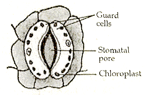
The stomata consists of an opening called the stomium. On either side of the stoma, two bean-shaped guard cells are present. Each guard cell is characterized by a thicker inner wall and a thinner outer wall.
| Important Topics | ||
|---|---|---|
| What is Tissue? | Protoplasm | Radicle |
| tendon | what is tissue? | Plumule |
Opening and Closing of Stomata
[Click Here for Sample Questions]
During the daytime plants prepare carbohydrates. They increase the turgidity of guard cells. The guard cells are opened. In the nighttime, the reverse happens.

Experiment To Show Stomata On The Leaf Surface
[Click Here for Sample Questions]
Aim: To show stomata under a microscope by preparing the temporary mount of a leaf peel
Principle: Plants are the green autotrophs. They prepare their own food by photosynthesis. Just like other organisms, they have to perform respiration also. To absorb water from the soil and to maintain the microclimate of the plant they have to perform transpiration also. The exchange of gases and water vapour takes place through stomata on the leaf.
Read more: Tendril
Materials required: Bryophyllum or Tradescantia plant, compound microscope, blotting paper, brush, coverslips, dropper, forceps, glycerine, needles, safranin solution, slides, and watch glass.
Procedure:
1) Select a healthy potted Bryophyllum or Tradescantia plant and pluck an undiseased and well-grown leaf from it.
2) Gently snip the leaf apart to get a thin film of epidermal peel from the lower surface of the leaf.
3) Place the peel in the watch glass by using forceps.
4) Pour a few drops of safranin solution into the watch glass so that the peel gets stained.
5) After a few minutes, take out the peel from the watch glass and place it on a glass slide by using a brush.
6) Add a few drops of glycerine over the peel to avoid desiccation and cover it gently with a coverslip.
7) Remove excess glycerine around the coverslip by using clean blotting paper.
8) Place the slide on the stage of the compound microscope and examine the peel under both low and high power magnification lenses.
Observations: Epidermal cells without intercellular spaces can be seen. A number of stomata are found to be scattered all over in between the epidermal cells. On either side of each stoma, two kidney-shaped guard cells can be noticed. Under high power lens chloroplasts and nucleus can be seen within guard cells. A thick concave-shaped outer wall of the guard cell is conspicuous.
Inference: Stomata are seen in between the guard cells on the lower surface of the dicot leaf.
Precautions:
1) Place the peel at the centre of the slide and coverslip.
2) Stain the peel moderately.
3) Do not leave any air bubbles inside the coverslip.
Read more: Difference between Mitochondria and Plastids
Things to Remember
[Click Here for Sample Questions]
- The tiny openings on the surface of leaves are called stomata.
- Stomata are the modified epidermal cells.
- Each stoma is flanked by two kidney- or dumbbell-shaped guard cells.
- The guard cell is characterized by a thicker inner wall and a thinner outer wall.
- In the dicot leaf, more stomata are present on the lower surface. In the monocot leaf, stomata are equally distributed on the lower and upper epidermis.
- Stomata are generally opened during the daytime and closed during nighttime.
- Stomata are useful for the exchange of gases and water vapour during photosynthesis, respiration, and transpiration.
- Bryophyllum or Tradescantia leaf peels are used to examine stomata under the microscope.
Sample Questions
Ques 1: Why do you do a temporary mount of a leaf peel experiment? (2 marks)
Ans: I do a temporary mount of a leaf peel experiment to examine stomata under a microscope. Leaves consist of microscopic pores on both surfaces called stomata. Stomata distribution varies on the lower and upper surfaces of leaves.
Ques 2: What are the differences between the stomata of a dicot leaf and a monocot leaf? (2 marks)
Ans: 1) In dicots lower surface of the leaf shows more stomata. In a monocot leaf, both the surfaces show equal distribution of stomata. 2) In dicots, the stomata are bean-or kidney-shaped. In monocots, the stomata are dumbbell-shaped.
Ques 3: What are three important functions performed through stomata? (2 marks)
Ans: 1) Plants get carbon dioxide for photosynthesis through stomata. 2) Plants get oxygen for respiration through stomata 3) Plants lose excess water in the form of water vapour through stomata during transpiration.
Ques 4: Describe the structure of typical stomata? (2 marks)
Ans: The typical stomata consists of an opening called the stoma. On either side of the stoma, there are two bean- or dumbbell-shaped guard cells. The guard cell is characterised by a thicker inner wall and a thinner outer wall.
Ques 5: Why do stomata open during the daytime and closed during nighttime? (3 marks)
Ans: Stomatal movements depend on the presence or absence of sugars and starch in guard cells. During the daytime, there is plenty of sunlight. Chloroplasts in plants prepare sugars by photosynthesis. Sugars increase the osmotic potential of guard cells, turgidity increases and consequently stomata open. In the nighttime, sugars will be converted into starch. So, the osmotic potential of guard cells decreases, flaccidity increases, and stomata close.
Ques 6:A student is observing the temporary mount of a leaf peel under a microscope. Draw a labelled diagram of the structure of the stomata as seen under the microscope. (3 marks) [CBSE Delhi, Set 1, 2019]
Ans: The following is the diagram of the stomata
Ques 7: What do you observe under the microscope during the Tradescantia leaf peel experiment? (3 marks)
Ans: There are three important features that can be studied under the microscope.
1) A number of epidermal cells all over the peel without intercellular spaces.
2) A number of stomata are scattered across the epidermal layer. The stomata consist of a stomatal pore, flanked by two kidney-shaped guard cells.
3) The guard cells consist of a number of chloroplasts, a nucleus, an inner thick cell wall and an outer thin cell wall.
Ques 8: Identify the observed various parts of the temporary mount of well-stained leaf peel, when focussed under the high power of a microscope. (3 marks) [CBSE, Term 1, Set 2, 2015]
- Parts of the temporary mount of well-stained leaf peel are
(i) Stomatal aperture (opened) or (closed)
- Guard cells
- Chloroplast
- Nucleus
- Epidermal cells:
Ques 9: List the steps of preparation of temporary mount of a leaf Peel to observe stomata. (3 marks) [CBSE, 2018]
- (i) Remove a peel from the lower surface of a healthy leaf using forceps and put it in water.
(ii) Put a few drops of safranin stain solution in a watch glass.
(iii) After 2-3 minutes take out the peel and put it on a clean glass slide.
(iv) Put a drop of glycerine solution and a coverslip over the peel.
(v) Avoid air bubbles in the coverslip
(v) Put the slide under a compound microscope.
Ques 10: Write the procedure that you follow to conduct an experiment to study stomata under a microscope? (5 marks)
Ans: The following steps are done in the experiment:
1) A healthy potted Bryophyllum or Tradescantia plant is selected and a healthy leaf is carefully plucked.
2) The leaf is gently snipped to get a thin film of epidermal peel from the lower surface.
3) The thin peel is placed on the watch glass by using forceps.
4) A few drops of safranin solution are poured into the surface of the peel.
5) The well-stained peel is placed on a glass slide by using a brush.
6) A few drops of glycerine and coverslip are put over the peel to avoid desiccation.
7) Excess glycerine around the coverslip can be removed by using clean blotting paper.
8) The peel is examined under both low and high power magnification lenses of the compound microscope.
Science Study Guides:



Comments