



Content Curator
Compound Microscope is a high-definition microscope that is used for the magnification of objects or samples. The term microscope can be broken down into two parts: 'micro' meaning tiny or small and ‘scope' meaning viewing or observation.
- A microscope can be thought of as a tool for observing small objects.
- The compound microscope has two or more double convex lenses that are fixed on the two ends of a metallic tube.
- The lower lens that is closest to the object is known as the objective lens; the upper lens that is nearest to the eye of the observer is known as the eyepiece.
- The microscope is used in hospitals and forensic labs to view bacteria, plant and animal cells, and tissues along with various microorganisms.
| Table of Content |
Key Terms: Microscope, Compound, Lens, Condenser, Light, Magnification, Eyepiece, Objective, Optical, Non-Optical, Simple microscope, compound microscope
Compound Microscope
[Click Here for Sample Questions]
A compound microscope is a high-resolution microscope that uses two sets of lenses to provide a 2-dimensional image of the object.
- The word ‘compound’ refers to the use of more than one lens in the microscope.
- It is a type of optical microscope along with a simple microscope.
- The difference between a simple and compound microscope is that a simple microscope only uses one lens while a compound microscope uses more than one lens.
- Two magnifying lenses are utilized instead of one to achieve better magnification.
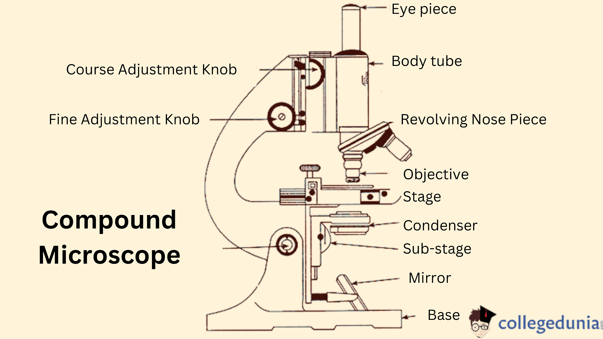
Compound Microscope Schematic
The basic components of a compound microscope are –
- Objective Lens: It is a convex lens placed near the object. The object is placed beyond the focal point and the image formed is real, inverted, and magnified.
- Fine adjustment screw: The screw is used to adjust the position of the objective lens. Even a small movement in the screw causes a huge change in magnification.
- Eyepiece: It is a convex lens that is placed near the eye. Its position is designed such that the image from the objective lens falls between its focus and center.
- Rough adjustment screw: The screw is used to form a clear image and focus the correct part of the object that is to be seen.
The objective lens (usually 4x, 10x, 40x, or 100x) is compounded (multiplied) by the eyepiece lens (often 10x) to give a high magnification of 40x, 100x, 400x, or 1000x in a compound microscope.
The video below explains this:
Compound Microscope Detailed Video Explanation:
Read More:
Working Principle of Compound Microscope
[Click Here for Previous Year Questions]
Compound microscopes use a combination of lenses to improve magnification and resolving power.
- The sample to be examined is mounted on a clear glass slide and placed between the condenser and objective lenses on the specimen stage.
- A condenser lens concentrates a beam of visible light from the base on the specimen.
- The objective lens collects the light emitted by the sample and magnifies it inside the body tube to form a primary image. The ocular lens or eyepiece enlarges this image once more.
- When a higher magnification is necessary, the nose piece of the compound microscope is turned after low-power focusing to align the higher magnification objective (usually 45X) with the lit section of the slide.
- It was sometimes necessary to use extremely high magnification. An oil immersion objective lens (often 100X) is used in this situation.
- As the image is created inside a brightly lighted field, the common light microscope is also known as a bright-field microscope.
Parts of Compound Microscope
[Click Here for Sample Questions]
A compound microscope gives information on the structural details of cells, tissues, and organs. The parts of a compound microscope are:
Optical Parts
There are 5 optical parts of a compound microscope. They are:
- Diaphragm: It controls the amount of light falling on the object. It is present below the stage. Disc and Iris are the two types of diaphragms.
- Condenser Lens: It is located below the diaphragm and facilitates the focusing of light.
- Reflector: It is attached above the base and helps in directing the light falling on the object with the help of the diaphragm and condenser.
- Objective lens: It is placed above the nosepiece and forms a real inverted image of the object inside the tube.
- Ocular Lens: It is also called the eyepiece and facilitates four types of magnifications - 5, 10, 15, and 20X.
Working
- A condenser gathers and focuses light from the illuminator onto the specimen being viewed.
- The diaphragm of Iris controls how much light reaches the specimen.
- A five-holed disc is put beneath the stage as the diaphragm.
Non-Optical Parts
The non-optical parts of a compound microscope are as follows:
Arm, Pillar, and Base
The compound microscope's Body Tube is connected to the Microscope's base by the Arm.
- The Microscope and its Illuminator are supported by the Base.
- The base and the arm are connected by a pillar.
Illuminator and Stage
The illuminator is the light source of a compound microscope.
- The illuminator in a compound light microscope is usually a low-voltage bulb.
- The slide is positioned on the stage, which is a level platform.
Nosepiece and Aperture
The objective lenses are held in place by a revolving turret known as the nosepiece.
- The viewer rotates the nosepiece of the compound microscope to change the objective lens.
- The aperture is the opening in the middle of the stage that permits illuminator light to reach the specimen.
Magnifying Power of Compound Microscope
[Click Here for Sample Questions]
The formula for the magnifying power of a compound microscope when the final image is formed at least distance of distinct vision is given by
\(M.P. = \frac {L}{f_o}[1+\frac {D}{f_e}]\)
The formula for the magnifying power of a compound microscope when the final image is formed at infinity is given by
\(M.P. = \frac {L}{f_o}\times \frac {D}{f_e}\)
Where
- L is the length of the compound microscope
- D is the least distance of distinct vision i.e. 25 cm.
- fo is the focal length of the object lens
- fe is the focal length of the eyepiece.
Uses of Compound Microscope
[Click Here for Previous Year Questions]
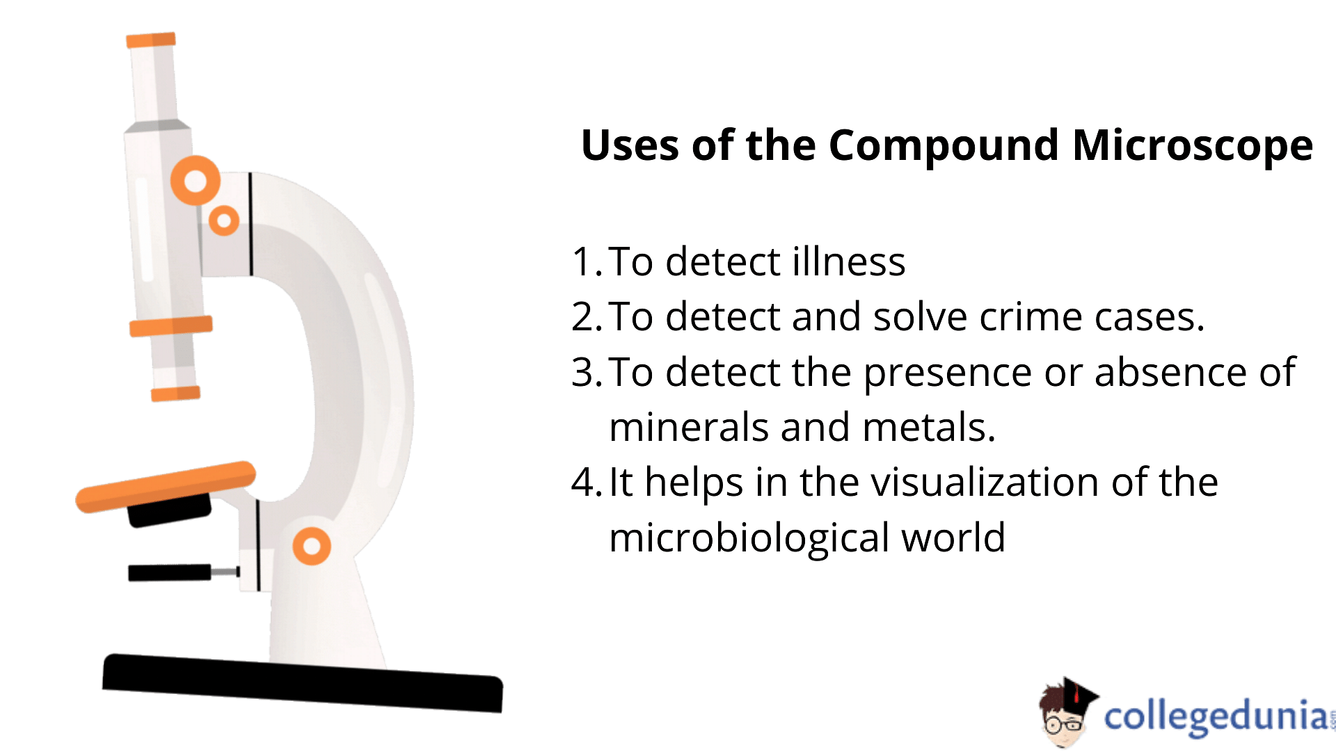
The major uses of the Compound Microscope are as follows:
- In pathology labs, a compound microscope is extremely useful for detecting illnesses.
- Human cells are drawn and examined under a microscope in forensic laboratories to detect and solve various crime cases.
- Compound microscopes can be used to detect the presence or absence of minerals as well as the presence of metals.
- The employment of a microscope in academic activities by students in schools and colleges has proven to be beneficial.
- It helps in the visualization and comprehension of the microbiological world of bacteria and viruses, which is otherwise undetectable to the naked eye.
Compound Microscope Uses
Advantages of Compound Microscope
[Click Here for Sample Questions]
The advantages of a compound microscope are:
- The ease of simplicity
- As a compound light microscope is compact, it is easy to use and store.
- It has its own light source.
- Compound microscopes may reveal a lot of information in samples because of the numerous lenses they have.
Precautions while using Compound Microscope
The Precautions to be taken care of while using a Compound Microscope are:
- Tilting or shaking the microscope while it is in use must be avoided.
- The light received by the lens is altered as a result of this bouncing, and the viewing of the focus item changes.
- The specimen must first be focused with a low-resolution lens before being targeted with a high-resolution lens.
- The lower power objective lens must be focused on the stage once you've finished using the compound microscope.
- Both arms and the base of the microscope should be used to lift the microscope.
- When using an oil immersion lens, one should use the oil to get the best results as the refractive index can be altered without the use of oil, and the reading of the specimen can be manipulated.
- Use a clean slide or clean the slide with suitable water and wipe it clean so that any stains do not show up under the microscope. Otherwise, it can be difficult for the user to distinguish between a stain or a component of the specimen.
Things to Remember
- A compound microscope magnifies the image under the microscope using two sets of lenses.
- The compound microscope has two or more convex lenses.
- It has a 4x, 10x, 40x, and 100x objective lens as well as a 10x eyepiece.
- Small samples that can't be seen with the naked eye are seen using compound microscopes.
- Under the microscope, these samples are usually mounted on a slide.
- Both arms and the base of the microscope should be used to lift the microscope.
Also, Check Out:
Previous Year Questions
- Frequency of Hollow cylinder when immersed in water. [WB JEE 2008]
- The ratio of frequencies of Pth overtone of a pipe. [MHT CET 2019]
- Calculate the maximum particle velocity. [MHT CET 2008]
- Calculate Brewster’s angle at the interface. [NEET 2020]
- Relation between the electric and magnetic field of a wave. [NEET 1994]
- Calculate the speed of sound in the air. [NEET 2019]
- Condition for the coherence of two light sources. [VITEEE 2010]
- The minimum distance in a bi-chromatic light. [JEE Advanced]
- Calculate the original frequency of the guitar springs. [NEET 2020]
- Calculate the speed of the wave on the spring. [NEET 1989]
- Calculate the intensity of sound when frequency and amplitude are changed. [NEET 1989]
- Calculate the frequency of sound from a vehicle. [NEET 1998]
- Calculate the frequency of the sound wave. [NEET 1990]
- Which electromagnetic wave does not emit electrons. [NEET 1990]
- Calculate the value of the wavelength of a transverse wave. [NEET 1998]
- Calculate the number of beats from the two strings. [NEET 2009]
- Calculate the velocity of sound in the air from a tuning fork. [NEET 2018]
- Calculate the frequency of a sinusoidal wave. [NEET 1992]
- Calculate the velocity of a wave from the wave equation. [NEET 2001]
- Calculate the phase difference between the two waves. [NEET 2001]
Sample Questions
Ques: What are the advantages of using a compound microscope? (2 Marks)
Ans. Compound microscopes are used to examine small samples that are difficult to recognize with the naked eye. Under a microscope, these samples are usually put on a slide. When utilizing a stereo microscope, there is more room beneath the microscope for larger materials like rocks or flowers, and slides are not necessary.
Ques: What are the characteristics of a compound microscope? (2 Marks)
Ans. Compound Microscope Characteristics:
- There are two or more convex lenses in this set.
- Magnification ranges from 40x to 1000x in most cases.
- At a time, only one objective is used.
- Images in only two dimensions.
- Monocular, binocular, trinocular, and multi-head setups are available.
Ques: What is the process of magnifying an object with a compound microscope? (2 Marks)
Ans: A microscope is a device that allows you to examine small objects, including cells. The picture of an object is enlarged in the microscope by at least one lens. This lens bends light toward the eye, giving the impression that an item is larger than it is.
Ques: What are some of the drawbacks of using a compound microscope? (2 Marks)
Ans: Compound Microscopes: What Are Their Limitations? Compound microscopes have two major drawbacks: light and working distance. Although compound microscopes may attain exceptionally high magnifications (up to 2,500x), some specimens are so minute that even at these extreme magnifications, they are not visible.
Ques: What are the three primary parts of a compound microscope? (3 Marks)
Ans: The head, base, and arm are the three structural components of a compound microscope.
- The optical optics are housed in the upper half of the microscope in the Head/Body.
- The illuminator is housed in the base of the microscope, which also serves as a support for the microscope.
- The arm attaches to the base and holds the microscope head in place.
Ques: In a compound microscope, how is an image formed? (2 Marks)
Ans: The intermediate image created by the objective lens in a compound microscope is real. The role of the eyepiece is to create a virtual, magnified image for your eye to observe in all instances. An objective lens and a magnifier, or eyepiece, make up the microscope.
Ques: In a microscope, what is the role of the eyepiece? (2 Marks)
Ans: The eyepiece, also known as an ocular, amplifies the primary image produced by the objective, allowing the eye to use the objective's full resolution. The microscope creates a virtual image of the specimen at the point when vision is the clearest, which is usually 250 mm.
Ques: A compound microscope has angular magnification equal to 10. If the object subtends an angle of 0.6∘ to the eye, what will be the angle subtended by the image to the eye? (3 Marks)
Ans: Given
- Magnifying power, MP = 10
- The angle subtended by the object to the eye, α = 0.6∘
The formula for magnifying power is given by
MP = β/α
⇒ β = MP x α
Where β is the angle subtended by the image to the eye.
On substituting the values, we get
β = 10 x 0.6 = 6∘
Ques: The tube of a microscope is of length 15 cm. Focal length of the eyepiece and objective are 3 cm and 2 cm. Find the magnifying power of the microscope for an image formed at infinity. (2 Marks)
Ans: When the final image is formed at infinity, then the magnifying power of a compound microscope is given by
MP = L/fo x D/fe
On substituting the values, we get
MP = 15/2 x 25/3 = 37.5
Ques: What are zooming microscopes? (2 Marks)
Ans: A zooming microscope is a type of microscope equipped with a knob for adjusting magnification. This feature makes it a flexible choice for medium and high-quality stereomicroscopes.
For Latest Updates on Upcoming Board Exams, Click Here: https://t.me/class_10_12_board_updates
Also Read:



Comments