




CBSE Class 12 Biology Set 1 - (57/3/1) Question Paper 2026 is available for download here. CBSE conducted Class 12 Biology exam on March 27, 2026 from 10:30 AM to 1:30 PM. The Biology theory paper is of 70 marks, and the internal assessment is of 30 marks.
Biology question paper includes MCQs (1 mark each), short-answer type questions (2 & 3 marks each), case-study based questions (4 marks each) and long-answer type questions (5 marks each) which makes up the total of 70 marks.
Download CBSE Class 12 Biology Set 1 - (57/3/1) Question Paper 2026 with detailed solutions from the links provided below.
CBSE Class 12 Biology Question Paper 2026 (Set 1 - 57/3/1) with Solution PDF
| CBSE Official PDF | CD PDF | CD PDF with Solutions |
|---|---|---|
| Download official PDF 1.13 MB | Download Collegedunia PDF 0.86 MB | Download solutions PDF 0.77 MB |






Foetal ejection reflex in human female triggers the release of which hormone?
View Solution
Step 1: Understanding the Question:
The question asks about the specific hormone whose release is triggered by the foetal ejection reflex in a human female, along with the precise anatomical site of its secretion.
The foetal ejection reflex is the neuroendocrine mechanism that initiates parturition (the process of childbirth).
Step 2: Key Formula or Approach:
The process of parturition is governed by a fully coordinated neuroendocrine reflex loop.
The signals for childbirth originate from the fully mature foetus and the placenta, which induce mild uterine contractions. This initial phase is formally defined as the foetal ejection reflex.
Step 3: Detailed Explanation:
When the foetus is completely developed, it, along with the placenta, generates signals that initiate mild, rhythmic contractions of the uterine myometrium.
These mild contractions stimulate the mechanical stretch receptors located in the cervix of the mother.
The activation of these receptors generates nerve impulses that travel via afferent pathways to the maternal hypothalamus.
In response, the maternal hypothalamus signals the posterior pituitary gland (neurohypophysis) to release the hormone oxytocin into the maternal bloodstream.
Oxytocin acts directly on the uterine smooth muscles, causing stronger and more vigorous uterine contractions. These enhanced contractions further stimulate the stretch receptors, leading to an increasing, positive-feedback loop that culminates in the expulsion of the baby.
While the foetal pituitary gland does secrete trace amounts of oxytocin during late pregnancy, it is the maternal pituitary gland that releases the massive surge of oxytocin required to drive the mechanical process of labour.
Therefore, options (A), (B), and (C) are incorrect because hCG maintains the corpus luteum, progesterone maintains pregnancy, and the essential oxytocin surge comes from the mother, not the foetus.
Step 4: Final Answer:
The foetal ejection reflex triggers the release of oxytocin from the maternal pituitary gland, which corresponds perfectly to option (D).
Quick Tip: Remember that labor and childbirth are controlled by a classic positive-feedback reflex loop. The signal starts from the foetus, but the heavy mechanical force (uterine contractions) is powered by oxytocin released directly from the maternal posterior pituitary gland.
Which of the following is not a functional unit of an ecosystem?
View Solution
Step 1: Understanding the Question:
The question asks us to identify which of the four given options is not classified as a functional unit (or component) of an ecosystem.
An ecosystem consists of structural components (how it is physically built) and functional components (how it dynamically operates).
Step 2: Key Formula or Approach:
According to standard ecological principles, the four primary functional attributes that allow an ecosystem to operate as a self-sustaining unit are:
1. Productivity (biomass synthesis)
2. Decomposition (nutrient recycling)
3. Energy flow (unidirectional movement of energy through trophic levels)
4. Nutrient cycling (biogeochemical cycles)
Step 3: Detailed Explanation:
Let's analyze each of the choices presented:
* Productivity (Option D): This is a critical functional attribute representing the rate of biomass production by autotrophs through photosynthesis per unit area over a specified time interval.
* Decomposition (Option B): This is the dynamic metabolic process where decomposers break down complex organic matter into inorganic nutrients, facilitating nutrient cycling.
* Energy flow (Option A): This describes the unidirectional thermodynamic transfer of energy from producers to various consumer levels.
All three of these options are actively dynamic processes and represent the functional machinery of an ecosystem.
* Stratification (Option C): This refers to the vertical distribution of different species occupying different levels within a biotic community. For example, in a forest ecosystem, trees occupy the top vertical strata, shrubs occupy the middle layer, and herbs/grasses occupy the bottom layers. Stratification is a structural property of a biological community, not a functional process.
Step 4: Final Answer:
Since stratification is a structural component and not a functional unit of an ecosystem, it is the correct choice, corresponding to option (C).
Quick Tip: To easily separate ecosystem components, ask yourself: Is it a physical arrangement or an ongoing process? Dynamic processes like energy transfer, decomposition, and production are functional units, whereas physical arrangements like vertical zoning (stratification) are purely structural features.
Match Column I with Column II and select the correct option:

View Solution
Step 1: Understanding the Question:
This problem requires matching key terms from biotechnology and genetic engineering listed in Column I with their correct functional descriptions listed in Column II.
Step 2: Detailed Explanation:
Let us evaluate each item in Column I one by one:
* a. Primers: In biotechnology, specifically during the Polymerase Chain Reaction (PCR), primers are essential components. They are defined as short, chemically synthesized oligonucleotides (usually about 18–22 nucleotides long) that are strictly complementary to the regions of the target template DNA strand. Therefore, a maps to iii.
* b. Insertional inactivation: This is a powerful selection method used to screen for recombinant plasmids. When a foreign gene of interest is cloned inside the coding sequence of an enzyme like \(\beta\)-galactosidase (encoded by the lacZ gene), the sequence is disrupted. As a result, the bacteria containing recombinant plasmids lose the ability to synthesize functional \(\beta\)-galactosidase and cannot produce a blue colour on X-gal media. Therefore, b maps to i.
* c. Bioreactor: A bioreactor is a large engineering vessel (typically holding 100 to 1000 liters) designed to provide optimal growth conditions (pH, temperature, oxygen, substrates) for the large-scale culture and production of specific proteins or metabolites by microbial, plant, or animal cells. Therefore, c maps to iv.
* d. Downstream processing: Once the biosynthetic stage in a bioreactor is finished, the product must go through a sequence of separation and purification steps before it can be formulated into a final market product. This post-fermentation phase is known as downstream processing. Therefore, d maps to ii.
Combining these mappings gives the sequence: a-iii, b-i, c-iv, d-ii.
Step 3: Final Answer:
The matching sequence is correctly represented by option (A).
Quick Tip: In matching matrix questions, pick the most distinct terms first. For instance, "Downstream processing" uniquely refers to "separation and purification," and "Primers" are always "oligonucleotides." Finding even one or two of these matches will instantly narrow down or solve the entire question.
Golden rice is a promising transgenic crop. When released for cultivation, it will help in :
View Solution
Step 1: Understanding the Question:
The question asks about the primary nutritional and agricultural purpose of developing and cultivating the genetically modified (transgenic) crop known as Golden Rice.
Step 2: Key Formula or Approach:
Golden Rice (\textit{Oryza sativa) is a genetically engineered variety designed for biofortification—the process of increasing the density of vitamins or minerals in a crop through genetic modification.
Step 3: Detailed Explanation:
Standard rice grains naturally produce \(\beta\)-carotene (a precursor of Vitamin A) in the green vegetative leaves, but the biosynthetic pathway is completely inactive in the edible endosperm part of the grain.
To address widespread micronutrient malnutrition in developing nations, scientists introduced two specific genes into the rice genome: the \textit{psy (phytoene synthase) gene from daffodils (\textit{Narcissus pseudonarcissus) and the \textit{crtI (phytoene desaturase) gene from the soil bacterium \textit{Erwinia uredovora.
The successful integration and expression of these foreign genes complete the metabolic pathway inside the endosperm cells, allowing the rice grains to accumulate \(\beta\)-carotene.
This accumulated pigment imparts a characteristic golden-yellow color to the grains, giving it the name "Golden Rice."
When ingested, \(\beta\)-carotene is cleaved and metabolized by enzymes in the human small intestine into active retinol (Vitamin A). Thus, its primary humanitarian and clinical objective is to counteract childhood blindness, xerophthalmia, and immune vulnerabilities associated with Vitamin A Deficiency (VAD).
It has no role in bio-fuel synthesis (A), insect pest resistance (C, which is a property of Bt crops), or herbicide tolerance (D, which is a property of HT crops).
Step 4: Final Answer:
Golden rice is cultivated to reduce Vitamin A deficiency in human populations, which is correctly matching option (B).
Quick Tip: Associate "Golden" with the rich yellow color of \(\beta\)-carotene (just like carrots!). This visual reminder helps you remember that Golden Rice is modified to produce Vitamin A precursors.
Four different transcription units are shown below. Choose the option with the correct image.
(A) 
(B) 
(C) 
(D) 
View Solution
Step 1: Understanding the Question:
The problem presents four different structural arrangements of a transcription unit in DNA. We must identify which schematic diagram correctly establishes the structural conventions governing the orientation of the promoter, terminator, template strand, and coding strand.
Step 2: Key Formula or Approach:
The convention for defining the structural components of a transcription unit is strictly based on the orientation of the coding strand (non-template strand running in the \(5' \rightarrow 3'\) direction):
1. The Promoter is located upstream, towards the \(5'\)-end of the coding strand.
2. The Terminator is located downstream, towards the \(3'\)-end of the coding strand.
3. The Template strand has a polarity of \(3' \rightarrow 5'\) because RNA polymerase synthesizes RNA only in the \(5' \rightarrow 3'\) direction.
4. The Coding strand runs in the \(5' \rightarrow 3'\) direction and shares an identical sequence with the synthesized mRNA (except U replaces T).
Step 3: Detailed Explanation:
Let's evaluate the structural logic of the diagrams based on these rules:
* Look closely at diagram (C):
* The top strand runs from left to right as \(3' \leftarrow 5'\) (which means \(5'\) is on the right and \(3'\) is on the left). It is explicitly labeled as the Template strand.
* The bottom strand runs from left to right as \(5' \rightarrow 3'\) (with \(5'\) on the left and \(3'\) on the right). It is labeled as the Coding strand.
* Since the coding strand has its \(5'\)-end on the left side, the Promoter must be placed on the left side. The diagram correctly shows the Promoter box on the left.
* Since the coding strand has its \(3'\)-end on the right side, the Terminator must be placed on the right side. The diagram correctly shows the Terminator box on the right.
* The template strand running \(3' \rightarrow 5'\) from right to left provides the correct sequence template for an RNA polymerase molecule to read as it moves from left to right.
Let us check why other options are structurally invalid:
* In option (A), the promoter is placed at the \(5'\)-end of the template strand instead of the coding strand, violating international convention.
* In option (B), the template and coding polarities do not match the standard upstream position of the promoter at the coding strand's \(5'\) terminus.
* In option (D), the positions of the template and coding labels are mixed up relative to the promoter location.
Therefore, diagram (C) represents the mathematically and structurally sound orientation of a transcription unit.
Step 4: Final Answer:
The transcription unit with the completely correct structural labels is given in option (C).
Quick Tip: To solve any transcription unit orientation problem instantly, remember this single rule: \textbf{All structural positions (Promoter and Terminator) are defined entirely with respect to the coding strand.} Promoter is always at the \(5'\)-end of the coding strand; Terminator is always at the \(3'\)-end of the coding strand.
Appearance of antibiotic-resistant bacteria is an example of evolution due to:
View Solution
Step 1: Understanding the Question:
The question asks us to identify the specific evolutionary driver or context that explains the rapid emergence of antibiotic-resistant strains of bacteria.
Step 2: Key Formula or Approach:
Evolution can occur via natural processes over geological time scales, or it can be accelerated within very short time frames due to human interventions. Changes driven directly or indirectly by human activities are classified as evolutionary phenomena due to anthropogenic action.
Step 3: Detailed Explanation:
Before antibiotics are widely used, a massive bacterial population naturally contains a few rare individuals with random genetic mutations that confer resistance to a particular chemical.
When humans introduce antibiotics into clinical and agricultural settings on a massive scale, an intense selective pressure is created.
The antibiotic kills off all sensitive bacteria (the vast majority). The few pre-existing resistant mutants survive and reproduce without competition.
Over a short period, these resistant individuals multiply rapidly, turning the entire population into a drug-resistant strain.
Because this natural selection is driven by the use of synthetic chemical agents created by humans, it is a clear example of evolution by anthropogenic action (human-induced evolution).
Let's review the other options:
* Adaptive radiation (Option A): This describes the evolutionary process where a single ancestral species rapidly diversifies into many different forms to fill different ecological niches (e.g., Darwin's finches).
* Divergent evolution (Option B): This refers to structural diversification from a common ancestral form over long geological periods (e.g., vertebrate limbs).
* Artificial selection (Option C): This involves intentional, deliberate breeding programs managed by humans to select for specific traits, such as breeding domesticated dogs or high-yield crops. Humans do not intentionally breed resistant bacteria; it is an accidental byproduct of medicine.
Step 4: Final Answer:
The appearance of antibiotic-resistant bacteria is an example of microevolution driven by anthropogenic action, corresponding to option (D).
Quick Tip: The word "Anthropogenic" comes from the Greek roots anthropos (human) and genesis (origin). Whenever you see rapid modern evolutionary shifts caused by chemical pollutants, pesticides, or medicines, it is always a direct result of anthropogenic action.
Identify the statements that correctly describe Darwin's Theory of Evolution.
(i) Overproduction of organisms leads to competition.
(ii) Variation is inherited and causes evolution.
(iii) Acquired characters are inherited.
(iv) Survival depends on favourable traits.
(v) New species arise due to accumulation of favourable variations.
Choose the correct option.
View Solution
Step 1: Understanding the Question:
The question requires us to evaluate five statements about evolutionary mechanics and select the option containing only the principles that align with Charles Darwin's Theory of Natural Selection.
Step 2: Key Formula or Approach:
Darwin's core framework rests on several interconnected logical observations:
1. Overproduction of offspring beyond what the environment can support.
2. Limited natural resources, which creates a competitive struggle for existence.
3. Pre-existing phenotypic variations within any population.
4. Natural selection, where individuals with favorable variations survive and reproduce more successfully.
5. Speciation via the gradual accumulation of these inherited favorable variations over generations.
Step 3: Detailed Explanation:
Let's analyze each statement based on Darwinian principles:
* Statement (i): "Overproduction of organisms leads to competition." This is a true statement. Darwin noted that populations grow geometrically, while resources grow arithmetically, causing a struggle for survival.
* Statement (ii): "Variation is inherited and causes evolution." This is true. Darwin recognized that variations must be heritable to serve as raw material for natural selection.
* Statement (iii): "Acquired characters are inherited." This is false. The inheritance of acquired characteristics (use and disuse) is the central concept of Jean-Baptiste Lamarck's evolutionary theory, which Darwin rejected.
* Statement (iv): "Survival depends on favourable traits." This is true. This statement describes differential survival, or "survival of the fittest."
* Statement (v): "New species arise due to accumulation of favourable variations." This is true. Darwin stated that gradual, continuous adaptations accumulate over long periods, leading to speciation.
Since statements (i), (ii), (iv), and (v) are completely accurate and statement (iii) is incorrect, option (A) is the correct choice.
Step 4: Final Answer:
The statements that correctly describe Darwin's theory are (i), (ii), (iv), and (v), corresponding to option (A).
Quick Tip: To solve questions about Darwin vs. Lamarck quickly, look for the phrase "acquired characters." This phrase is a hallmark of Lamarckism. You can immediately cross out any option containing statement (iii), which instantly leaves option (A) as the only possible answer.
Which of the following is not a symptom of Ascariasis ?
View Solution
Step 1: Understanding the Question:
The question asks us to identify which clinical sign or symptom listed among the choices is not caused by an infection of the roundworm parasite Ascaris lumbricoides.
Step 2: Key Formula or Approach:
Ascariasis is a parasitic infection of the human gastrointestinal tract caused by the common intestinal roundworm, \textit{Ascaris lumbricoides, which belongs to the phylum Aschelminthes.
Step 3: Detailed Explanation:
Let's review how \textit{Ascaris spreads and affects the body:
Eggs excreted with the feces of an infected individual contaminate soil, water, and crops. Healthy individuals ingest these eggs by consuming contaminated food or water.
Once inside the human host, the larvae hatch in the small intestine, pass through the intestinal wall, and migrate via the portal circulatory system through the liver and lungs before returning to the small intestine to mature into large adult worms.
The clinical symptoms of a heavy \textit{Ascaris infection include:
* Internal bleeding and Anaemia (Option C): The worms feed on the host's intestinal tissue and blood, which can lead to nutritional deficiencies and microcytic anemia.
* Muscular pain and fever (Option B): The migration of larvae through bodily tissues triggers systemic inflammatory responses, causing low-grade fever and muscle aches.
* Intestinal blockage (Option A): Because adult worms can grow up to 30 cm long, a dense mass of entangled worms can physically block the lumen of the small intestine, requiring emergency surgery.
\textit{ Skin ulcers (Option D): Ascaris lumbricoides* is strictly an internal parasite of the gut and visceral organs. It does not burrow into or colonize cutaneous skin tissue to cause open sores or skin ulcers. Skin ulcers are typical of cutaneous leishmaniasis or certain bacterial infections, not ascariasis.
Step 4: Final Answer:
Since skin ulcers are not a symptom of Ascariasis, option (D) is the correct answer.
Quick Tip: Think of \textit{Ascaris as a giant intestinal roundworm. Because it lives inside the digestive tract, its major symptoms are internal—such as abdominal pain, bleeding, anemia, and intestinal blockages—rather than superficial skin lesions.
Every trophic level has a certain mass of living material at a particular time. What is it called?
View Solution
Step 1: Understanding the Question:
The question asks for the correct ecological term used to describe the total mass of living organic material present at a specific trophic level in an ecosystem at any given time.
Step 2: Key Formula or Approach:
Ecosystem analysis requires distinguishing between biotic (living) and abiotic (non-living) pools of matter:
* The organic biogenic component is quantified as the standing crop.
* The inorganic nutrient component is quantified as the standing state.
Step 3: Detailed Explanation:
Let's define each ecological concept to see why the options match or differ:
* Standing crop (Option A): This is defined as the total biomass or mass of living organic matter present at a given trophic level per unit area at a specific time. It can be measured either as fresh weight or dry weight. Dry weight is more accurate because it excludes fluctuating water content.
* Standing state (Option C): This refers to the total amount of inorganic nutrients—such as nitrogen, phosphorus, calcium, and carbon—present in the soil or abiotic environment of an ecosystem at any given time.
* Primary productivity (Option B): This is a rate function, not a static mass measurement. It represents the velocity at which solar energy is captured and converted into organic matter by autotrophs per unit area over a year.
* Ecological efficiency (Option D): This is a ratio representing the percentage of energy transferred from one trophic level to the next higher level (usually averaging around 10% according to Lindeman's efficiency law).
Step 4: Final Answer:
The mass of living material at a specific trophic level is called the standing crop, which corresponds to option (A).
Quick Tip: Use this word association to remember the difference:
* Standing \textbf{Crop} = Living organic material (think of harvestable biological crops).
* Standing \textbf{State} = Inorganic mineral status of the soil environment.
The smallest part of DNA molecule that can be changed by point mutation is :
View Solution
Step 1: Understanding the Question:
The question asks for the absolute smallest structural unit within a double-stranded DNA molecule that can undergo a modification via a point mutation.
Step 2: Key Formula or Approach:
A point mutation is defined as a genetic mutation where a single base pair in the DNA sequence is altered, substituted, inserted, or deleted.
Step 3: Detailed Explanation:
Let us look at the structural hierarchy of DNA from largest to smallest among the choices:
* Gene (Option C): A large segment of DNA containing hundreds or thousands of base pairs that encodes a functional polypeptide or RNA molecule.
* Oligonucleotide (Option A): A short polymer chain composed of several linked nucleotide units (typically 10 to 50 base pairs).
* Codon (Option B): A specific sequence of three consecutive nucleotides in mRNA (or corresponding triplets in DNA) that specifies a single amino acid during translation.
* Nucleotide (Option D): The fundamental chemical monomer building block of nucleic acids, consisting of a nitrogenous base, a pentose sugar, and a phosphate group.
Since a point mutation involves a chemical alteration to a single nitrogenous base pair (for example, the transition of an Adenine-Thymine pair to a Guanine-Cytosine pair, as seen in Sickle Cell Anemia where a single base substitution occurs in the \(\beta\)-globin gene), the absolute limit of resolution for such a mutation is a single nucleotide site. This site is also referred to in molecular genetics as a muton—the smallest unit of genetic material mutable by a point mutation.
Step 4: Final Answer:
The smallest unit of DNA altered by a point mutation is a single nucleotide, which matches option (D).
Quick Tip: The definition of a point mutation contains the answer itself: "a change in a \textbf{single base pair." Since a base pair is an integral part of a single nucleotide unit, the nucleotide is the smallest unit of mutation.
Some statements regarding drugs and alcohol are given below :
(i) Adolescents are more vulnerable to peer pressure.
(ii) Drug addiction leads to increased tolerance level of the receptors present in our body.
(iii) Alcohol affects the central nervous system.
(iv) Excessive use of drugs helps to increase energy.
(v) Anabolic steroids decrease aggressiveness.
Choose the option with correct statement(s).
View Solution
Step 1: Understanding the Question:
We are given five statements concerning the physiological and behavioral impacts of drugs and alcohol. We must identify which statements are completely accurate and select the option that groups them together.
Step 2: Key Formula or Approach:
Evaluating these statements requires applying verified physiological and psychological principles of substance abuse as outlined in behavioral health and medical biology.
Step 3: Detailed Explanation:
Let us analyze each statement systematically:
* Statement (i): "Adolescents are more vulnerable to peer pressure." This is correct. Adolescence is a transitional psychological phase characterized by curiosity, a desire for experimentation, and a strong need for social acceptance, making teenagers highly susceptible to peer influence regarding drug use.
* Statement (ii): "Drug addiction leads to increased tolerance level of the receptors present in our body." This is correct. With repeated drug exposure, the cell surface receptors in the nervous system adapt to the substance. As their sensitivity drops, the body requires higher doses to achieve the same physiological or psychological effect.
* Statement (iii): "Alcohol affects the central nervous system." This is correct. Alcohol acts as a central nervous system depressant. It binds to GABA receptors, slows down brain activity, impairs motor coordination, delays reaction times, and disrupts emotional control.
* Statement (iv): "Excessive use of drugs helps to increase energy." This is incorrect. While certain stimulants can temporarily mask fatigue, chronic abuse damages metabolic pathways, exhausts organs, and causes widespread physiological decline rather than sustainable energy production.
* Statement (v): "Anabolic steroids decrease aggressiveness." This is incorrect. The abuse of synthetic anabolic-androgenic steroids is clinically linked to increased hostility, severe mood swings, and aggressive behavior (commonly referred to as "roid rage").
Statements (i), (ii), and (iii) are completely accurate, which corresponds directly to option (B).
Step 4: Final Answer:
The option containing the correct statements is (B).
Quick Tip: You can solve this quickly by looking at statement (v). It is well known that anabolic steroids increase aggressiveness rather than decreasing it. Since statement (v) is false, you can eliminate options (A) and (C). Statement (i) is clearly true, which eliminates option (D), leaving option (B) as the correct choice.
Select the correct option for the pair of plants pollinated by water.
View Solution
Step 1: Understanding the Question:
The question asks us to identify the specific option that lists a pair of plants where both species utilize water as their primary environmental agent for pollination (hydrophily).
Step 2: Key Formula or Approach:
Pollination by water, or hydrophily, is a specialized adaptation found in a limited number of aquatic plants. A critical point to remember is that simply living in an aquatic habitat does not mean a plant uses water to transfer its pollen grains.
Step 3: Detailed Explanation:
Let us look closely at the reproductive ecology of the plants mentioned in the choices:
Vallisneria and Hydrilla (Option A): Both of these are fully submerged freshwater aquatic plants that depend entirely on water to facilitate pollination. In *Vallisneria*, pollination takes place via a mechanism known as *ephydrophily*. The female flowers possess long, coiled stalks that uncoil to lift the flower to the surface of the water. Concurrently, male flowers detach and float freely on the water surface, releasing pollen grains that travel via passive water currents to reach the receptive female stigmas. In *Hydrilla*, pollen grains are similarly carried by water currents to complete the fertilization process. Therefore, this pair is fully correct.
Water lily and Water hyacinth (Options B, C, and D): Even though these species are classic aquatic plants, their large, colorful flowers emerge completely above the surface of the water. Because their reproductive organs are exposed to the air, they are pollinated by land-based vectors like insects (entomophily) or wind currents (anemophily), not by water.
Parthenium (Option C): Commonly known as carrot grass, this is a terrestrial weed that uses wind to scatter its pollen grains (anemophily).
Consequently, options (B), (C), and (D) are incorrect because they include species that emerge above the water line to interact with insects or wind.
Step 4: Final Answer:
The correct pair of plants that rely on water for pollination is Vallisneria and Hydrilla, matching option (A).
Quick Tip: Do not let aquatic habitats confuse you! Showy, emergent aquatic flowers like the \textbf{Water Lily} and \textbf{Water Hyacinth} raise themselves up to attract insects or catch the wind. Truly water-pollinated plants like \textbf{Vallisneria} and \textbf{Hydrilla} stay low or float on the water film to use current-driven transport.
Assertion (A): Activated sludge contains large population of bacteria.
Reason (R): These bacteria help in anaerobic digestion of human waste in biogas plant.
View Solution
Step 1: Understanding the Question:
The question evaluates two statements about secondary sewage treatment and biogas production. We need to determine if they are individually true and if the stated reason correctly explains the assertion.
Step 2: Key Formula or Approach:
* Activated Sludge: The sedimented mass of bacterial and fungal flocs formed during the aerobic biological treatment phase of sewage.
* Anaeriobic Digesters: Anaerobic bioreactors where anaerobic methanogenic microbes digest the sludge to produce biogas.
Step 3: Detailed Explanation:
Evaluating Assertion (A): During the secondary aeration phase of sewage treatment, useful aerobic heterotrophic microbes multiply into visible structural aggregates called "flocs" (masses of bacteria associated with fungal filaments). After the biochemical oxygen demand (BOD) is significantly reduced, the effluent is passed into a settling tank where these bacterial flocs are allowed to sediment. This sedimented biological mass is formally termed activated sludge. It contains a dense, highly active population of aerobic bacteria. Thus, Assertion (A) is true.
Evaluating Reason (R): The reason states that *these* bacteria help in the anaerobic digestion of human waste in a biogas plant. This is factually false. The bacteria dominant inside the activated sludge are strictly aerobic organisms. When transferred to an anaerobic sludge digester or biogas plant, these aerobic bacteria are broken down and digested by a completely different group of microbes—strictly anaerobic bacteria (such as *Methanobacterium*). Therefore, the bacteria making up the activated sludge do not carry out anaerobic digestion; they are the substrate being digested. Thus, Reason (R) is false.
Since the assertion is a true statement but the reason is completely false, it matches the configuration of option (C).
Step 4: Final Answer:
Assertion (A) is true, but Reason (R) is false, which corresponds to option (C).
Quick Tip: "Activated Sludge" is generated inside highly agitated aeration tanks to reduce BOD, meaning its microbial community is strictly \textbf{aerobic}. Because these microbes cannot function or drive processes in an \textbf{anaerobic} biogas digester, any statement claiming they actively perform anaerobic digestion is false.
Assertion (A): Ecosystems require a constant supply of energy to synthesize the molecules they require.
Reason (R): This is to counteract the universal tendency towards increasing disorderliness as per Second Law of Thermodynamics.
View Solution
Step 1: Understanding the Question:
The problem presents an assertion regarding the metabolic energy requirements of an ecosystem and a reason rooted in thermodynamic laws. We must assess their individual accuracy and determine if the physical law provides the correct underlying mechanism.
Step 2: Key Formula or Approach:
* The Second Law of Thermodynamics: States that any isolated physical system naturally progresses toward maximum thermodynamic entropy (disorder) unless work or energy is actively put into the system.
* Living organisms and ecological trophic networks are open systems that maintain internal molecular structural order by capturing, processing, and dissipating dynamic energy currents.
Step 3: Detailed Explanation:
Evaluating Assertion (A): An ecosystem is a complex network of living components that constantly carry out endergonic biochemical processes, such as biosynthesis, cellular active transport, growth, and cellular repair. These processes require a steady, continuous input of energy (mostly starting as solar radiation captured via photosynthesis) because energy cannot be recycled within an ecosystem. Therefore, Assertion (A) is true.
Evaluating Reason (R): According to the Second Law of Thermodynamics, all closed or undriven systems naturally drift toward a state of maximum entropy or global disorderliness. To maintain highly organized, complex macromolecular structures (like proteins, nucleic acids, and cellular membranes) and prevent metabolic decay, organisms must continuously perform thermodynamic work. This metabolic work requires a continuous flow of energy to push back against this spontaneous tendency toward increasing disorder. Thus, Reason (R) is true.
Evaluating the Link: Why do ecosystems require this constant stream of incoming energy? They require it specifically because it is the only physical mechanism that counteracts the natural increase in entropy dictated by the Second Law of Thermodynamics. Thus, the reason provides the direct physical explanation for the assertion.
Step 4: Final Answer:
Both Assertion (A) and Reason (R) are true, and Reason (R) is the correct explanation of Assertion (A), matching option (A).
Quick Tip: To test if a Reason correctly explains an Assertion, read them together using the word \textbf{"because"}: "Ecosystems require a constant supply of energy... \textbf{because} this counteracts the universal tendency towards increasing disorderliness." Since this combined statement makes complete physical and ecological sense, option (A) is the correct choice.
Assertion (A): DNA cannot pass through cell membrane of a bacterial cell.
Reason (R): DNA is a hydrophilic molecule.
View Solution
Step 1: Understanding the Question:
The question looks at the biochemical property of a DNA molecule that restricts its movement across biological plasma membranes, specifically the bacterial cell membrane. We must determine if both statements are correct and if the chemical property explains the membrane barrier.
Step 2: Key Formula or Approach:
The plasma membrane is a semi-permeable lipid bilayer composed primarily of amphipathic phospholipids. Its core interior is highly hydrophobic (lipophilic). Consequently, only non-polar, hydrophobic molecules can dissolve in and diffuse freely across the lipid core, while polar, charged, or hydrophilic molecules face an energetic barrier.
Step 3: Detailed Explanation:
Evaluating Assertion (A): In recombinant DNA technology and bacterial transformation experiments, exogenous DNA molecules cannot cross the bacterial plasma membrane through simple passive diffusion. To force a bacterial cell to take up an external plasmid, laboratory protocols must make the cells "competent" using chemical treatments (like divalent \(Ca^{2+}\) ions) combined with thermal shock. This demonstrates that the intact membrane acts as an impassable barrier to naked DNA. Thus, Assertion (A) is true.
* Evaluating Reason (R): A DNA molecule is a polymer composed of nucleotide units. Its structural backbone consists of alternating sugar and phosphate groups. Each phosphate group carries a full negative charge at physiological pH. Because of these abundant negative charges along its exterior backbone, DNA interacts strongly with water molecules, making it a highly hydrophilic (water-loving) and polar macromolecule. Thus, Reason (R) is true.
Evaluating the Link: Because the interior core of the bacterial cell membrane is made of non-polar fatty acid chains, it acts as an energetic barrier to highly charged, hydrophilic molecules like DNA. The hydrophilic nature of DNA directly explains why it cannot pass through the hydrophobic membrane bilayer without assistance. Therefore, the reason provides the correct chemical explanation for the assertion.
Step 4: Final Answer:
Both Assertion (A) and Reason (R) are true, and Reason (R) is the correct explanation of Assertion (A), matching option (A).
Quick Tip: Remember the rule of solubility: \textbf{"Like dissolves like."} Cell membranes have a greasy, hydrophobic interior. Because DNA is covered in negatively charged phosphate groups, it is highly hydrophilic and cannot dissolve into or pass through the greasy membrane core.
Assertion (A): Repetitive sequences are stretches of DNA sequences that are thought to have no direct coding functions.
Reason (R): They shed light on chromosome structure, dynamics and evolution.
View Solution
Step 1: Understanding the Question:
The question addresses the characteristics and scientific significance of repetitive DNA sequences as documented during the Human Genome Project (HGP). We need to determine if both statements are true and if they share a direct cause-and-effect relationship.
Step 2: Key Formula or Approach:
* Repetitive DNA: Genomic segments containing sequences of nucleotide bases that are repeated multiple times, ranging from tens to hundreds of times across the genome.
* They make up a large portion of the human genome, but most do not encode functional proteins. Instead, they serve as structural and evolutionary markers.
Step 3: Detailed Explanation:
Evaluating Assertion (A): Findings from the Human Genome Project showed that a large fraction of the human genome consists of repetitive sequences. These segments do not translate into proteins and have no direct coding function. They are often spread across non-coding introns or structural regions like centromeres and telomeres. Thus, Assertion (A) is true.
Evaluating Reason (R): Even though they do not code for proteins, repetitive sequences are highly valued in molecular biology. Comparing these sequences across individuals and species provides critical insights into chromosomal organization, the mechanics of cell division (dynamics), and ancestral evolutionary paths. Thus, Reason (R) is also true.
* Evaluating the Link: Let's read the two statements together with the connector "because": "Repetitive sequences are thought to have no direct coding functions because they shed light on chromosome structure, dynamics, and evolution." This connection is logically incorrect. The lack of a coding function is a property of their genetic nature (they do not recruit transcription factor complexes or contain open reading frames for translation). It is not caused by the fact that scientists use them to study chromosome evolution. Both statements are independent genetic facts established by genomic research.
Since both statements are true but the reason does not explain the assertion, option (B) is the correct choice.
Step 4: Final Answer:
Both Assertion (A) and Reason (R) are true, but Reason (R) is not the correct explanation of Assertion (A), matching option (B).
Quick Tip: When evaluating non-coding DNA questions, remember that a sequence's biochemical function (or lack thereof) is completely independent of its value as a research tool for scientists. Since both facts are true but describe different aspects of genomics, the answer defaults to option (B).
(a) A person complains of persistent itching in the groin region and scalp. The person also shows dry and scaly lesions on skin, nails, etc.
(i) Identify the disease.
(ii) Name its two causative agents.
(iii) Mention its mode of transmission.
View Solution
Step 1: Understanding the Question:
The question describes a patient presenting with persistent itching in warm, friction-prone body folds (groin) and the head (scalp), alongside dry, scaly lesions on the skin and nails. We need to identify this condition, its pathogens, and how it spreads. These subparts are grouped here as they form a single, unified clinical case profile.
Step 2: Key Formula or Approach:
Superficial fungal infections that target keratinized cutaneous structures (epidermis, hair, and nails) present with scaling and localized pruritus (itching). These are clinically categorized as dermatophytoses.
Step 3: Detailed Explanation:
(i) Identification of the Disease: The presence of dry, scaly lesions paired with intense itching in the groin area and scalp is the classic presentation of Ringworm (clinically known as Tinea).
(ii) Causative Agents: Ringworm is caused by a group of specialized, keratinophilic fungi called dermatophytes. The two most common genera responsible are \textit{Trichophyton and Microsporum (a third genus, \textit{Epidermophyton, is also a valid answer).
(iii) Mode of Transmission: The fungal spores are transmitted primarily through direct skin-to-skin contact with an infected person or animal. It can also spread via indirect contact with fomites, such as sharing contaminated towels, combs, clothing, or bed linen.
Step 4: Final Answer:
(i) The disease is Ringworm.
(ii) The two causative agents are \textit{Trichophyton and Microsporum.
(iii) The mode of transmission is direct contact or sharing contaminated personal items like towels and combs.
Quick Tip: Remember the acronym \textbf{MET} for ringworm-causing fungi: \textbf{M}icrosporum, \textbf{E}pidermophyton, and \textbf{T}richophyton. Any two of these will fulfill the causative agent requirement for this diagnostic question.
(b) A girl develops sneezing, watery eyes and difficulty in breathing every time she visits a flower garden.
(i) Identify the condition.
(ii) Name the immune component responsible for this condition.
(iii) Name the chemical released.
(iv) State one medicine used to treat such a condition.
View Solution
Step 1: Understanding the Question:
The question presents a scenario where a girl consistently develops acute respiratory and ocular symptoms (sneezing, runny eyes, dyspnea) when exposed to a specific outdoor environment (a flower garden). We must identify this physiological condition and its underlying immunological components, signaling molecules, and pharmaceutical treatments.
Step 2: Key Formula or Approach:
An exaggerated hypersensitivity reaction of the immune system to normally harmless environmental substances (allergens like plant pollen) is defined as an allergy. This reaction is mediated by specific immunoglobulins and tissue mast cells.
Step 3: Detailed Explanation:
(i) Identification of the Condition: The acute physical symptoms triggered by pollen grains in the garden indicate an Allergy (Allergic Rhinitis / Hay Fever).
(ii) Immune Component Responsible: The specific class of antibodies produced by the immune system in response to allergens is \(IgE\) antibodies (Immunoglobulin E).
(iii) Chemical Released: When allergens cross-link the \(IgE\) molecules bound to tissue mast cells, it causes degranulation and the rapid release of inflammatory chemicals, primarily Histamine and Serotonin.
(iv) Medicine Used for Treatment: To counteract these symptoms, patients are treated with Antihistamines (e.g., Cetirizine), or other emergency medications like adrenaline or steroids.
Step 4: Final Answer:
(i) The condition is an Allergy.
(ii) The immune component is \(IgE\) antibody.
(iii) The chemical released is Histamine.
(iv) The medicine used for treatment is an Antihistamine.
Quick Tip: Think of the cascade as a linear path: \textbf{Allergen (Pollen) \(\rightarrow\) \(IgE\) Antibody \(\rightarrow\) Mast Cell Degranulation \(\rightarrow\) Histamine Release \(\rightarrow\) Antihistamine Blocker.} Keeping this sequence in mind covers all parts of this question.
(a) (i) Mention any two characteristics of Neanderthal man that lived near East and Central Asia.
View Solution
Step 1: Understanding the Question:
The question asks for two distinct anatomical or cultural features that characterize Homo neanderthalensis (Neanderthal man), who lived in near East and Central Asia between roughly 100,000 and 400,000 years ago.
Step 2: Key Formula or Approach:
Neanderthals were advanced prehistoric hominids with significant brain development and specialized social habits that distinguished them from earlier hominids.
Step 3: Detailed Explanation:
Key characteristics documented in evolutionary biology for Neanderthal man include:
1. Large Cranial Capacity: They possessed a large brain size, averaging about \(1400 cc\), which is comparable to that of modern humans.
2. Cultural Practices (Burial): They were among the first hominids known to deliberately bury their dead with rituals, indicating structured social behaviors.
3. Use of Hides: They routinely used animal hides to shelter and protect their bodies from harsh, freezing environments.
Any two of these points provide an accurate description.
Step 4: Final Answer:
Two characteristics of Neanderthal man are a large cranial capacity of about \(1400 cc\) and the cultural practice of burying their dead.
Quick Tip: Neanderthal man is famously distinguished from earlier hominids by two milestones: their \textbf{\(1400 cc\) brain size and their cultural custom of \textbf{burying their dead}.
(a) (ii) Identify the following hominids with respect to human evolution:
(I) Brain capacity 900 cc, probably ate meat.
(II) First human-like with brain capacity 650 - 800 cc.
View Solution
Step 1: Understanding the Question:
We are asked to identify two specific ancestral hominid species based on their documented fossil cranial capacities and dietary characteristics.
Step 2: Key Formula or Approach:
The fossil record for human evolution shows a clear, progressive increase in cranial capacity over time: \textit{Homo habilis (\(650{-800 cc\)) \(\rightarrow\) \textit{Homo erectus (\(900 cc\)) \(\rightarrow\) \textit{\textit{Homo neanderthalensis (\(1400 cc\)).
Step 3: Detailed Explanation:
Hominid (I): Fossils with a brain capacity of approximately \(900 cc\) belong to \textit{Homo erectus. Existing around 1.5 million years ago, Homo erectus showed significant skeletal changes, walked fully upright, and fossils indicate they probably ate meat.
Hominid (II): The hominid identified as the first human-like primate tool-maker with a brain capacity ranging between \(650\) and \(800 cc\) is \textit{Homo habilis. They lived around 2 million years ago and are generally believed to have had a plant-based diet.
Step 4: Final Answer:
(I) The hominid is Homo erectus.
(II) The hominid is Homo habilis.
Quick Tip: Use this direct reference list for hominid cranial capacities:
\textbf{Homo habilis:} \(650{-}800 cc\) (First human-like tool maker)
\textbf{Homo erectus:} \(900 cc\) (Upright posture, meat consumer)
(b) The allele for brown eyes (B) is dominant over blue eyes (b). In a population, 36% of individuals have blue eyes. Assuming Hardy-Weinberg equilibrium, calculate:
(i) Frequency of allele B
(ii) Percentage of heterozygous individuals
(iii) Percentage of individuals that are homozygous dominant
(Working to be shown)
View Solution
Step 1: Understanding the Question:
We are given a population in Hardy-Weinberg equilibrium where brown eyes (\(B\)) are dominant over blue eyes (\(b\)). The recessive blue-eyed phenotype (\(bb\)) makes up \(36%\) of the population. We need to find the frequency of allele \(B\) and the percentages of the other genotypes. Since these calculations are mathematically dependent on one another, they are presented together.
Step 2: Key Formula or Approach:
The Hardy-Weinberg equilibrium expressions are:
1. Allele frequencies: \(p + q = 1\)
2. Genotype frequencies: \(p^2 + 2pq + q^2 = 1\)
Where:
\(p = \) frequency of the dominant allele (\(B\))
\(q = \) frequency of the recessive allele (\(b\))
\(p^2 = \) frequency of homozygous dominant individuals (\(BB\))
\(2pq = \) frequency of heterozygous individuals (\(Bb\))
\(q^2 = \) frequency of homozygous recessive individuals (\(bb\))
Step 3: Detailed Explanation:
Step 3A: Calculate allele frequencies (\(q\) and \(p\)) The percentage of blue-eyed individuals (\(bb\)) is given as \(36%\). Expressed as a decimal frequency:
\(\)q^2 = 36% = 0.36\(\)
Take the square root of both sides to find the frequency of the recessive allele \(b\):
\(\)q = \sqrt{0.36 = 0.6\(\)
Now, using \(p + q = 1\), find the frequency of the dominant allele \(B\):
\(\)p = 1 - q = 1 - 0.6 = 0.4\(\)
Step 3B: Calculate the percentage of heterozygous individuals (\(Bb\)) The frequency of heterozygotes is given by the term \(2pq\):
\(\)Frequency = 2pq = 2 \times 0.4 \times 0.6 = 0.48\(\)
Convert this frequency into a percentage:
\(\)\text{Percentage = 0.48 \times 100% = 48%\(\)
Step 3C: Calculate the percentage of homozygous dominant individuals (\(BB\)) The frequency of homozygous dominant individuals is given by the term \(p^2\):
\(\)\text{Frequency = p^2 = (0.4)^2 = 0.16\(\)
Convert this frequency into a percentage:
\(\)\text{Percentage = 0.16 \times 100% = 16%\(\)
Verification check: \(16% \text{ (BB) + 48% (Bb) + 36% (bb) = 100%\). The mathematics is completely consistent.
Step 4: Final Answer:
(i) The frequency of allele \(B\) is \(0.4\).
(ii) The percentage of heterozygous individuals is \(48%\).
(iii) The percentage of homozygous dominant individuals is \(16%\).
Quick Tip: In any Hardy-Weinberg problem, \textbf{always begin by writing down the recessive phenotype value (\(q^2\))}. Taking its square root gives you \(q\) immediately. Never start with the dominant trait percentage, because it contains a hidden mix of both \(p^2\) and \(2pq\) individuals.
What did Henking observe in his experiments on insects that led to the discovery of sex chromosome? How did his observation contribute to our understanding of sex determination?
View Solution
Step 1: Understanding the Question:
The question asks for a description of Hermann Henking's microscopic observations during insect spermatogenesis (1891). It also asks how this observation helped establish our modern understanding of chromosomal sex determination.
Step 2: Key Formula or Approach:
Henking identified a distinct nuclear structure that was present in exactly half of the male gametes, laying the groundwork for identifying the \(XX/XO\) genetic system of sex determination.
Step 3: Detailed Explanation:
Henking's Primary Observation: In 1891, Hermann Henking observed a specific nuclear structure while tracing the steps of spermatogenesis (sperm formation) in certain insects. He noted that this structure remained unpaired and did not separate like other chromosomes during meiosis. Crucially, he found that this structure was distributed unevenly among the resulting gametes: exactly \(50%\) of the sperm cells received this structure, while the remaining \(50%\) did not receive it. Because its function was unknown at the time, he named it the "X-body".
Contribution to Sex Determination: Henking did not establish its role in sex determination himself. However, later researchers recognized that Henking's "X-body" was an entire chromosome, which they renamed the X chromosome. This led directly to understanding the \(XO\) sex-determination system seen in insects like grasshoppers. Scientists discovered that eggs fertilized by a sperm carrying the X chromosome developed into females (\(XX\)), whereas eggs fertilized by a sperm lacking the X chromosome developed into males (\(XO\)). This proved for the first time that sex is determined by the specific chromosome combination passed down through the gametes.
Step 4: Final Answer:
Henking observed that exactly \(50%\) of insect sperm cells received a specific nuclear structure called the "X-body," while the other \(50%\) did not. This finding contributed to sex determination by revealing that the presence or absence of this specific sex chromosome in the fertilizing sperm decides the sex of the offspring.
Quick Tip: The letter "X" in the X chromosome originally stood for "unknown," much like an unknown variable \(x\) in mathematics. Henking called it the "X-body" simply because its function was a complete mystery under his microscope in 1891.
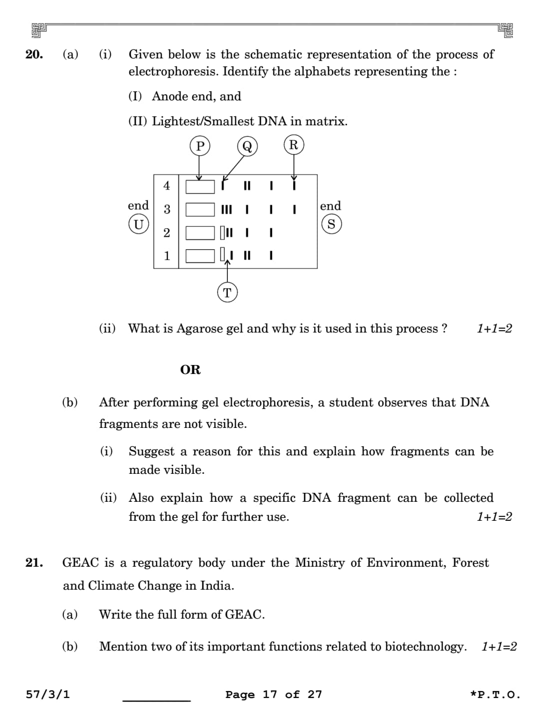
(a) (i) Given below is the schematic representation of the process of electrophoresis. Identify the alphabets representing the:
(I) Anode end, and
(II) Lightest/Smallest DNA in matrix.

View Solution
Step 1: Understanding the Question:
The question provides a diagram of an agarose gel electrophoresis plate showing sample loading wells and separated bands. We need to identify which letter corresponds to the positive electrode terminal (Anode end) and which letter points to the smallest migrated DNA fragment.
Step 2: Key Formula or Approach:
DNA molecules carry a uniform negative charge due to the phosphate groups in their structural backbone. Therefore, when placed in an electric field, they migrate away from the negative cathode terminal toward the positive Anode terminal.
Agarose gel functions as a molecular sieve. Smaller, lighter DNA fragments travel through the pores much faster and farther than larger, heavier fragments.
Step 3: Detailed Explanation:
(I) Identifying the Anode End: The sample loading wells are labeled as P on the left side near end U. Because DNA is loaded into these wells and moves from left to right toward end S, end U represents the negative cathode and end S represents the positive Anode terminal. Thus, S is the anode end.
(II) Identifying the Smallest DNA Fragment: The DNA fragments separate by size along the lane. The bands labeled Q are closest to the wells, meaning they are the largest, heaviest fragments that moved the least. The bands labeled R have migrated the farthest toward the positive terminal S. This indicates that they are the smallest, lightest DNA fragments. Thus, R represents the smallest DNA in the matrix.
Step 4: Final Answer:
(I) The Anode end is represented by the letter S.
(II) The lightest/smallest DNA fragment in the matrix is represented by the letter R.
Quick Tip: Remember the phrase \textbf{"Run towards the Red Anode."} Since DNA is negative, the smallest fragments will travel fastest and be found closest to the positive anode end (\textbf{S}), while heavy fragments lag behind near the loading wells.
(a) (ii) What is Agarose gel and why is it used in this process?
View Solution
Step 1: Understanding the Question:
The question asks for a definition of agarose gel and an explanation of its physical role during gel electrophoresis.
Step 2: Key Formula or Approach:
Agarose is a natural polysaccharide polymer that forms a porous cross-linked gel network when cooled, acting as a size-selective sieve for separating nucleic acids.
Step 3: Detailed Explanation:
What it is: Agarose is a natural linear polysaccharide polymer extracted from marine red seaweeds (such as Gelidium and Gracilaria).
Why it is used: When dissolved in hot buffer and allowed to cool, agarose forms a solid gel matrix containing microscopic pores. When an electric current is applied, these pores act as a molecular sieve. As negatively charged DNA molecules migrate through the gel, the pore network slows down larger DNA strands, while allowing smaller fragments to pass through quickly. This separation based on size allows researchers to analyze and isolate specific DNA fragments.
Step 4: Final Answer:
Agarose gel is a natural polymer extracted from seaweeds. It is used because its porous matrix acts as a molecular sieve that separates DNA fragments strictly by their physical size.
Quick Tip: Think of agarose gel as a molecular mesh screen. The dense network of fibers slows down large, bulky DNA pieces, while small, agile DNA fragments easily slip through the gaps and travel farther down the gel.
OR
Question 20:
(b) After performing gel electrophoresis, a student observes that DNA fragments are not visible.
(i) Suggest a reason for this and explain how fragments can be made visible.
(ii) Also explain how a specific DNA fragment can be collected from the gel for further use.
View Solution
Step 1: Understanding the Question:
The question addresses a common lab problem where DNA bands are invisible after running a gel. We need to explain why this occurs, how to visualize the hidden bands, and how to isolate a specific DNA band from the gel matrix for cloning work. These components are kept together as they form a continuous laboratory workflow.
Step 2: Key Formula or Approach:
Naked DNA molecules do not absorb light in the visible spectrum and appear transparent. Visualizing them requires staining the gel with an intercalating fluorescent dye and exposing it to ultraviolet (UV) radiation. Isolating the DNA requires an extraction method called elution.
Step 3: Detailed Explanation:
(i) Why DNA is Invisible and How to Visualize It: DNA fragments are clear and invisible to the naked eye under standard laboratory lighting. To see them, the gel must be stained with a fluorescent dye, most commonly Ethidium Bromide (EtBr). Once stained, the gel must be viewed under ultraviolet (UV) light illumination. Under UV light, the DNA bands fluoresce brightly, appearing as distinct, glowing bright orange bands.
(ii) Collecting the DNA Fragment: Once the target DNA band is located, it can be harvested from the gel through a process called Elution. First, a technician uses a clean blade to physically cut out the specific gel piece containing the desired DNA band. Then, the DNA is extracted and purified away from the agarose matrix using chemical reagents or spin columns, leaving pure DNA ready to be used in cloning or ligation.
Step 4: Final Answer:
(i) The DNA is invisible because it lacks a stain. It can be visualized by staining the gel with Ethidium Bromide and exposing it to UV light, which reveals the fragments as bright orange bands.
(ii) The specific DNA fragment is collected by cutting out the corresponding gel piece and separating the DNA from the gel via Elution.
Quick Tip: Remember two key terms for practical exams: \textbf{Ethidium Bromide + UV light} is the combination used to see the bands (look for the bright orange color), and \textbf{Elution} is the name of the process used to extract the DNA from the cut gel block.
GEAC is a regulatory body under the Ministry of Environment, Forest and Climate Change in India.
(a) Write the full form of GEAC.
(b) Mention two of its important functions related to biotechnology.
View Solution
Step 1: Understanding the Question:
The question asks for the official full name of the Indian regulatory body GEAC, along with two of its primary functions concerning biotechnology applications.
Step 2: Key Formula or Approach:
The commercial use and environmental release of Genetically Modified Organisms (GMOs) are strictly regulated by statutory bodies to protect public health and ecological safety.
Step 3: Detailed Explanation:
(a) Full Form of GEAC: Under the Ministry of Environment, Forest and Climate Change, the acronym GEAC stands for the Genetic Engineering Appraisal Committee.
(b) Important Functions Related to Biotechnology: The committee is responsible for two main regulatory tasks:
1. Safety Assessment: It evaluates the safety of research projects and industrial applications involving the large-scale use of genetically engineered microbes or hazardous pathogens.
2. Commercial Approval: It reviews environmental safety data and grants final clearance for introducing genetically modified organisms or crops (such as Bt cotton) into open agricultural fields for commercial farming. This helps prevent unintended ecological disruption or health hazards.
Step 4: Final Answer:
(a) The full form of GEAC is the Genetic Engineering Appraisal Committee.
(b) Two important functions are assessing the biosafety of genetic engineering research and approving the commercial release of genetically modified crops into public fields.
Quick Tip: Think of GEAC as the official regulatory gatekeeper for GMOs in India. No scientist or agricultural company can grow or sell a genetically modified crop in open fields without first passing safety reviews and obtaining approval from this committee.
(a) Explain the different ways by which apomictic seeds can be developed.
(b) Mention one advantage of apomictic seeds for farmers.
View Solution
Step 1: Understanding the Question:
The question asks for an explanation of the biological pathways through which apomictic (asexual) seeds develop in flowering plants. It also requires identifying a specific agricultural advantage that these seeds offer to farmers.
Step 2: Key Formula or Approach:
Apomixis is a reproductive mechanism where seeds are formed without going through fertilization. It acts as a mimic of sexual reproduction but preserves the exact genetic composition of the maternal plant.
Step 3: Detailed Explanation:
(a) Mechanisms of Apomictic Seed Development: Apomictic seeds can develop through two primary evolutionary pathways:
1. Apospory / Diplospory (Development from an unreduced egg cell): In several plant species, a diploid (\(2n\)) egg cell is formed without undergoing normal meiotic reduction division. This unreduced diploid egg cell then develops directly into an embryo through mitotic divisions without being fertilized by a male gamete.
2. Adventive Embryony (Development from nucellar or integumentary cells): In many citrus fruit varieties and mango (Mangifera indica), diploid somatic cells belonging to the surrounding maternal tissues—such as the nucellus or integuments—protrude directly into the embryo sac. These somatic cells divide mitotically and develop into fully functional embryos. This often leads to a phenomenon known as polyembryony, where a single seed contains multiple embryos.
(b) Advantage for Farmers: Farmers often grow hybrid crops because they provide high vigor, better resilience, and increased yields. However, if farmers collect seeds from a hybrid crop and plant them the following year, the alleles segregate during sexual reproduction, causing the offspring to lose those specialized hybrid traits. Consequently, farmers must buy expensive hybrid seeds every single season. If these hybrid seeds can be engineered to be apomictic, the embryos develop asexually without fertilization. This prevents genetic segregation, allowing farmers to keep saving and replanting their own high-yielding seeds year after year without losing hybrid performance.
Step 4: Final Answer:
(a) Apomictic seeds develop either by a diploid egg forming without meiosis and developing into an embryo without fertilization, or by surrounding maternal nucellar cells dividing and protruding into the embryo sac.
(b) The main advantage is that hybrid seeds do not segregate their traits, allowing farmers to replant saved seeds year after year without buying new ones every season .
Quick Tip: Think of apomixis as \textbf{"natural plant cloning inside a seed." Because there is no meiosis and no fusion of gametes, the resulting seed forms a exact genetic carbon copy of the high-performing maternal hybrid parent.
(a) What is meant by translation in protein synthesis?
(b) Explain charging of tRNA (aminoacylation of tRNA) and mention its importance in the process of translation.
View Solution
Step 1: Understanding the Question:
The question asks for a definition of translation within the context of gene expression. It also requires an explanation of the biochemical process of tRNA aminoacylation ("charging") and why this step is necessary for successful translation.
Step 2: Key Formula or Approach:
Translation converts an mRNA nucleotide sequence into a polypeptide chain. The chemical joining of amino acids into a protein requires energy. This energy is provided beforehand by linking each amino acid to its matching tRNA molecule.
Step 3: Detailed Explanation:
(a) Translation: Translation is the biochemical process where the genetic information carried in a linear sequence of nucleotides on a single-stranded messenger RNA (mRNA) molecule is decoded to direct the synthesis of a specific sequence of amino acids, forming a functional polypeptide chain. This process takes place within cellular ribosomes.
(b) Charging of tRNA (Aminoacylation): Amino acids cannot directly recognize nucleotide codons on an mRNA strand, nor can they spontaneously form peptide bonds without an input of energy.
1. The Charging Process: In the first phase of translation, an amino acid is activated by reacting with ATP in the presence of a specific enzyme called \textit{aminoacyl-tRNA synthetase. This reaction creates an intermediate complex (aminoacyl-AMP). The enzyme then transfers the activated amino acid to the \(3'\)-acceptor arm of its matching tRNA molecule, releasing AMP. This process is chemically written as:
\(\)\text{Amino Acid + \text{ATP + \text{tRNA \xrightarrow{\text{Synthetase \text{Aminoacyl-tRNA + \text{AMP + \text{PP_i\(\)
2. Importance in Translation: This process serves two critical roles. First, it ensures accuracy: the synthetase enzyme matches the correct amino acid with the right tRNA anticodon loop, preserving the genetic code. Second, it provides the necessary chemical energy: linking the amino acid to the tRNA creates a high-energy ester bond. When two charged tRNAs line up side by side inside the ribosome's A and P sites, the energy stored in this bond helps drive the spontaneous formation of a peptide bond between the adjacent amino acids without requiring a new ATP input during the elongation step.
Step 4: Final Answer:
(a) Translation is the decoding of an mRNA sequence into a specific sequence of amino acids to synthesize a protein .
(b) Charging of tRNA is the chemical attachment of an activated amino acid to its corresponding tRNA using ATP energy . Its importance is that it accurately pairs amino acids with codons and provides the structural energy needed to build peptide bonds during translation .
Quick Tip: Think of tRNA as a microscopic delivery truck. "Charging" is the process of loading a specific parcel (an amino acid) onto the truck bed using fuel (ATP). If the truck isn't loaded and fueled first, the assembly line at the ribosome factory stops completely.
(a) What is the carrying capacity of a species in a habitat?
(b) Explain the growth curve that takes this capacity into account.
View Solution
Step 1: Understanding the Question:
The question asks for a definition of the ecological term carrying capacity (\(K\)). It also requires an explanation of the population growth curve (logistic growth) that factors in this environmental limit.
Step 2: Key Formula or Approach:
When resources like food and space are limited, population growth follows a Verhulst-Pearl Logistic Growth pattern. This pattern is mathematically defined by the differential equation: \(\)\frac{dN{dt = rN \left(\frac{K - N{K\right)\(\)
Where \(N\) is population density, \(r\) is the intrinsic rate of natural increase, and \(K\) is the carrying capacity.
Step 3: Detailed Explanation:
(a) Carrying Capacity (\(K\)): Carrying capacity is defined as the maximum population size of a given biological species that a specific ecosystem can sustainably support over time, given the available resources like food, water, nesting territory, and space. Beyond this point, resource depletion increases mortality rates, causing population growth to level off.
(b) Logistic Growth Curve: When resources are limited, a population exhibits a Sigmoid (S-shaped) growth curve. This pattern consists of four distinct phases:
1. Lag Phase: Initial growth is slow while the population acclimates to the new environment.
2. Acceleration / Exponential Phase: As individuals reproduce, the population grows rapidly because resources are still relatively abundant.
3.
Deceleration Phase: Growth begins to slow down as resources become scarce and competition intensifies.
4.
Asymptote Phase: The population size stabilizes and reaches a plateau when it matches the carrying capacity (\(N = K\)), bringing net growth (\(\frac{dN}{dt}\)) to zero.
This model provides a more realistic representation of natural populations than exponential models, because resources in the real world are always limited.
Step 4: Final Answer:
(a) Carrying capacity is the maximum number of individuals of a species that a habitat can sustainably support with its limited resources .
(b) This model produces a Sigmoid (S-shaped) growth curve containing a lag phase, a rapid acceleration phase, a deceleration phase, and a flat asymptote phase where the population stabilizes at the carrying capacity (\(K\)).
Quick Tip: Remember that if the term \(\left(\frac{K - N}{K}\right)\) is included in a growth equation, it represents \textbf{environmental resistance}. As the population size (\(N\)) approaches the carrying capacity (\(K\)), this value drops toward zero, mathematically flattening the growth curve into an S-shape.
In vitro fertilization (IVF) is a popular method these days that is helping childless couples to bear a child.
(a) Write the different steps that are carried out in this technique.
(b) Would you consider gamete intra fallopian transfer as a type of IVF? Justify your answer.
View Solution
Step 1: Understanding the Question:
The question asks for an explanation of the sequential medical steps performed during In Vitro Fertilization (IVF). It also asks whether Gamete Intra Fallopian Transfer (GIFT) should be classified as an IVF technique, requiring a biological justification.
Step 2: Key Formula or Approach:
IVF (In Vitro Fertilization): Fertilization occurs outside the female body in a laboratory setting that mimics natural conditions, followed by embryo transfer (ET).
GIFT (Gamete Intra Fallopian Transfer): Unfertilized gametes are placed directly into the fallopian tube, meaning fertilization occurs inside the female body (in vivo).
Step 3: Detailed Explanation:
(a) Steps in the IVF Technique:
1. Ovarian Hyperstimulation: The female partner receives hormone injections (like FSH) to stimulate her ovaries to develop multiple mature follicles instead of the usual single egg per month.
2. Egg Retrieval: Once mature, the eggs are harvested from the ovarian follicles using a minor surgical procedure guided by ultrasound.
3. Sperm Collection: The male partner provides a semen sample, which is processed in the laboratory to isolate high-motility sperm.
4. Co-incubation / In Vitro Fertilization: The harvested eggs and prepared sperm are mixed together in a specialized petri dish containing nutrient media within a laboratory incubator. This allows fertilization to occur outside the mother's body. In cases of severe male infertility, a single sperm may be injected directly into an egg via Intra Cytoplasmic Sperm Injection (ICSI).
5. Embryo Culture and Transfer (ET): The fertilized eggs divide to form early embryos. At the 8-cell stage, the embryo can be transferred into the fallopian tube via ZIFT (Zygote Intra Fallopian Transfer). Alternatively, it is cultured to the blastocyst stage and transferred directly into the mother's uterus for implantation.
(b) Evaluation of GIFT: No, Gamete Intra Fallopian Transfer (GIFT) is not a type of IVF .
Justification: By definition, IVF requires fertilization to occur \textit{in vitro (outside the body in a laboratory environment). In contrast, during a GIFT procedure, unfertilized harvested eggs and washed sperm are mixed and immediately injected into the patient's fallopian tubes. This means actual fertilization occurs naturally \textit{inside the female body (\textit{in vivo), rather than in a laboratory dish. Therefore, GIFT is classified as an Assisted Reproductive Technology (ART), but it does not qualify as an IVF method.
Step 4: Final Answer:
(a) The primary steps of IVF are ovarian stimulation, egg retrieval, sperm preparation, laboratory fertilization (\textit{in vitro), and transferring the resulting embryo back into the female reproductive tract .
(b) No, GIFT is not a form of IVF because fertilization takes place \textit{in vivo (inside the mother's fallopian tubes) rather than in an external laboratory setting .
Quick Tip: To distinguish between these terms quickly, look at where fertilization occurs: \textit{ \textbf{IVF: Fertilization happens in a laboratory dish (}in vitro*).
\textbf{GIFT / In Vivo: Fertilization happens inside the living body (}in vivo*).
This difference helps prevent confusing these reproductive technologies.
Draw a neat diagram of a maize grain showing the internal structure and label any five parts.
View Solution
Step 1: Understanding the Question:
The question requires drawing a clear anatomical diagram showing the internal longitudinal structure of a monocotyledonous maize grain (Zea mays), including at least five accurate structural labels.
Step 2: Key Formula or Approach:
A maize grain is a single-seeded fruit known as a caryopsis, where the protective seed coat is completely fused with the outer pericarp wall. It features a large endosperm storage region and a lateral embryo.
Step 3: Detailed Explanation:
A complete structural drawing of a maize grain includes the following key parts:
1. Pericarp + Seed Coat: The outermost protective layer, formed by the tight fusion of the fruit wall (pericarp) and the seed coat.
2. Aleurone Layer: A specialized, protein-rich outer cell layer that surrounds the bulky starch endosperm storage tissue.
3. Endosperm: The main nutrient storage tissue, filled with starch, which feeds the embryo during germination.
4. Scutellum: The large, shield-shaped single cotyledon characteristic of monocots, which absorbs nutrients from the endosperm.
5. Coleoptile: A protective sheath that encloses and shields the developing young shoot apex (plumule).
6. Plumule: The embryonic shoot tip that will grow into the primary stem and leaves.
7. Radicle: The embryonic root tip that will grow downward to form the root system.
8. Coleorhiza: A protective undifferentiated sheath that covers and shields the embryonic root tip (radicle).
Step 4: Final Answer:
The student should sketch the longitudinal section of the maize grain, showing the large endosperm separated from the embryo by the scutellum, with the coleoptile protecting the plumule and the coleorhiza protecting the radicle.
Quick Tip: When labeling monocot seeds like maize, remember that protective sheaths always pair with specific embryonic tips: \textbf{Coleoptile protects the Plumule (both contain 'p' for shoot), and \textbf{Coleorhiza protects the Radicle} (both contain 'r' for root).
Cow dung and water are mixed and fed into a biogas plant to allow digestion of biowastes. The person performing this process says that there is no need to provide an inoculum.
(a) Do you agree with him? Justify your answer.
(b) What happens to the biowaste inside the digester ?
(c) Name the useful by-products obtained from this process and mention how they are used.
View Solution
Step 1: Understanding the Question:
The question presents a scenario involving a anaerobic biogas plant. We need to evaluate whether adding an external microbial starter (inoculum) is necessary, explain the breakdown of waste inside the digester, and identify the resulting useful by-products and their applications.
Step 2: Key Formula or Approach:
Biogas generation is a multi-step anaerobic process driven by methane-producing bacteria called methanogens, notably Methanobacterium. These bacteria naturally live inside the digestive tracts of cattle.
Step 3: Detailed Explanation:
(a) Evaluation of the Statement: Yes, I agree with the person's statement .
\textit{ Justification:There is no need to add an artificial starter or external microbial inoculum to the mixture. This is because methanogenic bacteria (specifically \textit{Methanobacterium) are already naturally present in large numbers inside the rumen (stomach) of cattle. When cow dung is collected and mixed into the plant, these native bacteria are carried along with it, automatically serving as an active biological starter culture to begin the fermentation process.
(b) Process Inside the Digester: Inside the sealed, oxygen-free digester tank, anaerobic microbes break down the organic biowaste through a multi-stage fermentation process:
1. Solubilization / Hydrolysis: Complex polymers in the dung (such as cellulose, hemicellulose, and proteins) are broken down into simpler soluble monomers.
2. \textit{Acidogenesis: Acid-producing bacteria convert these monomers into volatile fatty acids (like acetic acid), hydrogen, and carbon dioxide.
3. \textit{Methanogenesis: Finally, the strictly anaerobic methanogenic bacteria consume these organic acids and gases, converting them into a mixture of methane (\(CH_4\)), carbon dioxide (\(CO_2\)), and trace hydrogen sulfide (\(H_2S\)). This gas mixture is harvested as biogas.
(c) By-products and Their Uses:
1. Biogas: Collected from the top gas outlet of the digester tank. It is an excellent clean-burning fuel used directly for cooking and rural household lighting.
2. Spent Slurry / Residual Sludge: The leftover digested material is discharged through the outlet chamber. This sludge is rich in essential plant nutrients like nitrogen, phosphorus, and potassium. It is dried and used as a highly effective organic manure in agricultural fields to improve soil structure and boost crop yields.
Step 4: Final Answer:
(a) Yes, I agree, because methanogens are naturally present in cow dung, removing the need for an external inoculum .
(b) Inside the digester, anaerobic bacteria ferment and break down complex organic compounds into a mixture of methane and carbon dioxide gases .
(c) The useful by-products are biogas (used as a clean fuel for cooking and lighting) and spent slurry (used as a nutrient-rich organic manure for farming).
Quick Tip: Cattle eat large amounts of cellulose-rich grass, so their stomachs are packed with \textbf{methanogens to help break it down. Because these bacteria are already abundant in fresh cow dung, the raw material acts as its own active starter culture.
(a) Describe the experiment conducted by T.H. Morgan on Drosophila melanogaster involving eye colour and body colour.
(b) How did the results deviate from Mendelian inheritance pattern ?
(c) Explain the two genetic terms used by Morgan for his observations.
View Solution
Step 1: Understanding the Question:
The question asks for a description of Thomas Hunt Morgan's dihybrid crosses in fruit flies (Drosophila melanogaster) involving eye color and body color traits. It also asks how his data deviated from standard Mendelian ratios and requires an explanation of the terms linkage and recombination.
Step 2: Key Formula or Approach:
Mendel's Law of Independent Assortment predicts a \(9:3:3:1\) phenotypic ratio in the \(F_2\) generation of a dihybrid cross, or a \(1:1:1:1\) ratio in a dihybrid test cross. When genes are located close together on the same chromosome, they alter these expected proportions due to genetic linkage.
Step 3: Detailed Explanation:
(a) Morgan's Experiment: Morgan performed a dihybrid cross using fruit flies to study two X-linked genes: body color and eye color. He crossed yellow-bodied, white-eyed females (carrying recessive mutant alleles, \(yw\)) with wild-type brown-bodied, red-eyed males (carrying dominant alleles, \(y^+w^+\)). He then intercrossed the resulting \(F_1\) generation offspring to analyze the trait combinations in the \(F_2\) generation.
(b) Deviation from Mendel: According to Mendel's Law of Independent Assortment, the two genes should separate independently, producing an \(F_2\) generation with a \(9:3:3:1\) ratio, where \(37.5%\) of the offspring display new, non-parental trait combinations (recombinant phenotypes). However, Morgan observed a significant deviation: \(98.7%\) of the \(F_2\) offspring retained the exact parental combinations, while only \(1.3%\) showed recombinant traits. This proved that the genes did not sort independently.
(c) Morgan's Genetic Terms: To explain these findings, Morgan introduced two key concepts:
1. Linkage: This describes the physical association of two or more genes located on the same chromosome. Because they are physically linked on the same DNA strand, they tend to be inherited together as a unit during meiosis, keeping parental trait combinations intact.
2. Recombination: This describes the generation of non-parental gene combinations in offspring. It occurs during prophase I of meiosis, when homologous chromosomes cross over and exchange genetic material, swapping alleles between chromatids. Morgan noted that the rate of recombination depends directly on the distance between genes: genes located very close together show tight linkage and low recombination rates (like eye and body color at \(1.3%\)), while genes farther apart show weaker linkage and higher recombination rates.
Step 4: Final Answer:
(a) Morgan crossed yellow-bodied, white-eyed female flies with brown-bodied, red-eyed male flies, and then intercrossed their \(F_1\) offspring to study how the traits were passed down.
(b) The results deviated from the expected Mendelian dihybrid ratios because parental phenotypes appeared at a very high rate (\(98.7%\)), while recombinants appeared at a very low rate (\(1.3%\)), showing that independent assortment did not occur.
(c) Linkage refers to the physical association of genes on the same chromosome, which keeps them together. Recombination refers to the formation of new, non-parental gene combinations due to crossing over during meiosis.
Quick Tip: Remember this rule for genetic distance: \textbf{Recombination frequency is directly proportional to the physical distance between genes. - Close together = Tight Linkage = Low Recombination (\(1.3%\) for body/eye color).
Far apart = Loose Linkage = High Recombination (\(37.2%\) for eye color/wing size).
Read the following passage and answer the questions that follow:
The data below shows the concentration of nicotine smoked by a smoker taking 10 puffs/minute.

(a) (i) With reference to the above graph, explain the concentration of nicotine in the blood at 10 minutes.
(ii) How will this affect the concentration of carbon monoxide and haem-bound oxygen at 10 minutes?
(b) How does cigarette smoking result in high blood pressure and increase in heart rate?
(c) To which class of compounds does nicotine belong? Name one other drug from the same class.
View Solution
Step 1: Understanding the Question:
This case-based question requires analyzing a clinical data graph that plots blood nicotine concentration against time for a smoker. We must interpret the peak concentration value at the \(10\)-minute mark , relate this data to systemic gas-exchange physiology (carbon monoxide and oxyhaemoglobin levels) , explain the cardiovascular mechanism behind smoking-induced hypertension , and classify the compound nicotine pharmacologically.
Step 2: Key Formula or Approach:
Graph Interpretation: Read the coordinate value on the y-axis corresponding to \(x = 10 minutes\).
Gas Dynamics: Cigarette smoke introduces carbon monoxide (\(CO\)), which binds to haemoglobin with a much higher affinity than oxygen, shifting the biochemical equilibrium away from oxyhaemoglobin (\(HbO_2\)).
Neurochemical Pathway: Nicotine acts as an agonist that stimulates adrenal medullary secretion, releasing catecholamines into the bloodstream.
Step 3: Detailed Explanation:
(a) (i) Interpretation at 10 Minutes: Looking at the provided line graph, the concentration of nicotine in the blood increases steadily from \(0 mg/cm^3\) and reaches its absolute maximum peak at the \(10\)-minute mark. Following the grid line at \(10 minutes\) vertically to the curve and reading horizontally across to the y-axis reveals that the peak concentration achieved is exactly \(45 mg/cm^3\). This peak represents the point where the rate of nicotine absorption from the lungs into the pulmonary circulation is at its highest, just as active smoking concludes.
(a) (ii) Effects on Gases: At the \(10\)-minute peak of active cigarette smoking, the concentration of inhaled carbon monoxide (\(CO\)) in the blood increases significantly. Because carbon monoxide has an affinity for haemoglobin that is roughly 200 times stronger than oxygen, it binds preferentially to form carboxyhaemoglobin (\(COHb\)). This process displaces oxygen molecules and blocks them from binding to haemoglobin sites. Consequently, the concentration of haem-bound oxygen (oxyhaemoglobin) drops sharply, reducing oxygen delivery to peripheral tissues.
(b) High Blood Pressure and Heart Rate Mechanism: Inhaled nicotine travels through the bloodstream and binds to nicotinic acetylcholine receptors located on the adrenal medulla. This binding stimulates the adrenal glands to secrete large amounts of the catecholamine hormones adrenaline (epinephrine) and noradrenaline (norepinephrine) into systemic circulation. These hormones act on the cardiovascular system by binding to \(\alpha_1\) and \(\beta_1\) adrenergic receptors, which causes:
1. Systemic vasoconstriction (narrowing of arterial blood vessels), which increases peripheral resistance and raises blood pressure.
2. Direct stimulation of the heart's sinoatrial node, which increases the heart rate and forces the heart to work harder.
(c) Classification of Nicotine: Nicotine belongs to the chemical class of Alkaloids (specifically, it is a plant-derived nitrogenous organic base that acts as a potent CNS stimulant). Another widely known drug belonging to this exact same functional class of central nervous system stimulants is Caffeine (or Cocaine / Amphetamines).
Step 4: Final Answer:
(a) (i) At 10 minutes, the blood nicotine concentration hits its maximum peak value of \(45 mg/cm^3\).
(ii) This causes the carbon monoxide concentration to rise and the haem-bound oxygen level to drop significantly.
(b) Nicotine stimulates the adrenal medulla to release adrenaline and noradrenaline, which trigger vasoconstriction and accelerate the heart rate, causing high blood pressure.
(c) Nicotine belongs to the class of Alkaloids (Stimulants), and another member of this class is Caffeine (or Cocaine).
Quick Tip: Remember the cardiotoxic cascade of smoking: \textbf{Nicotine \(\rightarrow\) Adrenal Medulla \(\rightarrow\) Adrenaline Surge \(\rightarrow\) Vasoconstriction + Tachycardia \(\rightarrow\) Hypertension.} At the same time, carbon monoxide acts as a chemical competitor that crowds oxygen off your haemoglobin molecules.
Read the following passage and answer the questions that follow:
India is one of the megadiverse countries housing around 8·1 per cent of global species diversity, although its land area is only 2·4 per cent of the world’s land area. Many of the species are highly threatened due to human activities like deforestation, mining and habitat fragmentation. Laws like Wildlife (Protection) Act, 1972 were enacted by the Government of India to preserve our biological wealth. Various conservation measures are being implemented to save the threatened species.
The following bar graph shows the number of species conserved under different biodiversity conservation methods.

(a) Which method conserves the highest number of species? Is it ex situ or in situ conservation?
(b) Which other methods shown in the diagram are opposite to the one identified by you in question (a)? How are these two conservation approaches different?
(c) (i) Write two features of Biodiversity hotspots.
(c) (ii) To which category do sacred groves belong and how do they help in bio-conservation?
View Solution
Step 1: Understanding the Question:
This case-based question centers on analyzing biodiversity conservation methods using a provided bar graph. We need to identify the highest-performing strategy and categorize it , contrast it with opposing approaches shown in the data , and explain either the features of biodiversity hotspots or the ecological role of sacred groves. As requested, both choices for part (c) are solved.
Step 2: Key Formula or Approach:
Graph Analysis: Identify the tallest bar on the chart to determine the highest number of conserved species.
Classification: - In situ (on-site): Protecting an endangered species within its natural habitat.
\textit{Ex situ (off-site): Removing an endangered species from its natural habitat and placing it in a specialized, human-managed facility for protection.
Step 3: Detailed Explanation:
(a) Identifying the Highest Method: Looking at the heights of the bars on the graph, the bar labeled Wildlife Sanctuaries is significantly taller than the rest, showing a value of \(2500\) species conserved. This strategy is classified as an \textit{in situ (on-site) conservation approach because it protects wild species by securing their existing natural habitat from human exploitation.
(b) Identifying and Contrasting the Opposite Approach: The strategies shown on the graph that are conceptually opposite to in situ habitat protection are Zoological Parks (\(990\) species) and Botanical Gardens (\(1100\) species). These two methods represent \textit{ex situ (off-site) conservation.
Key Differences: 1. \textit{\textit{In situ approach: Protects the entire ecosystem as a whole by managing the species directly within its native environment (e.g., National Parks, Sanctuaries, Biosphere Reserves). This allows natural evolutionary processes and ecological interactions to continue undisturbed.
2. \textit{\textit{Ex situ approach: Involves taking threatened plants or animals out of their natural, endangered habitats and moving them into artificial, human-managed enclosures or facilities (e.g., zoos, botanical gardens, gene banks). This strategy focuses on providing intensive individual care, veterinary support, and managed captive breeding programs away from wild threats.
(c) (i) Features of Biodiversity Hotspots: Biodiversity hotspots are specific geographical areas that receive high priority for conservation because they meet two strict ecological criteria:
1. High degree of Endemism: They house a large number of endemic species—plants and animals that live natively in that specific region and are found nowhere else on Earth.
2. Severe Accelerated Habitat Loss: These regions face extreme environmental threats; they must have already lost at least \(70%\) of their original primary native vegetation due to human activities.
(c) (ii) Sacred Groves:
Category: Sacred groves belong to the category of \textit{in situ conservation methods, driven by traditional, community-led cultural protection.
Mechanism of Bio-conservation: Sacred groves are patches of pristine forest that are set aside and protected by local indigenous communities due to deep-rooted religious beliefs and cultural traditions. Deities are believed to reside within these forests, making any logging, hunting, or resource extraction strictly taboo. Because these areas are shielded from human interference, they serve as natural ecological sanctuaries that preserve rare, threatened, and endemic plant and animal species that have been cleared from the surrounding landscape.
Step 4: Final Answer:
(a) Wildlife Sanctuaries conserve the highest number of species (\(2500\)). This strategy is an \textit{in situ conservation method.
(b) The opposite methods are Zoological Parks and Botanical Gardens (ex situ). \textit{\textit{In situ protects species within their natural habitats, while \textit{\textit{ex situ removes them to human-controlled facilities for intensive care.
(c) (i) Hotspots feature extreme levels of species richness and high endemism, along with severe, ongoing accelerated habitat destruction.
(c) (ii) Sacred groves are \textit{in situ measures where traditional religious taboos ban all human interference, preserving rare and endangered species within untouched forest patches.
Quick Tip: To keep these two main conservation approaches straight, think of their Latin roots: \textbf{In situ = "In position"} (protecting the wild home, like a sanctuary), while \textbf{Ex situ = "Out of position"} (moving the species to an artificial home, like a zoo or botanical garden).
(a) (i) Describe the series of experiments conducted by Frederick Griffith. Comment on the significance of the result obtained.
(ii) State the contribution of Avery, MacLeod and McCarty.
View Solution
Step 1: Understanding the Question:
This question asks for a detailed description of Frederick Griffith's transforming experiments (1928) using Streptococcus pneumoniae and an explanation of the significance of his findings. It also requires stating how Avery, MacLeod, and McCarty chemically identified the transforming principle.
Step 2: Key Formula or Approach:
Griffith's Framework: Uses two distinct bacterial strains—the virulent, smooth (S) strain containing a polysaccharide capsule, and the non-virulent, rough (R) strain which lacks a capsule.
Biochemical Characterization: Uses selective enzymatic digestion (Protease, RNase, and DNase) to identify the specific macromolecule that transfers genetic traits.
Step 3: Detailed Explanation:
(i) Griffith's Transforming Principle Experiments: Griffith worked with Streptococcus pneumoniae, a bacterium that causes pneumonia in mammals, and observed two distinct phenotypic strains:
1. \textit{Smooth (S) Strain: Produces smooth, shiny colonies because the cells synthesize an outer polysaccharide capsule. When injected into mice, they develop pneumonia and die (virulent strain).
2. \textit{Rough (R) Strain: Produces rough colonies because they lack this protective capsule. When injected into mice, the host's immune system clears the bacteria, and the mice survive (non-virulent strain).
3. \textit{Heat-Killed S Strain: Griffith heated the virulent S strain to kill the cells. When injected into mice, the mice survive, showing that the dead bacteria cannot cause disease.
4. \textit{Mixture of Heat-Killed S + Live R Strains: Griffith mixed the harmless heat-killed S bacteria with harmless living R bacteria and injected the mixture into healthy mice. Surprisingly, the mice developed pneumonia and died. Furthermore, he isolated living, fully capsuled S-strain bacteria from the blood of these dead mice.
\textit{Significance of the Result: Griffith concluded that the living R-strain bacteria had absorbed a hereditary material from the dead, heat-killed S-strain cells. This material permanently transformed the R strain, enabling it to synthesize a smooth polysaccharide coat and become virulent. He named this active chemical component the "Transforming Principle," which provided the first experimental proof that cells contain a transferable genetic material.
(ii) Contribution of Avery, MacLeod, and McCarty (1933-44): Griffith's work did not reveal the chemical identity of the transforming principle. Avery, MacLeod, and McCarty purified major macromolecules (proteins, RNA, carbohydrates, and DNA) from heat-killed S-strain cells to see which fraction could transform live R cells:
They treated the purified fractions with protein-digesting enzymes (proteases) or RNA-digesting enzymes (RNases) and found that transformation still occurred normally. This proved that neither proteins nor RNA carried the transforming information.
However, when they treated the mixture with a DNA-digesting enzyme (DNase), transformation stopped completely, and no live S-strain bacteria were formed.
This rigorous biochemical experiment proved that DNA is the transforming principle and provided the first definitive proof that DNA is the genetic material of living organisms.
Step 4: Final Answer:
(i) Griffith demonstrated that heat-killed S-strain bacteria can transfer a "transforming principle" to living R-strain bacteria, permanently changing them into virulent S-strain cells.
(ii) Avery, MacLeod, and McCarty discovered that this transforming principle is pure DNA by showing that DNA-digesting enzymes (DNase) completely halt the transformation process.
Quick Tip: Remember that Griffith discovered \textit{what transformation does (introducing the concept of a transforming principle), but Avery, MacLeod, and McCarty discovered how it works chemically by using targeted digestive enzymes to prove that DNA is the genetic material.
OR
Question 31:
(b) (i) Work out the crosses between:
(I) Normal female and Haemophilic male
(II) Carrier female and Normal male
(III) Carrier female and Haemophilic male
(ii) Write the conclusions you draw from these crosses. Comment on the type of inheritance of the disease.
(Use: \(X\) - Normal, \(X^h\) - Haemophilic)
View Solution
Step 1: Understanding the Question:
The question asks to model three genetic crosses for haemophilia using specific sex-chromosome notation (\(X\) and \(X^h\)). It also requires analyzing the inheritance patterns and proportions of offspring generated from these crosses.
Step 2: Key Formula or Approach:
Haemophilia is an X-linked recessive genetic disorder. Because the gene resides on the X chromosome, males are hemizygous (\(XY\) or \(X^hY\)) and will express the disease if they inherit a single mutated copy. Females must inherit two copies (\(X^hX^h\)) to show the disease; a single copy makes them normal carriers (\(XX^h\)).
Step 3: Detailed Explanation:
(i) Working out the Crosses: - Cross (I): Normal female (\(XX\)) \(\times\) Haemophilic male (\(X^hY\))

Offspring Proportions: \(100%\) of daughters are normal carriers (\(XX^h\)), and \(100%\) of sons are completely normal (\(XY\)).
Cross (II): Carrier female (\(XX^h\)) \(\times\) Normal male (\(XY\))

\textit{Offspring Proportions: Daughters: \(50%\) normal (\(XX\)), \(50%\) carriers (\(XX^h\)). Sons: \(50%\) normal (\(XY\)), \(50%\) haemophilic (\(X^hY\)).
Cross (III): Carrier female (\(XX^h\)) \(\times\) Haemophilic male (\(X^hY\))

\textit{Offspring Proportions: Daughters: \(50%\) carriers (\(XX^h\)), \(50%\) haemophilic (\(X^hX^h\)). Sons: \(50%\) normal (\(XY\)), \(50%\) haemophilic (\(X^hY\)).
(ii) Evolutionary and Inheritance Conclusions: 1. Sex-Linked Recessive Pattern: These crosses illustrate why sex-linked recessive conditions affect males far more often than females. Since males have only one X chromosome, receiving a single \(X^h\) chromosome from their mother leads directly to the disease. Females are protected by their second X chromosome and will only express the disease if an affected father crosses with a carrier or affected mother (as shown in Cross III).
2. Criss-Cross Inheritance: A haemophilic father cannot pass the disease directly to his sons (Cross I), because he only passes his Y chromosome to them. Instead, he transmits his affected X chromosome to his daughters, making them carriers who can then pass the condition down to his grandsons.
Step 4: Final Answer:
(i) Cross I produces all normal sons and carrier daughters. Cross II leads to a \(50%\) chance of haemophilia for sons while all daughters are unaffected. Cross III gives a \(50%\) risk of haemophilia for both sons and daughters.
(ii) These results prove that haemophilia follows a criss-cross, X-linked recessive inheritance pattern, which heavily skews the active disease toward male offspring.
Quick Tip: To double-check your work on X-linked inheritance, remember that \textbf{fathers pass their Y chromosome exclusively to their sons and their X chromosome exclusively to their daughters. This means a male can never inherit an X-linked condition from his father.
(a) (i) Explain any two basic principles/core techniques on which biotechnology is based.
(ii) Describe any three key tools used in Recombinant DNA technology.
View Solution
Step 1: Understanding the Question:
The prompt asks for an explanation of the two core engineering principles behind modern biotechnology. It also requires a descriptive overview of three essential molecular tools used to build recombinant DNA molecules in the laboratory.
Step 2: Key Formula or Approach:
Foundational Core Principles: Built on the dual pillars of genetic engineering and sterile process bioprocess engineering.
Recombinant Toolsets: Uses restriction endonucleases (molecular scissors), DNA ligases (molecular glue), and cloning vectors (molecular delivery vehicles).
Step 3: Detailed Explanation:
(i) Two Core Principles of Biotechnology: Modern biotechnology stands on two fundamental engineering techniques:
1.
Genetic Engineering: This involves techniques to deliberately alter the chemistry of genetic material (DNA and RNA) in a laboratory setting. By modifying the nucleotide sequences, scientists can insert foreign desirable genes into a host organism, permanently changing its phenotype to produce a target product (such as engineering bacteria to manufacture human insulin).
2.
Bioprocess Engineering: This focuses on maintaining strictly sterile, contamination-free conditions inside large-scale chemical industrial vessels. This ensures that only the desired genetically modified microbe or cell line grows, allowing the efficient mass production of pure biotech products like vaccines, antibiotics, and enzymes.
(ii) Three Key Tools in Recombinant DNA Technology: To splice and insert genes successfully, scientists rely on three primary tools:
1. Restriction Enzymes (Endonucleases): Often called "molecular scissors," these bacterial enzymes scan double-stranded DNA to find specific palindromic nucleotide sequences and cut the sugar-phosphate backbone. This cutting often leaves short single-stranded overhangs known as "sticky ends," which readily base-pair with complementary sequences cut by the same enzyme.
2. DNA Ligase: Known as the "molecular glue," this enzyme catalyzes the formation of covalent phosphodiester bonds to securely join separate DNA fragments together. It patches the structural gaps between the host vector DNA and the foreign gene insert, creating a continuous recombinant DNA molecule.
3.
Cloning Vectors: These act as molecular vehicles used to carry the foreign gene insert into a living host cell. Plasmids and bacteriophages are commonly used because they have an origin of replication (ori) for independent copying inside the host, unique restriction target sites, and selectable markers (such as antibiotic-resistance genes) to easily identify cells that have taken up the recombinant DNA.
Step 4: Final Answer:
(i) Biotechnology is built on genetic engineering (modifying host genomes) and bioprocess engineering (maintaining sterile conditions for industrial product synthesis).
(ii) The three primary tools are restriction endonucleases (molecular scissors), DNA ligases (molecular glue), and cloning vectors (molecular delivery vehicles).
Quick Tip: Think of recombinant DNA technology like a editing film reel: \textbf{Restriction enzymes act as the scissors to cut out the scene you want, \textbf{DNA ligase} serves as the splicing tape to join it into a new reel, and the \textbf{Cloning vector} is the projector used to run and replicate the final cut inside a theater (the host cell).
OR
Question 32:
(b) "Early and accurate diagnosis of diseases is vital in medical technology."
(i) Name the conventional methods of diagnosis and their disadvantages.
(ii) Which three diagnostic techniques have been developed through Biotechnology ? Explain how each one helps in detecting diseases.
View Solution
Step 1: Understanding the Question:
The question asks to identify conventional medical diagnostic methods and discuss their limitations in early disease detection. It also requires listing three advanced diagnostic techniques developed through biotechnology and explaining how each one detects diseases.
Step 2: Key Formula or Approach:
Traditional Limitations: Rely on physical symptoms or high concentrations of a pathogen before a test can detect it.
Biotech Advantage: Focuses on amplifying trace amounts of genetic material or exploiting highly specific molecular interactions (antigen-antibody binding) to catch infections early.
Step 3: Detailed Explanation:
(i) Conventional Diagnostic Methods and Their Disadvantages: Traditional clinical diagnostics rely on methods such as Serum (blood) analysis, Urine analysis, and standard stool cultures.
Disadvantages: These methods can only detect a pathogen after it has multiplied significantly inside the host's body and altered blood chemistry or caused physical symptoms. Because they require a high concentration of the pathogen to show results, conventional methods cannot provide an early diagnosis, which often delays vital treatment during the early stages of an infection.
(ii) Three Advanced Biotechnological Diagnostic Techniques: Biotechnology has introduced highly sensitive tools that can detect diseases long before symptoms appear:
1.
Polymerase Chain Reaction (PCR): PCR is a molecular technique that can amplify a single target DNA or RNA sequence millions of times within a few hours. Even if a patient has a very low concentration of a pathogen in their body (such as early-stage HIV or a viral infection before symptoms start), PCR can amplify and detect the pathogen's unique genetic signature. This makes it an invaluable tool for early diagnostics.
2.
Enzyme-Linked Immunosorbent Assay (ELISA): ELISA is an immunodiagnostic assay based on the specific binding between antigens and antibodies. It can detect either the presence of specific viral proteins (antigens) in a patient's sample or the specific antibodies produced by the patient's immune system in response to an infection. The binding event triggers an enzyme-catalyzed color change that can be measured precisely, allowing rapid screening for conditions like HIV or hepatitis.
3.
Recombinant DNA Technology (using Nucleic Acid Probes): This method uses a single-stranded segment of radioactive or fluorescently labeled DNA/RNA, known as a probe, which has a nucleotide sequence complementary to a specific mutated disease gene. When this probe is mixed with a patient's single-stranded genomic DNA, it binds exclusively to its matching mutated sequence (hybridization). The sample is then exposed to photographic film via autoradiography. If the target gene has mutated, the probe cannot bind cleanly, which shows up clearly on the film. This technique allows for the early detection of genetic disorders and specific cancer mutations before tumors physically develop.
Step 4: Final Answer:
(i) Conventional testing uses blood and urine analysis, which fail to detect infections early because they require a high pathogen load or visible symptoms to show results.
(ii) The three biotechnology methods are PCR (which amplifies trace pathogen nucleic acids), ELISA (which detects specific antigen-antibody binding), and Nucleic Acid Probes (which use autoradiography to find gene mutations early).
Quick Tip: To easily remember the three advanced biotech diagnostics, think of them by their primary targets: 1. \textbf{PCR searches for and amplifies the pathogen's \textbf{DNA/RNA}.
2. \textbf{ELISA} tests for \textbf{Proteins} (Antigens or Antibodies).
3. \textbf{Probes} identify specific \textbf{Genetic Mutations} using radioactive tags and autoradiography.
(a) Oogenesis is a discontinuous process that begins before birth and is completed after puberty.
(i) Trace the development of a gamete mother cell till its release from the ovary during ovulation.
(ii) Name the two pituitary hormones that play an important role in the process.
View Solution
Step 1: Understanding the Question:
The question requires detailing the step-by-step developmental stages of oogenesis, tracing the process from embryonic gamete mother cells up to the point of mature ovulation. It also asks for the names of the two pituitary hormones that regulate this female reproductive cycle.
Step 2: Key Formula or Approach:
Oogenesis is a highly regulated, discontinuous process. It starts during embryonic development, pauses at prophase I during childhood, resumes monthly after puberty, and pauses again at metaphase II until fertilization occurs.
Step 3: Detailed Explanation:
(i) Developmental Pathway of Oogenesis: 1. Embryonic Phase: Millions of gamete mother cells, called oogonia, are formed within each fetal ovary during early embryonic development. No new oogonia are created or added after birth.
2. \textit{Formation of Primary Oocyte: These oogonia undergo mitotic division and differentiate into primary oocytes. They enter prophase I of meiosis but become temporarily arrested at this stage (diplotene stage).
3. \textit{Primary Follicle Stage: Each arrested primary oocyte is surrounded by a layer of granulosa cells, forming a primary follicle. Many of these follicles degenerate between birth and puberty, leaving only 60,000 to 80,000 primary follicles in each ovary at puberty.
4. \textit{Secondary and Tertiary Follicle Stage: At puberty, cyclical hormonal changes stimulate a few primary follicles to resume development each month. The primary oocyte becomes surrounded by additional granulosa layers and a outer cell coat (theca), forming a secondary follicle. This soon matures into a tertiary follicle, characterized by a fluid-filled cavity called the antrum.
5. \textit{Meiotic Resumption: Within the tertiary follicle, the primary oocyte completes its first meiotic division. This unequal division produces a large haploid secondary oocyte and a tiny, transient first polar body.
6. \textit{Graafian Follicle and Ovulation: The tertiary follicle develops into a mature Graafian follicle. The secondary oocyte synthesizes a new protective glycoprotein membrane around itself, called the zona pellucida. The secondary oocyte then enters meiosis II but arrests at metaphase II. Finally, a surge in luteinizing hormone causes the Graafian follicle to rupture, releasing the secondary oocyte from the ovary into the fallopian tube—a process known as ovulation.
(ii) Two Pituitary Hormones involved: The entire process is strictly controlled by gonadotropin hormones secreted by the anterior pituitary gland:
1. Follicle Stimulating Hormone (FSH): Stimulates the growth, proliferation, and maturation of ovarian follicles during the early follicular phase.
2. Luteinizing Hormone (LH): Triggers the final maturation of the follicle, induces the completion of meiosis I, and a sharp spike in its levels (LH surge) causes the follicle to rupture to release the egg during ovulation.
Step 4: Final Answer:
(i) The developmental pathway follows the sequence: Oogonium \(\rightarrow\) Primary Oocyte (arrested in Prophase I) \(\rightarrow\) Primary Follicle \(\rightarrow\) Secondary Follicle \(\rightarrow\) Tertiary Follicle \(\rightarrow\) Secondary Oocyte + First Polar Body (via completed Meiosis I) \(\rightarrow\) Mature Graafian Follicle \(\rightarrow\) Ovulation (released at Metaphase II).
(ii) The two primary pituitary hormones are Follicle Stimulating Hormone (FSH) and Luteinizing Hormone (LH).
Quick Tip: Remember that the egg released during ovulation is \textbf{not a fully mature ovum. It is actually a \textbf{secondary oocyte arrested in metaphase II}. It will only complete its second meiotic division if a sperm successfully penetrates its outer membranes.
OR
Question 33:
(b) (i) Draw a labelled two-celled structure of male gametophyte of an angiosperm.
(ii) Name the three layers that surround the cytoplasm of a male gametophyte starting from innermost to outermost layer.
(iii) Which organic material makes the outermost layer? Mention its advantage.
(iv) Why is the outermost layer of male gametophyte not continuous ?
View Solution
Step 1: Understanding the Question:
The question requires drawing a clear diagram of a mature, 2-celled pollen grain (male gametophyte) in angiosperms. It also asks to list the layers surrounding its cytoplasm from the inside out , identify the chemical component of the outer wall along with its biological advantages , and explain why this outer wall has structural gaps.
Step 2: Key Formula or Approach:
Two-Celled State: Consists of a large vegetative cell with an asymmetric nucleus and a small generative cell floating within its cytoplasm.
Wall Envelope (Sporoderm): Composed of an inner intine layer and an outer exine layer.
Chemical Resistance: Powered by sporopollenin, one of the most chemically resistant organic materials known.
Step 3: Detailed Explanation:
(i) Drawing of a Two-Celled Male Gametophyte: A sketch of a mature 2-celled pollen grain must clearly show:
1. A large Vegetative Cell containing abundant food reserves and a large, irregular or amoeboid-shaped nucleus.
2. A small, spindle-shaped Generative Cell with dense cytoplasm and a distinct nucleus, floating freely inside the vegetative cell's cytoplasm.
3. The inner wall layer (Intine) and the sculpted outer wall layer (Exine), along with small openings called Germ Pores.
(ii) Three Layers Surrounding the Cytoplasm: Starting from the innermost layer next to the plasma membrane and moving outward, the structural layers are:
1. Plasma Membrane: The selective lipid bilayer that encloses the cytoplasm.
2.
Intine: The inner wall layer, which is thin, flexible, and composed of a mixture of cellulose and pectin.
3.
Exine: The outermost wall layer, which is thick, rigid, and highly sculpted.
(iii) Organic Material of the Outermost Layer (Exine): The exine layer is composed of a specialized organic polymer called Sporopollenin.
Advantages: Sporopollenin is one of the most chemically resistant organic materials known in the biological world. It can withstand extremely high temperatures, strong laboratory acids, and harsh alkaline treatments. Furthermore, no known biological enzyme can degrade or digest sporopollenin. This incredible chemical resistance protects the male gametes from drying out or being damaged by the environment during wind or insect transport, allowing pollen grains to remain well-preserved as fossils for millions of years.
(iv) Why the Outermost Layer is Not Continuous: The exine layer is not continuous because it contains small, circular gaps or apertures where sporopollenin is completely absent. These openings are called Germ Pores. Because sporopollenin is an impenetrable chemical barrier, a continuous outer wall would trap the cells inside forever. The germ pores provide designated exit points that allow the intine layer to grow outward and form a pollen tube during germination on a compatible flower stigma, enabling the male gametes to travel down to the ovary for fertilization.
Step 4: Final Answer:
(i) The student should sketch a pollen grain showing the large vegetative cell and the spindle-shaped generative cell enclosed within the intine and exine walls.
(ii) From inside to outside, the layers are the plasma membrane, the intine, and the exine.
(iii) The exine is made of sporopollenin, which protects the pollen from environmental stress, heat, and enzymes, allowing it to survive fossilization.
(iv) The exine has gaps called germ pores where sporopollenin is absent, which allows the pollen tube to emerge during germination.
Quick Tip: To remember the functions of a pollen grain's walls, think of them this way: the outer \textbf{Exine wall provides \textbf{Ex}treme environmental protection using sporopollenin, while the inner \textbf{Intine} wall grows \textbf{In}ward and outward to build the pollen tube through a germ pore.
CBSE Class 12 Biology Unit-Wise Weightage
| Unit No. | Unit Name | Marks |
|---|---|---|
| VI | Reproduction | 16 |
| VII | Genetics and Evolution | 20 |
| VIII | Biology and Human Welfare | 12 |
| IX | Biotechnology and Its Applications | 12 |
| X | Ecology and Environment | 10 |
| Total | — | 70 |



Comments