



Content Curator
Neurons and neuroglia are two different types of cells that form the nervous system. Neurons are specialized to transmit impulses, while neuroglia (also referred to as glial cells or glia) provides support and nutrition for neurons. Neurons are the key elements of the nervous system. These cells can be characterized by their elongated morphology, high rate of metabolism and ability to differentiate and multiply. On the other hand the role of neuroglia is to support neuronal activity. The majority of neurons in the mature nervous system are not capable of differentiating and multiplying in contrast to neuroglia which can multiply themselves.
Read More: Animals Nervous System
| Table of Content |
Key Terms: Neurons, Neuroglia, Central Nervous System, Axons, Dendrites, Neurotransmitters
Neurons
[Click Here for Sample Questions]
Neurons are the structural and functional units of the nervous system. They are long, slender nerve cells in the central nervous system (brain and spinal cord) that transmit and interpret information. They are capable of generating electrical signals that travel along their axons to other neurons or target organs, where they influence their target cells to respond. Neurons have an enormous capacity for self-repair and can regenerate when injured. Neurons send messages using an electrochemical process known as chemical synaptic transmission, which is based on the movement of ions across cellular membranes.
Structure of Neurons
The structure of a neuron can be divided into three parts: cell body, dendrites and axon. Neurons consist of a cell body and dendrites connected by an axon. Some neurons have additional fine branching extensions called dendritic spines or axonal spines, which increase surface area and either receive incoming signals or send outgoing signals via axon collaterals.
They exist in three types: sensory, motor, and interneurons. Sensory neurons carry messages from sensory organs to the CNS (Central Nervous System), where they are processed into sense data. Motor neurons carry signals from the brain to other parts of the body (muscles, glands) through the PNS (Peripheral Nervous System). Interneurons connect different types of neurons at their neural structure.
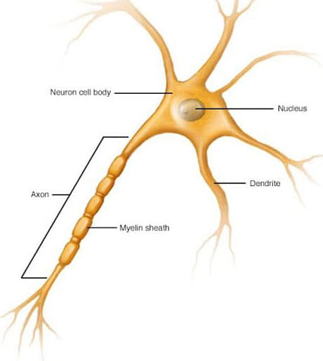
Structure of a Neuron
Function of Neurons
The main function of the neuron is to transmit electrical signals throughout the body. They do so in the following manner:
- The electrical impulses created by neurons are transmitted across the gap between neurons by the discharge and uptake of chemicals called neurotransmitters.
- These cells relay information through the chemicals that they release into the space between them called synaptic clefts.
- The human conscious experience arises from activities of neurons in the brain.
- Most of these nerve cells produce chemicals called neurotransmitters and release them into the gap between neurons, a synapse.
- The receiving neuron has receptors that are shaped to receive specific types of neurotransmitters, and when these connect with the receptors, an electrical signal is sent along to its dendrites and via an axon from one cell to another.
- This connection between two neurons allows for communication within the human brain, which helps us think and feel emotions.
- When neurons communicate with one another, they transmit information for things like motor control and thought processes.
Neurons are the functional units of information processing. Sensory neurons convert sensory input into electrical signals, and these signals travel to their target organ via nerve fibres. Motor neurons convert neural signals into muscle activation and transmit these commands to the effector organs. Interneurons are located within the medulla spinalis, where they connect sensory and motor neurons together.
Read Also:
Neuroglia
[Click Here for Sample Questions]
Neuroglia are the support cells of the nervous system. They are of different shapes and sizes and perform different functions including holding neurons in place, supplying nutrients and oxygen to neurons and removing toxins from the brain.
There are mainly three types of neuroglia: astrocytes, oligodendrocytes and microglia. Astrocytes are star shaped cells that form a barrier around capillaries in the brain called the Blood-Brain Barrier which regulates what substances enter the brain. Oligodendrocytes form myelin sheaths around axons. Microglial cells are also phagocytic and respond to damage within the CNS.
The term neuroglia refers to all the cells that support and protect nerve cells. Neuroglia can be derived from precursor cells, such as radial glial cells, or they can be derived directly from neuronal progenitor cells.

Neuroglial Cell
Types of Neuroglia
There are mainly three types of neuroglia: oligodendrocytes (make myelin), astrocytes (support neurons) and microglia (clean up). Neuroglia do not generate electrical impulses but they communicate with neurons by releasing neurotransmitters.
- Astrocytes - Astrocytes are star-shaped neuroglia cells that play important roles in the brain. Astrocytes support the neurons by adjusting their external chemical environment, transporting nutrients and oxygens to them, removing impurities as well as recycling neurotransmitters.
Astrocytes play a major role in maintaining homeostasis, especially in parts of your brain related to movement, sensation and memory.
- Oligodendrocytes - Oligodendrocytes are the neurons that wrap several times around the axons of neurons. Their projections are not as prominent as other glial cells but they play a vital role in insulating the axons and preventing them from hyperpolarization, which reduces signal transduction through it. This also causes oligodendrocytosis in which a person experiences impaired thinking and muscle coordination.
Oligodendrocytes are large, myelinated glial cells that form myelin, a fatty insulating sheath, around axons. The oligodendrocytes perform three tasks - the support of neurons by producing myelin and providing nutrients to them; they secrete materials that develop and repair the axons of neurons; they act as a cellular barrier or shield (similarly to endothelial cells)
- Microglial Cells - Microglial cells are the innate immune cells of the central nervous system (CNS). They are capable of detecting injuries and defects in both the CNS and peripheral nervous system (PNS). Microglia find their source of nutrition through phagocytosis, which is when they engulf foreign particles.
In addition to engulfing, they also secrete factors that promote healing responses in the brain. Microglia account for 10 to 15 percent of all cells found within the brain, and their primary function is to maintain homeostasis within the central nervous system through multiple functions that include phagocytosis, antigen presentation, cytokine production, and regulation of synaptic plasticity.
- Ependymal Cells - The ependymal cells are the epithelial lining of the central canal and the four ventricles of the brain. These cells function as a permeable barrier between the underlying cells and the humor (CSF). The movement of cilia within these cells are involved within the circulation of CSF.
Read Also:
| Spinal Nerves | Difference Between Nervous System and Endocrine System | Difference Between Spinal Cord and Backbone |
Difference Between Neuron and Neuroglia
[Click Here for Sample Questions]
The key point of differences between neuron and neuroglia are tabulated below:
| Parameter | Neurons | Neuroglia |
|---|---|---|
| Definition | Neurons are the functional unit of the nervous system. They form synapses between two neurons, between neurons and sensory or effector organs, or even between sensory or effector organs. Neurons are involved in the signal transduction process, which takes place at synapses. | Neuroglia provides nutrients to neurons and produces the packaging media between neurons. They also support neuronal communication through forming scaffold bodies around them and protecting them from a mechanical injury such as impact damage. |
| Significance | Neurons are the structural and functional unit of the nervous system. | Neuroglia is the supporting cells of neurons. |
| Functions | Neurons are involved in signal transduction. | Neuroglia provides nutrients to neurons and also forms packaging media between neurons. |
| Granules | Nissl’s granules are present in neurons. | They do have granules. |
| Axons | Neurons have long, slender projections called axons, which conduct nerve impulses. | Neuroglia do not have axons. However, some neuroglia comprises small, slender projections. |
| Synapse | Synapses form between two or more neurons, or between a neuron and sensory or effector organs. | Synapses are absent in Neuroglia. |
| Size | Neurons can be as small as 4 micrometres. | Neuroglia is smaller than neurons |
| Quantity | The number of neurons present in humans is around 100 billion. | The number of neuroglia is 5 to 10 times higher than that of neurons. |
| Transmission | Neurons transmit information through electrical signals. | Neuroglia does not transmit electrical signals. |
Read Also: NCERT Solutions for Class 11 Biology Chapter 21: Neural Control and Coordination
Things to Remember
- Neurons are the functional unit of the nervous system while neuroglia is the supporting cells of neurons.
- Synapses are the junctions between two neurons. These junctions transmit signals from one nerve cell to another through electrical pulses or chemical messengers called neurotransmitters.
- Neuroglia is of three types: Oligodendrocytes, astrocytes and microglia.
- Neuroglia gives structural support and nourishment to neurons and surrounds them with a protective layer, like insulation around an electrical wire.
Sample Questions
Ques. What is the function of acetylcholine? (2 Marks)
Ans. Nerve impulses are transmitted from one neuron to another at a synapse by neurotransmitters. Acetylcholine is the neurotransmitter secreted by both pre-and post-ganglionic parasympathetic cholinergic neurons. The enzyme cholinesterase inactivates excess acetylcholine into its chemical components acetic acid and choline.
Ques. What is the function of an axon? (3 Marks)
Ans. The main function of the axon is the transmission of nerve impulses. Axons are long extensions of a neuron that process information and transmit it to other neurons. Axons conduct impulses away from a cell's body. The axon has an insulating myelin sheath, which is made of fat, protein and cholesterol that serves to accelerate the conduction of action potentials. The sheath also plays a role in isolating individual neurons from one another.
Ques. The rate at which a nerve impulse travels along a nerve fibre is dependent upon which factor? (3 Marks)
Ans. The ability of a nerve fibre to propagate an action potential along its length is dependent upon the diameter of the axon, which determines its internal resistance. A larger diameter decreases this resistance and allows faster conduction. The closer two points on the pathway are to each other, the faster an action potential will travel from one location to another. In other words, it takes less time for an electrical impulse to travel across longer distances than it does through smaller distances.
Ques. What is a synapse? (2 Marks)
Ans. The junction between a nerve cell and another cell is called Synapse. Messages, in the form of electrical action potentials, travel within the neuron. The space between two neurons is known as the synaptic cleft. Neurotransmitters are stored in small synaptic vesicles present at the tip of the axon and are released into the synaptic cleft.
Ques. What are Nissl's bodies? (2 Marks)
Ans. Nissl’s bodies found in the cell body of the neurons are the granules of rough endoplasmic reticulum (RER) with rosettes of free ribosomes.The main function of Nissl bodies within neurons is to help in the production and dispersal of chemical substances like proteins and peptides.
Ques. Describe the general structure of a neuron. (3 Marks)
Ans. The structure of a neuron is divided into three parts: Cell body, dendrites and axon. The structure of a neuron will vary based on the function of the neuron. Some neurons are designed to send messages to other neurons, while others convey messages from sensory organs to the central nervous system. Regardless of their specific role, they all contain cell bodies and one or more axons. The axon is a long fibre that conducts electrical impulses away from the cell body toward another neuron or an effector organ such as muscle or glandular tissue.
Ques. Explain the relationship between central nervous system and peripheral nervous system. (3 Marks)
Ans. The central nervous system (CNS) is a network of nerves that controls the actions and responses of the body. It consists of the brain and spinal cord. This network of nerves is also known as the "thinking machine", and it controls all voluntary movements and involuntary responses.
The peripheral nervous system (PNS) controls all activities that require action potentials generated by individual neurons, such as sensation and muscle movement.
Ques. Distinguish between myelinated and unmyelinated nerve fibers. (2 Marks)
Ans. Myelinated nerve fibers are bound by Schwann cells longitudinally along their length to form the myelin sheath. This white, fatty layer completely covers the nerve fiber and often gives it a salt and pepper appearance. Unmyelinated nerve fibers lack these sheaths, making them less efficient at transmitting impulses.
Ques. Explain how a membrane becomes polarized? (2 Marks)
Ans. The membrane of a cell is usually polarized. This means that more negative ions are on one side of the membrane and more positive ions are on the other side. For example, in an excitable cell such as a muscle or nerve cell, the inside is negative and the outside has more positive ions. These forces help transmit an impulse from one end of the cell to another.
Ques. Explain the difference between a polarized neuron and a depolarized neuron. (3 Marks)
Ans. A polarized neuron is resting, or inactive, and has fewer positive ions on the inner face of its plasma membrane than on its outer face. This is due to the fact that the inside of a neuron is negatively charged whereas the outside is positively charged. A depolarized neuron can be thought of as an excited component of a nerve impulse in which sodium ions rush inward to change the polarity at this site. When this happens, there are more positive ions inside than outside, thus reversing the negative charge seen previously with a polarized neuron.
Read Also:
Check Also:



Comments