



Education Content Expert
Monocots and Dicots are the two classifications of vascular plants. The classification is based on the number of cotyledons. Monocotyledons and Dicotyledons are two types of cotyledons. Monocotyledons or monocots are flowering plants or angiosperms in which the seeds typically contain only one embryonic leaf or Cotyledon. For example Ginger, onions, wheat, and grass. Dicotyledons or Dicots are flowering plants in which the seeds typically contain two embryonic leaves or cotyledons. Examples are legumes, including beans, lentils, peas, and peanuts.
Plant Anatomy refers to the study of the interior structure of a plant. Understanding the sectioning and staining methods used with plant materials is crucial to studying the structural specifics of the stem or root of a monocot or dicot plant. It is preferable to stain the piece with suitable stains in order to observe the tissues clearly, as different stains give the tissues different colors.
Read More: Anatomy of Flowering Plants
| Table of Content |
Key Terms: Monocot Root, Monocot Stem, Dicot Root, Dicot Stem, Transverse Section Epidermis, Vascular Bundles, Pith, Cortex
Aim
[Click Here for Sample Questions]
To prepare a temporary stained mount of a transverse section of dicot and monocot stem and root to study various plant tissues.
Material Required
[Click Here for Previous Year Questions]
- Preserved material of sunflower root and stem
- Preserved or fresh material of maize root and stem
- Microscope
- Sharp blade
- Slides
- Watch glass
- Coverslips
- Safranin (1gm in 100 ml of 50% ethanol)
- Brush
- Glycerine
- Blotting paper
Read More: Difference Between Dicotyledons and Monocotyledons
Procedure
[Click Here for Sample Questions]
The procedure includes the following steps:
Preparing Sections
- Hold the cut plant material between your thumb and index finger, maintaining the razor's edge parallel to the plant's longitudinal axis.
- Cut it into teeny-tiny pieces.
- These pieces should be moved into a watch glass with the aid of a brush and the blade's edge.
- Water must stay in the watch glass.
Process of Staining
- Choose 2 to 4 nice, thin transverse pieces. Move it to another watch glass that has a safranin stain on it.
- Give the entire set a few minutes to soak up the stain.
- Drain the stain sections after a time, then rinse them with water one more to remove any remaining residue.
Mounting
- Place a stained part in the center of a slide that has been cleaned and mount it with glycerin or water.
- The coverslip is slowly placed with a needle.
- Blotting paper can be used to remove extra water or glycerine off the edge of the coverslip.
- Make sure no air bubbles develop when mounting.
Precautions
[Click Here for Previous Year Questions]
- The blade and the material should both have access to enough water during cutting the segment.
- Use a brush while working in parts.
- Place the coverslip carefully to prevent air bubbles.
- Filter paper can be used to remove extra glycerine.
Read More:
Transverse Section of the Dicot Root
[Click Here for Sample Questions]
Aim
To study the transverse section of the dicot root a sunflower root.
Theory
- Epidermis
- A single layer of densely packed cells with thin cell walls forms the top layer.
- Several cells have root hairs, which are slender outgrowths.
- Cortex
- Thin-walled parenchyma cells present in multiple layers under the epidermis.
- Endodermis
- They compose the cortex's deepest layer and have a barrel-like structure.
- They are a single-cell layer that is tightly packed.
- The thicker, radially oriented cell walls produce a Casparian band.
- Pericycle
- A layer of cells with a thin wall known as the pericycle lies beneath the endodermis.
- Vascular Bundles
- It consists of radial bundles with a 2 to 6 alternate configuration of phloem and xylem bundles.
- Bundles of xylem are called exarch, and they have protoxylem on the outside and metaxylem in the core.
- Xylem vessels are polygonal in shape.
- Phloem Bundles
- Phloem bundles comprise of parenchyma, companion cells, and sieve tubes.
- Conjunctive Tissue
- The parenchyma cells that divide the xylem and phloem bundles from one another are called Conjunctive Tissue.
- Pith
- They don't happen very often or don't happen very often.
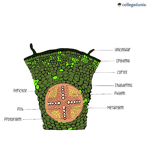
T.S. of Dicot Root
Identification
- Unicellular hair can be seen in the epidermis.
- Absence of hypodermis.
- Vascular bundles oriented radially.
- There are no more than 6 phloem/xylem bundles.
- The xylem is an exarch.
- Absence or infrequent occurrence of pith.
Conclusion
A sample of the dicot root is included in the specimen.
Read More: Anatomy of Flowering Plants Important Questions
Transverse Section of the Dicot Stem
[Click Here for Previous Year Questions]
Aim
To prepare a temporary stained mount of a transverse section of the dicot stem to study various plant tissues.
Theory
Epidermis
- A single layer of parenchymatous cells without intercellular gaps makes up this layer's protective outermost layer.
- A cuticle layer and multicellular hairs are present on the epidermal cells' outside walls (trichomes).
Cortex
- Cortex differentiates into a few layers of collenchyma cells below the epidermis to form the hypodermis, which gives the stem its mechanical strength.
- Chlorenchyma cells are arranged in a few layers with obvious intercellular gaps. Also present are several resin ducts.
- Cells from the parenchyma make up the third zone. These cells hold food-related components.
Endodermis (Starch Sheath)
- This layer's cells are tightly packed and barrel-shaped, with no intercellular gaps.
- This layer is also known as the starch sheath because the starch grains in these cells are plentiful.
Stele
In tissue, it forms the central core having a vascular bundle. It comprises vascular bundles, pericycles, medullary rays, and pith.
(a) Pericycle
- Between the endodermis and the vascular bundles.
- Has recurring sclerenchyma and parenchymal areas.
- Hard-blast flames connect every sclerenchyma patch.
(b) Medullary Rays
- It is found in the spaces between the vascular bundles. In this space, parenchymatous cells may be seen. They have thin walls and are oriented radially in 4-5 rows.
- Aids in the lateral conduction of water and food and the storage of food materials.
(c) Pith
- In the stem, it creates the middle section.
- Large parenchymatous cells make up its origin, which extends from beneath the vascular bundle to its centre.
Vascular Bundles
- They are arranged in a ring-like configuration and are open, collateral, and conjoint.
- Each vascular bundle is made up of cambium, phloem, and xylem.
- Under the pericycle, on the outside side, is where the phloem is found. It conducts food material and is made of partner cells with thin cell walls, sieve tubes, and phloem parenchyma.
- The vascular bundle's interior contains xylem, with a smaller protoxylem located toward the center and a larger metaxylem located toward the periphery. Xylem is hence endarch. These cells are lignified and lifeless. Water and mineral conductivity is Xylem.
- Between the phloem and xylem is where the cambium is located. The cell walls of the rectangular cells are thin. Meristematic tissues make up their structure and produce new cells.
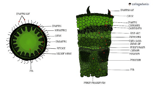
T.S. of Dicot Stem
Identification
- There are multicellular hairs on the epidermis.
- It has collenchymatous hypodermis.
- Protoxylem is located in the center of the xylem, which is endarch, while the metaxylem is located on the periphery.
- The open, collateral, and conjoint vascular bundles.
- They are set up in a ring-like arrangement.
- Pith is located in the middle.
Conclusion
A sample of the dicot stem is included in the specimen.
Read More: Morphology of Flowering Plants
Transverse Section of the Monocot Root
[Click Here for Sample Questions]
Aim
To prepare a temporary stained mount of a transverse section of monocot root to study various plant tissues.
Theory
- Epidermis or Epiblema
- It has a single, non-cuticle outer layer.
- Tightly packed cells.
- Some cells may show the emergence of unicellular root hair.
- Cortex
- Found beneath the epidermis.
- It has many layers of parenchyma cells and is wide.
- Endodermis
- Cortical tissue's deepest layer.
- Arrangement of ring-like barrel-shaped cells.
- Casparian bands are present.
- Pericycle
- A single layer of parenchymatous cells is found below the endodermis.
- Vascular Bundles
- Radial bundles are a group of eight or more alternating phloem and xylem bundles.
- Bundles of the Xylem are exarch.
- Oval or circular shape is there in the xylem bundles.
- Companion cells, sieve tubes, and parenchyma make up phloem, which can be found beneath the pericycle.
- Conjunctive Tissue
- The parenchyma tissue serves to distinguish between the xylem and phloem bundles.
- Pith
- A pith with good development is seen.
- It consists of the parenchyma in the root's middle.
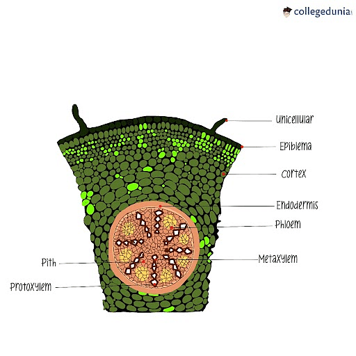
T.S. of Monocot Root
Identification
- Unicellular hair is visible in the epidermis, and there is no hypodermis.
- Vascular bundles are placed radially.
- 8 or more bundles of phloem and xylem.
- A well-developed pith.
Conclusion
A sample of the monocot root is included in the specimen.
Read More:
| Related Topics | ||
|---|---|---|
| Difference Between Xylem And Phloem | Symplast | Transportation in Plants |
| Difference Between Tracheids And Vessels | Anatomy of Flowering Plants | Plant Tissue Vs Animal Tissue |
Transverse Section of the Monocot Stem
[Click Here for Previous Year Questions]
Aim
To prepare a temporary stained mount of a transverse section of monocot stem to study various plant tissues.
Theory
The circular transverse section has a smooth surface. Numerous dispersed vascular bundles make up the foundation tissue. The transverse section shows the tissue structure in a particular way, as follows, from the periphery to the center:
- Epidermis
- A single, outermost layer.
- Live cells with thin walls. A thick cuticle is present on the outermost surface.
- Stomata are uncommonly found.
- Epidermal hair is absent.
- The function is Internal tissue protection.
- Hypodermis
- Present beneath the epidermis.
- Consists of the dead layer of sclerenchymatous cells with thick walls. Comprises two of these three layers.
- Ground Tissue
- Below the hypodermis, there is ground tissue.
- consists of parenchymatous cells, thin-walled live cells.
- intercellular gaps and a loosely packed cell structure.
- There is no distinction between the cortex, pericycle, and endodermis, as can be seen in the dicotyledonous stem.
- Vascular Bundles
- In the ground tissue, there are a few sporadic closed-type, collateral, and conjoint vascular bundles.
- Compared to the core, the vascular bundles are more numerous and tightly packed at the perimeter.
- Vascular bundles in the core are larger than those in the periphery.
- They have an oval shape and a sclerenchymatous bundle sheath surrounds them.
- It is made up of phloem and xylem.
(a) Xylem
- Y-shaped.
- The two lateral arms are metaxylem.
- At the base, a protoxylem is found.
- A cavity called the lysogenic cavity is created by the lower protoxylem particles.
- Function: supplies water
(b) Phloem
- Contains live cells and is located near the edge.
- Companion cells, sieve tubes, and the phloem parenchyma are included in it.
- Function: Transports food ingredients.
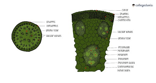
T.S. of Monocot Stem
Conclusion
A sample of the monocot stem is included in the specimen.
Read More: Sexual Reproduction in Flowering Plants
Things to Remember
- The epidermis, cortex, endodermis, pericycle, xylem, phloem, and pith make up the monocot root.
- Monocot stems are protected by an outer layer of skin tissue called the epidermis, similar to how monocot roots are.
- Dicot stems have a dermal layer, multicellular stem hair, and a clearly defined epidermis with a cuticle.
- A dicot stem's internal structure is primarily made up of pith, pericycle, hypodermis, cortex endodermis, and epidermis.
- Monocot stems, like their roots, are covered in an outer layer of dermal tissue known as the epidermis. Ground tissue and vascular tissue make up the remaining portion of the stem.
- Monocot stems' vascular bundles have chambers that hold water.
- A monocot root has a pith in the stele, unlike dicot roots. Additionally, it has vascular bundles made of both the xylem and phloem.
Previous Years’ Questions (PYQs)
- As compared to a dicot root, a monocot root has… (NEET 2012)
- As secondary growth proceeds, in a dicot stem, the thickness of… (JKCET 2015)
- Grafting is not possible in monocots because they…
- Which is not true for the anatomy of the dicot stem…
- In a mature dicot stem that has undergone secondary growth…
- In the diagram of T.S. of Stele of Dicot Root, the different…
- Which of the following is incorrect regarding dicot root… (JIPMER 2015)
- Why are vascular bundles termed closed-in monocots…
- Saccharum officinarum, a monocot plant has… (JIPMER 2010)
- In the monocotyledonous seeds, the endosperm is… (KEAM)
Sample Questions
Ques. What components make up the ground tissue of the monocot stem? (2 Marks)
Ans. A massive parenchymatous ground tissue and sclerenchymatous bundle sheath surround the vascular bundles that make up the ground tissue of the Monocot stem. Vascular bundles are arranged in a closed, connected fashion with no cambium layer in between.
Ques. When does the vascular tissue of a Dicot stem stop functioning? (1 Mark)
Ans. The vascular tissues of a dicot stem quit working as they age and are replaced by new vascular tissues. To create secondary xylem and secondary phloem, they go through secondary growth.
Ques. Why is the layer of the endodermis termed the starch sheath? (3 Marks)
Ans. Starch granules are only found in the innermost layer of the cortex of the Dicot stem, which is why this layer is known as the starch sheath. These are used by the plant to produce ATP and occasionally when it is under stress. The Monocot stem lacks this sheath because, other than ground tissues, there is no further development of tissue layers following the Hypodermis.
Ques. What layers are found in a Dicot stem between the epidermis and pericycle? (3 Marks)
Ans. Between the epidermis and the pericycle layer of the Dicot stem, there are three layers. The cortex is made up of these three layers, which are as follows:
- Just below the epidermis lies the outer hypodermis, which is made up of a few layers of collenchymatous cells.
- The cortical layer follows, which consists of parenchymatous cells with thin walls and tiny intercellular spaces between them.
- The starch sheath is present in the last layer of the endodermis.
Ques. Why don't monocot stems develop secondary growth? (2 Marks)
Ans. Because there is no cambium in the vascular bundle between the xylem and phloem, monocot stems do not experience secondary growth. As a result, they do not experience secondary xylem and secondary phloem formation.
Ques. What function does the phloem parenchyma provide in the dicot stem? (2 Marks)
Ans. An essential part of phloem tissue is the phloem parenchyma. It serves as a storage space for food items and other substances including latex, resin, and mucin. They move the food produced by photosynthesis in the plant system in both the upper and lower directions.
Ques. What characteristics does the dicot root possess? (2 Marks)
Ans. The center of dicot roots contains vascular structures. In contrast to monocots, dicots differ in how their xylem and phloem are arranged. Phloem bundles are grouped around the central portion of the dicot root's xylem, which is completely enclosed by the vascular cambium.
Ques. Briefly describe the anatomy of the dicot root. (5 Marks)
Ans. Sunflower root is the most typical instance of a dicot root: The epidermis is the skin's outermost layer. Unicellular root hairs are a common form of outgrowth for epidermal cells.
- A number of thin layers of thin-walled parenchyma cells with intercellular gaps make up the cortex.
- The term "endodermis" refers to the cortex's deepest layer. Without any intercellular gaps, it is made up of a single layer of barrel-shaped cells.
- Endodermal cells feature strips of a waxy, water-impermeable substance called suberin deposited on both their tangential and radial walls.
- A few layers of thick-walled parenchymatous cells, known as the pericycle, are located next to the endodermis between the xylem and phloem.
- These cells initiate the lateral roots and vascular cambium during secondary growth. The pitch is small or unnoticeable.
- Conjunctive tissue refers to the parenchymatous cells that are located between the xylem and the phloem.
- Typically, there are two to four phloem and xylem patches. A cambium ring eventually forms to form the stele.
Ques. Discuss about the cortex of monocot roots. (3 Marks)
Ans. The multi-layered structure known as the cortex serves as a storage area for food and nutrients for the plant. It comprises parenchyma tissues, which are living tissues that aid in photosynthesis and storage. These cells, which contain intercellular spaces between them, are found just beneath the tissues of the epidermis.
Ques. Write a few features of the dicot stem. (3 Marks)
Ans. The stems of dicots feature a distinct epidermis with a cuticle, a layer of the dermis, and multicellular stem hair. Epidermis, hypodermis, cortex endodermis, pericycle, vascular strand, and pith make up the majority of a dicot stem's internal anatomy. Dicot stems include plants like sunflower and cucurbit.
Check-More:



Comments